USOO6593292B1 (12) United States Patent (10) Patent No.: US 6,593,292 B1 Rothbard et al. (45) Date of Patent: Jul. 15, 2003 (54) COMPOSITIONS AND METHODS FOR 4,532.207 A 7/1985 Brewer et al. ................ 435/68 ENHANCING DRUG DELIVERY ACROSS 4,631,190 A 12/1986 Shen et al. ................... 424/85 AND INTO EPTHELLAL TISSUES (List continued on next page.) (75) Inventors: Jonathan B. Rothbard, Cupertino, CA FOREIGN PATENT DOCUMENTS (US); Paul A. Wender, Menlo Park, CA (US); P. Leo McGrane, Mountain 5. 2. go. View, CA (US); Lalitha V. S. Sista, EP OOO9498 4/1980 Sunnyvale, CA (US); Thorsten A. Kirschberg, Mountain View, CA (US) (List continued on next page.) (73) Assignee: CellGate, Inc., Sunnyvale, CA (US) OTHER PUBLICATIONS - Arbuck, et al. “Taxol: Clinical Results and Current Issues in c: s (*) Notice: Subiyility Development'; Chapter 14 in TAXOL(R): Science and Appli S.C. 154(b) by 104 d cations, M. Suffness ed., CRC Press (New York), pp. a -- y ayS. 379-415 (1995). (21) Appl. No.: 09/648,400 Balicki, et al. “Histone H2A Significantly Enhances. In Vitro 1-1. DNA Transfection”; Molecular Medicine vol. 3, No. 11 pp. Related U.S. Application Data Boussif, et al. "A versatile vector for gene and oligonucle (60) Provisional application No. 60/150,510, filed on Aug. 24, otide transfer into cells in culture and in vivo: Polyethylen 1999. imine"; Proc. Natl. Acad Sci. USA vol. 92, pp. 7297-7301 (51) Int. Cl." ...................... A61K 31/496. A61K 38/13. ("9 '') A61K 47/16; A61K 47/42 (List continued on next page.) (52) U.S. Cl. ............................... 514/2; 514/11; 514/12; Primary Examiner Jeffrey E. Russel 5 1. lo E.8. 3.5 E. (74) Attorney, Agent, or Firm Townsend and Townsend .U/, and Crew LLP 514/456; 514/458; 514/634; 514/635; 514/636; 530/300; 530/321; 530/328; 530/329; 530/330; (57) ABSTRACT 544/366 This invention provides compositions and methods for (58) Field of Search ............................. 424/1.69, 9.322, enhancing delivery of drugs and other agents COSS epithe 424/9.323, 9.34, 9.341, 9.1, 91. 433, lial tissues, including the skin, gastrointestinal tract, pulmo 434, 435, 436, 449, 427, 428,437; 514/2, nary epithelium, and the like. The compositions and meth 3, 11, 12, 13, 14, 15, 16, 17, 634, 636, ods are also useful for delivery acroSS endothelial tissues, 169, 456, 291, 252.13, 252.14, 252.19, including the blood brain barrier. The compositions and 383, 423, 263, 166, 457, 458, 269, 19s, methods employ a delivery enhancing transporter that has 199, 200, 152, 25, 262, 635, 159, 254.07; Sufficient guanidino or amidino Sidechain moieties to 530/300, 302,303, 315, 31, 324, enhance delivery of a compound conjugated to the reagent 325, 326, 327, 328, 329, 330,5625, acroSS one or more layers of the tissue, compared to the 230, 236, 243, 233; 544/366 non-coniugated compound. The deliverV-enhancing polv Jug p y g poly (56) References Cited mers include, for example, poly-arginine molecules that are U.S. PATENT DOCUMENTS preferably between about 6 and 25 residues in length. 4,046,722 A 9/1977 Rowland .................... 530/362 134 Claims, 23 Drawing Sheets % O HX % |W c. HY O H'gh ". ''' her." C -CONH. of H -- 3 5 5 -(-ni?fy N. N N-, & O t Sh C o t n O X a's y-NH. DEA (10x), DMF, rt n = 5, 7, 9, 0 (control)
Transcript
USOO6593292B1
(12) United States Patent (10) Patent No.: US 6,593,292 B1 Rothbard et al. (45) Date of Patent: Jul. 15, 2003
(54) COMPOSITIONS AND METHODS FOR 4,532.207 A 7/1985 Brewer et al. ................ 435/68 ENHANCING DRUG DELIVERY ACROSS 4,631,190 A 12/1986 Shen et al. ................... 424/85 AND INTO EPTHELLAL TISSUES (List continued on next page.)
(75) Inventors: Jonathan B. Rothbard, Cupertino, CA FOREIGN PATENT DOCUMENTS (US); Paul A. Wender, Menlo Park, CA (US); P. Leo McGrane, Mountain 5. 2. go. View, CA (US); Lalitha V. S. Sista, EP OOO9498 4/1980 Sunnyvale, CA (US); Thorsten A. Kirschberg, Mountain View, CA (US) (List continued on next page.)
(73) Assignee: CellGate, Inc., Sunnyvale, CA (US) OTHER PUBLICATIONS - Arbuck, et al. “Taxol: Clinical Results and Current Issues in c: s
(*) Notice: Subiyility Development'; Chapter 14 in TAXOL(R): Science and Appli S.C. 154(b) by 104 d cations, M. Suffness ed., CRC Press (New York), pp.
a -- y ayS. 379-415 (1995).
(21) Appl. No.: 09/648,400 Balicki, et al. “Histone H2A Significantly Enhances. In Vitro 1-1. DNA Transfection”; Molecular Medicine vol. 3, No. 11 pp.
Related U.S. Application Data Boussif, et al. "A versatile vector for gene and oligonucle (60) Provisional application No. 60/150,510, filed on Aug. 24, otide transfer into cells in culture and in vivo: Polyethylen
1999. imine"; Proc. Natl. Acad Sci. USA vol. 92, pp. 7297-7301 (51) Int. Cl." ...................... A61K 31/496. A61K 38/13. ("9 '')
A61K 47/16; A61K 47/42 (List continued on next page.) (52) U.S. Cl. ............................... 514/2; 514/11; 514/12; Primary Examiner Jeffrey E. Russel
5 1. lo E.8. 3.5 E. (74) Attorney, Agent, or Firm Townsend and Townsend .U/, and Crew LLP
544/366 This invention provides compositions and methods for (58) Field of Search ............................. 424/1.69, 9.322, enhancing delivery of drugs and other agents COSS epithe
424/9.323, 9.34, 9.341, 9.1, 91. 433, lial tissues, including the skin, gastrointestinal tract, pulmo 434, 435, 436, 449, 427, 428,437; 514/2, nary epithelium, and the like. The compositions and meth
3, 11, 12, 13, 14, 15, 16, 17, 634, 636, ods are also useful for delivery acroSS endothelial tissues, 169, 456, 291, 252.13, 252.14, 252.19, including the blood brain barrier. The compositions and
383, 423, 263, 166, 457, 458, 269, 19s, methods employ a delivery enhancing transporter that has 199, 200, 152, 25, 262, 635, 159, 254.07; Sufficient guanidino or amidino Sidechain moieties to
530/300, 302,303, 315, 31, 324, enhance delivery of a compound conjugated to the reagent 325, 326, 327, 328, 329, 330,5625, acroSS one or more layers of the tissue, compared to the
230, 236, 243, 233; 544/366 non-coniugated compound. The deliverV-enhancing polv Jug p y g poly (56) References Cited mers include, for example, poly-arginine molecules that are
U.S. PATENT DOCUMENTS preferably between about 6 and 25 residues in length.
''' her." C -CONH. of H -- 3 5 5 -(-ni?fy N. N N-, & O t Sh
C o t n
O X a's y-NH.
DEA (10x), DMF, rt
n = 5, 7, 9, 0 (control)
US 6,593.292 B1 Page 2
U.S. PATENT DOCUMENTS
4,701,521 A 10/1987 Ryser et al. ................ 530/322 4,847,240 A 7/1989 Ryser et al. .................. 514/12 4,880,911 A 11/1989 Brewer et al. .... ... 530/351 5,028,707 A 7/1991 Nichols et al. ... ... 546/156 5,162,505 A 11/1992 Dean et al. .... 530/391.5 5,354,844 A 10/1994 Beuget al. ................. 530/345 5,362.831 A 11/1994 Mongelli et al. ........... 526/304 5,633.230 A 5/1997 Twist et al. ................... 514/15 5,646,120 A 7/1997 Sumner-Smith et al. ...... 514/14 5,674,849. A 10/1997 Twist et al. ................... 514/15 5,716,614 A 2/1998 Katz et al. ................. 424/94.3 5,783,178 A 7/1998 Kabanov et al. ......... 424/78.31 5,789,531 A 8/1998 Sumner-Smith et al. .... 530/328 5,795,909 A 8/1998 Shashoua .................... 514/449 5,804.604 A 9/1998 Frankel et al. . ... 530/324 5,831,001 A 11/1998 Twist et al. .... ... 530/328 5,977,163 A 11/1999 Li et al. ........ ... 514/449 6,077,835 A 6/2000 Hanson et al. ................ 514/44 6,306,993 B1 * 10/2001 Rothbard et al. ........... 526/304
FOREIGN PATENT DOCUMENTS
EP O 599 303 6/1994 JP 10 O95738 4/1998 WO WO 79/00515 8/1979 WO WO 91/O9958 7/1991 WO WO 92,07871 5/1992 WO WO 93/04701 3/1993 WO WO 93/21941 11/1993 WO WO 94/04686 3/1994 WO WO 94/14464 7/1994 WO WO95/11038 4/1995 WO WO 96/21036 7/1996 WO WO 97/33552 9/1997 WO WO 97/40854 11/1997 WO WO 98/52614 11/1998 WO WO 01/13957 3/2001
OTHER PUBLICATIONS
Burton, et al. "Basic polyelectrolytes and protein transport acroSS the new-born pig intestine'; Physiological Society p. 27P–28P (Dec. 1970). vol. 211, No. 2. Brugidou, et al. “The Retro-Inverso Form of a Homeobox-Derived Short Peptide is Rapidly Internalised by Cultured Neurones: A New Basis For An Efficient Intra cellular Delivery System”; Biochemical and Biophysical Research Communications vol. 214, No. 2 pp. 685-693 (Sep.1995). Buschle, et al. “Transloading of tumor antigen-derived peptides into antigen-presenting cells”; PNAS Vol. 94, pp. 3256-3261 (Apr. 1997). Chen, et al. “Galactosylated Histone-Mediated Gene Trans fer and Expression; Human Gene Therapy vol. 5, pp. 429–435 (1994). Cooke, et al. “Nitric Oxide Synthase: Role in the Genesis of Vascular Disease”; Annu. Rev. Med. Vol. 48, pp. 489-509 (1997). Dattilo, et al. “Inducible Nitric Oxide Synthase Expression in Human Vein Grafts”; Am J Surg. vol. 174, pp. 177-180 (1997). de Bont, et al. “Synthesis and Biological Activity of B-Glu curonyl Carbamate-Based Prodrugs of Paclitaxel as Poten tial Candidates for ADEPT'; Bioorganic & Medicinal Chemistry vol. 5, No. 2 pp. 405–414 (1997).
Derossi, et al. “The Third Helix of the Antennapedia Home odomain Translocates through Biological Membranes'; The Journal of Biological Chemistry vol. 269, No. 14 pp. 10444–10450 (1994). Elferink, “Changes of Plasma Membrane Permeability in Neutrophils Treated With Polycations”; Inflammation vol. 15, No. 2 pp. 103-115 (Apr. 1991). Emi, et al. “Gene Transfer Mediated by Polyarginine Requires a Formation of Big Carrier-Complex of DNA Aggregate”; Biochemical and Biophysical Research Com munications vol. 231, pp. 421-424 (1997). Fawell, et al. “Tat-mediated delivery of heterologous pro teins into cells”; Proc. Natl. Acad. Sci. USA vol. 91, pp. 664-668 (Jan. 1994). Fletcher, et al. “Partially Modified Retro-Inverso Peptides: Development, Synthesis, and Conformational Behavior”; Chem. Rey: vol. 96 pp. 763–795 (1998). Garg, et al. "Nitric Oxide-generating Vasodilators and 8-Bromo-Cyclic Guanosine Monophosphate Inhibit Mito genesis and Proliferation of Cultured Rat Vascular Smooth Muscle Cells”, J. Clin. Invest. vol. 83, pp. 1774–1777 (May 1989). George, et al. “The Medicinal Chemistry of Taxol'; Chapter 13 in TAXOL(R): Science and Applications, M. Suffness ed., CRC Press (New York), pp. 317–375 (1995). Golik, et al. “Synthesis and Antitumor Evaluation of Pacli taxel Phosphonooxymethyl Ethers: A Novel Class of Water Soluble Paclitaaxel Pro-Drugs’ Bioorganic & Medicinal Chemistry Letters vol. 6, No. 15 pp. 1837–1842 (1996). Greenwald, et al. “Drug Delivery Systems: Water Soluble Taxol. 2'-Poly(ethylene glycol) Ester Prodrugs-Design and in Vivo Effectiveness'; J. Med. Chem. 39(2):424–431 (Jan. 1996). Kessler “Peptoids-A New Approach to the Development of Pharmaceuticals; Angew Chem. Int. Ed. Engl. vol. 32, No. 4 pp. 543–544 (1993). Kingston “Natural Toxoids: Structure and Chemistry’; Chapter 12 in TAXOL(R): Science & Applications, M. Suf fnessed, CRC Press (New York), pp. 287-315 (1995). Lam, et al. “The “One-Bead-One-Compound” Combina torial Library Method”; Chem. Re: vol. 97, pp. 411-448 (1997). Lloyd-Jones, M.D., et al. “The Vascular Biology of Nitric Oxide and Its Role in Atherogenesis”; Annu. Rev. Med. Vol. 47, pp. 365-375 (1996). Mauersberger, et al. “UnterSuchungen Zur Zytotoxizität von Poly-L-Arginin, Poly-L-Lysin and DEAE-Dextran bei L-Zellen und Mäuseembryofibroblasten'; Exp. Path vol. 18 pp. 268-274 (1977). Murphy, et al. “A combinatorial approach to the discovery of efficient cationic peptoid reagents for gene delivery; Proc. Natl. Acad. Sci. USA vol. 95, pp. 1517–1522 (Feb. 1998). Natsume, et al. “Screening of Absorption Enhancers for Nasal Peptide and Protein Delivery”; Proceed. Intern. Symp. Control. Rel. Bioact. Mater. vol. 23, pp. 481-482 (Jul. 1996). Nicolaou, et al. “Design, Synthesis and biological activity of protaxols"; Nature vol. 364, pp. 464–466 (Jul. 1993). Rodrigues, et al. “Synthesis and B-lactamase-mediated acti Vation of a cephalosporin-taxol prodrug Chemistry and Biology vol. 2, pp. 223-227 (Apr. 1995). Rose, “Preclinical Antitumor Activity ot Taxanes”; Chapter in TAXOLOR: Science & Applications, M. Suffness ed., CRC Press (New York), pp. 209-235 (1995).
US 6,593.292 B1 Page 3
Simon, et al. "Peptoids: A modular approach to drug dis covery”; Proc. Natl. Acad. Sci. USA vol. 89 pp. 9367–9371 (Oct. 1992). Straubinger “Biopharmaceutics of Paclitaxel (Taxol): For mulation, Activity, and Pharmacokinetics”; Chapter 9 in in TAXOL(RScience & Applications, M. Suffness ed., CRC Press (New York), pp. 237–258 (1995). Sumner-Smith, et al. “123: 79357m Antiherpetic activities of N-O-acetyl-nona-D-=arginine amide acetate'; 6001 Chemical Abstracts vol. 123, No. 7 p. 606 (1995). Thompson, et al. “Synthesis and Applications of Small Molecule Libraries”; Chem. Rev. 96:555–600 (1996). Tsao, et al. “Nitric Oxide Regulates Monocyte Chemotactic Protein-1”; Circulation 96:,934-940 (1997). Uchida, et al. “Polycations Decrease the Transepithelial Resistance of Cultured Tracheal Epithelial Cells”; Chest vol. 101, No. 3, p. 33S (Mar. 1992). Ueda, et al. “Synthesis and Antitumor Evaluation of 2'-Oxy carbonylpaclitaxels (Paclitaxel-2'-Carbonates)”; Bioor ganic & Medicinal Chemistry Letters vol. 4 No. 15 pp. 1861–1864 (Aug. 1994). Ueda, et al. “Novel Water Soluble Phosphate Prodrugs of Taxole Possessing In Vivo Antitumor Activity”; 3(8):1761–1766 (May 1993). Vyas, et al. “Synthesis and Antitumor Evaluation of Water Soluble Taxol Phosphates”; Bioorganic & Medicinal Chem istry Letters vol. 3, No. 6 pp. 1357–1360 (1993). Vyas, et al. “Phosphatase-Activated Prodrugs of Paclitaxel’; Chapter 9 in Taxane Anticancer Agents American Chemical Society p. 124 (1995). Wolf, et al. “Dietary L-Arginine Supplementation Normal izes Platelet Aggregation in Hypercholesterolemic Humans'; JACC vol. 29, No. 3 pp. 479–485 (Mar. 1997). Zuckermann, et al. “Efficient Method for the Preparation of Peptoids Oligo(N-substituted glycines) by Submonomer Solid-Phase Synthesis”; Chemtracts-Macromolecular Chemistry vol. 4 pp. 80-83 (1993). Aoyagi, et al. "Polymerization of Benzalkonium Chloride-Type Monomer and Application to Percutaneous Drug Absorption Enhancer'; Journal of Controlled Release, vol. 13, No. 1 pp. 63–71 (Jul 1990). Babiuk, et al., “Cutaneous vaccination: the Skin as an immunologically active tissue and the challenge of antigen delivery'; Journal of Controlled Release vol. 66 pp. 199-214 (2000). Colin, et al. "Liposomes enhance delivery and expression of an RGD-oligolysine gene transfer vector in human tracheal cells"; Gene Therapy vol. 5 pp. 1488–1498 (1998).
Coyle, e tal. “Role of Cationic Proteins in the Airway Hyperresponsiveness Due to Airway Inflammation; Am. Respir: Crit. Care Med. vol. 150 pp. 563-571 (1994). Gama, et al. "CA"-sensing receptors in intestinal epithe lium”; American Journal of Physiology vol. 273, No. 4, Part 1 pp. C1168–C1175 (Oct. 1997). Hosoya, et al. “Effect of Several Hydrophilic Polymers on the Permeation of Morphine and Salicylic Acid through Excised Hairless Rat Skin"; Chem. Pharm. Bull. 46(5) 882–885 (1998). Hulsmann, “Permeability of Human Isolated Airways Increases after Hydrogen Peroxide and Poly-L-arginine'; Am. J. Respir: Crit. Care Med. vol. 153 pp. 841-846 (1996). Koji Kobayashi, “Composition for Transmucosally Absorb able Preparation”; Patent Abstracts of Japan, Publication No. 10095738, Publication Date Apr. 4, 1998. Perr, et al. “Protamine Selectively Inhibits Collagen Syn thesis by Human Intestinal Smooth Muscle Cells and Other Mesenchymal Cells'; Journal of Cellular Physiology 140:463-470 (1989). Peterson, et al. “Polyamino Acid Enhancement of Bacterial Phagocytosis by Human Polymorphonuclear Leukocytes and Peritoneal Macrophages”; Infection and Immunity vol. 43, No. 2, pp. 561-566 (Feb. 1984). Santana, et al., “Inflammatory responses induced by poly-L-arginine in rat lungs in Vivo”, Agents Actions Vol. 39, No. 3-4 pp. 104-110 (1993). Tzan, et al. “Mammalian urinary bladder permeability is altered by cationic proteins modulation by divalent cations'; American Journal of Physiology vol. 267, No. 4, Part 1, pp. C1013–C1026 (1994). Tzan, et al., “Modification of Epithelial Permeability by Cationic Polypeptides'; American Journal of Physiology vol. 265, No. 6, Part 1, pp. C1637–C1647 (1993). Uchida, et. al., Cationic Proteins Increase the Permeability of Cultured Rabbit Trachael Epthihelial Cells: Modification by Heparin and Extracellular Calcium: Experimental Lung Research vol. 22, No. 1, pp. 85–99 (1996). Wei, et al. “Synthesis of Oligoarginine-Oligonucleotide Conjugates and Oligoarginine-Bridge Oligonucleotide Pairs”; Bioconjugate Chem. Vol. 5, pp. 468-474 (Sep./Oct. 1994).
* cited by examiner
U.S. Patent Jul. 15, 2003 Sheet 1 of 23 US 6,593,292 B1
Figure l
5ul O (C-AC)2O, Pyr N
O ". N N - - 1. 2. N Yo O N
Figure 2
\ O t O
-N
figy i N Yo
a \ \ Q treo
U.S. Patent Jul. 15, 2003 Sheet 2 of 23 US 6,593,292 B1
Figure 3
HX
l y-NH. HY O O HN NH -N-NH
s O s H
DEA (10x), DMF, rt V - - -r N NE
o’s o g
U.S. Patent Jul. 15, 2003 Sheet 3 of 23 US 6,593,292 B1
Figure 4
U.S. Patent Jul. 15, 2003 Sheet 4 of 23 US 6,593,292 B1
ACHN N 1 NH
Fig. 5A "T. , , , N Sry, N=/
ON
/-N CONH-"T"
N -s Fig. SED HNNN
H O N
NO2
Fig. SC "T"-HN CO-Z-Drug
PhCOHN Q h OV YYS-O
P 5 6H6BOAC Fig. G D
O
OP(O)(OH)2 V
"T"-HN
U.S. Patent Jul. 15, 2003 Sheet 5 of 23 US 6,593,292 B1
acQ 9 OH ? y
SÉ PhCOHN Q. Y F. C. k-fisco 9 Ph O\ 2. SN1
Fig. 5 G
Fig - B -
US 6,593,292 B1 Sheet 7 of 23 Jul. 15, 2003 U.S. Patent
t Figure
-rr+----r+-----------+-------r------r--------
U.S. Patent Jul. 15, 2003 Sheet 8 of 23 US 6,593,292 B1
Figure 8 CO COH 2
CO2H r 2 CO2H r Cu?" ls N u 993 - 1N NN-CO2H 1N1 N1)-N HO HOC N
l ls 2 2 CO2H COH
Figure 9 O O O O
H H
C - d', 'so TFA 2. 5 ro HO2C a y HO2C
H O
cuso, Cu2" ch 5 ro -1 HO HOC 2 O
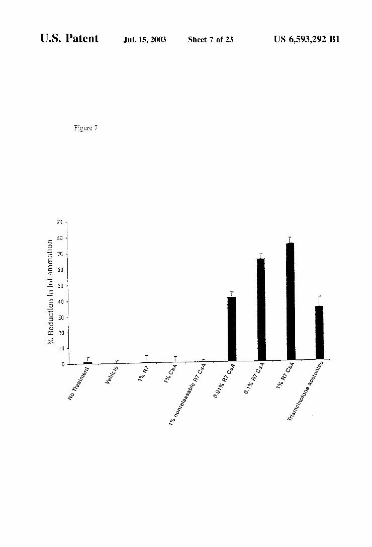
Figure 11
O
-( (oro C Biotin-aca-ra-AAC-CONH2 s DEA, DMF
n = 5 and 7
U.S. Patent Jul. 15, 2003 Sheet 9 of 23 US 6,593,292 B1
Figure 12
OC(O)Cl DIEA, DCM 52%
COOBn
O OBn
Figure 13
B-aca-r-K-CONH2, DEA, DMAP 0.3ed, DMF, rt
O OBn
Figure 14
NH2-r-COOH, DIEA, DMAP 0.3ed, DMF, rt
O OBn
U.S. Patent Jul. 15, 2003 Sheet 10 Of 23 US 6,593,292 B1
Figure 15A
H2, Pd/C, EtOAC
Figure 15B
NHS, DCC, CH2Cl2, it
U.S. Patent Jul. 15, 2003 Sheet 11 of 23 US 6,593,292 B1
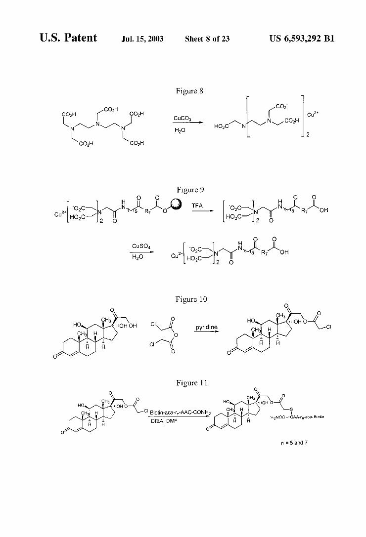
Figure 15C AcO O OH
NH-R7-COOH, DIEA, DMAP 0.3eq, DMF, rt
U.S. Patent Jul. 15, 2003 Sheet 12 of 23 US 6,593,292 B1
Figure 16 HS
TRANSPORTER O H2N
O DRUG-X-H -> DRUG-X
C
O
-N-S DRUG-X RELEASE AS INDICATED TRANSPORTER al-Ho-Ho
O
S
of transporter DRUG-X-H H
Figure 17
U.S. Patent Jul. 15, 2003 Sheet 13 of 23 US 6,593,292 B1
Figure 18
HN N HN N HN N 2 H 2 H 2 H H2N
U.S. Patent Jul. 15, 2003 Sheet 14 of 23 US 6,593,292 B1
Figure 19
H H NH2 N. NH2 NNNH2
l l HN N 2 H HN N
U.S. Patent Jul. 15, 2003 Sheet 15 of 23 US 6,593,292 B1
Figure 20
MTT Cytotoxicity Assay 3 day assay
100
80
2n is 60 CG 1040 S. - IC = 1, 1 nM O ---
> O CG 1062 O' 40 IC = 6 nM S v Taxol
IC = 3 nM 20
(). I () ()()
concentration (nM)
U.S. Patent Jul. 15, 2003 Sheet 16 of 23 US 6,593,292 B1
Figure 21
2OO
1.1. O5 OO 5 O
Tat Tat Tat Tat Tat 49-57 49-56 49-55 50-57 51-57
Figure 22
200
1. 5 O
1. O O
Tat A- A - A- A- A- A - A- A- A 49- 49 50 51 52 53 54 55 56 57 57
U.S. Patent Jul. 15, 2003 Sheet 18 of 23 US 6,593,292 B1
Figure 26
600
500
400
300
200
100
rS r/ rg N- N- N arg5 arg7 arg9
U.S. Patent Jul. 15, 2003 Sheet 19 of 23 US 6,593,292 B1
Figure 27
2OOO
1500
1000
500
O O.1 1.O 10.0 100.0
Concentration (uM)
Figure 28
1000
800 O C
8 3 600 s
400 C
3 > 200
O Uh di N. c. 2 2 2 2 2
X X X X X O O (O C CO U OY N OO VO
U.S. Patent Jul. 15, 2003 Sheet 20 of 23 US 6,593,292 B1
Figure 29
1400
1200
1000
9 N- N- N- N- N chg9 etg9 arg9 btg9 hxg9
U.S. Patent Jul. 15, 2003 Sheet 22 of 23 US 6,593,292 B1
Figure 31A
Synthetic Schemes for FK506 Conjugates
Scheme I (Linker: 6-Maleimido caproic hydrazide, TFA salt)
O AcOH, Aco P O + Henr-n-no- reflux, 24h ~OH
O 45% O O O
1) EtN, CICOOCH(CH), NHNHtBoc, 2h, (81%)
O GE) G)
-----ms- ~NH-NHCFCoo 2) TFA, CHCl, 3h, (95%) O
O
Ref. Willner et al; Bioconjugate Chemistry, 1993, 4,521-527
COMPOSITIONS AND METHODS FOR ENHANCING DRUG DELIVERY ACROSS
AND INTO EPTHELLAL TISSUES
CROSS-REFERENCE TO RELATED APPLICATIONS
This application claims priority to U.S. Provisional Patent Application No. 60/150,510, filed Aug. 24, 1999. This application is related to U.S. patent application Ser. No. 09/645,689 filed on even date herewith. Both of these applications are incorporated herein by reference for all purposes.
BACKGROUND OF THE INVENTION
1. Field of the Invention This invention pertains to the field of compositions and
methods that enhance the delivery of drugs and other compounds acroSS the dermal and epithelial membranes, including, for example, Skin, the gastrointestinal epithelium and the bronchial epithelium.
2. Background Transdertnal or transmucosal drug delivery is an attractive
route of drug delivery for Several reasons. Gastrointestinal drug degradation and the hepatic first-pass effect are avoided. In addition, transdermal and transmucosal drug delivery is well-Suited to controlled, Sustained delivery (see, e.g., Elias, In Percutaneous Absorption. Mechanisms Methodology-Drug Delivery, Bronaugh & Maibach, Eds., pp. 1-12, Marcel Dekker, New York, 1989.). For many applications, traditional methods of administering drugs are not optimal because of the very large initial concentration of the drug. Transdermal delivery could allow a more uniform, slower rate of delivery of a drug. Moreover, patient com pliance is encouraged because Such delivery methods are easy to use, comfortable, convenient and non-invasive.
These advantages of transdermal and transmucosal deliv ery have not led to many clinical applications because of the low permeability of epithelial membranes, the skin in particular, to drugs. The difficulties in delivering drugs acroSS the Skin result from the barrier property of skin. Skin is a structurally complex thick membrane that represents the body's border to the external hostile environment. The skin is composed of the epidermis, the dermis, the hypodermis, and the adenexal Structures (epidermal appendages). The epidermis, the outermost epithelial tissue of the Skin, is itself composed of Several layers-the Stratum corneum, the Stra tum granulosum, the Stratum Spinosum, and the Stratum basale. Compounds that move from the environment into and
through intact skin must first penetrate the Stratum corneum, the Outermost layer of Skin, which is compact and highly keratinized. The Stratum corneum is composed of Several layers of keratin-filled skin cells that are tightly bound together by a "glue” composed of cholesterol and fatty acids. The thickness of the Stratum corneum varies depending upon body location. It is the presence of Stratum corneum that results in the impermeability of the skin to pharmaceutical agents. The Stratum comeum is formed naturally by cells migrating from the basal layer toward the skin Surface where they are eventually Sloughed off. AS the cells progreSS toward the Surface, they become progressively more dehy drated and keratinized. The penetration acroSS the Stratum corneum layer is generally the rate-limiting Step of drug permeation acroSS Skin. See, e.g., Flynn, G. L., In Percuta neous Absorption. Mechanisms-Methodology-Drug Delivery, Supra. at pages 27–53.
15
25
35
40
45
50
55
60
65
2 After penetration through the Stratum corneum layer,
Systemically acting drug molecules then must pass into and through the epidermis, the dermis, and finally through the capillary walls of the bloodstream. The epidermis, which lies under the Stratum corneum, is composed of three layers. The Outermost of these layerS is the Stratum granulosum, which lies adjacent to the Stratum corneum, is composed of cells that are differentiated from basal cells and keratinocytes, which make up the underlying layers. Having acquired additional keratin and a more flattened shape. The cells of this layer of the epidermis, which contain granules that are composed largely of the protein filaggrin. This protein is believed to bind to the keratin filaments to form the keratin complex. The cells also Synthesize lipids that function as a “cement” to hold the cells together. The epidermis, in particular the Stratum granulosum, contains enzymes Such as aminopeptidases. The next-Outermost layer of the epidermis is the Stratum
Spinosum, the principal cells of which are keratinocytes, which are derived from basal cells that comprise the basal cell layer. Langerhans cells, which are also found in the Stratum Spinosum, are antigen-presenting cells and thus are involved in the mounting of an immune response against antigens that pass into the Skin. The cells of this layer are generally involved in contact Sensitivity dermatitis. The innermost epidermal layer is the Stratum basale, or
basal cell layer, which consists of one cell layer of cuboidal cells that are attached by hemi-desmoSomes to a thin base ment membrane which Separates the basal cell layer from the underlying dermis. The cells of the basal layer are relatively undifferentiated, proliferating cells that Serve as a progenitor of the outer layers of the epidermis. The basal cell layer includes, in addition to the basal cells, melanocytes. The dermis is found under the epidermis, which is sepa
rated from the dermis by a basement membrane that consists of interlocking rete ridges and dermal papillae. The dermis itself is composed of two layers, the papillary dermis and the reticular dermis. The dermis consists of fibroblasts, histiocytes, endothelial cells, perivascular macrophages and dendritic cells, mast cells, Smooth muscle cells, and cells of peripheral nerves and their endorgan receptors. The dermis also includes fibrous materials. Such as collagen and reticulin, as well as a ground Substance (principally glycosaminoglycans, including hyaluronic acid, chondroitin Sulfate, and dermatan Sulfate).
Several methods have been proposed to enhance trans dermal transport of drugs. For example, chemical enhancers (Burnette, R. R. In Developmental Issues and Research Initiatives; Hadgraft J., Ed., Marcel Dekker: 1989; pp. 247-288), iontophoresis, and others have been used. However, in spite of the more than thirty years of research that has gone into delivery of drugs across the skin in particular, fewer than a dozen drugs are now available for transdermal administration in, for example, skin patches.
Transport of drugs and other molecules acroSS the blood brain barrier is also problematic. The brain capillaries that make up the blood-brain barrier are composed of endothelial cells that form tight junctions between themselves (Goldstein et al., Scientific American 255:74-83 (1986); Pardridge, W. M., Endocrin. Rev. 7: 314-330 (1986)). The endothelial cells and the tight intercellular junctions that join the cells form a barrier against the passive movement of many molecules from the blood to the brain. The endothelial cells of the blood-brain barrier have few pinocytotic vesicles, which in other tissues can allow Somewhat unse lective transport across the capillary wall. Nor is the blood
US 6,593,292 B1 3
brain barrier interrupted by continuous gaps or channels that run through the cells, thus allowing for unrestrained passage of drugs and other molecules.
Thus, a need exists for improved reagents and methods for enhancing delivery of compounds, including drugs, acroSS epithelial tissues and endothelial tissueS Such as the skin and the blood-brain barrier. The present invention fulfills this and other needs.
SUMMARY OF THE INVENTION
The present invention provides methods for enhancing delivery of a compound into and acroSS one or more layers of an animal epithelial or endothelial tissue. The methods involve contacting tissue with a conjugate that includes the compound and a delivery-enhancing transporter. The delivery-enhancing transporters, which are also provided by the invention, have Sufficient guanidino or amidino moieties to increase delivery of the conjugate into and acroSS one or more intact epithelial or endothelial tissue layers compared to delivery of the compound in the absence of the delivery enhancing transporter. Typically, the delivery-enhancing transporters have from 6 to 25 guanidino or amidino moieties, and more preferably between 7 and 15 guanidino moieties.
The delivery-enhancing transporters and methods of the invention are useful for delivering drugs, diagnostic agents, and other compounds of interest acroSS epithelial tissues Such as the Skin and mucous membranes. Delivery acroSS the blood-brain barrier is also enhanced by the conjugates and methods of the invention. The methods and compositions of the invention can be used not only to deliver the compounds to the particular site of administration, but also provide Systemic delivery.
In Some embodiments, the delivery-enhancing transporter comprises 7-15 arginine residues or analogs of arginine. The delivery-enhancing transporter can have at least one arginine that is a D-arginine and in Some embodiments, all arginines are D-arginine. The delivery-enhancing transporter can con sist essentially of 5 to 50 amino acids, at least 50 percent of which are arginine. In some embodiments, at least 70% of the amino acids are arginines or arginine analogs. In Some embodiments, the delivery-enhancing transporter comprises at least 5 contiguous arginines or arginine analogs. The compound to be delivered can be connected to the
delivery enhancing transporter by a linker. In Some embodiments, the linker is a releasable linker which releases the compound, in biologically active form, from the delivery-enhancing transporter after the compound has passed into and through one or more layers of the epithelial and/or endothelial tissue. In Some embodiments, the com pound is released from the linker by Solvent-mediated cleavage. The conjugate is, in Some embodiments, Substan tially stable at acidic pH but the compound is substantially released from the delivery-enhancing transporter at physi ological pH. In some embodiments, the half-life of the conjugate is between 5 minutes and 24 hours upon contact with the skin or other epithelial or endothelial tissue. For example, the half-life can be between 30 minutes and 2 hours upon contact with the skin or other epithelial or endothelial tissue.
Examples of conjugate Structures of the invention include those having Structures Such as 3, 4, or 5, as follows:
5
1O
15
25
35
40
45
50
55
60
65
O R2
R-X-C-(CH2): Y-C-(CH2). NH-(CH2) -C-Rs
O O
O R5
R-X- C- (CH) R- (CH2) CH- C-Rs
O
O Rs
R-X-C-O-(CH2)-CH-C-Rs
O
where R-X comprises the compound; X is a functional group on the compound to which the linker is attached; Y is N or C.; R is hydrogen, alkyl, aryl, acyl, or allyl, R comprises the delivery-enhancing transporter; R is Substi tuted or unsubstituted S, O, N or C; R is OH, SH or NHR; R is hydrogen, alkyl, aryl, acyl or allyl, k and m are each independently Selected from 1 and 2; and n is 1 to 10. Preferably, X is selected from the group consisting of N, O, S, and CRRs, wherein R, and Rs are each independently Selected from the group consisting of H and alkyl. In Some embodiments, R is S.; Rs is NHR, and R is hydrogen, methyl, allyl, butyl or phenyl. In Some embodiments, R is benzyl, k, m, and n are each 1, and X is O. In Some embodiments, the conjugate comprises Structure 3 and R is Selected to obtain a conjugate half-life of between 5 minutes and 24 hours. In Some embodiments, R is Selected to obtain a conjugate half-life of between 5 minutes and 24 hours. In Some embodiments, the conjugate comprises structure 4, R is S.; Rs is NHR, and R is hydrogen, methyl, allyl, butyl or phenyl. In Some embodiments, the conjugate comprises Structure 4, Rs is NHR, R is hydrogen, methyl, allyl, butyl or phenyl; and k and m are each 1. One example of a conjugate is:
Ph
O CH2
R-O-C-CH-N-C-CH-NH-CH-C-R,
O O
where Ph is phenyl.
The invention also provides conjugates in which the release of the linker from the biological agent involves a first, rate-limiting intramolecular reaction, followed by a faster intramolecular reaction that results in release of the linker. The rate-limiting reaction can, by appropriate choice of substituents of the linker, be made to be stable at a pH that is higher or lower than physiological pH. However, once the conjugate has passed into and acroSS one or more layers of an epithelial or endothelial tissue, the linker will be cleaved from the agent. An example of a compound that has this type of linker is structure 6, as follows:
US 6,593,292 B1
O O R5
R-X-C-OCH-Ar-O-C-(CH2) R-(CH2) CH-i-R, O
wherein R-X comprises the compound to be delivered acroSS one or more layers of an epithelial and/or endothelial tissue, X is a functional group on the compound to which the linker is attached; Ar is an aryl group having the attached radicals arranged in an Ortho or para configuration, which aryl group can be Substituted or unsubstituted; R comprises the delivery-enhancing transporter; R is Substituted or unsubstituted S, O, N or C; R is OH, SH or NHR, R is hydrogen, alkyl, aryl, acyl or allyl, and k and m are each independently Selected from 1 and 2. In Some embodiments, X is Selected from the group consisting of N, O, S, and CR7Rs, wherein R, and Rs are each independently Selected from the group consisting of H and alkyl. In Some embodiments, R is S.; Rs is NHR, and R is hydrogen, methyl, allyl, butyl or phenyl. In Some embodiments, the conjugate comprises:
O O NH2
R-O-C-OCH-Ar-O-C-CH2-S-CH-CH-C-R.
O
In preferred embodiments, the compositions of the inven tion comprise a linker Susceptible to Solvent-mediated cleav age. For example, a preferred linker is Substantially stable at acidic pH but is Substantially cleaved at physiological pH.
Additional embodiments of the invention provide trans dermal drug formulations. These formulations include a therapeutically effective amount of a therapeutic agent, a delivery-enhancing transporter that includes Sufficient guanidino or amidino Sidechain moieties to increase delivery of the conjugate acroSS one or more layers of an animal epithelial tissue compared to the trans-epithelial tissue deliv ery of the biologically active agent in non-conjugated form; and a vehicle Suited to transdermal drug administration.
BRIEF DESCRIPTION OF THE FIGURES
FIG. 1 shows a reaction Scheme for the preparation of an C-chloroacetyl cycloSporin A derivative.
FIG. 2 shows a general procedure for the coupling of cysteine-containing peptides to the C-chloro acetyl cyclosporin A derivative.
FIG. 3 shows a reaction scheme for the coupling of the cyclosporin A derivative to a biotin-labeled peptide.
FIG. 4 shows a reaction Scheme for coupling of a cyclosporin A derivative to an unlabeled peptide (SEQ ID NO:7).
FIGS. 5A-H show various types of cleavable linkers that can be used to link a delivery-enhancing transporter to a biologically active agent or other molecule of interest. FIG. 5A shows an example of a disulfide linkage. FIG. 5B shows a photocleavable linker which is cleaved upon exposure to electromagnetic radiation. FIG. 5C shows a modified lysyl residue used as a cleavable linker. FIG. 5D shows a conju gate in which the delivery-enhancing transporter T is linked to the 2'-OXygen of the anticancer agent, paclitaxel. The linking moiety includes (i) a nitrogen atom attached to the delivery-enhancing transporter, (ii) a phosphate monoester
15
25
35
40
45
50
55
60
65
6 located para to the nitrogen atom, and (iii) a carboxymethyl group meta to the nitrogen atom, which is joined to the 2'-OXygen of paclitaxel by a carboxylate ester linkage. FIG. 5E a linkage of a delivery-enhancing transporter to a bio logically active agent, e.g., paclitaxel, by an aminoalkyl carboxylic acid; a linker amino group is joined to a delivery enhancing transporter by an amide linkage and to a pacli taxel moiety by an ester linkage. FIGS. 5F and G show chemical Structures and conventional numbering of constitu ent backbone atoms for paclitaxel and “TAXOTERETM” (R'=H, R'—BOC). FIG. 5G shows the general chemical Structure and ring atom numbering for taxoid compounds.
FIG. 6 displays a Synthetic Scheme for a chemical con jugate between a heptamer of L-arginine (SEQ ID NO:3) and cyclosporin A (panel A) and its pH dependent chemical release (SEQ ID NO:6) (panel B). The C-chloro ester (2) was treated with benzylamine in the presence of Sodium iodide to effect Substitution, giving the Secondary amine (5). Amine (5) was treated with anhydride (6) and the resultant crude acid (7) was converted to its corresponding NHS ester (8). Ester (8) was then coupled with the amino terminus of hepta-L-arginine (SEQ ID NO:3), giving the N-Boc pro tected CSA conjugate (9). Finally, removal of the Boc protecting group with formic acid afforded the conjugate (10) as its octatrifluoroacetate salt after HPLC purification.
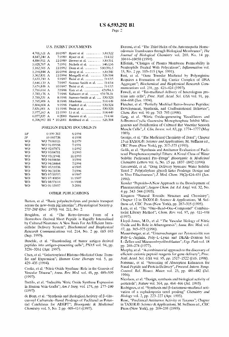
FIG. 7 displays inhibition of inflammation in murine contact dermatitis by releasable R7 CSA. Balb/c (6-7 weeks) mice were painted on the abdomen with 100 ul of 0.7% DNFB in acetone olive oil (95:5). Three days later both ears of the animals were restimulated with 0.5% DNFB in acetone. Mice were treated one, five, and twenty hours after restimulation with either vehicle alone, 1% R7 peptide alone, 1% CSA, 1% nonreleasable R7 CSA, 0.01%/0.1% /1.0% releasable R7 CSA, and the fluorinated steroid posi tive control 0.1% triamcinolone acetonide. Ear inflammation was measured 24 hours after restimulation using a Spring loaded caliper. The percent reduction of inflammation was calculated using the following formula (t-n)/(u-n), where t=thickness of the treated ear, n=the thickness of a normal untreated ear, and u=thickness of an inflamed ear without any treatment. N=20 animals in each group.
FIG. 8 shows a procedure for the preparation of a copper diethylene-triaminepentaacetic acid complex (Cu-DTPA).
FIG. 9 shows a procedure for linking the Cu-DTPA to a transporter through an aminocaproic acid.
FIG. 10 shows a reaction for the acylation of hydrocor tisone with chloroacetic anhydride.
FIG. 11 shows a reaction for linking the acylated hydro cortisone to a transporter.
FIG. 12 shows a reaction for preparation of C-2 deriva tives of taxol.
FIG. 13 shows a schematic of a reaction for coupling of a taxol derivative to a biotin-labeled peptide.
FIG. 14 shows a reaction for coupling of an unlabeled peptide to a C-2 derivative of taxol. FIG.15A-C shows a reaction Scheme for the formation of
other C-2 taxol-peptide conjugates. FIG. 16 shows a general Strategy for Synthesis of a
conjugate in which a drug or other biological agent is linked to a delivery-enhancing transporter by a pH-releasable linker.
FIG. 17 shows a schematic diagram of a protocol for Synthesizing a taxol. 2'-chloroacetyl derivative.
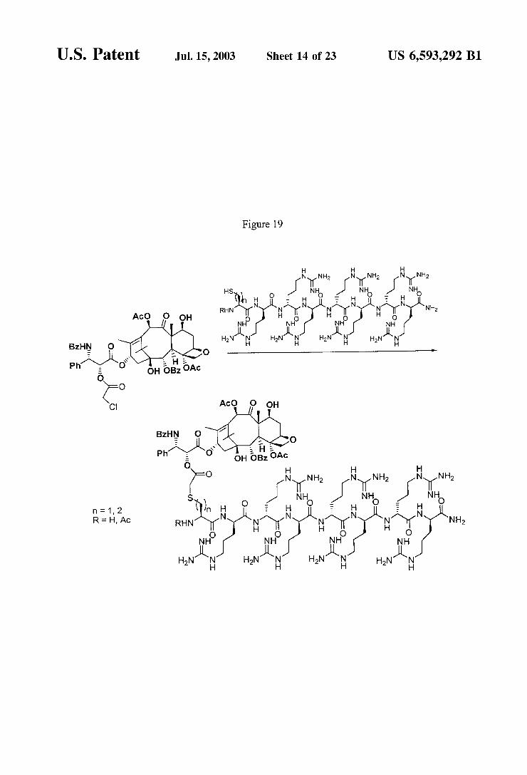
FIG. 18 shows a strategy by which a taxol 2'-chloroacetyl derivative is linked to an arginine heptamer delivery enhancing transporter.
US 6,593,292 B1 7
FIG. 19 shows three additional taxol-r7 conjugates that can be made using the reaction conditions illustrated in FIG. 18.
FIG. 20 shows the results of a 3 day MTT cytotoxicity assay using taxol and two different linkers.
FIG. 21 FACS cellular uptake assay of truncated analogs of Tatos, (Fl-ahx-RKKRRQRRR, SEQID NO:8): Tatlos (Fl-ahx-RKKRRQRR, SEQ ID NO:9), Tatless (F1-ahx RKKRRQR, SEQ ID NO:10), Tatsos, (F1-ahx KKRRQRRR, SEQ ID NO:11), and Tats, (Fl-ahx KRRQRRR, SEQ ID NO:12). Jurkat cells were incubated with varying concentrations (12.5uM shown) of peptited for 15 min at 23° C.
FIG. 22 shows FACS cellular uptake assay of alanine Substituted analogs of Tatos 7: A-49 (Fl-ahx AKKRRQRRR, SEQ ID NO:13), A-50 (F1-ahx RAKRRQRRR, SEQ ID NO:14), A-51 (F1-ahx RKARRQRRR, SEQ ID NO:15), A-52 (F1-ahx RKKARQRRR, SEQ ID NO:16), A-53 (F1-ahx RKKRAQRRR, SEQ ID NO:17), A-54 (F1-ahx RKKRRARRR, SEQ ID NO:18), A-55 (F1-ahx RKKRRQARR, SEQ ID NO:19), A-56 (F1-ahx RKKRRQRAR, SEQ ID NO:20), and A-57 (Fl-ahx RKKRRQRRA; SEQ ID NO:21). Jurkat cells were incubated with varying concentrations (12.5uM shown) of peptides for 12 min at 23° C.
FIG. 23: FACS cellular uptake assay of d- and retro isomers of Tattos:7: d-Tatto-s7 (Fl-ahx-rkkrrqrrr), Tats, to (Fl-ahx-RRRQRRKKR, SEQ ID NO:22), and d-Tats (Fl-ahx-rrrqrrkkr). Jurkat cells were incubated with varying concentrations (12.5uM shown) of peptides for 15 min at 23° C.
FIG. 24 FACS cellular uptake of a series of arginine oligomers and Tattos: R5 (Fl-ahx-RRRRR, SEQ ID NO:23), R6 (Fl-ahx-RRRRRR, SEQ ID NO:24), R7 (Fl ahx-RRRRRRR, SEQ ID NO:25), R8 (Fl-ahx-RRRRRRR; SEQ ID NO:26), R9 (F1-ahx-RRRRRRRRR, SEQ ID NO:27), rS (Fl-ahx-rrrrrr), ré (Fl-ahx-rrrrrr), r7 (Fl-ahx rrrrrrr), r8 (Fl-ahx-rrrrrrrr), r9 (Fl-ahx-rrrrrrrrr). Jurkat cells were incubated with varying concentrations (12.5 uM shown) of peptides for 4 min at 23° C.
FIG. 25: Preparation of guanidine-substituted peptoids. FIG. 26: FACS cellular uptake of polyguanidine peptoids
and d-arginine oligomers. Jurkat cells were incubated with varying concentrations (12.5 uM shown) of peptoids and peptides for 4 min at 23° C.
FIG. 27: FACS cellular uptake of d-arginine oligomers and polyguanidine peptoids. Jurkat cells were incubated with varying concentrations (12.5 uM shown) of fluores cently labeled peptoids and peptides for 4 min at 23° C.
FIG. 28: FACS cellular uptake of and d-arginine oligo merS and N-hxg peptoids. Jurkat cells were incubated with varying concentrations (6.3 uM shown) of fluorescently labeled peptoids and peptides for 4 min at 23° C.
FIG. 29: FACS cellular uptake of d-arginine oligomers and N-chg peptoids. Jurkat cells were incubated with vary ing concentrations (12.5uM shown) of fluorescently labeled peptoids and peptides for 4 min at 23° C.
FIG. 30 shows a general Strategy for attaching a delivery enhancing transporter to a drug that includes a triazole ring Structure.
FIG. 31A and FIG. 31B show synthetic schemes for making conjugates in which FK506 is attached to a delivery enhancing transporter.
DETAILED DESCRIPTION
Definitions
An “epithelial tissue” is the basic tissue that covers Surface areas of the Surface, Spaces, and cavities of the body.
15
25
35
40
45
50
55
60
65
8 Epithelial tissues are composed primarily of epithelial cells that are attached to one another and rest on an extracellular matrix (basement membrane) that is typically produced by the cells. Epithelial tissues include three general types based on cell shape: Squamous, cuboidal, and columnar epithe lium. Squamous epithelium, which lines lungs and blood vessels, is made up of flat cells. Cuboidal epithelium lines kidney tubules and is composed of cube shaped cells, while columnar epithelium cells line the digestive tract and have a columnar appearance. Epithelial tissues can also be classi fied based on the number of cell layers in the tissue. For example, a simple epithelial tissue is composed of a Single layer of cells, each of which sits on the basement membrane. A “stratified” epithelial tissue is composed of several cells Stacked upon one another, not all cells contact the basement membrane. A "pseudoStratified' epithelial tissue has cells that, although all contact the basement membrane, appear to be Stratified because the nuclei are at various levels. The term “trans-epithelial delivery or administration
refers to the delivery or administration of agents by perme ation through one or more layers of a body Surface or tissue, Such as intact skin or a mucous membrane, by topical administration. Thus, the term is intended to include both transdermal (e.g., percutaneous adsorption) and transmu cosal administration. Delivery can be to a deeper layer of the tissue, for example, and/or delivery to the bloodstream.
"Delivery enhancement, “penetration enhancement' or "permeation enhancement” as used herein relates to an increase in amount and/or rate of delivery of a compound that is delivered into and acroSS one or more layers of an epithelial or endothelial tissue. An enhancement of delivery can be observed by measuring the rate and/or amount of the compound that passes through one or more layers of animal or human skin or other tissue. Delivery enhancement also can involve an increase in the depth into the tissue to which the compound is delivered, and/or the extent of delivery to one or more cell types of the epithelial or other tissue (e.g., increased delivery to fibroblasts, immune cells, and endot helial cells of the skin or other tissue). Such measurements are readily obtained by, for example, using a diffusion cell apparatus as described in U.S. Pat. No. 5,891,462. The amount or rate of delivery of an agent acroSS and/or
into Skin or other epithelial or endothelial membrane is Sometimes quantitated in terms of the amount of compound passing through a predetermined area of skin or other tissue, which is a defined area of intact unbroken living skin or mucosal tissue. That area will usually be in the range of about 5 cm to about 100 cm, more usually in the range of about 10 cm to about 100 cm, still more usually in the range of about 20 cm to about 60 cm. The terms "guanidyl, guanidinyl" and “guanidino' are
used interchangeably to refer to a moiety having the formula -HN=C(NH2)NH (unprotonated form). As an example, arginine contains a guanidyl (guanidino) moiety, and is also referred to as 2-amino-5-guanidinovaleric acid or CL-amino Ö-guanidinovaleric acid. "Guanidium” refers to the posi tively charged conjugate acid form. The term “guanidino moiety' includes, for example, guanidine, guanidinium, guanidine derivatives such as (RNHC(NH)NHR'), mono Substituted guanidines, monoguanides, biguanides, bigu anide derivatives such as (RNHC(NH)NHC(NH)NHR'), and the like. In addition, the term "guanidino moiety encom passes any one or more of a guanide alone or a combination of different guanides.
“Amidinyl' and “amidino” refer to a moiety having the formula-C(=NH)(NH). “Amidinium” refers to the posi tively charged conjugate acid form.
US 6,593,292 B1
The term “trans-barrier concentration” or “trans-tissue concentration” refers to the concentration of a compound present on the Side of one or more layers of an epithelial or endothelial barrier tissue that is opposite or “trans' to the Side of the tissue to which a particular composition has been added. For example, when a compound is applied to the skin, the amount of the compound measured Subsequently acroSS one or more layers of the skin is the trans-barrier concentration of the compound.
“Biologically active agent” or “biologically active Sub stance” refers to a chemical Substance, Such as a Small molecule, macromolecule, or metal ion, that causes an observable change in the Structure, function, or composition of a cell upon uptake by the cell. Observable changes include increased or decreased expression of one or more mRNAS, increased or decreased expression of one or more proteins, phosphorylation of a protein or other cell component, inhibition or activation of an enzyme, inhibition or activation of binding between members of a binding pair, an increased or decreased rate of Synthesis of a metabolite, increased or decreased cell proliferation, and the like. The terms “the rapeutic agent”, “the rapeutic
composition', and “therapeutic Substance” refer, without limitation, to any composition that can be used to the benefit of a mammalian Species. Such agents may take the form of ions, Small organic molecules, peptides, proteins or polypeptides, oligonucleotides, and oligosaccharides, for example.
The term "macromolecule' as used herein refers to large molecules (MW greater than 1000 daltons) exemplified by, but not limited to, peptides, proteins, oligonucleotides and polynucleotides of biological or Synthetic origin.
"Small organic molecule” refers to a carbon-containing agent having a molecular weight (MW) of less than or equal to 1000 daltons.
The terms “non-polypeptide agent' and “non-polypeptide therapeutic agent” refer to the portion of a conjugate that does not include the delivery-enhancing transporter, and that is a biologically active agent other than a polypeptide. An example of a non-polypeptide agent is an anti-Sense oligonucleotide, which can be conjugated to a poly-arginine peptide to form a conjugate for enhanced delivery into and acroSS one or more layers of an epithelial or endothelial tissue. A "Subunit,” as used herein, is a monomeric unit that are
joined to form a larger polymeric compound. The Set of amino acids are an example of Subunits. Each amino acid shares a common backbone (-C-C-N-), and the dif ferent amino acids differ in their sidechains. The backbone is repeated in a polypeptide. A Subunit represents the shortest repeating pattern of elements in a polymer backbone. For example, two amino acids of a peptide are not considered a peptide because two amino acids would not have the shortest repeating pattern of elements in the polymer backbone.
The term “polymer” refers to a linear chain of two or more identical or non-identical Subunits joined by covalent bonds. A peptide is an example of a polymer; peptides can be composed of identical or non-identical amino acid Subunits that are joined by peptide linkages (amide bonds).
The term "peptide' as used herein refers to a compound made up of a single chain of D- or L- amino acids or a mixture of D- and L-amino acids joined by peptide bonds. Generally, peptides contain at least two amino acid residues and are less than about 50 amino acids in length. D-amino acids are represented herein by a lower-case one-letter amino acid symbol (e.g., r for D-arginine), whereas L-amino
15
25
35
40
45
50
55
60
65
10 acids are represented by an upper case one-letter amino acid Symbol (e.g., R for L-arginine). Homopolymer peptides are represented by a one-letter amino acid symbol followed by the number of consecutive occurrences of that amino acid in the peptide- (e.g., R7 represents a heptamer that consists of L-arginine residues; SEQ ID NO:3). The term “protein’ as used herein refers to a com-pound
that is composed of linearly arranged amino acids linked by peptide bonds, but in contrast to peptides, has a well-defined conformation. Proteins, as opposed to peptides, generally consist of chains of 50 or more amino acids.
“Polypeptide' as used herein refers to a polymer of at least two amino acid residues and which contains one or more peptide bonds. "Polypeptide' encompasses peptides and proteins, regardless of whether the polypeptide has a well-defined conformation.
Description of the Preferred Embodiments The present invention provides compositions and meth
ods that enhance the transfer of compounds, including drugs and other biologically active compounds, into and acroSS one or more layers of an animal epithelial or endothelial tissue. The methods involve contacting the tissue with a conjugate that includes the compound of interest linked to a delivery-enhancing transporter. The delivery enhancing transporters provided by the invention are molecules that include Sufficient guanidino or amidino moieties to increase delivery of the conjugate into and acroSS one or more intact epithelial and endothelial tissue layers. The methods and compositions are useful for trans-epithelial and trans endothelial delivery of drugs and other biologically active molecules, and also for delivery of imaging and diagnostic molecules. The methods and compositions of the invention are particularly useful for delivery of compounds that require trans-epithelial or trans-endothelial transport to exhibit their biological effects, and that by themselves (without conjugation to a delivery-enhancing transporters or Some other modification), are unable, or only poorly able, to croSS Such tissues and thus exhibit biological activity. The delivery-enhancing transporters and methods of the
invention provide Significant advantages over previously available methods for obtaining trans-epithelial and trans endothelial tissue delivery of compounds of interest. The transporters make possible the delivery of drugs and other agents acroSS tissues that were previously impenetrable to the drug. For example, while delivery of drugs acroSS Skin was previously nearly impossible for all but a few compounds, the methods of the invention can deliver com pounds not only into cells of a first layer of an epithelial tissue Such as Skin, but also acroSS one or more layers of the skin. The blood brain barrier is also resistant to transport of drugs and other diagnostic and therapeutic reagents, the methods and transporters of the invention provide means to Such transport. The delivery-enhancing transporers increase delivery of
the conjugate into and acroSS one or more intact epithelial or endothelial tissue layers compared to delivery of the com pound in the absence of the delivery-enhancing transporter. The delivery-enhancing transporters can, in Some embodiments, increase delivery of the conjugate signifi cantly over that obtained using the tat protein of HIV-1 (Frankel et al. (1991) PCT Pub. No. WO 91/09958). Deliv ery is also increased significantly over the use of Shorter fragments of the tat protein containing the tat basic region (residues 49-57 having the sequence RKKRRQRRR, SEQ ID NO:28) (Barsoum et al. (1994) WO94/04686 and Fawell
US 6,593,292 B1 11
et al. (1994) Proc. Natl. Acad. Sci. USA 91: 664-668). Preferably, delivery obtained using the transporters of the invention is increased more than 2-fold, Still more preferably six-fold, still more preferably ten-fold, and still more pref erably twenty-fold, over that obtained with tat residues 49-57.
Similarly, the delivery-enhancing transporters of the invention can provide increased delivery compared to a 16 amino acid peptide-cholesterol conjugate derived from the Antennapedia homeodomain that is rapidly internalized by cultured neurons (Brugidou et al. (1995) Biochem. BiophyS. Res. Commun. 214: 685-93). This region, residues 43-58 at minimum, has the amino acid sequence ROIKIWFONR RMKWKK (SEQ ID NO:29). The Herpes simplex protein VP22, like tat and the Antennapedia domain, was previously known to enhance transport into cells, but was not known to enhance transport into and acroSS endothelial and epithelial membranes (Elliot and O'Hare (1997) Cell 88: 223–33; Dilber et al. (1999) Gene Ther. 6: 12–21; Phelanet al. (1998) Nature Biotechnol. 16: 440-3). In presently preferred embodiments, the delivery-enhancing transporters provide Significantly increased delivery compared to the Antenna pedia homeodomain and to the VP22 protein.
Structure of Delivery-Enhancing Transporters The delivery-enhancing transporters of the invention are
molecules that have Sufficient guanidino and/or amidino moieties to increase delivery of a compound to which the delivery-enhancing transporter is attached into and acroSS one or more layers of an epithelial tissue (e.g., skin or mucous membrane) or an endothelial tissue (e.g., the blood brain barrier). The delivery-enhancing transporters generally include a backbone Structure to which is attached the guani dino and/or amidino Sidechain moieties. In Some embodiments, the backbone is a polymer that consists of Subunits (e.g., repeating monomer units), at least Some of which Subunits contain a guanidino or amidino moiety. A. Guanidino and/or Amidino Moieties
The delivery-enhancing transporters typically display at least 5 guanidino and/or amidino moieties, and more pref erably 7 or more such moieties. Preferably, the delivery enhancing transporters have 25 or fewer guanidino and/or amidino moieties, and often have 15 or fewer of Such moieties. In Some embodiments, the delivery-enhancing transporter consists essentially of 50 or fewer Subunits, and can consist essentially of 25 or fewer, 20 or fewer, or 15 or fewer Subunits. The delivery-enhancing transporter can be as short as 5 Subunits, in which case all Subunits include a guanidino or amidino Sidechain moiety. The delivery enhancing transporters can have, for example, at least 6 Subunits, and in Some embodiments have at least 7 or 10 subunits. Generally, at least 50% of the subunits contain a guanidino or amidino Sidechain moiety. More preferably, at least 70% of the Subunits, and sometimes at least 90% of the Subunits in the delivery-enhancing transporter contain a guanidino or amidino Sidechain moiety. Some or all of the guanidino and/or amidino moieties in
the delivery-enhancing transporters can be contiguous. For example, the delivery-enhancing transporters can include from 6 to 25 contiguous guanidino and/or amidino containing Subunits. Seven or more contiguous guanidino and/or amidino-containing Subunits are present in Some embodiments. In Some embodiments, each Subunit that contains a guanidino moiety is contiguous, as exemplified by a polymer containing at least Six contiguous arginine residues.
The delivery-enhancing transporters are exemplified by peptides. Arginine residues or analogs of arginine can con
15
25
35
40
45
50
55
60
65
12 Stitute the Subunits that have a guanidino moiety. Such an arginine-containing peptide can be composed of either all D-, all L- or mixed D- and L-amino acids, and can include additional amino acids, amino acid analogs, or other mol ecules between the arginine residues. Optionally, the delivery-enhancing transporter can also include a non arginine residue to which a compound to be delivered is attached, either directly or through a linker. The use of at least one D-arginine in the delivery-enhancing transporters can enhance biological Stability of the transporter during transit of the conjugate to its biological target. In Some cases the delivery-enhancing transporters are at least about 50% D-arginine residues, and for even greater Stability transport erS in which all of the Subunits are D-arginine residues are used. If the delivery enhancing transporter molecule is a peptide, the transporter is not attached to an amino acid Sequence to which the amino acids that make up the delivery enhancing transporter molecule are attached in a naturally occurring protein.
Preferably, the delivery-enhancing transporter is linear. In a preferred embodiment, an agent to be delivered into and acroSS one or more layers of an epithelial tissue is attached to a terminal end of the delivery-enhancing transporter. In Some embodiments, the agent is linked to a single transport polymer to form a conjugate. In other embodiments, the conjugate can include more than one delivery-enhancing transporter linked to an agent, or multiple agents linked to a Single delivery-enhancing transporter. More generally, it is preferred that each Subunit contains
a highly basic Sidechain moiety which (i) has a pKa of greater than 11, more preferably 12.5 or greater, and (ii) contains, in its protonated State, at least two geminal amino groups (NH) which share a resonance-stabilized positive charge, which gives the moiety a bidentate character. The guanidino or amidino moieties extend away from the
backbone by virtue of being linked to the backbone by a Sidechain linker. The Sidechain atoms are preferably pro Vided as methylene carbon atoms, although one or more other atoms Such as Oxygen, Sulfur or nitrogen can also be present. For example, a linker that attaches a guanidino moiety to a backbone can be shown as:
HN-CH-C-OH O
(CH2)
NH
CFNH
(CH2)
NH
CENH
NH2
In these formulae, n is preferably at least 2, and is preferably between 2 and 7. In Some embodiments, n is 3 (arginine for Structure 1). In other embodiments, n is between 4 and 6; most preferably n is 5 or 6. Although the sidechain in the exemplified formulae is shown as being attached to a peptide
US 6,593,292 B1 13
backbone (i.e., a repeating amide to which the Sidechain is attached to the carbon atom that is cc to the carbonyl group, Subunit 1) and a peptoid backbone (i.e., a repeating amide to which the Sidechain is attached to the nitrogen atom that is f to the carbonyl group, Subunit 2), other non-peptide backbones are also Suitable, as discussed in more detail herein. Thus, Similar Sidechain linkers can be attached to nonpeptide backbones (e.g., peptoid backbones).
In Some embodiments, the delivery-enhancing transport erS are composed of linked Subunits, at least Some of which include a guanidino and/or amidino moiety. Examples of Suitable Subunits having guanidino and/or amidino moieties are described below. Amino acids. In Some embodiments, the delivery
enhancing transporters are composed of D or Lamino acid residues. The amino acids can be naturally occurring or non-naturally occurring amino acids. Arginine (C-amino-8- guanidinovaleric acid) and C.-amino-e-amidino-hexanoic acid (isosteric amidino analog) are examples of Suitable guanidino- and amidino-containing amino acid Subunits. The guanidinium group in arginine has a pKa of about 12.5. In Some preferred embodiments the transporters are com prised of at least Six contiguous arginine residues.
Other amino acids, Such as C.-amino-B-guanidino propionic acid, C.-amino-y-guanidino-butyric acid, or C.-amino-e-guanidino-caproic acid (containing 2, 3 or 5 Sidechain linker atoms, respectively, between the backbone chain and the central guanidinium carbon) can also be used.
D-amino acids can also be used in the delivery enhancing transporters. Compositions containing exclusively D-amino acids have the advantage of decreased enzymatic degrada tion. However, they can also remain largely intact within the target cell. Such stability is generally not problematic if the agent is biologically active when the polymer is still attached. For agents that are inactive in conjugate form, a linker that is cleavable at the site of action (e.g., by enzyme or Solvent-mediated cleavage within a cell) should be included within the conjugate to promote release of the agent in cells or organelles.
Other Subunits. Subunits other than amino acids can also be selected for use in forming transport polymers. Such Subunits can include, but are not limited to, hydroxy amino acids, N-methyl-amino acids amino aldehydes, and the like, which result in polymers with reduced peptide bonds. Other Subunit types can be used, depending on the nature of the Selected backbone, as discussed in the next Section. B. Backbones The guanidino and/or amidino moieties that are included
in the delivery-enhancing transporters are generally attached to a linear backbone. The backbone can comprise a variety of atom types, including carbon, nitrogen, oxygen, Sulfur and phosphorus, with the majority of the backbone chain atoms typically consisting of carbon. A plurality of Sidechain moieties that include a terminal guanidino or amidino group are attached to the backbone. Although spacing between adjacent Sidechain moieties is typically consistent, the delivery-enhancing transporters used in the invention can also include variable spacing between Sidechain moieties along the backbone. A more detailed backbone list includes N-Substituted
14 tetrazole (CN), retrothioamide (NHCS), retroreduced (NHCH), Sulfonamido (SONH), methylenesulfonamido (CHRSONH), retrosulfonamide (NHSO), and peptoids (N-substituted amides), and backbones with malonate and/or gem-diamino-alkyl Subunits, for example, as reviewed by Fletcher et al. (1998) Chem. Rev. 98:763) and detailed by references cited therein. Many of the foregoing Substitutions result in approximately isosteric polymer backbones relative to backbones formed from C-amino acids. AS mentioned above, in a peptoid backbone, the Sidechain
is attached to the backbone nitrogen atoms rather than the carbon atoms. (See e.g., Kessler (1993) Angew. Chem. Int. Ed. Engl. 32:543; Zuckerman et al. (1992) Chemtracts Macromol. Chem. 4:80; and Simon et al. (1992) Proc. Natl. Acad. Sci. USA 89.9367.) An example of a suitable peptoid backbone is poly-(N-Substituted)glycine (poly-NSG). Syn thesis of peptoids is described in, for example, U.S. Pat. No. 5,877,278. As the term is used herein, transporters that have a peptoid backbone are considered "non-peptide' transporters, because the transporters are not composed of amino acids having naturally occurring Sidechain locations. Non-peptoid backbones, including peptoid backbones, pro vide enhanced biological Stability (for example, resistance to enzymatic degradation in vivo). C. Synthesis of Delivery-enhancing Transporters
Delivery-enhancing transporters are constructed by any method known in the art. Exemplary peptide polymers can be produced Synthetically, preferably using a peptide Syn thesizer (e.g., an Applied Biosystems Model 433) or can be synthesized recombinantly by methods well known in the art. Recombinant Synthesis is generally used when the delivery enhancing transporter is a peptide which is fused to a polypeptide or protein of interest.
N-methyl and hydroxy-amino acids can be substituted for conventional amino acids in Solid phase peptide Synthesis. However, production of delivery-enhancing transporters with reduced peptide bonds requires Synthesis of the dimer of amino acids containing the reduced peptide bond. Such dimers are incorporated into polymers using Standard Solid phase Synthesis procedures. Other Synthesis procedures are well known and can be found, for example, in Fletcher et al. (1998) Chem. Rev. 98.763, Simon et al. (1992) Proc. Natl. Acad. Sci. USA 89:9367, and references cited therein.
The delivery-enhancing transporters of the invention can be flanked by one or more non-guanidino/non-amidino Subunits (Such as glycine, alanine, and cysteine, for example), or a linker (Such as an aminocaproic acid group), that do not significantly affect the rate of trans-tissue layer transport of the corresponding delivery-enhancing transporter-containing conjugates. Also, any free amino ter minal group can be capped with a blocking group, Such as an acetyl or benzyl group, to prevent ubiquitination in Vivo. Where the transporter is a peptoid polymer, one Synthetic
method involves the following Steps: 1) a peptoid polyamine is treated with a base and pyrazole-1-carboxamidine to provide a mixture; 2) the mixture is heated and then allowed to cool; 3) the cooled mixture is acidified; and 4) the acidified mixture is purified. Preferably the base used in step 1 is a carbonate, Such as Sodium carbonate, and heating Step 2 involves heating the mixture to approximately 50° C. for between about 24 hours and about 48 hours. The purification Step preferably involves chromatography (e.g., reverse phase HPLC). D. Attachment of Transport Polymers To Biologically Active Agents The agent to be transported can be linked to the delivery
enhancing transporter according to a number of embodi
US 6,593,292 B1 15
ments. In one embodiment, the agent is linked to a single delivery-enhancing transporter, either via linkage to a ter minal end of the delivery-enhancing transporter or to an internal Subunit within the reagent via a Suitable linking grOup.
In a Second embodiment, the agent is attached to more than one delivery-enhancing transporter, in the same manner as above. This embodiment is Somewhat leSS preferred, Since it can lead to crosslinking of adjacent cells.
In a third embodiment, the conjugate contains two agent moieties attached to each terminal end of the delivery enhancing transporter. For this embodiment, it is presently preferred that the agent has a molecular weight of less than 10 kDa. With regard to the first and third embodiments just
mentioned, the agent is generally not attached to one any of the guanidino or amidino Sidechains So that they are free to interact with the target membrane.
The conjugates of the invention can be prepared by Straightforward Synthetic Schemes. Furthermore, the conju gate products are usually Substantially homogeneous in length and composition, So that they provide greater con Sistency and reproducibility in their effects than heteroge neous mixtures.
According to an important aspect of the present invention, it has been found by the applicants that attachment of a Single delivery-enhancing transporter to any of a variety of types of biologically active agents is Sufficient to Substan tially enhance the rate of uptake of an agent into and acroSS one or more layers of epithelial and endothelial tissues, even without requiring the presence of a large hydrophobic moi ety in the conjugate. In fact, attaching a large hydrophobic moiety can Significantly impede or prevent croSS-layer trans port due to adhesion of the hydrophobic moiety to the lipid bilayer of cells that make up the epithelial or endothelial tissue. Accordingly, the present invention includes conju gates that do not contain Substantially hydrophobic moieties, Such as lipid and fatty acid molecules.
Delivery-enhancing transporters of the invention can be attached covalently to biologically active agents by chemical or recombinant methods.
1. Chemical Linkages Biologically active agents Such as Small organic mol
ecules and macromolecules can be linked to delivery enhancing transporters of the invention via a number of methods known in the art (see, for example, Wong, S. S., Ed., Chemistry of Protein Conjugation and Cross-Linking, CRC Press, Inc., Boca Raton, Fla. (1991), either directly (e.g., with a carbodiimide) or via a linking moiety. In particular, carbamate, ester, thioether, disulfide, and hydra Zone linkages are generally easy to form and Suitable for most applications. Ester and disulfide linkages are preferred if the linkage is to be readily degraded in the cytosol, after transport of the Substance across the cell membrane.
Various functional groups (hydroxyl, amino, halogen, etc.) can be used to attach the biologically active agent to the transport polymer. Groups which are not known to be part of an active site of the biologically active agent are preferred, particularly if the polypeptide or any portion thereof is to remain attached to the Substance after delivery.
Polymers, such as peptides produced as described in PCT application US98/10571 (Publication No. WO 9852614), are generally produced with an amino terminal protecting group, Such as FMOC. For biologically active agents which can Survive the conditions used to cleave the polypeptide from the Synthesis resin and deprotect the Sidechains, the FMOC may be cleaved from the N-terminus of the com
15
25
35
40
45
50
55
60
65
16 pleted resin-bound polypeptide So that the agent can be linked to the free N-terminal amine. In Such cases, the agent to be attached is typically activated by methods well known in the art to produce an active ester or active carbonate moiety effective to form an amide or carbamate linkage, respectively, with the polymeramino group. Of course, other linking chemistries can also be used. To help minimize side-reactions, guanidino and amidino
moieties can be blocked using conventional protecting groups, Such as carbobenzyloxy groups (CBZ), di-t-BOC, PMC, Pbf, N-NO, and the like.
Coupling reactions are performed by known coupling methods in any of an array of Solvents, Such as N,N- dimethyl formamide (DMF), N-methyl pyrrolidinone, dichloromethane, water, and the like. Exemplary coupling reagents include, for example, O-benzotriazolyloxy tetram ethyluronium hexafluorophosphate (HATU), dicyclohexyl carbodiimide, bromo-tris(pyrrolidino) phosphonium bro mide (PyBroP), etc. Other reagents can be included, such as N,N-dimethylamino pyridine (DMAP), 4-pyrrollidino pyridine, N-hydroxy Succinimide, N-hydroxybenzotriazole, and the like.
2. Fusion Polypeptides Delivery-enhancing transporters of the invention can be
attached to biologically active polypeptide agents by recom binant means by constructing vectors for fusion proteins comprising the polypeptide of interest and the delivery enhancing transporter, according to methods well known in the art. Generally, the delivery-enhancing transporter com ponent will be attached at the C-terminus or N-terminus of the polypeptide of interest, optionally via a short peptide linker.
3. Releasable Linkers The biologically active agents are, in presently preferred
embodiments, attached to the delivery-enhancing trans porter using a linkage that is specifically cleavable or releasable. The use of Such linkages is particularly important for biologically active agents that are inactive until the attached delivery-enhancing transporter is released. In Some cases, Such conjugates that consist of a drug molecule that is attached to a delivery-enhancing transporter can be referred to as prodrugs, in that the release of the delivery enhancing transporter from the drug results in conversion of the drug from an inactive to an active form. AS used herein, “cleaved' or “cleavage' of a conjugate or linker refers to release of a biological agent from a transporter molecule, thereby releasing an active biological agent. "Specifically cleavable” or “specifically releasable” refers to the linkage between the transporter and the agent being cleaved, rather than the transporter being degraded (e.g., by proteolytic degradation).
In Some embodiments, the linkage is a readily cleavable linkage, meaning that it is Susceptible to cleavage under conditions found in Vivo. Thus, upon passing into and through one or more layers of an epithelial and/or endothe lial tissue, the agent is released from the delivery-enhancing transporter. Readily cleavable linkages can be, for example, linkages that are cleaved by an enzyme having a specific activity (e.g., an esterase, protease, phosphatase, peptidase, and the like) or by hydrolysis. For this purpose, linkers containing carboxylic acid esters and disulfide bonds are Sometimes preferred, where the former groups are hydro lyzed enzymatically or chemically, and the latter are Severed by disulfide exchange, e.g., in the presence of glutathione. The linkage can be Selected So it is cleavable by an enzy matic activity that is known to be present in one or more layers of an epithelial or endothelial tissue. For example, the
US 6,593,292 B1 17
Stratum granulosum of skin has a relatively high concentra tion of N-peptidase activity. A specifically cleavable linker can be engineered onto a
transporter molecule. For example, amino acids that consti tute a protease recognition Site, or other Such specifically recognized enzymatic cleavage Site, can be used to link the transporter to the agent. Alternatively, chemical or other types of linkers that are cleavable by, for example, exposure to light or other Stimulus can be used to link the transporter to the agent of interest. A conjugate in which an agent to be delivered and a
delivery-enhancing transporter are linked by a specifically cleavable or specifically releasable linker will have a half life. The term “half-life” in this context refers to the amount of time required after applying the conjugate to an epithelial or endothelial membrane for one half of the amount of conjugate to become dissociated to release the free agent. The half-life for some embodiments is typically between 5 minutes and 24 hours, and more preferably is between 30 minutes and 2 hours. The half-life of a conjugate can be “tuned' or modified, according to the invention, as described below.
In Some embodiments, the cleavage rate of the linkerS is pH dependent. For example, a linker can form a stable linkage between an agent and a delivery-enhancing trans porter at an acidic pH (e.g., pH 6.5 or less, more preferably about 6 or less, and still more preferably about 5.5 or less). However, when the conjugate is placed at physiological t pH (e.g., pH 7 or greater, preferably about pH 7.4), the linker will undergo cleavage to release the agent. Such pH Sensi tivity can be obtained by, for example, including a functional group that, when protonated (i.e., at an acidic pH), does not act as a nucleophile. At a higher (e.g., physiological) pH, the functional group is no longer protonated and thus can act as a nucleophile. Examples of Suitable functional groups include, for example, N and S. One can use Such functional groups to fine-tune the pH at which Self-cleavage occurs.
The cleavable linker can be self-immolating. Such linkers contain a nucleophile (e.g., oxygen, nitrogen or Sulfur) distal to the agent and a cleavable group (e.g., ester, carbonate, carbamate, thiocarbamate) proximal to the agent. Intramo lecular attack of the nucleophile on the cleavable group either directly or indirectly releases the agent. In general, the nucleophile is 5 to 6 atoms distal from the cleaved group, thereby forming a 5-6 member ring as a product of immo lation.
Such linkers include those having a structure 3, 4, or 5, as follows:
3
R-X-C-(CH)-Y- f (CH) -NH-(CH2) -R O O
4
i R-X-C-(CH) R-(CH2). CH- i-R
O 5
O Rs
R-X- C-O- (CH2) CH- i-R O
15
25
35
40
45
50
55
60
65
18 wherein:
R-X comprises the agent to be delivered; X is a functional group on the agent, to which functional
group the linker is attached; Y is N or C; R is hydrogen, alkyl, aryl, acyl, or allyl, R comprises the delivery-enhancing transporter; R is substituted or unsubstituted S, O, N or C; R is OH, SH or NHR; R is hydrogen, alkyl, aryl, acyl or allyl, k and m are each independently Selected from 1 and 2; and n is 1 to 10. The agent to be delivered (e.g., a drug or diagnostic agent)
generally includes a functional group (designated as X in the formulae above) by which the linker is attached to the delivery-enhancing transporter. Examples of Suitable func tional groups for X include, for example, N, O, S, and CRRs, wherein R, and Rs are each independently Selected from the group consisting of H and alkyl. If X is O, for example, release of the agent from the delivery-enhancing transporter and linker will yield the agent in its free alcohol form; if X is N, the free amine will result. Similarly, if X is S, release of the linker will yield the agent in the thiol form.
For a linker having the Structure 3, one can adjust the half-life of a conjugate that includes the linker by the choice of the R. Substituent. By using an R of increased or decreased size, one can obtain a conjugate that has a longer or shorter half-life, respectively. R is preferably methyl, ethyl, propyl, butyl, allyl, benzyl or phenyl. Similar modu lation of half-life can be accomplished in an analogous manner with other linkers (e.g., Rs on structure 4).
Structure 3 also provides an example of how the linker Structure can affect the pH Sensitivity of a conjugate. The backbone amino group is protonated at acidic pH (e.g., pH 5.5), and thus is stable in that the amine does not function as a strong nucleophile. Upon raising the pH to physiological pH (e.g., pH 7.4), however, the amine is no longer proto nated and thus can act as a nucleophile. The resulting nucleophilic attack by the amine upon the carbonyl adjacent to the agent of interest then results in release of the agent from the linker and delivery-enhancing transporter. Again, this rationale also applies to other linker Structures. In an example of a preferred linker of Structure 3, R is benzyl, k, m, and n are each 1, and X is O. An example of a Suitable linker of Structure 4 has Rs as
NHR, R selected from hydrogen, methyl, allyl, butyl or phenyl; and k and m each being 1.
Another example of a Self-immolating linker is repre Sented by Structure 5 as shown above. In presently preferred conjugates that include linkers of this structure, R is S and Rs is NHR, wherein R is hydrogen, methyl, allyl, butyl or phenyl; and k is 2.
For example, a presently preferred conjugate comprises the Structure:
Ph
O CH2
R-O-C-CH-N-C-CH-NH-CH-C-Rs:
O O
wherein Ph is phenyl. The invention also provides linkers in which cleavage
occurs in two stages. The first stage of cleavage is rate
US 6,593,292 B1 19
limiting and can also be fine-tuned for pH Sensitivity and half-life. Once this initial rearrangement has occurred, the Second Stage of the intramolecular reaction occurs relatively quickly. An example of a conjugate having this type of linker is represented as Structure 6:
O O R5
R-X-C-OCH-Ar-O-C-(CH) R-(CH2) CH--R, O
wherein:
R-X is the agent to be delivered; X is a functional group on the agent, to which functional
group the linker is attached; Ar is an aryl group having the attached radicals arranged
in an Ortho or para configuration, which aryl group can be substituted or unsubstituted;
R is the delivery-enhancing transporter; R is substituted or unsubstituted S, O, N or C; R is OH, SH or NHR; R is hydrogen, alkyl, aryl, acyl or allyl, and k and m are each independently Selected from 1 and 2. Examples of preferred conjugates of Structure 6 include
those in which R is S, R is NHR, and R is hydrogen, methyl, allyl, butyl or phenyl. For example, a Suitable conjugate of Structure 6 is:
O O NH2
R-O-C-OCH-Ar-O-C-CH-S-CH-CH-C-Rs.
O
The Self-immolating linkers typically undergo intramo lecular cleavage with a half-life between about 10 minutes and about 24 hours in water at a pH of approximately 7.4. Preferably, the cleavage half-life is between about 20 min utes and about 4 hours in water at a pH of approximately 7.4. More preferably, the cleavage half-life is between about 30 minutes and about 2 hours in water at a pH of approximately 74.
In another preferred embodiment, the cleavable linker contains a first cleavable group that is distal to the agent, and a Second cleavable group that is proximal to the agent, Such that cleavage of the first cleavable group yields a linker agent conjugate containing a nucleophilic moiety capable of reacting intramolecularly to cleave the Second cleavable group, thereby releasing the agent from the linker and polymer. This embodiment is illustrated by various small molecule conjugates discussed below and in PCT applica tion US98/10571 (Publication No. WO9852614).
In one approach, the conjugate can include a disulfide linkage, as illustrated in FIG. 5A (see also, PCT application US98/10571 (Publication No. WO9852614)), which shows a conjugate (I) containing a transport polymer T which is linked to a cytotoxic agent, 6-mercaptopurine, by an N-acetyl-protected cysteine group which Serves as a linker. Thus, the cytotoxic agent is attached by a disulfide bond to the 6-mercapto group, and the transport polymer is bound to the cysteine carbonyl moiety via an amide linkage. Cleavage of the disulfide bond by reduction or disulfide exchange results in release of the free cytotoxic agent. A method for Synthesizing a disulfide-containing conju
gate is provided in Example 9A of PCT application US98/
15
25
35
40
45
50
55
60
65
20 10571. The product contains a heptamer of Arg residues which is linked to 6-mercaptopurine by an N-acetyl-CyS Ala-Ala linker, where the Ala residues are included as an additional Spacer to render the disulfide more accessible to thiols and reducing agents for cleavage within a cell. The linker in this example also illustrates the use of amide bonds, which can be cleaved enzymatically within a cell.
In another approach, the conjugate includes a photocleav able linker which is cleaved upon eXposure to electromag netic radiation. An exemplary linkage is illustrated in FIG. 5B, which shows a conjugate (II) containing a transport polymer T which is linked to 6-mercaptopurine via a meta nitrobenzoate linking moiety. Polymer T is linked to the nitrobenzoate moiety by an amide linkage to the benzoate carbonyl group, and the cytotoxic agent is bound via its 6-mercapto group to the p-methylene group. The compound can be formed by reacting 6-mercaptopurine with p-bromomethyl-m-nitrobenzoic acid in the presence of NaOCH/methanol with heating, followed by coupling of the benzoate carboxylic acid to a transport polymer, Such as the amino group of a Y-aminobutyric acid linker attached to the polymer (see e.g., Example 9B of PCT application US98/10571). Photo-illumination of the conjugate causes release of the 6-mercaptopurine by virtue of the nitro group that is ortho to the mercaptomethyl moiety. This approach finds utility in phototherapy methods as are known in the art, particularly for localizing drug activation to a Selected area of the body.
Preferably, the cleavable linker contains first and second cleavable groups that can cooperate to cleave the polymer from the biologically active agent, as illustrated by the following approaches. That is, the cleavable linker contains a first cleavable group that is distal to the agent, and a Second cleavable group that is proximal to the agent, Such that cleavage of the first cleavable group yields a linker-agent conjugate containing a nucleophilic moiety capable of react ing intramolecularly to cleave the Second cleavable group, thereby releasing the agent from the linker and polymer.
FIG. 5C shows a conjugate (III) containing a transport polymer T linked to the anticancer agent, 5-fluorouracil (5FU). Here, the linkage is provided by a modified lysyl residue. The transport polymer is linked to the C-amino group, and the 5-fluorouracil is linked via the C-carbonyl. The lysyl e-amino group has been modified to a carbamate ester of o-hydroxymethyl nitrobenzene, which comprises a first, photolabile cleavable group in the conjugate. Photo illumination severs the nitrobenzene moiety from the conjugate, leaving a carbamate that also rapidly decomposes to give the free C-amino group, an effective nucleophile. Intramolecular reaction of the e-amino group with the amide linkage to the 5-fluorouracil group leads to cyclization with release of the 5-fluorouracil group. FIG.5D illustrates a conjugate (IV) containing a delivery
enhancing transporter T linked to 2'-oxygen of the antican cer agent, paclitaxel. The linkage is provided by a linking moiety that includes (i) a nitrogen atom attached to the delivery-enhancing transporter, (ii) a phosphate monoester located para to the nitrogen atom, and (iii) a carboxymethyl group meta to the nitrogen atom, which is joined to the 2'-OXygen of paclitaxel by a carboxylate ester linkage. Enzymatic cleavage of the phosphate group from the con jugate affords a free phenol hydroxyl group. This nucleo philic group then reacts intramolecularly with the carboxy late ester to release free paclitaxel, fully capable of binding to its biological target. Example 9C of PCT application US98/10571 describes a synthetic protocol for preparing this type of conjugate.
US 6,593,292 B1 21
FIG. 5E illustrates yet another approach wherein a delivery-enhancing transporter is linked to a biologically active agent, e.g., paclitaxel, by an aminoalkyl carboxylic acid. Preferably, the linker amino group is linked to the linker carboxyl carbon by from 3 to 5 chain atoms (n=3 to 5), preferably either 3 or 4 chain atoms, which are preferably provided as methylene carbons. As seen in FIG. 5E, the linker amino group is joined to the delivery-enhancing transporter by an amide linkage, and is joined to the pacli taxel moiety by an ester linkage. Enzymatic cleavage of the amide linkage releases the delivery-enhancing transporter and produces a free nucleophilic amino group. The free amino group can then react intramolecularly with the ester group to release the linker from the paclitaxel.
In another approach, the conjugate includes a linker that is labile at one pH but is stable at another pH. For example, FIG. 6 illustrates a method of Synthesizing a conjugate with a linker that is cleaved at physiological pH but is stable at acidic pH. Preferably, the linker is cleaved in water at a pH of from about 6.6 to about 7.6. Preferably the linker is stable in water at a pH from about 4.5 to about 6.5.
Uses of Delivery-enhancing Transporters The delivery-enhancing transporters find use in
therapeutic, prophylactic and diagnostic applications. The delivery-enhancing transporters can carry a diagnostic or biologically active reagent into and acroSS one or more layers of skin or other epithelial tissue (e.g., gastrointestinal, lung, and the like), or across endothelial tissues Such as the blood brain barrier. This property makes the reagents useful for treating conditions by delivering agents that must pen etrate acroSS one or more tissue layers in order to exert their biological effect.
Compositions and methods of the present invention have particular utility in the area of human and Veterinary thera peutics. Generally, administered dosages will be effective to deliver picomolar to micromolar concentrations of the thera peutic composition to the effector Site. Appropriate dosages and concentrations will depend on factorS Such as the therapeutic composition or drug, the site of intended delivery, and the route of administration, all of which can be derived empirically according to methods well known in the art. Further guidance can be obtained from Studies using experimental animal models for evaluating dosage, as are known in the art.
Administration of the compounds of the invention with a Suitable pharmaceutical excipient as necessary can be car ried out via any of the accepted modes of administration. Thus, administration can be, for example, intravenous, topical, Subcutaneous, transcutaneous, intramuscular, oral, intra-joint, parenteral, peritoneal, intranasal, or by inhala tion. Suitable sites of administration thus include, but are not limited to, Skin, bronchial, gastrointestinal, anal, vaginal, eye, and ear. The formulations may take the form of Solid, Semi-Solid, lyophilized powder, or liquid dosage forms, Such as, for example, tablets, pills, capsules, powders, Solutions, Suspensions, emulsions, Suppositories, retention enemas, creams, ointments, lotions, aerosols or the like, preferably in unit dosage forms Suitable for Simple administration of precise dosages.
The compositions typically include a conventional phar maceutical carrier or excipient and may additionally include other medicinal agents, carriers, adjuvants, and the like. Preferably, the composition will be about 5% to 75% by weight of a compound or compounds of the invention, with the remainder consisting of Suitable pharmaceutical excipi
15
25
35
40
45
50
55
60
65
22 ents. Appropriate excipients can be tailored to the particular composition and route of administration by methods well known in the art, e.g., Remington's Pharmaceutical Sciences, 18TH ED., Mack Publishing Co., Easton, Pa. (1990).
For oral administration, Such excipients include pharma ceutical grades of mannitol, lactose, Starch, magnesium Stearate, Sodium Saccharine, talcum, cellulose, glucose, gelatin, Sucrose, magnesium carbonate, and the like. The composition may take the form of a Solution, Suspension, tablet, pill, capsule, powder, Sustained-release formulation, and the like.
In Some embodiments, the pharmaceutical compositions take the form of a pill, tablet or capsule, and thus, the composition can contain, along with the biologically active conjugate, any of the following: a diluent Such as lactose, Sucrose, dicalcium phosphate, and the like; a disintegrant Such as Starch or derivatives thereof; a lubricant Such as magnesium Stearate and the like; and a binder Such a Starch, gum acacia, polyvinylpyrrollidone, gelatin, cellulose and derivatives thereof. The active compounds of the formulas may be formulated
into a Suppository comprising, for example, about 0.5% to about 50% of a compound of the invention, disposed in a polyethylene glycol (PEG) carrier (e.g., PEG 1000 96% and PEG 4000 (4%).
Liquid compositions can be prepared by dissolving or dispersing compound (about 0.5% to about 20%), and optional pharmaceutical adjuvants in a carrier, Such as, for example, aqueous Saline (e.g., 0.9% w/v Sodium chloride), aqueous dextrose, glycerol, ethanol and the like, to form a Solution or Suspension, e.g., for intravenous administration. The active compounds may also be formulated into a retention enema.
If desired, the composition to be administered may also contain minor amounts of non-toxic auxiliary Substances Such as wetting or emulsifying agents, pH buffering agents, Such as, for example, Sodium acetate, Sorbitan monolaurate, or triethanolamine oleate.
For topical administration, the composition is adminis tered in any Suitable format, Such as a lotion or a transdermal patch. For delivery by inhalation, the composition can be delivered as a dry powder (e.g., Inhale Therapeutics) or in liquid form via a nebulizer. Methods for preparing Such dosage forms are known or
will be apparent to those skilled in the art; for example, See Remington's Pharmaceutical Sciences, Supra., and Similar publications. The composition to be administered will, in any event, contain a quantity of the pro-drug and/or active compound(s) in a pharmaceutically effective amount for relief of the condition being treated when administered in accordance with the teachings of this invention.
Generally, the compounds of the invention are adminis tered in a therapeutically effective amount, i.e., a dosage Sufficient to effect treatment, which will vary depending on the individual and condition being treated. Typically, a therapeutically effective daily dose is from 0.1 to 100 mg/kg of body weight per day of drug. Most conditions respond to administration of a total dosage of between about 1 and about 30 mg/kg of body weight per day, or between about 70 mg and 2100 mg per day for a 70 kg perSon.
Stability of the conjugate can be further controlled by the composition and Stereochemistry of the backbone and Sidechains of the delivery-enhancing transporters. For polypeptide delivery-enhancing transporters, D-isomers are generally resistant to endogenous proteases, and therefore
US 6,593,292 B1 23
have longer half-lives in Serum and within cells. D-polypeptide polymers are therefore appropriate when longer duration of action is desired. L-polypeptide polymers have shorter half-lives due to their susceptibility to proteases, and are therefore chosen to impart shorter acting effects. This allows side-effects to be averted more readily by withdrawing therapy as Soon as Side-effects are observed. Polypeptides comprising mixtures of D and L-residues have intermediate Stabilities. Homo-D-polymers are generally preferred. A. Application to Skin
The delivery-enhancing transporters of the invention make possible the delivery of biologically active and diag nostic agents acroSS the Skin. Surprisingly, the transporters can deliver an agent acroSS the Stratum comeum, which previously had been a nearly impenetrable barrier to drug delivery. The Stratum corneum, the outermost layer of the skin, is composed of Several layers of dead, keratin-filled skin cells that are tightly bound together by a “glue” composed of cholesterol and fatty acids. Once the agents are delivered through the Stratum corneum by the transporters of the invention, the agents can enter the viable epidermis, which is composed of the Stratum granulosum, Stratum lucidum and Stratum germinativum which, along with the Stratum corneum, make up the epidermis. Delivery in Some embodiments of the invention is through the epidermis and into the dermis, including one or both of the papillary dermis and the reticular dermis.
This ability to obtain penetration of one or more layers of the skin can greatly enhance the efficacy of compounds Such as antibacterials, antifungals, antivirals, antiproliferatives, immunosuppressives, Vitamins, analgesics, hormones, and the like. Numerous Such compounds are known to those of skill in the art (see, e.g., Hardman and Limbird, Goodman & Gilman's The Pharmacological Basis of Therapeutics, McGraw-Hill, New York, 1996).
In Some embodiments, the agent is delivered into a blood vessel that is present in the epithelial tissue, thus providing a means for delivery of the agent Systemically. Delivery can be either intrafollicular or interfollicular, or both. Pretreat ment of the skin is not required for delivery of the conju gateS.
In other embodiments, the delivery-enhancing transport erS are useful for delivering cosmetics and agents that can treat skin conditions. Target cells in the skin that are of interest include, for example, fibroblasts, epithelial cells and immune cells. For example, the transporters provide the ability to deliver compounds Such as antiinflammatory agents to immune cells found in the dermis.
Glucocorticoids (adrenocorticoid steroids) are among the compounds for which delivery acroSS Skin can be enhanced by the delivery-enhancing transporters of the invention. Conjugated glucocorticoids of the invention are useful for treating inflammatory skin diseases, for example. Exem plary glucocorticoids include, e.g., hydrocortisone, pren isone (deltaSone) and predrisonlone (hydeltaSol). Examples of particular conditions include eczema (including atopic dermatitis, contact dermatitis, allergic dermatitis), bullous disease, collagen vascular diseases, Sarcoidosis, Sweet's disease, pyoderma gangrenoSum, Type I reactive leprosy, capillary he mangiomas, lichen planus, exfoliative dermatitis, erythema nodosum, hormonal abnormalities (including acne and hirsutism), as well as toxic epidermal necrolysis, erythema multiforme, cutaneous T-cell lymphoma, discoid lupus erythematosus, and the like.
Retinoids are another example of a biologically active agent for which one can use the delivery-enhancing trans
15
25
35
40
45
50
55
60
65
24 porters of the invention to enhance delivery into and acroSS one or more layers of the Skin or other epithelial or endot helial tissue. Retinoids that are presently in use include, for example retinol, tretinoin, isotretinoin, etretinate, acitretin, and arotinoid. Conditions that are treatable using retinoids conjugated to the delivery-enhancing transporters of the invention include, but are not limited to, acne, keratinization disorders, skin cancer, precancerous conditions, psoriasis, cutaneous aging, discoid lupus erythematosus, Scleromyxedema, Verrucous epidermal nevus, Subcorneal pustular dermatosis, Reiter's Syndrome, warts, lichen planus, acanthosis nigricans, Sarcoidosis, Grover's disease, porokeratosis, and the like.
Cytotoxic and immunosuppressive drugs constitute an additional class of drugs for which the delivery-enhancing transporters of the invention are useful. These agents are commonly used to treat hyperproliferative diseases Such as pSoriasis, as well as for immune diseases Such as bullous dermatoses and leukocytoclastic vasculitis. Examples of Such compounds that one can conjugate to the delivery enhancing transporters of the invention include, but are not limited to, antimetabolites Such as methotrexate, azathioprine, fluorouracil, hydroxyurea, 6-thioquanine, mycophenolate, chlorambucil, Vinicristine, vinblasrine and dactinomycin. Other examples are alkylating agents Such as cyclophosphamide, mechloroethamine hydrochloride, carmustine, taxol, tacrolimus and vinblastine are additional examples of useful biological agents, as are dapsone and SulfaSalazine. Ascomycins, Such as CycloSporine, FK506 (tacrolimus), and rapamycin (e.g., U.S. Pat. No. 5,912,253) and analogs of Such compounds are of particular interest (e.g., Mollinson et al., Current Pharm. Design 4(5):367-380 (1998); U.S. Pat. Nos. 5,612,350; 5,599,927; 5,604.294; 5.990,131; 5,561,140; 5,859,031; 5,925,649; 5,994,299; 6,004,973 and 5,508.397). Cyclosporins include cyclosporin A, B, C, D, G and M. See, e.g., U.S. Pat. Nos. 6,007,840; and 6,004,973. For example, such compounds are useful in treating psoriasis, eczema (including atopic dermatitis, con tact dermatitis, allergic dermatitis) and alopecia areata. The delivery-enhancing transporters can be conjugated to
agents that are useful for treating conditions Such as lupus erythematosus (both discoid and Systemic), cutaneous dermatomyositis, porphyria cutanea tarda and polymor phous light eruption. Agents useful for treating Such condi tions include, for example, quinine, chloroquine, hydroxychloroquine, and quinacrine. The delivery-enhancing transporters of the invention are
also useful for transdermal delivery of antiinfective agents. For example, antibacterial, antifungal and antiviral agents can be conjugated to the delivery-enhancing transporters. Antibacterial agents are useful for treating conditions Such as acne, cutaneous infections, and the like. Antifungal agents can be used to treat tinea corporis, tinea pedis, onychomycosis, candidiasis, tinea Versicolor, and the like. Because of the delivery-enhancing properties of the conjugates, these conjugates are useful for treating both localized and widespread infections. Antifungal agents are also useful for treating onychomycosis. Examples of anti fungal agents include, but are not limited to, azole antifun gals Such as itraconazole, myconazole and fluconazole. Examples of antiviral agents include, but are not limited to, acyclovir, famciclovir, and Valacyclovir. Such agents are useful for treating viral diseases, e.g., herpes.
Another example of a biologically active agent for which enhancement of delivery by conjugation to the delivery enhancing transporters of the invention is desirable are the antihistamines. These agents are useful for treating condi
US 6,593,292 B1 25
tions Such as pruritus due to urticaria, atopic dermatitis, contact dermatitis, psoriasis, and many others. Examples of Such reagents include, for example, terfenadine, astemizole, lorotadine, cetirizine, acrivastine, temelastine, cimetidine, ranitidine, famotidine, nizatidine, and the like. Tricyclic antidepressants can also be delivered using the delivery enhancing transporters of the invention.
Topical antipsoriasis drugs are also of interest. Agents Such as corticosteroids, calcipotriene, and anthralin can be conjugated to the delivery-enhancing transporters of the invention and applied to skin.
The delivery-enhancing transporters of the invention are also useful for enhancing delivery of photochemotherapeu tic agents into and acroSS one or more layers of skin and other epithelial tissues. Such compounds include, for example, the pSoralens, and the like. SunScreen components are also of interest; these include p-aminobenzoic acid esters, cinnamates and Salicylates, as well as benzophenones, anthranilates, and avobenzone.
Pain relief agents and local anesthetics constitute another class of compounds for which conjugation to the delivery enhancing transporters of the invention can enhance treat ment. Lidocaine, bupibacaine, novocaine, procaine, tetracaine, benzocaine, cocaine, and the opiates, are among the compounds that one can conjugate to the delivery enhancing transporters of the invention.
Other biological agents of interest include, for example, minoxidil, keratolytic agents, destructive agents Such as podophyllin, hydroquinone, capsaicin, masoprocol, colchicine, and gold. B. Gastrointestinal Administration The delivery-enhancing transporters of the invention are
also useful for delivery of conjugated drugs by gastrointes tinal administration. Gastrointestinal administration can be used for both Systemically active drugs, and for drugs that act in the gastrointestinal epithelium. Among the gastrointestinal conditions that are treatable
using appropriate reagents conjugated to the delivery enhancing transporters are Crohn's disease (e.g., cyclosporin and FK506), ulcerative colitis, gastrointestinal ulcers, peptic ulcer disease, imbalance of Salt and water absorption (can lead to constipation, diarrhea, or malnutrition), abnormal proliferative diseases, and the like. Ulcer treatments include, for example, drugs that reduce gastric acid Secretion, Such as H histamine inhibitors (e.g., cymetidine and ranitidine) and inhibitors of the proton potassium ATPase (e.g., lanSoprazle amd omeprazle), and antibiotics directed at Helicobacter pylori.
Antibiotics are among the biologically active agents that are useful when conjugated to the delivery-enhancing trans porters of the invention, particularly those that act on invasive bacteria, Such as Shigella, Salmonella, and Yers inia. Such compounds include, for example, norfloxacin, ciprofloxacin, trimethoprim, Sulfamethyloxazole, and the like.
Anti-neoplastic agents can also be conjugated to the delivery-enhancing transporters of the invention and admin istered by the gastrointestinal route. These include, for example, cisplatin, methotrexate, taxol, fluorouracil, mercaptopurine, donorubicin, bleomycin, and the like. C. Respiratory Tract Administration The delivery-enhancing transporters of the invention can
also used to enhance administration of drugs through the respiratory tract. The respiratory tract, which includes the nasal mucosa, hypopharynx, and large and Small airway Structures, provides a large mucosal Surface for drug absorp tion. The enhanced penetration of the conjugated agents into
15
25
35
40
45
50
55
60
65
26 and acroSS one or more layers of the epithelial tissue that is provided by the delivery-enhancing transporters of the invention results in amplification of the advantages that respiratory tract delivery has over other delivery methods. For example, lower doses of an agent are often needed to obtain a desired effect, a local therapeutic effect can occur more rapidly, and Systemic therapeutic blood levels of the agent are obtained quickly. Rapid onset of pharmacological activity can result from respiratory tract administration. Moreover, respiratory tract administration generally has relatively few side effects. The transporters of the invention can be used to deliver
biological agents that are useful for treatment of pulmonary conditions. Examples of conditions treatable by nasal administration include, for example, asthma. These com pounds include antiinflammatory agents, Such as corticosteroids, cromolyn, and nedocromil, bronchodialators Such as B2-selective adronergic drugs and theophylline, and immunosuppressive drugs (e.g., cyclosporin and FK506). Other conditions include, for example, allergic rhinitis (which can be treated with glucocorticoids), and chronic obstructive pulmonary disease (emphysema). Other drugs that act on the pulmonary tissues and can be delivered using the transporters of the invention include beta-agonists, mast cell Stabilizers, antibiotics, antifungal and antiviral agents, Surfactants, vasoactive drugs, Sedatives and hormones.
Respiratory tract administration is useful not only for treatment of pulmonary conditions, but also for delivery of drugs to distant target organs via the circulatory System. A wide variety of Such drugs and diagnostic agents can be administered through the respiratory tract after conjugation to the delivery-enhancing transporters of the invention. D. Delivery of Agents across the Blood Brain Barrier The delivery-enhancing transporters are also useful for
delivering biologically active and diagnostic agents acroSS the blood brain barrier. The agents are useful for treating ischemia (e.g., using an anti-apoptotic drug), as well as for delivering neurotransmitters and other agents for treating various conditions Such as Schizophrenia, Parkinson's disease, pain (e.g., morphine, the opiates). The 5-hydroxytryptamine receptor antagonist is useful for treat ing conditions Such as migraine headaches and anxiety. E. Diagnostic Imaging and Contrast Agents The delivery-enhancing transporters of the invention are
also useful for delivery of diagnostic imaging and contrast agents into and acroSS one or more layers of an epithelial and/or endothelial tissue. Examples of diagnostic agents include Substances that are labeled with radioactivity, Such as 'mTc glucoheptonate, or Substances used in magnetic resonance imaging (MRI) procedures Such as gadolinium doped chelation agents (e.g. Gd-DTPA). Other examples of diagnostic agents include marker genes that encode proteins that are readily detectable when expressed in a cell (including, but not limited to, (B-galactosidase, green fluo rescent protein, luciferase, and the like. A wide variety of labels may be employed, Such as radionuclides, fluors, enzymes, enzyme Substrates, enzyme cofactors, enzyme inhibitors, ligands (particularly haptens), etc.
Biologically Active and Diagnostic Molecules useful with the Delivery-enhancing Transporters
The delivery-enhancing transporters can be conjugated to a wide variety of biologically active agents and molecules that have diagnostic use. A. Small Organic Molecules
Small organic molecule therapeutic agents can be advan tageously attached to linear polymeric compositions as
US 6,593,292 B1 27
described herein, to facilitate or enhance transport acroSS one or more layers of an epithelial or endothelial tissue. For example, delivery of highly charged agents, Such as levodopa (L-3,4-dihydroxy-phenylalanine; L-DOPA) may benefit by linkage to delivery-enhancing transporters as described herein. Peptoid and peptidomimetic agents are also contemplated (e.g., Langston (1997) DDT2:255; Gian nis et al. (1997) Advances Drug Res. 29:1). Also, the invention is advantageous for delivering Small organic mol ecules that have poor Solubilities in aqueous liquids, Such as Serum and aqueous Saline. Thus, compounds whose thera peutic efficacies are limited by their low solubilities can be administered in greater dosages according to the present invention, and can be more efficacious on a molar basis in conjugate form, relative to the non-conjugate form, due to higher uptake levels by cells.
Since a significant portion of the topological Surface of a Small molecule is often involved, and therefore required, for biological activity, the Small molecule portion of the con jugate in particular cases may need to be Severed from the attached delivery-enhancing transporter and linker moiety (if any) for the Small molecule agent to exert biological activity after crossing the target epithelial tissue. For Such Situations, the conjugate preferably includes a cleavable linker for releasing free drug after passing through an epithelial tissue.
FIG. 5D and FIG. 5E are illustrative of another aspect of the invention, comprising taxane- and taxoid anticancer conjugates which have enhanced trans-epithelial tissue transport rates relative to corresponding non-conjugated forms. The conjugates are particularly useful for inhibiting growth of cancer cells. Taxanes and taxoids are believed to manifest their anticancer effects by promoting polymeriza tion of microtubules (and inhibiting depolymerization) to an extent that is deleterious to cell function, inhibiting cell replication and ultimately leading to cell death. The term “taxane” refers to paclitaxel (FIG. 5F,
R'=acetyl, R"=benzyl) also known under the trademark “TAXOL) and naturally occurring, Synthetic, or bioengi neered analogs having a backbone core that contains the A, B, C and D rings of paclitaxel, as illustrated in FIG.5G. FIG. 5F also indicates the Structure of “TAXOTERETMTM' (R'=H, R"=BOC), which is a somewhat more soluble synthetic analog of paclitaxel sold by Rhone-Poulenc. “Tax oid” refers to naturally occurring, Synthetic or bioengineered analogs of paclitaxel that contain the basic A, B and C rings of paclitaxel, as shown in FIG. 5H. Substantial synthetic and biological information is available on Syntheses and activi ties of a variety of taxane and taxoid compounds, as reviewed in Suffness (1995) Taxol. Science and Applications, CRC Press, New York, N.Y., pp. 237-239, particularly in Chapters 12 to 14, as well as in the Subsequent paclitaxel literature. Furthermore, a host of cell lines are available for predicting anticancer activities of these com pounds against certain cancer types, as described, for example, in Suffness at Chapters 8 and 13.
The delivery-enhancing transporter is conjugated to the taxane or taxoid moiety via any Suitable Site of attachment in the taxane or taxoid. Conveniently, the transport polymer is linked via a C2'-oxygen atom, C7-Oxygen atom, using linking Strategies as above. Conjugation of a transport polymer via a C7-Oxygen leads to taxane conjugates that have anticancer and antitumor activity despite conjugation at that position. Accordingly, the linker can be cleavable or non-cleavable. Conjugation via the C2'-OXygen significantly reduces anticancer activity, So that a cleavable linker is preferred for conjugation to this site. Other sites of attach ment can also be used, Such as C10.
15
25
35
40
45
50
55
60
65
28 It will be appreciated that the taxane and taxoid conju
gates of the invention have improved water Solubility rela tive to taxol (s().25 ug/mL) and taxotere (6-7 ug/mL). Therefore, large amounts of Solubilizing agents Such as “CREMOPHOREL" (polyoxyethylated castor oil), polysor bate 80 (polyoxyethylene Sorbitan monooleate, also known as "TWEEN 80°), and ethanol are not required, so that Side-effects typically associated with these Solubilizing agents, Such as anaphylaxis, dyspnea, hypotension, and flushing, can be reduced. B. Metals
Metals can be transported into and acroSS one or more layers of epithelial and endothelial tissues using chelating agents Such as texaphyrin or diethylene triamine pentacetic acid (DTPA), conjugated to a delivery-enhancing transporter of the invention, as illustrated by Example. These conjugates are useful for delivering metal ions for imaging or therapy. Exemplary metal ions include Eu, Lu, Pr, Gd, Tc99m, GaG7, In 111, Y90, Cu67, and Co57. Preliminary membrane transport Studies with conjugate candidates can be per formed using cell-based assayS. Such as described in the Example Section below. For example, using europium ions, cellular uptake can be monitored by time-resolved fluores cence measurements. For metal ions that are cytotoxic, uptake can be monitored by cytotoxicity. C. Macromolecules The enhanced transport methods of the invention are
particularly Suited for enhancing transport into and acroSS one or more layers of an epithelial or endothelial tissue for a number of macromolecules, including, but not limited to proteins, nucleic acids, polysaccharides, and analogs thereof. Exemplary nucleic acids include oligonucleotides and polynucleotides formed of DNA and RNA, and analogs thereof, which have Selected Sequences designed for hybrid ization to complementary targets (e.g., antisense Sequences for Single- or double-stranded targets), or for expressing nucleic acid transcripts or proteins encoded by the Sequences. Analogs include charged and preferably uncharged backbone analogs, Such as phosphonates (preferably methyl phosphonates), phosphoramidates (N3' or N5"), thiophosphates, uncharged morpholino-based polymers, and protein nucleic acids (PNAS). Such molecules can be used in a variety of therapeutic regimens, including enzyme replacement therapy, gene therapy, and anti-Sense therapy, for example. By way of example, protein nucleic acids (PNA) are
analogs of DNA in which the backbone is structurally homomorphous with a deoxyribose backbone. The back bone consists of N-(2-aminoethyl)glycine units to which the nucleobases are attached. PNAS containing all four natural nucleobases hybridize to complementary oligonucleotides obeying Watson-Crick base-pairing rules, and is a true DNA mimic in terms of base pair recognition (Egholm et al. (1993) Nature 365:566–568. The backbone of a PNA is formed by peptide bonds rather than phosphate esters, making it well-Suited for anti-Sense applications. Since the backbone is uncharged, PNA/DNA or PNA/RNA duplexes that form exhibit greater than normal thermal stability. PNAS have the additional advantage that they are not recognized by nucleases or proteases. In addition, PNAS can be Syn thesized on an automated peptides Synthesizer using Stan dard t-Boc chemistry. The PNA is then readily linked to a transport polymer of the invention.
Examples of anti-Sense oligonucleotides whose transport into and acroSS epithelial and endothelial tissues can be enhanced using the methods of the invention are described, for example, in U.S. Pat. No. 5,594,122. Such oligonucle
US 6,593,292 B1 29
otides are targeted to treat human immunodeficiency virus (HIV). Conjugation of a transport polymer to an anti-sense oligonucleotide can be effected, for example, by forming an amide linkage between the peptide and the 5'-terminus of the oligonucleotide through a Succinate linker, according to well-established methods. The use of PNA conjugates is further illustrated in Example 11 of PCT Application PCT/ US98/10571. FIG. 7 of that application shows results obtained with a conjugate of the invention containing a PNA Sequence for inhibiting Secretion of gamma-interferon (Y-IFN) by T cells, as detailed in Example 11. As can be Seen, the anti-Sense PNA conjugate was effective to block Y-IFN Secretion when the conjugate was present at levels above about 10 uM. In contrast, no inhibition was seen with the Sense-PNA conjugate or the non-conjugated antisense PNA alone.
Another class of macromolecules that can be transported acroSS one or more layers of an epithelial or endothelial tissue is exemplified by proteins, and in particular, enzymes. Therapeutic proteins include, but are not limited to replace ment enzymes. Therapeutic enzymes include, but are not limited to, alglucerase, for use in treating lySOZomal gluco cerebrosidase deficiency (Gaucher's disease), alpha-L- iduronidase, for use in treating mucopolysaccharidosis I, alpha-N-acetylglucosamidase, for use in treating Sanfilippo B Syndrome, lipase, for use in treating pancreatic insufficiency, adenosine deaminase, for use in treating Severe combined immunodeficiency Syndrome, and triose phosphate isomerase, for use in treating neuromuscular dysfunction associated with triose phosphate isomerase defi ciency.
In addition, and according to an important aspect of the invention, protein antigens may be delivered to the cytosolic compartment of antigen-presenting cells (APCs), where they are degraded into peptides. The peptides are then transported into the endoplasmic reticulum, where they associate with nascent HLA class I molecules and are displayed on the cell Surface. Such “activated APCs can serve as inducers of class I restricted antigen-Specific cytotoxic T-lymphocytes (CTLs), which then proceed to recognize and destroy cells displaying the particular antigen. APCs that are able to carry out this process include, but are not limited to, certain macrophages, B cells and dendritic cells. In one embodiment, the protein antigen is a tumor antigen for eliciting or promoting an immune response against tumor cells. The transport of isolated or soluble proteins into the cytosol of APC with subsequent activation of CTL is exceptional, Since, with few exceptions, injection of isolated or soluble proteins does not result either in activation of APC or induction of CTLS. Thus, antigens that are conjugated to the transport enhancing compositions of the present inven tion may serve to Stimulate a cellular immune response in vitro or in vivo.
In another embodiment, the invention is useful for deliv ering immunospecific antibodies or antibody fragments to the cytosol to interfere with deleterious biological processes Such as microbial infection. Recent experiments have shown that intracellular antibodies can be effective antiviral agents in plant and mammalian cells (e.g., Tavladoraki et al. (1993) Nature 366:469; and Shaheen et al. (1996) J. Virol. 70:3392. These methods have typically used single-chain variable region fragments (ScFv), in which the antibody heavy and light chains are Synthesized as a Single polypeptide. The variable heavy and light chains are usually Separated by a flexible linker peptide (e.g., of 15 amino acids) to yield a 28 kDa molecule that retains the high affinity ligand binding Site. The principal obstacle to wide application of this
15
25
35
40
45
50
55
60
65
30 technology has been efficiency of uptake into infected cells. But by attaching transport polymers to ScFV fragments, the degree of cellular uptake can be increased, allowing the immunospecific fragments to bind and disable important microbial components, such as HIV Rev, HIV reverse transcriptase, and integrase proteins. D. Peptides
Peptides to be delivered by the enhanced transport meth ods described herein include, but should not be limited to, effector polypeptides, receptor fragments, and the like. Examples include peptides having phosphorylation Sites used by proteins mediating intra-cellular Signals. Examples of Such proteins include, but are not limited to, protein kinase C, RAF-1, p21 Ras, NF-KB, C-JUN, and cytoplasmic tails of membrane receptorS Such as IL-4 receptor, CD28, CTLA-4, V7, and MHC Class I and Class II antigens. When the delivery-enhancing transporter is also a peptide,
Synthesis can be achieved either using an automated peptide Synthesizer or by recombinant methods in which a poly nucleotide encoding a fusion peptide is produced, as men tioned above.
EXAMPLES
The following examples are offered to illustrate, but not to limit the present invention.
Example 1
Penetration of Biotinylated Polymers of D-arginine into the Skin of Nude Mice
This Example demonstrates that poly-arginine heptamers can deliver conjugated biotin into and acroSS layers of the skin, both follicularly and interfollicularly, and into the dermis.
Methods
Biotinylated peptides were Synthesized using Solid phase techniques and commercially available Fmoc amino acids, resins, and reagents (PE BioSystems, Foster City Calif., and Bachem Torrence, Calif.) on a Applied Biosystems 433 peptide Synthesizer. Fastmoc cycles were used with O-(7- aZabenzotriazol-1-yl)-1,1,3,3-tetramethyluronium hexfluo rophosphate (HATU) substituted for HBTU/HOBt as the coupling reagent. Prior to the addition of biotin to the amino terminus of the peptide, amino caproic acid (aca) was conjugated and acted as a Spacer. The peptides were cleaved from the resin using 96% trifluoroacetic acid, 2% triisopro pylsilane, and 2% phenol for between 1 and 12 hours. The longer reaction times were necessary to completely remove the Pbf protecting groups from the polymers of arginine. The peptides Subsequently were filtered from the resin, precipi tated using diethyl ether, purified using HPLC reverse phase columns (Alltech Altima, Chicago, Ill.) and characterized using either electrospray or matrix assisted laser desorption mass spectrometry (Perceptive BioSystems, Boston, Mass.).
Varying concentrations (1 mM-10 uM) of a heptamer of D-arginine with biotin covalently attached to the amino terminus using an amino caproic acid spacer (bio r7), dissolved in phosphate buffered saline (PBS), were applied to the back of anesthetized nude mice. Samples (100 ul) were applied as a liquid without excipient, prevented from dispersing by a Vaseline TM barrier, and allowed to penetrate for fifteen minutes. At the end of this period the animal was Sacrificed, the relevant Sections of skin were excised, embedded in mounting medium (OCT), and frozen. Frozen Sections (5 microns) were cut using a cryostat, collected on
US 6,593,292 B1 31
Slides, and Stained with fluorescently labeled Streptavidin (Vector Laboratories, Burlingame, Calif.). The slides were fixed in acetone at 4 C. for ten minutes, air dried, Soaked in PBS for five minutes, blocked with normal goat serum for five minutes, and washed with PBS for five minutes. The section was stained by incubation with fluorescently labeled streptavidin at 30 tug/ml for thirty minutes, washed with PBS, counterstained with propidium iodide (1 lug/ml) for two minutes, and the Section was mounted with Vectash ieldTM mounting media. Slides were analyzed by fluorescent microScopy. Parallel Studies were done using Streptavidin horse radish peroxidase rather than fluorescein-Streptavidin. The biotinylated peptide was visualized by treatment of the Sections with the horseradish peroxidase Substrate diaminobenzadine, and Visualization with light microscopy.
Results
Biotinylated arginine heptamer crossed into and acroSS the epidermis and into the dermis. The cytosol and nuclei of all cells in the field were fluorescent, indicating penetration into virtually every cell of the nude mouse skin in the Section. The Staining: pattern was consistent with unantici pated transport that was both follicular and interfollicular. In addition, positive cells were apparent in papillary and reticu lar dermis. In contrast, no Staining was apparent in mice treated with biotin alone, or phosphate buffered Saline alone.
Example 2
Penetration of Biotinylated Polymers of D-arginine into the Skin of Normal Balb/C Mice
Varying concentrations (1 mM-100 uM) of a heptamer of D-arginine with biotin covalently attached to the amino terminus using an amino caproic acid spacer (bio r7), dissolved in PBS, were applied to a skin of the groin of an anesthetized Balb/C mice. Sample (100 ul) was applied as a liquid within excipient and prevented from dispersing by a Vaseline TM barrier and allowed to penetrate for thirty min utes. At the end of this period animal was Sacrificed, the relevant Section of skin was excised, embedded in mounting medium (OCT) and frozen. Frozen sections were cut using a cryostat, collected on Slides, and Stained with fluorescently labeled Streptavidin (Vector Laboratories, Burlingame, Calif.) as described in Example 1. Slides were analyzed by fluorescent microscopy.
Results
AS with the Skin from nude mice, biotinylated arginine heptamer crossed into and across the epidermis and into the dermis. The cytosol and nuclei of all cells in the field were fluorescent, indicating penetration into Virtually every cell of the nude mouse skin in the Section. The Staining pattern was consistent with unanticipated transport that was both folli cular and interfollicular. In addition, positive cells were apparent in papillary and reticular dermis. In contrast, no Staining was apparent in mice treated with biotin alone, or phosphate buffered Saline alone.
Example 3
Penetration of Biotinylated Polymers of D-arginine into Normal Human Skin Grafted onto Nude Mice
Varying concentrations (1 mM-100 uM) of a heptamer of D-arginine with biotin covalently attached to the amino
15
25
35
40
45
50
55
60
65
32 terminus using an amino caproic acid spacer (bio r7), dissolved in PBS, were applied to human foreskin grafts on the back of SCID mice (see, e.g., Deng et al. (1997) Nature Biotechnol. 15: 1388–1391; Khavariet al. (1997) Adv. Clin. Res. 15:27–35; Choate and Khavari (1997) Human Gene Therapy 8:895–901). Samples (100 ul) were applied as a liquid within excipient and prevented from dispersing by a Vaseline TM barrier and allowed to penetrate for fifteen min utes. At the end of this period animal was Sacrificed, the relevant Section of skin was excised, embedded in mounting medium (OCT) and frozen. Frozen sections were cut using a cryostat, collected on Slides, and Stained with fluorescently labeled Streptavidin (Vector Laboratories, Burlingame, Calif.) as described in Example 1. Slides were analyzed by fluorescent microScopy.
Results
AS with the skin from nude and normal mice, biotinylated arginine heptamer crossed into and across the epidermis and into the dermis of the human skin. The cytosol and nuclei of all cells in the field were fluorescent, indicating penetration into and through the epidermis and dermis. Intense Staining was seen at both 20x and 40x magnification. The Staining pattern was consistent with unanticipated transport that was both follicular and interfollicular. In addition, positive cells were apparent in papillary and reticular dermis. In contrast, no Staining was apparent in mice treated with biotin alone, or phosphate buffered Saline alone, and very little Staining was observed with the biotinylated arginine pentamer conjugate, either at low or high magnification.
Example 4
Increased Penetration of Biotinylated Polymers of D-arginine into Skin of Nude Mouse using Plastic
Wrap or a Lotion Excipient
Varying concentrations (1 mM-100 uM) of a heptamer of D-arginine with biotin covalently attached to the amino terminus using an amino caproic acid spacer (bio r7), dissolved in PBS, and mixed with an equal volume of LubridermTM. The lotion mixture was then applied to the back of nude mice and allowed to penetrate for thirty, Sixty, and 120 minutes. Alternatively, Sample (100 ul) was applied as a liquid without excipient and prevented from evaporating by wrapping plastic wrap over the Sample Sealed with Vaseline T.M. The samples were allowed to penetrate for thirty, sixty, and 120 minutes. At the end of this period animal was Sacrificed, the relevant Section of skin was excised, embedded in mounting medium (OCT) and frozen. Frozen Sections were cut using a cryostat, collected on Slides, and Stained with fluorescently labeled Streptavidin (Vector Laboratories, Burlingame, Calif.) as described in Example 1. Slides were analyzed by fluorescent microScopy.
Results
Both lotion and plastic wrap resulted in increased uptake compared with Staining without excipient. Lotion was more effective than plastic wrap in enhancing uptake of the conjugate. Biotinylated arginine pentamers crossed into and acroSS Several skin layers, reaching both the cytosol and nuclei of epidermal cell layers, both follicular and interfol licular. In addition, positive cells were apparent in papillary and reticular dermis.
US 6,593,292 B1 33
Example 5
Penetration of CycloSporin Conjugated to a Biotinylated Pentamer, Heptamer, and Nonamer of
D-arginine into the Skin of Nude Mice Methods
A. Linking CycloSporin to Delivery-enhancing Transporters 1. Preparation of the C-chloroacetyl Cyclosporin A
derivative. The C-chloroacetyl cycloSporin Aderivative was prepared
as shown in FIG.1. Cyclosporin A (152.7 mg, 127 umol) and chloroacetic acid anhydride (221.7 mg; 1300 umol) were placed into a dry flask under N-atmosphere. Pyridine (1.0 mL) was added and the solution was heated to 50° C. (oil bath). After 16 hours the reaction was cooled to room temperature and quenched with water (4.0 mL). The result ing Suspension was extracted with diethylether (X.15 mL). The combined organic layers were dried over MgSO. Filtration and evaporation of Solvents in vacuo delivered a yellow oil, which was purified by flash chromatography on silica gel (eluent: EtOAc/hexanes: 40%–80%) yielding 136 mg (106.4 umol, 83%) of the desired product.
2. Coupling to Transporter Molecules A general procedure for the coupling of cysteine contain
ing peptides to the C-chloro acetyl CycloSporin A derivative is shown in FIG. 2.
a. Labeled Peptides The cyclosporin A derivative and the labeled peptide (1
equivalent) were dissolved in DMF (~10 mmol of Cyclosporin A derivative/mL DMF) under an N-atmosphere. Diisopropylethylamine (10 equivalents) was added and Stirring at room temperature was continued until all starting material was consumed (usually after 16 hours) (FIG.3). The solvents were removed in vacuo and the crude reaction product was dissolved in water and purified by reversed phase high preSSure liquid chromatography (RP-HPLC) (eluent: water/MeCNTFA). The products were obtained in the following yields:
B-aca-ris-Ala-Ala-Cys-O-acyl-Cyclosporin A: 47% B-aca-r7-Cys-O-acyl-Cyclosporin A: 43% B-aca-r9-Cys-O-acyl-Cyclosporin A: 34% B-aca-Cys-O-acyl-Cyclosporin A: 55% b. Unlabeled Peptides The peptide (34.7 mg, 15.3 umol) and the Cyclosporin A
derivative (19.6 mg, 15.3 umol) were dissolved in DMF (1.0 mL) under an N-atmosphere (FIG. 4). Diisopropylethy lamine (19.7 mg, 153 umol) was added and stirring at room temperature was continued. After 12 hours the Solvent was removed in vacuo. The crude material was dissolved in water and purified by RP-HPLC (eluent: water/ MeCNTFA) yielding the pure product (24.1 mg, 6.8 mmol, 44%). B. Analysis of transport acroSS Skin Varying concentrations (1 mM-100 uM) of cyclosporin conjugated to either bioti nylated pentamer, heptamer, or nonamers of D-arginine (bio rS, r7, or r9), dissolved in PBS, were applied to the back of nude mice. Samples (100 ul) were applied as a liquid within excipient and prevented from dispersing by a Vaseline TM barrier and allowed to penetrate for thirty, sixty, and 120 minutes. At the end of this period animal was Sacrificed, the relevant Section of skin was excised, embedded in mounting medium (OCT) and frozen. Frozen sections were cut using a cryostat, collected on Slides, and Stained with fluorescently labeled Streptavidin (Vector Laboratories, Burlingame, Calif.) as described in Example 1. Slides were analyzed by fluorescent microscopy
15
25
35
40
45
50
55
60
65
34 Results
The conjugates of cycloSporin with biotinylated heptam erS and nonamers of D-arginine effectively entered into and acroSS the epidermis and into the dermis of the Skin of nude mice. In contrast, very little uptake was seen using a conjugate between a pentamer of arginine and cycloSporin, and no staining was seen with a PBS control. The cytosol and nuclei of all cells in the field were fluorescent, indicating penetration into and through the epidermis and dermis. The Staining pattern was consistent with unanticipated transport that was both follicular and interfollicular. In addition, positive cells were apparent in papillary and reticular der mis. These results demonstrate remarkable uptake only when Sufficient guanidinyl groups are included in the delivery-enhancing transporter.
Example 6
Demonstration that a D-arginine Heptamer can Penetrate Human Skin
Human and murine skin differ Significantly in a number of ways, with human epidermis being considerably thicker. To determine if the D-arginine heptamerS/cycloSporin A (r7 CSA) conjugate could also penetrate human skin, biotin r7 CSA was applied to full thickness human skin grafted onto the back of a SCID mouse. AS in murine skin, conjugated cyclosporin A penetrated human epidermis and dermis. Fluorescence was observed in both the cytosol and the nuclei of cells in tissue exposed to biotinylated peptides alone, but in sections stained with biotin r7 CSA the majority of fluorescence was cytosolic, consistent with r7 CSA bind ing to cyclosporin A's known cytoplasmic targets.
Example 7
Demonstration that Cyclosporin A-transporter Conjugates Enter T Cells in the Dermis
Methods
Inhibition of IL-2 secretion by releasable R-CSA conju gate. Jurkat cells (5x10") were incubated with varying concentrations of a nonreleasable or releasable R7-CSA conjugate or CSA overnight at 37 C. to allow for the release of the active form of CSA prior to stimulation with PMA and ionomycin. T cells Subsequently were Stimulated to produce IL-2 by addition of 10 ng/ml PMA (Sigma, St. Louis, Mo.) and 1 uM ionomycin (CalBiochem, San Diego, Calif.). Cultures were incubated overnight at 37 C. and Superna tants were collected and IL-2 was measured using a fluo rescent ELISA. Briefly, plates were coated with 4 ug/ml anti-human IL-2 antibody (BD Pharmingen, San Diego, Calif.), blocked with PBS containing 10% FBS for 1 hour at room temperature, washed, and Supernatants added and incubated for 1 hour. Media was removed and biotinylated anti-human IL-2 (1.6 ug/ml), was added for one hour. The plates were washed, and then europium labeled Streptavidin (0.04 ng/ml) was added for one hour. After another wash, enhancement Solution was added and the resulting fluores cence was measured using a Wallac plate reader (Wallac, Turku, Finland).
Results
To determine whether biotinylated D-arginine heptamer cyclosporin (r7 CSA) conjugate would reach infiltrating T cells within inflamed skin in vivo, biotin r7 CSA was applied to the site of inflammation on the back of a mouse with
US 6,593,292 B1 35
experimentally induced contact dermatitis. Inflamed skin was stained with rhodamine labeled goat anti-mouse CD3 to localize T-cells and with fluorescein labeled streptavidin to localize the biotin r7 CSA. Biotin r7 CSA was found in all CD3+ T cells in the tissue in addition to a variety of other cells that probably represent other inflammatory cells as well as resident fibroblasts. These data indicate that biotin r7 CSA penetrates inflamed Skin to reach key target T lymphocytes.
Example 8
Synthesis, In Vitro and In Vivo Activity of a Releasable Conjugate of a Short Oligomer of
Arginine and CSA
Modification of the 2 alcohol of Cyclosporin A results in Significant loSS of its biological activity. See, e.g., R. E. Handschumacher, et al., Science 226, 544-7 (1984). Consequently, to ensure release of free CycloSporin A from its conjugate after transport into cells, CycloSporin A was conjugated to an oligo-arginine transporter through a pH sensitive linker as shown in FIG. 10. The resultant conjugate is stable at acidic pH but at pH>7 it undergoes an intramo lecular cyclization involving addition of the free amine to the carbonyl adjacent to Cyclosporin A (FIG. 6), which results in the release of unmodified CycloSporin A.
Another modification in the design of the releasable conjugate was the use of L-arginine (R), and not D-arginine (r) in the transporter. While the oligo-D-arginine transporters were used for the histological experiments to ensure maxi mal Stability of the conjugate and therefore accuracy in determining its location through fluorescence, oligomers of L-arginine were incorporated into the design of the releas able conjugate to minimize its biological half-life. Consis tent with its design, the resultant releasable conjugate was shown to be stable at acidic pH, but labile at physiological pH in the absence of serum. This releasable Cyclosporin A conjugate’s half-life in pH 7.4 PBS was 90 minutes.
Results
The releasable guanidino-heptamer conjugate of CycloSporin A was shown to be biologically active by inhibiting IL-2 secretion by the human T cell line, Jurkat, stimulated with PMA and ionomycin in vitro. See R. Wiskocil, et al., J. Immunol 134, 1599–603 (1985). The conjugate was added 12 hours prior to the addition of PMA/ionomycin and dose dependent inhibition was observed by the releasable R7 CSA conjugate. This inhibi tion was not observed with a nonreleasable analog (FIG. 6) that differed from the releasable conjugate by retention of the t-Boc protecting group, which prevented cyclization and resultant release of the active drug. The ECso of the releas able R7 cycloSporin conjugate was approximately two fold higher than CSA dissolved in alcohol and added at the same time as the releasable conjugate.
The releasable R7 CSA conjugate was assayed in vivo for functional activity using a murine model of contact derma titis. Treatment with the 1% releasable R7 CSA conjugate resulted in 73.9%+4.0 reduction in ear inflammation (FIG. 7). No reduction in inflammation was seen in the untreated ear, indicating that the effect Seen in the treated ear was local and not Systemic. LeSS inhibition was observed in the ears of mice treated with 0.1 and 0.01% R7-CSA (64.8%+4.0 and 40.9%+3.3 respectively), demonstrating that the effect was titratable. Treatment with the fluorinated corticosteroid posi tive control resulted in reduction in ear Swelling (34.1%+6.3), but significantly less than that observed for
15
25
35
40
45
50
55
60
65
36 0.1% releasable R7 CSA (FIG. 7). No reduction of inflam mation was observed in any of the mice treated with unmodified Cyclosporin A, vehicle alone, R7, or nonreleas able R7 CSA.
Example 9
The Penetration of Copper and Gadolinium-DTPA r7 Complexes into the Skin of Nude Mice
Methods 1. Preparation of Metal Complexes Step 1 - Preparation of copper
diethylenetriaminepentaacetic acid complex (Cu-DTPA) Copper carbonate (10 mmol) and diethylenetriaminpen
tacetic acid (10 mmol) were dissolved in water (150 mL) (FIG. 8). After 18 h, the solution was centrifuged to removed any Solids. The blue Solution was decanted and lyophilized to provide a blue powder (yields>90%).
Step 2- Preparation of DTPA transporter The Cu-DTPA was linked to a transporter through an
aminocaproic acid spacer using a PE Applied BioSystems Peptide Synthesizer (ABI 433A) (FIG. 9). The material was cleaved from the resin by treatment with trifluoroacetic acid (TFA) (40 mL), triisopropylsilane (100 uL) and phenol (100 uL) for 18 h. The resin was filtered off and the peptide was precipitated by addition of diethyl ether (80 mL). The Solution was centrifuged and the Solvent decanted off. The crude solid was purified by reverse-phase HPLC using a water/acetonitrile gradient. Treatment with TFA resulted in loss of Cut" ion which needed to be reinserted. DTPA-aca-R7-CO2H (10 mg, 0.0063 mmol) and copper
sulfate (1.6 mg, 0.0063 mmol) were dissolved in water (1 mL). Let gently stir for 18 h and lyophilized to provide product as a white powder (10 mg). 2. Analysis of Ttransport Across Skin
Metal diethylenetriaminepentaacetic acid (DTPA) com plexes were formed by mixing equimolar amounts of metal salts with DTPA in water for 18 hours. At the end of this time, the Solutions were centrifuged, frozen and lyophilized. The dried powder was characterized by mass spectrometry and used in solid phase peptide synthesis. The metal-DTPA complexes were attached to polymers of D- or L-arginine that were still attached to Solid-phase resin used in peptide synthesis. The metal-DTPA complexes were attached using an aminocaproic acid Spacer. The Solid phase peptide Syn thesis techniques were described in Example 1, with the exception that cleavage of the peptide-DTPA-metal complex in trifluoroacetic acid released the metal. The metal is replaced after HPLC purification and lyophilization of the peptide-DTPA complex. Replacement of the metal involved incubation of equimolar amounts of the metal Salt with the peptide-aminocaproic acid-DTPA complex and Subsequent lyophilization.
Varying concentrations (1 uM to 1 mM) of the Cu-DTPA aca-r7 complex were applied to the abdominal region of nude mice for 15, 30 and 45 minutes. AS controls, an equimolar amount of the Cu-DTPA complex was spotted onto the abdominal region. At the end of the incubation period, the Samples were simply wiped off and intense blue color was apparent on the skin where the Cu-DTPA-aca-r7 complex was spotted and not where the Cu-DTPA alone was spotted. In the case of the application of 1 mM, visible blue dye was seen for three days, decreasing with time, but being apparent for the full period.
Varying concentrations (1 uM to 1 mM) of the Gd-DTPA -aca-r7 complex are injected into the tail vein of BALB/c mice in 100 ul. Distribution of the Gd is observed in real
US 6,593,292 B1 37
time using magnetic resonance imaging. Distribution of the dye is apparent throughout the bloodstream, entering liver, Spleen, kidney, and heart. When injected into the carotid artery of rabbits, the dye is seen to cross the blood brain barrier.
Example 10
Penetration of Hydrocortisone Conjugated to a Biotinylated Pentamer, Heptamer, and Nonamer of
D-arginine into the Skin of Nude Mice
Methods A. Linking of Hydrocortisone to Delivery-enhancing Trans porters
Step 1 - Acylation of hydrocortisone with chloroacetic anhydride. A solution of hydrocortisone (200 mg, 0.55 mmol) and
chloroacetic anhydride (113 mg, 0.66 mmol) in pyridine (5 mL) was stirred at room temperature for 2 h (FIG. 10). The Solvent was evaporated off and the crude product was chromatographed on Silica using 50% hexanes/ethyl acetate as the eluent. Product isolated a whites solid (139 mg, 58%).
Step 2- Linking to transporter. A Solution of the chloroacetic ester of hydrocortisone
(0.0137 mmol), a transporter containing a cysteine residue (0.0137) and diisopropylethylamine (DIEA) (0.0274 mmol) in dimethylformamide (DMF) (1 mL) was stirred at room temperature for 18 h (FIG. 11). The material was purified via reverse-phase HPLC using a water/acetonitrile gradient and lyophilized to provide a white powder.
B. Analysis of Transport Across Skin Varying concentrations (1 mM-100 uM) of hydrocorti
Sone conjugated to either biotinylated pentamer, heptamer, or nonamers of D-arginine (bio riS, r7, or r9), dissolved in PBS, were applied to the back of nude mice. Samples (100 All) were applied as a liquid within excipient and prevented from dispersing by a Vaseline TM barrier and allowed to penetrate for thirty, sixty, and 120 minutes. At the end of this period animal was Sacrificed, the relevant Section of skin was excised, embedded in mounting medium (OCT) and frozen. Frozen Sections were cut using a cryostat, collected on Slides, and Stained with fluorescently labeled Streptavidin (Vector Laboratories, Burlingame, Calif.) as described in Example 1. Slides were analyzed by fluorescent microscopy.
Results
The conjugates of hydrocortisone with biotinylated hep tamers of D-arginine effectively entered into and acroSS the epidermis and into the dermis of the skin of nude mice. In contrast, very little uptake was seen using a conjugate between a pentamer of arginine and hydrocortisone, and no staining was seen with a PBS control. The cytosol and nuclei of all cells in the field were fluorescent, indicating penetra tion into and through the epidermis and dermis. The Staining pattern was consistent with unanticipated transport that was both follicular and interfollicular. In addition, positive cells were apparent in papillary and reticular dermis. These results demonstrate remarkable uptake only when Sufficient guanidinyl groups are included in the delivery-enhancing transporter.
1O
15
25
35
40
45
50
55
60
65
38 Example 11
Penetration of Taxol Conjugated to a Biotinylated Pentamer, Heptamer, and Nonamer of D-arginine
into the Skin of Nude Mice
Methods 1. Conjugation of C-2' Activated Taxol Derivatives to Biotin-labeled Peptides
Synthesis of C-2' Derivatives Taxol (48.7 mg, 57.1 umol) was dissolved in CHCl (3.0
mL) under an N-atmosphere. The solution was cooled to 0° C. A stock solution of the chloroformate of benzyl-(p- hydroxy benzoate) (200 mmol, in 2.0 mL CHCl-freshly prepared from benzyl-(p-hydroxy benzoate) and diphosgene) was added at 0°C. and stirring at that tempera ture was continued for 5 hours, after which the Solution was warmed to room temperature (FIG. 12). Stirring was con tinued for additional 10 hours. The solvents were removed in vacuo and the crude material was purified by flash chromatography on Silica gel (eluent: EtOAc/hexanes 30%–70%) yielding the desired taxol C-2 carbonate (36.3 mg, 32.8 umol, 57.4%).
Coupling to Biotin-labeled Peptides. A procedure for coupling to biotin-labeled peptides is
shown in FIG. 13. The taxol derivative and the biotin labeled peptide (1.2 equivalents) were dissolved in DMF (~10 umol/mL DMF) under an N-atmosphere. Stock solutions of diisopropylethylamine (1.2 equivalents in DMF) and DMAP (0.3 equivalents in DMF) were added and stirring at room temperature was continued until all Starting material was consumed. After 16 hours the Solvent was removed in vacuo. The crude reaction mixture was dissolved in water and purified by RP-HPLC (eluent: water/MeCN*TFA) yielding the conjugates in the indicated yields:
beled peptide (27.1 mg, 13.4 umol) were dissolved in DMF (1.5 mL) under an N-atmosphere (FIG. 14). Diisopropyl ethylamine (1.7 mg, 13.4 umol) was added as a stock solution in DMF, followed by DMAP (0.68 mg, 5.6 umol) as a stock solution in DMF. Stirring at room temperature was continued until all Starting material was consumed. After 16 hours the Solvent was removed in vacuo. The crude material was dissolved in water and purified by RP-HPLC (eluent: water/MeCNTFA) yielding the desired product (16.5 mg, 5.9 tumol, 53%).
Other C-2 Conjugates The taxol derivative (8.7 mg, 7.85umol) was dissolved in
EtOAc (2.0 mL). Pd/C (10%, 4.0 mg) was added and the reaction flask was purged with H five times (FIG. 15A). Stirring under an atmosphere of hydrogen was continued for 7 hours. The Pd/C was filtered and the solvent was removed in vacuo. The crude material (6.7 mg, 6.58 umol, 84%) obtained in this way was pure and was used in the next Step without further purification. The free acid taxol derivative (18.0 mg, 17.7 umol) was
dissolved in CHCl (2.0 mL). Dicyclohexylcarbodiimide (4.3 mg, 21.3 umol) was added as a stock Solution in CH2Cl2 (0.1 mL). N-Hydroxysuccinimide (2.0 mg, 17.7 umol) was added as a stock solution in DMF (0.1 mL) (FIG. 15B). Stirring at room temperature was continued for 14 hours. The Solvent was removed in vacuo and the resultant crude material was purified by flash chromatography on Silica gel
The activated taxol derivative (14.0 mg, 12.6 umol) and the peptide (30.6 mg, 15.1 umol) were dissolved in DMF (3.0 mL) under an N-atmosphere (FIG. 15C). Diisopropy lethylamine (1.94 mg, 15.1 umol) was added as a stock solution in DMF (0.1 mL), followed by DMAP (0.76 mg, 6.3 umol) as a stock solution in DMF 0.1 mL). Stirring at room temperature was continued until all the Starting material was consumed. After 20 hours the Solvent was removed in vacuo. The crude material was dissolved in water and purified by RP-HPLC (eluent: water/MeCN*TFA) yielding the two depicted taxol conjugates in a ration of 1:6 (carbonate VS carbamate, respectively). 2. Analysis of Transport AcroSS Skin
Varying concentrations (1 mM-100 uM) of taxol conju gated to either biotinylated pentamer, heptamer, or nonarn ers of D-arginine (bio ris, r7, or r9), dissolved in PBS, were applied to the back of nude mice. Samples (100 ul) were applied as a liquid within excipient and prevented from dispersing by a Vaseline TM barrier and allowed to penetrate for thirty, sixty, and 120 minutes. At the end of this period animal was Sacrificed, the relevant Section of skin was excised, embedded in mounting medium (OCT) and frozen. Frozen Sections were cut using a cryostat, collected on Slides, and Stained with fluorescently labeled Streptavidin (Vector Laboratories, Burlingame, Calif.) as described in Example 1. Slides were analyzed by fluorescent microscopy.
Results
The conjugates of taxol with biotinylated heptamers and nonamers of D-arginine effectively entered into and acroSS the epidermis and into the dermis of the skin of nude mice. In contrast, very little uptake was seen using a conjugate between a pentamer of arginine and taxol, and no staining was seen with a PBS control. The cytosol and nuclei of all cells in the field were fluorescent, indicating penetration into and through the epidermis and dermis. The Staining pattern was consistent with unanticipated transport that was both follicular and interfollicular. In addition, positive cells were apparent in papillary and reticular dermis. These results demonstrate remarkable uptake only when Sufficient guanidinyl groups are included in the delivery-enhancing transporter.
Example 12
Conjugate of Taxol and Delivery-enhancing Transporter with pH-Releasable Linker
This Example demonstrates the use of a general Strategy for Synthesizing prodrugs that have a delivery-enhancing transporter linked to a drug by a linker that releases the drug from the delivery-enhancing transporter upon exposure to physiological pH. In general, a Suitable site on the drug is derivatized to carry an a-chloroacetyl residue. Next, the chlorine is displaced with the thiol of a cysteine residue that carries an unprotected amine. This Scheme is shown in FIG. 16.
Methods Synthesis of Taxol-2'-chloroacetyl
Taxol (89.5 mg, 104.9 umol) was dissolved in CHCI (3.5 mL). The solution was cooled to 0° C. under an N-atmosphere. C.-Chloroacetic anhydride (19.7 mg, 115.4 umol) was added, followed by DIEA (14.8 mg, 115.4 umol). The Solution was allowed to warm to room temperature. After thin layer chromatography (tlc) analysis indicated
15
25
35
40
45
50
55
60
65
40 complete consumption of Starting material, the Solvent was removed in vacuo and the crude material was purified by flash chromatography on silica gel (eluent: EtOAC/Hex 20%-50%) yielding the desired material (99.8 mg, quantitative) (FIG. 17).
(1.0 mL) under an N-atmosphere. DIEA (2.8 mg, 22.4 umol) was added. A solution of taxol-2'-chloroacetate (20.8 mg, 22.4 umol) in DMF (1.0 mL) was added. Stirring at room temperature was continued for 6 hours. Water con taining 0.1% TFA (1.0 mL) was added, the sample was frozen and the solvents were lyophilized. The crude material was purified by RP-HPLC (eluent: water/MeCN* 0.1%TFA: 85%–15%). A schematic of this reaction is shown in FIG. 18. Synthesis of Related Conjugates
Using the conjugation conditions outlined above, the three additional conjugates shown in were Synthesized.
Cytotoxicity ASSay
The taxol conjugates were tested for cytotoxicity in a 3-(4,5-dimethylthiazol-2-yl)-2,5-diphenyl-tetrazolium bromide (MTT) dye reduction. Results, which are shown in FIG. 20, demonstrate that the taxol conjugated to r7 with a readily pH-releasable linker (CG 1062; R=Ac in the struc ture shown in FIG. 19) is significantly more cytotoxic than either taxol alone or taxol conjugated to r7 with a leSS readily pH-releasable linker (CG 1040; R=H in the struc ture shown in FIG. 19).
Example 13
Structure-Function Relationships of Fluorescently Labeled Peptides Derived from Tats,
Methods
General. Rink amide resin and Boc-O were purchased from Novabiochem. Diisopropylcarbodiimide, bromoacetic acid, fluoresce in isothiocyanate (FITC-NCS), ethylenediamine, 1,3-diaminopropane, 1,4-diaminobutane, 1,6-diaminohexane, trans-1,6-diaminocyclohexane, and pyrazole-1-carboxamidine were all purchased from Ald richE). All Solvents and other reagents were purchased from commercial Sources and used without further purification. The mono-Boc amines were Synthesized from the commer cially available diamines using a literature procedure (10 equiv. of diamine and 1 equiv. of Boc-O in chloroform followed by an aqueous work up to remove unreacted diamine) (34).
General Procedure for Peptide Synthesis. Tatos, (RKKRRQRRR, SEQ ID NO:28), truncated and alanine Substituted peptides derived from Tatos, Antennapedia. ss (RQIKIWFQNRRMKWKK; SEQ ID NO:29), and homopolymers of arginine (R5-R9; SEQ ID NOS:1-5) and d-arginine (r3-r9) were prepared with an automated peptide synthesizer (ABI433) using standard solid-phase Fmoc chemistry (35) with HATU as the peptide coupling reagent. The fluorescein moiety was attached via a aminohexanoic acid spacer by treating a resin-bound peptide (1.0 mmol) with fluorescein isothiocyanate (1.0 mmol) and DIEA (5 mmol) in DMF (10 mL) for 12 h. Cleavage from the resin was achieved using 95.5 TFA/triisopropylsilane. Removal of the Solvent in vacuo gave a crude oil which was triturated with cold ether. The crude mixture thus obtained was centrifuged, the ether was removed by decantation, and the resulting orange Solid was purified by reverse-phase HPLC (H2O/CHCN in 0.1%TFA). The products were isolated by lyophilization and characterized by electrospray mass Spec trometry. Purity of the peptides was >95% as determined by analytical reverse-phase HPLC (HO/CHCN in 0.1%TFA).
All peptides and peptoids Synthesized contain an amino hexanoic (ahx) acid moiety attached to the N-terminal amino group with a fluorescein moiety (F1) covalently linked to the amino group of the aminohexanoic acid Spacer. The car boxyl terminus of every peptide and peptoid is a carboxa mide.
Cellular Uptake ASSay. The arginine homopolymers and guanidine-substituted peptoids were each dissolved in PBS buffer (pH 7.2) and their concentration was determined by absorption of fluorescein at 490 nm (e=67,000). The accu racy of this method for determining concentration was established by weighing Selected Samples and dissolving them in a known amount of PBS buffer. The concentrations determined by UV spectroscopy correlated with the amounts weighed out manually. Jurkat cells (human T cell line), murine B cells (CH27), or human PBL cells were grown in 10% fetal calf serum and DMEM and each of these were used for cellular uptake experiments. Varying amounts of arginine and oligomers of guanidine-Substituted peptoids were added to approximately 3x10 cells in 2% FCS/PBS (combined total of 200 uL) and placed into microtiter plates (96 well) and incubated for varying amounts of time at 23 C. or 4 C. The microtiter plates were centrifuged and the cells were isolated, washed with cold PBS (3x250 uL), incubated with 0.05% trypsin/0.53 mM EDTA at 37° C. for 5 min, washed with cold PBS, and resuspended in PBS containing 0.1% propidium iodide. The cells were analyzed using fluorescent flow cytometry (FACScan, Becton Dickinson) and cells Staining with propidium iodide were excluded from the analysis. The data presented is the mean fluorescent signal for the 5000 cells collected.
Inhibition of Cellular Uptake with Sodium Azide. The assays were performed as previously described with the exception that the cells used were preincubated for 30 min with 0.5% sodium azide in 2% FCS/PBS buffer prior to the addition of fluorescent peptides and the cells were washed with 0.5% sodium azide in PBS buffer. All of the cellular uptake assays were run in parallel in the presence and absence of Sodium azide.
Cellular Uptake Kinetics ASSay. The assays were per formed as previously described except the cells were incu bated for 0.5, 1, 2, and 4 min at 4 C. in triplicate in 2%
15
25
35
40
45
50
55
60
65
42 FCS/PBS (50 ul) in microtiter plates (96 well). The reactions were quenched by diluting the samples into 2% FCS/PBS (5 mL). The assays were then worked up and analyzed by fluorescent flow cytometry as previously described.
Results
To determine the Structural requirements for the cellular uptake of Short arginine-rich peptides, a Series of fluorescently-labeled truncated analogues of Tatos, were Synthesized using Standard Solid-phase chemistry. See, e.g., Atherton, E. et al. SOLID-PHASE PEPTIDE SYNTHESIS (IRL: Oxford, Engl. 1989). A fluorescein moiety was attached via an aminohexanoic acid Spacer on the amino termini. The ability of these fluorescently labeled peptides to enter Jurkat cells was then analyzed using fluorescent acti vated cell sorting (FACS). The peptide constructs tested were Tats, (Fl-ahx-RKKRRQRRR, SEQID NO:8): Tate. 56 (Fl-ahx-RKKRRQRR, SEQ ID NO:9), Tats (F1-ahx RKKRRQR, SEQ ID NO:10), Tatsos, (F1-ahx KKRRQRRR, SEQ ID NO:11), and Tats, (Fl-ahx KRRQRRR, SEQ ID NO:12). Differentiation between cell Surface binding and internalization was accomplished throughout by running a parallel Set of assays in the presence and absence of Sodium azide. Because Sodium azide inhibits energy-dependent cellular uptake but not cell Surface binding, the difference in fluorescence between the two assays provided the amount of fluorescence resulting from internalization.
Deletion of one arginine residue from either the amine terminus (Tatso-57) or the carboxyl terminus (Tato.se) resulted in an 80% loss of intracellular fluorescence com pared to the parent Sequence (Tato-s7). From the one amino acid truncated analogs, further deletion of R-56 from the carboxyl terminus (Tatloss) resulted in an additional 60% loss of intracellular fluorescence, while deletion of K-50 from the amine terminus (Tatsis,) did not further diminish the amount of internalization. These results indicate that truncated analogs of Tatos, are significantly less effective at the transcellular delivery of fluorescein into Jurkat cells, and that the arginine residues appear to contribute more to cellular uptake than the lysine residues. To determine the contribution of individual amino acid
residues to cellular uptake, analogs containing alanine Sub Stitutions at each site of Tatos, were Synthesized and assayed by FACS analysis (FIG. 22). The following con structs were tested: A-49 (Fl-ahx-AKKRRQRRR, SEQ ID NO:13), A-50 (F1-ahx-RAKRRQRRR, SEQ ID NO:14), A-51 (Fl-ahx-RKARRORRR, SEQ ID NO:15), A-52 (F1 ahx-RKKARQRRR, SEQ ID NO:16), A-53 (Fl-ahx RKKRAQRRR, SEQ ID NO:17), A-54 (F1-ahx RKKRRARRR, SEQ ID NO:18), A-55 (F1-ahx RKKRRQARR, SEQ ID NO:19), A-56 (F1-ahx RKKRRQRAR; SEQ ID NO:20), and A-57 (Fl-ahx RKKRRQRRA; SEQ ID NO:21). Substitution of the non charged glutamine residue of Tats, with alanine (A-54) resulted in a modest decrease in cellular internalization. On the other hand, alanine Substitution of each of the cationic residues individually produced a 70-90% loss of cellular uptake. In these cases, the replacement of lysine (A-50, A-51) or arginine (A-49, A-52, A-55, A-56, A-57) with alanine had similar effects in reducing uptake. To determine whether the chirality of the transporter
peptide was important, the corresponding d-(d-Tatto-s), retro-1-(Tats7-49), and retro-inverSo isomers (d-Tats, lo) were synthesized and assayed by FACS analysis (FIG. 23). Importantly, all three analogs were more effective at entering
US 6,593,292 B1 43
Jurkat cells then Tatosz. These results indicated that the chirality of the peptide backbone is not crucial for cellular uptake. Interestingly, the retro-1 isomer (Tatszo) which has three arginine residues located at the amine terminus instead of one arginine and two lysines found in Tatos, demon Strated enhanced cellular uptake. Thus, residues at the amine terminus appear to be important and that arginines are more effective than lysines for internalization. The improved cellular uptake of the unnatural d-peptides is most likely due to their increased stability to proteolysis in 2% FCS (fetal calf serum) used in the assays. When serum was excluded, the d- and 1-peptides were equivalent as expected.
These initial results indicated that arginine content is primarily responsible for the cellular uptake of Tatosz. Furthermore, these results were consistent with our previous results where we demonstrated that short oligomers of arginine were more effective at entering cells then the corresponding Short oligomers of lysine, ornithine, and histidine. What had not been established was whether argi nine homo-oligomers are more effective than Tatosz. To address this point, Tatos, was compared to the l-arginine (R5-Ro) and d-arginine (ris-r9) oligomers. Although Tats, contains eight cationic residues, its cellular internalization was between that of R6 and R7 (FIG. 24) demonstrating that the presence of Six arginine residues is the most important factor for cellular uptake. Significantly, conjugates contain ing 7-9 arginine residues exhibited better uptake than Tatao-s7. To quantitatively compare the ability of these arginine
oligomers and Tatos, to enter cells, Michaelis-Menton kinetic analyses were performed. The rates of cellular uptake were determined after incubation (3 C.) of the peptides in Jurkat cells for 30, 60, 120, and 240 seconds (Table 1). The resultant K values revealed that r9 and R9 entered cells at rates approximately 100-fold and 20-fold faster than Tatz-so respectively. For comparison, Antennapediass was also analyzed and was shown to enter cells approximately 2-fold faster than Tatz-so, but significantly slower than r9 or R9.
TABLE 1.
Michaelis-Menton kinetics: Antennapedia ss (Fl-ahx ROKIWFONRRMKWKK SEOID NO:3O).
peptide K (uM) Vmax
Tatags 7 770 O.38 Antennapedia 43 ss 427 O41 R9 44 O.37 r 7.6 O.38
Example 14
Design and Synthesis of Peptidomimetic Analogs of Tatao-s7 Methods
General Procedure for Peptoid Polyamine Synthesis. Pep toids were Synthesized manually using a fritted glass appa ratus and positive nitrogen pressure for mixing the resin following the literature procedure developed by Zucker mann. See, e.g., Murphy, J. E. et al., Proc. Natl. Acad. Sci. USA 95, 1517–1522 (1998); Simon, R. J. et al., Proc. Natl. Acad. Sci. USA 89,9367–9371 (1992); Zuckermarm, R. N.et al., J. Am. Chem. Soc. 114, 10646–10647 (1992). Treatment of Fmoc-substituted Rink amide resin (0.2 mmol) with 20% piperidine/DMF (5 mL) for 30 min (2x) gave the free
15
25
35
40
45
50
55
60
65
44 resin-bound amine which was washed with DMF (3x5 mL). The resin was treated with a solution of bromoacetic acid (2.0 mmol) in DMF (5 mL) for 30 min. This procedure was repeated. The resin was then washed (3x5 mL DMF) and treated with a solution of mono-Boc diamine (8.0 mmol) in DMF (5 mL) for 12 hrs. These two steps were repeated until an oligomer of the required length was obtained (Note: the solution of mono-Boc diamine in DMF could be recycled without appreciable loss of yield). The resin was then treated with N-Fmoc-aminohexanoic acid (2.0 mmol) and DIC (2.0 mmol) in DMF for 1 h and this was repeated. The Fmoc was then removed by treatment with 20% piperidine/DMF (5 mL) for 30 min. This step was repeated and the resin was washed with DMF (3x5 mL). The free amine resin was then treated with fluorescein isothiocyanate (0.2 mmol) and DIEA (2.0 mmol) in DMF (5 mL) for 12 hrs. The resin was then washed with DMF (3x5 mL) and dichloromethane (5x5 mL). Cleavage from the resin was achieved using 95:5 TFA/triisopropylsilane (8 mL). Removal of the solvent in vacuo gave a crude oil which was triturated with cold ether (20 mL). The crude mixture thus obtained was centrifuged, the ether was removed by decantation, and the resulting orange solid was purified by reverse-phase HPLC (HO/ CHCN in 0.1% TFA). The products were isolated by lyophilization and characterized by electrospray mass Spec trometry and in selected cases by H NMR spectroscopy.
General Procedure for Perguanidinylation of Peptoid Polyamines. A solution of peptoid amine (0.1 mmol) dis solved in deionized water (5 mL) was treated with sodium carbonate (5 equivalents per amine residue) and pyrazole 1-carboxamidine (5 equivalents per amine residue) and heated to 50° C. for 24–48 hr. The crude mixture was then acidified with TFA (0.5 mL) and directly purified by reverse phase HPLC (HO/CHCN in 0.1% TFA). The products were characterized by electrospray mass spectrometry and isolated by lyophilization and further purified by reverse phase HPLC. The purity of the guanidine-substituted pep toids was >95% as determined by analytical reverse-phase HPLC (HO/CHCN in 0.1% TFA).
Results
Utilizing the Structure-function relationships that had been determined for the cellular uptake of Tatz-so, we designed a set of polyguanidine peptoid derivatives that preserve the 1,4 backbone Spacing of Side chains of arginine oligomers, but have an oligoglycine backbone devoid of Stereogenic centers. These peptoids incorporating arginine like Side chains on the amide nitrogen were Selected because of their expected resistance to proteolysis, and potential ease and Significantly lower cost of Synthesis (Simon et al., Proc. Natl. Acad. Sci. USA 89:9367–9371 (1992); Zuckermann, et al., J. Am. Chem. Soc. 114: 10646-10647 (1992). Furthermore, racemization, frequently encountered in pep tide Synthesis, is not a problem in peptoid Synthesis, and the “Sub-monomer' peptoid approach allows for facile modifi cation of Side-chain Spacers. Although the preparation of an oligurea and peptoid-peptide hybrid (Hamy, et al., Proc. Natl. Acad. Sci. USA 94:3548–3553 (1997)) derivatives of Tate. 57 have been previously reported, their cellular uptake was not explicitly Studied. The desired peptoids were prepared using the "Sub
monomer approach (Simon et al., Zuckermann et al.) to peptoids followed by attachment of a fluorescein moiety via an aminohexanoic acid spacer onto the amine termini. After cleavage from the Solid-phase resin, the fluorescently labeled polyamine peptoids thus obtained were converted in good yields (60-70%) into polyguanidine peptoids by treat
US 6,593,292 B1 45
ment with excess pyrazole-1-carboxamidine (Bematowicz, et al., J. Org. Chem. 57:2497-2502 (1992) and sodium carbonate (as shown in FIG. 25). Previously reported syn theses of peptoids containing isolated N-Arg units have relied on the synthesis of N-Arg monomers (5-7 steps) prior to peptoid Synthesis and the use of specialized and expensive guanidine protecting groups (Pmc, Pbf) (Kruijtzer, et al., Chem. Eur: J. 4:1570–1580 (1998); Heizmann, et al. Peptide Res. 7:328-332 (1994). The compounds reported here rep resent the first examples of polyguanidinylated peptoids prepared using a perguanidinylation Step. This method pro vides easy access to polyguanidinylated compounds from the corresponding polyamines and is especially useful for the Synthesis of perguanidinylated homooligomers. Furthermore, it eliminates the use of expensive protecting groups (Pbf, Pmc). An additional example of a perguanidi nylation of a peptide Substrate using a novel triflyl Substituted guanylating agent has recently been reported (Feichtinger, et al., J. Org. Chem. 63:8432-8439 (1998)).
The cellular uptake of fluorescently labeled polyguani dine N-arg5,7,9 peptoids was compared to the correspond ing d-arginine peptides riš,7,9 (Similar proteolytic properties) using Jurkat cells and FACS analysis. The amount of fluorescence measured inside the cells with N-arg5,7,9 was proportional to the number of guanidine residues: N-arg9>N-arg7>N-arg5 (FIG. 26), analogous to that found for ris,7,9. Furthermore, the N-arg5,7,9 peptoids showed only a slightly lower amount of cellular entry compared to the corresponding peptides, ri5,7,9. The results demonstrate that the hydrogen bonding along the peptide backbone of Tats, or arginine oligomers is not a required Structural element for cellular uptake and oligomeric guanidine-Substituted peptoids can be utilized in place of arginine-rich peptides as molecular transporters. The addi tion of Sodium azide inhibited internalization demonstrating that the cellular uptake of peptoids was also energy depen dent.
Example 15
The Effect of Side Chain Length on Cellular Uptake
After establishing that the N-arg peptoids efficiently crossed cellular membranes, the effect of Side chain length (number of methylenes) on cellular uptake was investigated. For a given number of guanidine residues (5,7,9), cellular uptake was proportional to Side chain length. Peptoids with longer Side chains exhibited more efficient cellular uptake. A nine-mer peptoid analog with a six-methylene Spacer between the guanidine head groups and the backbone (N-hxg9) exhibited remarkably higher cellular uptake than the corresponding d-arginine oligomer (r9). The relative order of uptake was N-hxg9 (6 methylene)>N-btg9 (4 methylene)>r9 (3 methylene)>N-arg9 (3 methylene)>N- etg9 (2 methylene) (FIG. 27). Of note, the N-hxg peptoids showed remarkably high cellular uptake, even greater than the corresponding d-arginine oligomers. The cellular uptake of the corresponding heptamers and pentamerS also showed the same relative trend. The longer Side chains embodied in the N-hxg peptoids improved the cellular uptake to Such an extent that the amount of internalization was comparable to the corresponding d-arginine oligomer containing one more guanidine residue (FIG. 28). For example, the N-hxg7 peptoid showed comparable cellular uptake to r8. To address whether the increase in cellular uptake was due
to the increased length of the Side chains or due to their hydrophobic nature, a Set of peptoids was Synthesized con
15
25
35
40
45
50
55
60
65
46 taining cyclohexyl Side chains. These are referred to as the N-chg5,7,9 peptoids. These contain the same number of side chain carbons as the N-hxg peptoids but possess different degrees of freedom. Interestingly, the N-chg peptoid showed much lower cellular uptake activity than all of the previously assayed peptoids, including the N-etg peptoids (FIG. 29). Therefore, the conformational flexibility and sterically unen cumbered nature of the Straight chain alkyl spacing groups is important for efficient cellular uptake.
DISCUSSION
The nona-peptide, Tato-s7, has been previously shown to efficiently translocate through plasma membranes. The goal of this research was to determine the structural basis for this effect and use this information to develop simpler and more effective molecular transporters. Toward this end, truncated and alanine Substituted derivatives of Tatos, conjugated to a fluoroscein label was prepared. These derivatives exhib ited greatly diminished cellular uptake compared to Tatosz, indicating that all of the cationic residues of Tatos, are required for efficient cellular uptake. When compared with our previous Studies on short oligomers of cationic oligomers, these findings Suggested that an oligomer of arginine might be Superior to Tatos, and certainly more easily and cost effectively prepared. Comparison of short arginine oligomers with Tatos, showed that members of the former were indeed more efficiently taken into cells. This was further quantified for the first time bt Michaelis-Menton kinetics analysis which showed that the R9 and r9 oligomers had Km values 30-fold and 100-fold greater than that found for Tatao-s7.
Given the importance of the guanidino head group and the apparent insensitivity of the oligomer chirality revealed in our peptide Studies, we designed and Synthesized a novel Series of polyguanidine peptoids. The peptoids N-argiš,7,9, incorporating the arginine Side chain, exhibited comparable cellular uptake to the corresponding d-arginine peptides rS,7,9, indicating that the hydrogen bonding along the pep tide backbone and backbone chirality are not essential for cellular uptake. This observation is consistent with molecu lar models of these peptoids, arginine oligomers, and Tato 57, all of which have a deeply embedded backbone and a guanidinium dominated Surface. Molecular models further reveal that these Structural characteristics are retained in varying degree in oligomers with different alkyl Spacers between the peptoid backbone and guanidino head groups. Accordingly, a Series of peptoids incorporating 2-(N-etg), 4-(N-btg), and 6-atom (N-hxg) spacers between the back bone and Side chain were prepared and compared for cellular uptake with the N-arg peptoids (3-atom spacers) and d-arginine oligomers. The length of the Side chains had a dramatic affect on cellular entry. The amount of cellular uptake was proportional to the length of the Side chain with N-hxg-N-btg-N-arg>N-etg. Cellular uptake was improved when the number of alkyl Spacer units between the guani dine head group and the backbone was increased. Significantly, N-hxg.9 was superior to r9, the latter being 100-fold better than Tatosz. This result led us to prepare peptoid derivatives containing longer octyl spacers (N-ocg) between the guanidino groups and the backbone. ISSues related to Solubility prevented us from testing these com pounds.
Because both perguanidinylated peptides and perguanidi nylated peptoids efficiently enter cells, the guanidine head group (independent of backbone) is apparently the critical Structural determinant of cellular uptake. However, the pres ence of Several (over six) guanidine moieties on a molecular
US 6,593,292 B1 47
Scaffold is not Sufficient for active transport into cells as the N-chg peptoids did not efficiently translocate into cells. Thus, in addition to the importance of the guanidine head group, there are structure/conformiational requirements that are significant for cellular uptake.
In Summary, this investigation identified a Series of Struc tural characteristics including Sequence length, amino acid composition, and chirality that influence the ability of Tato 57 to enter cells. These characteristics provided the blueprint for the design of a series of novel peptoids, of which 17 members were Synthesized and assayed for cellular uptake. Significantly, the N-hxg.9 transporter was found to be Supe rior in cell uptake to r9 which was comparable to N-btg9. Hence, these peptoid transporters proved to be Substantially better than Tatosz. This research established that the pep tide backbone and hydrogen bonding along that backbone are not required for cellular uptake, that the guanidino head group is Superior to other cationic Subunits, and most Significantly, that an extension of the alkyl chain between the backbone and the head group provides Superior transporters. In addition to better uptake performance, these novel pep toids offer several advantages over Tats, including cost effectiveness, ease of Synthesis of analogs, and protease Stability. These features along with their significant water solubility (>100 mg/mL) indicate that these novel peptoids could serve as effective transporters for the molecular deliv ery of drugs, drug candidates, and other agents into cells.
Example 16
Synthesis of Itraconazole-Transporter Conjugate This Example provides one application of a general
Strategy for attaching a delivery-enhancing transporter to a compound that includes a triazole Structure. The Scheme, using attachment of itraconazole to an arginine (r7) delivery enhancing transporter as an example, is shown in FIG. 30. In the Scheme, R is H or alkyl, n is 1 or 2, and X is a halogen. The reaction involves making use of quaternization of a
nitrogen in the triazole ring to attach an acyl group that has a halogen (e.g., Br, Fl, I) or a methyl ester. Compound 3 was isolated by HPLC. Proton NMR in DO revealed itracona Zole and transporter peakS.
The methyl ester provided yields of 70% and greater, while yields obtained using the Br-propionic acid/ester pair were 40-50%. The acyl derivative is then reacted with the amine of the delivery-enhancing transporter to form the conjugate. Alternatively, the halogenated acyl group can first be attached to the transporter molecule through an amide linkage, after which the reaction with the drug compound is conducted.
Example 17
Preparation of FK506 Conjugates This Example describes the preparation of conjugates in
which FK506 is attached to a delivery-enhancing trans porter. Two different linkers were used, each of which released FK506 at physiological pH (pH 5.5 to 7.5), but had longer half-lives at more acidic pH. These Schemes are diagrammed in FIGS. 31A and B.
Linker 1: 6-maleimidocaproic Hydrazide Trifluroacetate (Scheme I and II)
A solution of FK506 (1) (0.1 g, 124.4 umol), 6-maleimidocaproic hydrazide trifluoroacetate (2) (0.126 g, 373.2 umol) and trifluoroacetic acid (catalytic, 1 uD) in
15
25
35
40
45
50
55
60
65
48 anhydrous methanol (5 mL) was stirred at room temperature for 36 h. The reaction was monitored by thin layer chroma tography that showed almost complete disappearance of the Starting material. TLC Solvent System-dichloromethane (95): methanol (5), R=0.3). The reaction mixture was con centrated to dryness and dissolved in ethyl acetate (20 mL). The organic layer was washed with water and 10% sodium bicarbonate Solution and then dried over Sodium Sulfate, filtered and concentrated. The residue was purified by col umn chromatography using dichloromethane (96): methanol (4) as eluent to give the hydrazone 3 (0.116 g., 92%).
A solution of the above hydrazone (3) (0.025 g, 24.7 umol), transporter (1x, Bacaro CCONH2.9TFA, Bacar, CCONH2.7TFA, Baca CCONE, NH r,CCONH2.8TFA, NHR,CCONH.8TFA) and diisopropy lethylamine (1x) in anhydrous dimethylformamide (1 mL) were Stirred under nitrogen at room temperature for 36 h when TLC indicated the complete disappearance of the Starting hydrazone. Solvent was evaporated from the reac tion mixture and the residue purified by reverse phase HPLC using trifluoroacetic acid buffered water and acetonitrile.
Yields of conjugates with various transporters:
Conjugate with BacaroCCONH.9TFA (4)–73% Bacar CCONH.7TFA (5)–50% BacaCCONH(6)–52.9% NHr,CCONH.8TFA (7)–43.8% NHRCCONH.8TFA (8)–62.8% Structures of all the products were confirmed by 1H-NMR
spectra and TOF MS analysis.
Linker 2: 2-(2-pyridinyldithio) Ethyl Hydrazine Carboxylate (Scheme III and IV)
A solution of FK506 (1) (0.1 g, 124.4 umol), 2-(2- pyridinyldithio) ethyl hydrazine carboxylate (9) (0.091 g, 373.2 umol) and trifluoroacetic acid (catalytic, 1 uD) in anhydrous methanol (5 mL) was stirred at room temperature for 16 h. The reaction was monitored by thin layer chroma tography that showed almost complete disappearance of the Starting material. TLC solvent System-ethyl acetate R=0.5). The reaction mixture was concentrated to dryness and dissolved in ethyl acetate (20 mL). The organic layer was washed with water and 10% sodium bicarbonate Solu tion and then dried over Sodium Sulfate, filtered and con centrated. The residue was purified by column chromatog raphy using dichloromethane (97): methanol (3) as eluent to give the hydrazone 10 (0.091 g, 71%)
It is understood that the examples and embodiments described herein are for illustrative purposes only and that various modifications or changes in light thereof will be Suggested to perSons Skilled in the art and are to be included within the Spirit and purview of this application and Scope of the appended claims. All publications, patents, and patent applications cited herein are hereby incorporated by refer ence for all purposes.
US 6,593,292 B1 49
SEQUENCE LISTING
<160> NUMBER OF SEQ ID NOS: 30
<210> SEQ ID NO 1 &2 11s LENGTH 5 &212> TYPE PRT <213> ORGANISM: Artificial Sequence &220s FEATURE <223> OTHER INFORMATION: Description of Artificial Sequence: Arg
homopolymer R5 Arg oligomer
<400 SEQUENCE: 1
Arg Arg Arg Arg Arg 1 5
<210> SEQ ID NO 2 &2 11s LENGTH 6 &212> TYPE PRT
<213> ORGANISM: Artificial Sequence &220s FEATURE <223> OTHER INFORMATION: Description of Artificial Sequence: Arg
homopolymer R6 Arg oligomer
<400 SEQUENCE: 2
Arg Arg Arg Arg Arg Arg 1 5
<210> SEQ ID NO 3 &2 11s LENGTH 7 &212> TYPE PRT <213> ORGANISM: Artificial Sequence &220s. FEATURE <223> OTHER INFORMATION: Description of Artificial Sequence: Arg
homopolymer R7, heptamer of L-Arg, hepta-L-Arg, Arg oligomer
<400 SEQUENCE: 3
Arg Arg Arg Arg Arg Arg Arg 1 5
<210> SEQ ID NO 4 &2 11s LENGTH 8 &212> TYPE PRT <213> ORGANISM: Artificial Sequence &220s FEATURE <223> OTHER INFORMATION: Description of Artificial Sequence: Arg
homopolymer R8, Arg oligomer
<400 SEQUENCE: 4
Arg Arg Arg Arg Arg Arg Arg Arg 1 5
<210 SEQ ID NO 5 &2 11s LENGTH 9 &212> TYPE PRT <213> ORGANISM: Artificial Sequence &220s FEATURE <223> OTHER INFORMATION: Description of Artificial Sequence: Arg
homopolymer R9, Arg oligomer
<400 SEQUENCE: 5
Arg Arg Arg Arg Arg Arg Arg Arg Arg 1 5
<210> SEQ ID NO 6 &2 11s LENGTH 7 &212> TYPE PRT <213> ORGANISM: Artificial Sequence
SO
<400
US 6,593,292 B1 S1
-continued
FEATURE:
OTHER INFORMATION: Description of Artificial Sequence: heptamer of L-Arg chemical conjugate after pH dependent chemical release FEATURE: NAME/KEY: MODRES LOCATION: (1) OTHER INFORMATION Xaa = 2-4-benzyl-2,5-diketopiperazinyl-acetyl-Arg
SEQUENCE: 6
Xaa Arg Arg Arg Arg Arg Arg 1
<400
5
SEQ ID NO 7 LENGTH 10 TYPE PRT
ORGANISM: Artificial Sequence FEATURE:
OTHER INFORMATION: Description of Artificial Sequence: unlabeled peptide FEATURE: NAME/KEY: MODRES LOCATION: (10) OTHER INFORMATION: Xaa = cysteinamine
SEQUENCE: 7
Arg Arg Arg Arg Arg Arg Arg Gly Gly Xaa 1
<400
5 10
SEQ ID NO 8 LENGTH 10 TYPE PRT
ORGANISM: Artificial Sequence FEATURE:
OTHER INFORMATION: Description of Artificial Sequence: analog of HIV-1 tat protein basic region Tat-49-57 FEATURE: NAME/KEY: MODRES LOCATION: (1) OTHER INFORMATION: Xaa = fluorescein linked to amino group of aminohexanoic acid (Fl-ahx)
SEQUENCE: 8
Xaa Arg Lys Lys Arg Arg Glin Arg Arg Arg 1
<400
5 10
SEQ ID NO 9 LENGTH 9 TYPE PRT
ORGANISM: Artificial Sequence FEATURE:
OTHER INFORMATION: Description of Artificial Sequence: Tat-49-56 truncated analog of HIV-1 tat protein basic region Tat-49-57 FEATURE:
NAME/KEY: MODRES LOCATION: (1) OTHER INFORMATION: Xaa = fluorescein linked to amino group of aminohexanoic acid (Fl-ahx)
SEQUENCE: 9
Xaa Arg Lys Lys Arg Arg Glin Arg Arg 1 5
SEQ ID NO 10 LENGTH 8 TYPE PRT
ORGANISM: Artificial Sequence FEATURE:
OTHER INFORMATION: Description of Artificial Sequence: Tat-49-55 truncated analog of HIV-1 tat protein basic region
52
US 6,593,292 B1 S3
-continued
Tat- 49-57 &220s FEATURE <221 NAME/KEY: MOD RES <222> LOCATION: (1) <223> OTHER INFORMATION: Xaa = fluorescein linked to amino group of
aminohexanoic acid (Fl-ahx)
<400 SEQUENCE: 10
Xaa Arg Lys Lys Arg Arg Glin Arg 1 5
<210> SEQ ID NO 11 &2 11s LENGTH 9 &212> TYPE PRT <213> ORGANISM: Artificial Sequence &220s FEATURE
<223> OTHER INFORMATION: Description of Artificial Sequence: Tat-50-57 truncated analog of HIV-1 tat protein basic region Tat- 49-57
&220s FEATURE <221 NAME/KEY: MODRES <222> LOCATION: (1) <223> OTHER INFORMATION: Xaa = fluorescein linked to amino group of
aminohexanoic acid (Fl-ahx)
<400 SEQUENCE: 11
Xaa Lys Lys Arg Arg Glin Arg Arg Arg 1 5
<210> SEQ ID NO 12 &2 11s LENGTH 8 &212> TYPE PRT <213> ORGANISM: Artificial Sequence &220s. FEATURE <223> OTHER INFORMATION: Description of Artificial Sequence: Tat-51-57
truncated analog of HIV-1 tat protein basic region Tat- 49-57
&220s FEATURE <221 NAME/KEY: MODRES <222> LOCATION: (1) <223> OTHER INFORMATION: Xaa = fluorescein linked to amino group of
aminohexanoic acid (Fl-ahx)
<400 SEQUENCE: 12
Xaa Lys Arg Arg Glin Arg Arg Arg 1 5
<210> SEQ ID NO 13 &2 11s LENGTH 10 &212> TYPE PRT <213> ORGANISM: Artificial Sequence &220s FEATURE <223> OTHER INFORMATION: Description of Artificial Sequence: A-49
Ala- substituted analog of HIV-1 tat protein basic region Tat-49-57
&220s FEATURE <221 NAME/KEY: MODRES <222> LOCATION: (1) <223> OTHER INFORMATION: Xaa = fluorescein linked to amino group of
aminohexanoic acid (Fl-ahx)
<400 SEQUENCE: 13
Xaa Ala Lys Lys Arg Arg Glin Arg Arg Arg 1 5 10
<210> SEQ ID NO 14 &2 11s LENGTH 10 &212> TYPE PRT
<213> ORGANISM: Artificial Sequence &220s FEATURE <223> OTHER INFORMATION: Description of Artificial Sequence: A-50
US 6,593,292 B1 SS
-continued
Ala- substituted analog of HIV-1 tat protein basic region Tat-49-57
&220s FEATURE <221 NAME/KEY: MODRES <222> LOCATION: (1) <223> OTHER INFORMATION: Xaa = fluorescein linked to amino group of
aminohexanoic acid (Fl-ahx)
<400 SEQUENCE: 14
Xaa Arg Ala Lys Arg Arg Glin Arg Arg Arg 1 5 10
<210 SEQ ID NO 15 &2 11s LENGTH 10 &212> TYPE PRT <213> ORGANISM: Artificial Sequence &220s FEATURE <223> OTHER INFORMATION: Description of Artificial Sequence: A-51
Ala- substituted analog of HIV-1 tat protein basic region Tat-49-57
&220s FEATURE <221 NAME/KEY: MODRES <222> LOCATION: (1) <223> OTHER INFORMATION: Xaa = fluorescein linked to amino group of
aminohexanoic acid (Fl-ahx)
<400 SEQUENCE: 15
Xaa Arg Lys Ala Arg Arg Glin Arg Arg Arg 1 5 10
<210> SEQ ID NO 16 &2 11s LENGTH 10 &212> TYPE PRT
<213> ORGANISM: Artificial Sequence &220s FEATURE <223> OTHER INFORMATION: Description of Artificial Sequence: A-52
Ala- substituted analog of HIV-1 tat protein basic region Tat-49-57
&220s FEATURE <221 NAME/KEY: MODRES <222> LOCATION: (1) <223> OTHER INFORMATION: Xaa = fluorescein linked to amino group of
aminohexanoic acid (Fl-ahx)
<400 SEQUENCE: 16
Xaa Arg Lys Lys Ala Arg Glin Arg Arg Arg 1 5 10
<210 SEQ ID NO 17 &2 11s LENGTH 10 &212> TYPE PRT
<213> ORGANISM: Artificial Sequence &220s FEATURE <223> OTHER INFORMATION: Description of Artificial Sequence: A-53
Ala- substituted analog of HIV-1 tat protein basic region Tat-49-57
&220s FEATURE <221 NAME/KEY: MODRES <222> LOCATION: (1) <223> OTHER INFORMATION: Xaa = fluorescein linked to amino group of
aminohexanoic acid (Fl-ahx)
<400 SEQUENCE: 17
Xaa Arg Lys Lys Arg Ala Glin Arg Arg Arg 1 5 10
<210> SEQ ID NO 18 &2 11s LENGTH 10 &212> TYPE PRT <213> ORGANISM: Artificial Sequence &220s FEATURE
US 6,593,292 B1 57
-continued
<223> OTHER INFORMATION: Description of Artificial Sequence: A-54 Ala- substituted analog of HIV-1 tat protein basic region Tat-49-57
&220s FEATURE <221 NAME/KEY: MODRES <222> LOCATION: (1) <223> OTHER INFORMATION: Xaa = fluorescein linked to amino group of
aminohexanoic acid (Fl-ahx)
<400 SEQUENCE: 18
Xaa Arg Lys Lys Arg Arg Ala Arg Arg Arg 1 5 10
<210 SEQ ID NO 19 &2 11s LENGTH 10 &212> TYPE PRT
<213> ORGANISM: Artificial Sequence &220s FEATURE <223> OTHER INFORMATION: Description of Artificial Sequence: A-55
Ala- substituted analog of HIV-1 tat protein basic region Tat-49-57
&220s FEATURE <221 NAME/KEY: MODRES <222> LOCATION: (1) <223> OTHER INFORMATION: Xaa = fluorescein linked to amino group of
aminohexanoic acid (Fl-ahx)
<400 SEQUENCE: 19
Xaa Arg Lys Lys Arg Arg Glin Ala Arg Arg 1 5 10
<210> SEQ ID NO 20 &2 11s LENGTH 10 212s. TYPE PRT <213> ORGANISM: Artificial Sequence &220s FEATURE <223> OTHER INFORMATION: Description of Artificial Sequence: A-56
Ala- substituted analog of HIV-1 tat protein basic region Tat-49-57
&220s FEATURE <221 NAME/KEY: MOD RES <222> LOCATION: (1) <223> OTHER INFORMATION: Xaa = fluorescein linked to amino group of
aminohexanoic acid (Fl-ahx)
<400 SEQUENCE: 20
Xaa Arg Lys Lys Arg Arg Glin Arg Ala Arg 1 5 10
<210> SEQ ID NO 21 &2 11s LENGTH 10 &212> TYPE PRT <213> ORGANISM: Artificial Sequence &220s FEATURE <223> OTHER INFORMATION: Description of Artificial Sequence: A-57
Ala- substituted analog of HIV-1 tat protein basic region Tat-49-57
&220s FEATURE <221 NAME/KEY: MOD RES <222> LOCATION: (1) <223> OTHER INFORMATION: Xaa = fluorescein linked to amino group of
aminohexanoic acid (Fl-ahx)
<400 SEQUENCE: 21
Xaa Arg Lys Lys Arg Arg Glin Arg Arg Ala 1 5 10
<210> SEQ ID NO 22 &2 11s LENGTH 10 &212> TYPE PRT <213> ORGANISM: Artificial Sequence
58
<400
US 6,593,292 B1 59
-continued
FEATURE:
OTHER INFORMATION: Description of Artificial Sequence: Tat-57-49 retro-isomer of HIV-1 tat protein basic region Tat- 49-57 FEATURE: NAME/KEY: MODRES LOCATION: (1) OTHER INFORMATION: Xaa = fluorescein linked to amino group of aminohexanoic acid (Fl-ahx)
SEQUENCE: 22
Xaa Arg Arg Arg Glin Arg Arg Lys Lys Arg 1
<400
5 10
SEQ ID NO 23 LENGTH 6 TYPE PRT
ORGANISM: Artificial Sequence FEATURE:
OTHER INFORMATION: Description of Artificial Sequence: Arg oligomer R5, Arg homopolymer FEATURE: NAME/KEY: MODRES LOCATION: (1) OTHER INFORMATION: Xaa = fluorescein linked to amino group of aminohexanoic acid (Fl-ahx)
SEQUENCE: 23
Xaa Arg Arg Arg Arg Arg 1
<400
5
SEQ ID NO 24 LENGTH 7 TYPE PRT
ORGANISM: Artificial Sequence FEATURE:
OTHER INFORMATION: Description of Artificial Sequence: Arg oligomer R6 Arg homopolymer FEATURE: NAME/KEY: MODRES LOCATION: (1) OTHER INFORMATION: Xaa = fluorescein linked to amino group of aminohexanoic acid (Fl-ahx)
SEQUENCE: 24
Xaa Arg Arg Arg Arg Arg Arg 1
<400
5
SEQ ID NO 25 LENGTH 8 TYPE PRT
ORGANISM: Artificial Sequence FEATURE:
OTHER INFORMATION: Description of Artificial Sequence: Arg oligomer R7, Arg homopolymer FEATURE:
NAME/KEY: MODRES LOCATION: (1) OTHER INFORMATION: Xaa = fluorescein linked to amino group of aminohexanoic acid (Fl-ahx)
SEQUENCE: 25
Xaa Arg Arg Arg Arg Arg Arg Arg 1 5
SEQ ID NO 26 LENGTH 9 TYPE PRT
ORGANISM: Artificial Sequence FEATURE:
60
US 6,593,292 B1 61
-continued
<223> OTHER INFORMATION: Description of Artificial Sequence: Arg oligomer R8 Arg homopolymer
&220s FEATURE <221 NAME/KEY: MODRES <222> LOCATION: (1) <223> OTHER INFORMATION: Xaa = fluorescein linked to amino group of
aminohexanoic acid (Fl-ahx)
<400 SEQUENCE: 26
Xaa Arg Arg Arg Arg Arg Arg Arg Arg 1 5
<210 SEQ ID NO 27 &2 11s LENGTH 10 &212> TYPE PRT
<213> ORGANISM: Artificial Sequence &220s FEATURE
<223> OTHER INFORMATION: Description of Artificial Sequence: Arg oligomer R9, Arg homopolymer
&220s FEATURE
<221 NAME/KEY: MOD RES <222> LOCATION: (1) <223> OTHER INFORMATION: Xaa = fluorescein linked to amino group of
aminohexanoic acid (Fl-ahx)
<400 SEQUENCE: 27
Xaa Arg Arg Arg Arg Arg Arg Arg Arg Arg 1 5 10
<210> SEQ ID NO 28 &2 11s LENGTH 9 &212> TYPE PRT
<213> ORGANISM: Artificial Sequence &220s FEATURE
<223> OTHER INFORMATION: Description of Artificial Sequence: HIV-1 tat protein basic region Tat-49-57
<400 SEQUENCE: 28
Arg Lys Lys Arg Arg Glin Arg Arg Arg 1 5
<210 SEQ ID NO 29 &2 11s LENGTH 16 &212> TYPE PRT
<213> ORGANISM: Artificial Sequence &220s FEATURE
<223> OTHER INFORMATION: Description of Artificial Sequence: Antennapedia homeodomain Antennapedia- 43-58
<400 SEQUENCE: 29
Arg Glin Ile Lys Ile Trp Phe Glin Asn Arg Arg Met Lys Trp Llys Lys 1 5 10 15
<210 SEQ ID NO 30 &2 11s LENGTH 17 &212> TYPE PRT
<213> ORGANISM: Artificial Sequence &220s FEATURE
<223> OTHER INFORMATION: Description of Artificial Sequence: Antennapedia homeodomain Antennapedia- 43-58
&220s FEATURE
<221 NAME/KEY: MODRES <222> LOCATION: (1) <223> OTHER INFORMATION: Xaa = fluorescein linked to amino group of
aminohexanoic acid (Fl-ahx)
62
US 6,593,292 B1 63 64
-continued
<400 SEQUENCE: 30
Xaa Arg Glin Ile Lys Ile Trp Phe Glin Asn Arg Arg Met Lys Trp Lys 1 5 10
Lys
What is claimed is: 1. A method for enhancing delivery of a compound into
and acroSS one or more layers of an animal epithelial or endothelial tissue, the method comprising:
contacting the endothelial or epithelial tissue with a conjugate that comprises the compound attached to a delivery-enhancing transporter by a linker,
wherein the delivery-enhancing transporter comprises Sufficient guanidino or amidino moieties to increase delivery of the conjugate into and acroSS one or more intact epithelial or endothelial tissue layerS compared to delivery of the compound in the absence of the delivery-enhancing transporter; and
the conjugate has a structure Selected from the group consisting of Structures 3, 4, 5 and 6, as follows:
3
R-X-C-(CH)-Y- f (CH) -NH-(CH2) -R O O
4
i R-X-C-(CH) R-(CH2). CH- i-R
O 5
R-x-c-o-c-ch-- O
6
s R-X-C-OCH-Ar-O-C-(CH2) R-(CH2)n-CH- -Rs
O
wherein: R-X comprises the compound; X is a functional group on the compound to which the
linker is attached; Y is N or CH; R is hydrogen, alkyl, aryl, acyl, or allyl, R comprises the delivery-enhancing transporter; R is unsubstituted O, Substituted N or C or substituted
or unsubstituted S; R is OH, SH or NHR; R is hydrogen, alkyl, aryl, acyl or allyl, k and m are each independently Selected from 1 and 2, n is 1 to 10; and Ar is an aryl group having the attached radicals
arranged in an ortho or para configuration, which aryl group can be Substituted or unsubstituted.
2. The method of claim 1, wherein X is selected from the group consisting of NH, O, S, and CRRs, wherein R, and Rs are each independently Selected from the group consist ing of H and alkyl.
15
25
35
40
45
50
55
60
65
15
3. The method of claim 1, wherein the conjugate com prises Structure 3 and R2 is Selected to obtain a conjugate half-life of between 5 minutes and 24 hours.
4. The method of claim 1, wherein the conjugate com prises Structure 3, Y is N, and R is methyl, ethyl, propyl, butyl, allyl, benzyl or phenyl.
5. The method of claim 1, wherein R is benzyl, k, m, and in are each 1, and X is O.
6. The method of claim 1, wherein the conjugate com prises Structure 4, R is S.; Rs is NHR, and R is hydrogen, methyl, allyl, butyl or phenyl.
7. The method of claim 1, wherein the conjugate com prises Structure 4, Rs is NHR, R is hydrogen, methyl, allyl, butyl or phenyl; and k and m are each 1.
8. The method of claim 1, wherein the conjugate com prises Structure 6 and X is Selected from the group consisting of NH, O, S, and CR-7Rs, wherein R, and Rs are each independently Selected from the group consisting of H and alkyl.
9. The method of claim 1, wherein the conjugate com prises structure 6 and R is S.; Rs is NHR, and R is hydrogen, methyl, allyl, butyl or phenyl.
10. The method of claim 1, wherein the delivery enhancing transporter comprises a non-peptide backbone.
11. The method of claim 1, wherein the delivery enhancing transporter comprises from 5 to 25 guanidino or amidino moieties.
12. The method of claim 11, wherein the delivery enhancing transporter comprises between 7 and 15 guani dino moieties.
13. The method of claim 1, wherein the delivery enhancing transporter comprises at least 6 contiguous guani dino and/or amidino moieties.
14. The method of claim 1, wherein the delivery enhancing transporter consists essentially of 5 to 50 amino acids, at least 50 percent of which amino acids are arginines or analogs thereof.
15. The method of claim 14, wherein the delivery enhancing transporter comprises 5 to 25 arginine residues or analogs thereof.
16. The method of claim 15, wherein at least one arginine is a D-arginine.
17. The method of claim 16, wherein all of the arginines are D-arginines.
18. The method of claim 14, wherein at least 70 percent of the amino acids that comprise the delivery-enhancing transporter are arginines or arginine analogs.
19. The method of claim 14, wherein the delivery enhancing transporter comprises at least 5 contiguous argin ines or arginine analogs.
20. The method of claim 1, wherein delivery of the compound into and acroSS the intact epithelial tissue layers is at least two-fold greater than that of the compound conjugated to a basic HIV tat peptide consisting of residues 49-57.
21. The method of claim 1, wherein the compound is Selected from the group consisting of immunosuppressives,
US 6,593,292 B1 65
antibacterials, antifungals, antivirals, antiproliferatives, hormones, antuinflammatories, Vitamins, and analgesics.
22. The method of claim 20, wherein the compound is an immunosuppressive.
23. The method of claim 22, wherein the immunosup pressive is cycloSporin.
24. The method of claim 20, wherein the compound is an analgesic.
25. The method of claim 1, wherein the epithelial tissue is the skin.
26. The method of claim 1, wherein the epithelial tissue is an eye.
27. The method of claim 1, wherein the epithelial tissue is a gastrointestinal epithelium.
28. The method of claim 1, wherein the epithelial tissue is a bronchial epithelium.
29. The method of claim 1, wherein the compound is cyclosporin.
30. The method of claim 1, wherein the compound is a glucocorticoid or ascomycin.
31. A method for enhancing delivery of a compound into and acroSS one or more layers of an animal epithelial and endothelial tissue, the method comprising:
contacting the epithelial tissue with a conjugate that comprises the compound and a delivery-enhancing transporter,
wherein the delivery-enhancing transporter comprises Sufficient guanidino or amidino moieties to increase delivery of the conjugate into and acroSS one or more intact epithelial or endothelial tissue layerS compared to delivery of the compound in the absence of the delivery-enhancing transporter; and
wherein the epithelial tissue comprises a blood vessel and the compound enters the blood vessel from the epithe lial tissue.
32. The method of claim 31, wherein the compound exerts its biological effect after entry into the capillary System.
33. The method of claim 31, wherein the delivery enhancing transporter comprises a non-peptide backbone.
34. The method of claim 31, wherein the delivery enhancing transporter is not attached to an amino acid Sequence to which the delivery enhancing transporter mol ecule is attached in a naturally occurring protein.
35. The method of claim 31, wherein the delivery enhancing transporter comprises from 5 to 25 guanidino or amidino moieties.
36. The method of claim 31, wherein the delivery enhancing transporter comprises between 7 and 15 guani dino moieties.
37. The method of claim 31, wherein the delivery enhancing transporter comprises at least 6 contiguous guani dino and/or amidino moieties.
38. The method of claim 31, wherein the delivery enhancing transporter consists essentially of 5 to 50 amino acids, at least 50 percent of which amino acids are arginines or analogs thereof.
39. The method of claim 38, wherein the delivery enhancing transporter comprises 5 to 25 arginine residues or analogs thereof.
40. The method of claim 39, wherein at least one arginine is a D-arginine.
41. The method of claim 40, wherein all of the arginines are D-arginines.
42. The method of claim 38, wherein at least 70 percent of the amino acids that comprise the delivery-enhancing transporter are arginines or arginine analogs.
43. The method of claim 38, wherein the delivery enhancing transporter comprises at least 5 contiguous argin ines or arginine analogs.
15
25
35
40
45
50
55
60
65
66 44. The method of claim 31, wherein the compound is
attached to the delivery enhancing transporter by a linker. 45. The method of claim 44, wherein the linker is a
releasable linker which releases the compound from the delivery-enhancing transporter after the compound has passed into and through one or more layers of an epithelial or endothelial tissue.
46. The method of claim 45, wherein the compound is biologically active upon release from the linker.
47. The method of claim 31, wherein the compound is Substantially inactive when conjugated to the delivery enhancing transporter.
48. The method of claim 45, wherein the half-life of the conjugate is between 5 minutes and 24 hours upon contact with the epithelial or endothelial tissue.
49. The method of claim 48, wherein half-life of the conjugate is between 30 minutes and 2 hours upon contact with the epithelial or endothelial tissue.
50. The method of claim 45, wherein the compound is released from the linker by Solvent-mediated cleavage.
51. The method of claim 44, wherein the conjugate is substantially stable at acidic pH but the compound is sub Stantially released from the delivery-enhancing transporter at physiological pH.
52. The method of claim 31, wherein the conjugate has a Structure Selected from the group consisting of Structures 3, 4, 5 and 6, as follows:
3
f R-X-C-(CH2): Y-C-(CH2)-NH- culti
y O 4
R-X-C-(CH2) -R-(CH2)- ci-- O
5 O R5
R-X-C-O-(CH) CH-C-Rs O
I O
wherein: R-X comprises the compound; X is a functional group on the compound to which the
remainder of the conjugate is attached; Y is N or CH; R is hydrogen, alkyl, aryl, acyl, or allyl, R comprises the delivery-enhancing transporter; R is unsubstituted O, substituted N or C or substituted or
unsubstituted S; R is OH, SH or NHR; R is hydrogen, alkyl, aryl, acyl or allyl, k and m are each independently Selected from 1 and 2; and n is 1 to 10; and Ar is an aryl group having the attached radicals arranged
in an Ortho or para configuration, which aryl group can be substituted or unsubstituted.
US 6,593,292 B1 67
53. The method of claim 52, wherein X is selected from the group consisting of NH, O, S, and CR-7Rs, wherein R, and Rs are each independently Selected from the group consisting of H and alkyl.
54. The method of claim 52, wherein the conjugate comprises structure 3 and R is selected to obtain a conju gate half-life of between 5 minutes and 24 hours.
55. The method of claim 52, wherein the conjugate comprises structure 3, Y is N, and R is methyl, ethyl, propyl, butyl, allyl, benzyl or phenyl.
56. The method of claim 53, wherein R is benzyl, k, m, and n are each 1, and X is O.
57. The method of claim 52, wherein the conjugate comprises Structure 4, R is S.; Rs is NHR, and R is hydrogen, methyl, allyl, butyl or phenyl.
58. The method of claim 52, wherein the conjugate comprises Structure 4, Rs is NHR, R is hydrogen, methyl, allyl, butyl or phenyl; and k and m are each 1.
59. The method of claim 52, wherein the conjugate comprises Structure 6 and X is Selected from the group consisting of NH, O, S, and CR7Rs, wherein R, and Rs are each independently Selected from the group consisting of H and alkyl.
60. The method of claim 52, wherein the conjugate comprises Structure 6 and R is S.; Rs is NHR, and R is hydrogen, methyl, allyl, butyl or phenyl.
61. A method for enhancing delivery of a compound into and acroSS one or more layers of an animal epithelial or endothelial tissue, the method comprising:
contacting the epithelial or endothelial tissue with a conjugate that comprises the compound and a delivery enhancing transporter,
wherein the delivery-enhancing transporter comprises Sufficient guanidino or amidino moieties to increase delivery of the conjugate into and acroSS one or more intact epithelial or endothelial tissue layerS compared to delivery of the compound in the absence of the delivery-enhancing transporter, and
wherein the compound is Selected from the group con Sisting of: an anti-inflammatory agent, caffeine, proline, Salicylic acid, vitamin E:
a therapeutic agent for treating cystic fibrosis, asthma, allergic rhinitis or chronic obstructive pulmonary dis ease,
a compound that acts in the gastrointestinal epithelium; and
a compound that acts upon immune cells in the dermis. 62. The method of claim 61, wherein the compound is an
anti-inflammatory agent. 63. The method of claim 61, wherein the compound is
caffeine, proline, Salicylic acid, or Vitamin E. 64. The method of claim 61, wherein the compound is a
therapeutic agent for treating cystic fibrosis, asthma, allergic rhinitis or chronic obstructive pulmonary disease.
65. The method of claim 61, wherein the compound is a compound that acts in the gastrointestinal epithelium.
66. The method of claim 61, wherein the compound is a compound that acts upon immune cells in the dermis.
67. The method of claim 61, wherein the compound is Selected from the group consisting of corticosteroids, NSIADs, cromolyn, and nedocromil.
15
25
35
40
45
50
55
60
65
68 68. The method of claim 61, wherein the therapeutic agent
for treating asthma is Selected from the group consisting of an antiinflammatory agent, a bronchodilator, and an immu noSuppressive drug.
69. The method of claim 61, wherein the therapeutic agent is an antiinflammatory agent Selected from the group con Sisting of a corticosteroid, cromolyn, and nedocromil.
70. The method of claim 61, wherein the compound is a therapeutic for a condition Selected from the group consist ing of ulcerative colitis, gastrointestinal ulcers, Crohn's disease, peptic ulcer disease, and abnormal proliferative diseases.
71. The method of claim 61, wherein the compound is a therapeutic for ulcers and is Selected from the group con sisting of an H histamine inhibitor, an inhibitor of the proton-potassium ATPase, and an antibiotic directed at Heli cobacter pylori.
72. The method of claim 61, wherein the delivery enhancing transporter comprises a non-peptide backbone.
73. The method of claim 61, wherein the delivery enhancing transporter is not attached to an amino acid Sequence to which the delivery enhancing transporter mol ecule is attached in a naturally occurring protein.
74. The method of claim 61, wherein the delivery enhancing transporter comprises from 5 to 25 guanidino or amidino moieties.
75. The method of claim 74, wherein the delivery enhancing transporter comprises between 7 and 15 guani dino moieties.
76. The method of claim 61, wherein the delivery enhancing transporter comprises at least 6 contiguous guani dino and/or amidino moieties.
77. The method of claim 61, wherein the delivery enhancing transporter consists essentially of 5 to 50 amino acids, at least 50 percent of which amino acids are arginines or analogs thereof.
78. The method of claim 77, wherein the delivery enhancing transporter comprises 5 to 25 arginine residues or analogs thereof.
79. The method of claim 78, wherein at least one arginine is a D-arginine.
80. The method of claim 79, wherein all of the arginines are D-arginines.
81. The method of claim 77, wherein at least 70 percent of the amino acids that comprise the delivery-enhancing transporter are arginines or arginine analogs.
82. The method of claim 77, wherein the delivery enhancing transporter comprises at least 5 contiguous argin ines or arginine analogs.
83. The method of claim 61, wherein the compound is attached to the delivery enhancing transporter by a linker.
84. The method of claim 83, wherein the linker is a releasable linker which releases the compound from the delivery-enhancing transporter after the compound has passed into and through one or more layers of an epithelial or endothelial tissue.
85. The method of claim 84, wherein the compound is biologically active upon release from the linker.
86. The method of claim 61, wherein the compound is Substantially inactive when conjugated to the delivery enhancing transporter.
US 6,593,292 B1 69
87. The method of claim 84, wherein the half-life of the conjugate is between 5 minutes and 24 hours upon contact with the epithelial or endothelial tissue.
88. The method of claim 84, wherein half-life of the conjugate is between 30 minutes and 2 hours upon contact with the epithelial or endothelial tissue.
89. The method of claim 84, wherein the compound is released from the linker by Solvent-mediated cleavage.
90. The method of claim 83, wherein the conjugate is substantially stable at acidic pH but the compound is sub Stantially released from the delivery-enhancing transporter at physiological pH.
91. The method of claim 61, wherein the conjugate has a Structure Selected from the group consisting of Structures 3, 4, 5 and 6, as follows:
3
R-X-C-(CH2): Y- (CH2). NH-(CH2) i-R O O
4
R-X-C-(CH2) -R-(CH2)-CH- i O
5
R-x-c-o-cha-ch-i-R, O
6
s R-X-C-OCH-Ar-O-C-(CH2) -R-(CH2)n- CH-i-R,
O
wherein: R-X comprises the compound; X is a functional group on the compound to which the
remainder of the conjugate is attached; Y is N or CH; R is hydrogen, alkyl, aryl, acyl, or allyl, R comprises the delivery-enhancing transporter; R is unsubstituted O, substituted N or C or substituted or
unsubstituted S; R is OH, SH or NHR; R is hydrogen, alkyl, aryl, acyl or allyl, k and m are each independently Selected from 1 and 2, and n is 1 to 10; and Ar is an aryl group having the attached radicals arranged
in an Ortho or para configuration, which aryl group can be substituted or unsubstituted.
92. The method of claim 91, wherein X is selected from the group consisting of NH, O, S, and CRRs, wherein R7 and Rs are each independently Selected from the group consisting of H and alkyl.
93. The method of claim 91, wherein the conjugate comprises Structure 3 and R is Selected to obtain a conju gate half-life of between 5 minutes and 24 hours.
94. The method of claim 91, wherein the conjugate comprises Structure 3, Y is N, and R is methyl, ethyl, propyl, butyl, allyl, benzyl or phenyl.
15
25
35
40
45
50
55
60
65
70 95. The method of claim 94, wherein R is benzyl, k, m,
and n are each 1, and X is O. 96. The method of claim 91, wherein the conjugate
comprises Structure 4, R is S.; Rs is NHR, and R is hydrogen, methyl, allyl, butyl or phenyl.
97. The method of claim 96, wherein the conjugate comprises Structure 4, Rs is NHR, R is hydrogen, methyl, allyl, butyl or phenyl; and k and m are each 1.
98. The method of claim 91, wherein the conjugate comprises Structure 6 and X is Selected from the group consisting of NH, O, S, and CR-7Rs, wherein R, and Rs are each independently Selected from the group consisting of H and alkyl.
99. The method of claim 91, wherein the conjugate comprises Structure 6 and R is S.; Rs is NHR, and R is hydrogen, methyl, allyl, butyl or phenyl.
100. The method of claim 61, wherein delivery of the compound into and acroSS the intact epithelial tissue layers is at least two-fold greater than that of the compound conjugated to a basic HIV tat peptide consisting of residues 49-57.
101. The method of claim 61, wherein the epithelial tissue is the skin.
102. The method of claim 61, wherein the epithelial tissue an eye.
103. The method of claim 61, wherein the epithelial tissue a gastrointestinal epithelium.
104. The method of claim 61, wherein the epithelial tissue a bronchial epithelium.
105. A method for enhancing delivery of a compound into and acroSS one or more layers of an animal epithelial or endothelial tissue, the method comprising:
contacting the epithelial or endothelial tissue with a conjugate that comprises the compound and a delivery enhancing transporter,
wherein the delivery-enhancing transporter comprises Sufficient guanidino or amidino moieties to increase delivery of the conjugate into and acroSS one or more intact epithelial or endothelial tissue layerS compared to delivery of the compound in the absence of the delivery-enhancing transporter; and
wherein the conjugate is administered topically and the compound is taken up by cells that comprise the follicular or interfollicular epidermis.
106. A conjugate comprising a) a compound to be deliv ered into and acroSS one or more layers of an animal epithelial or endothelial tissue, and b) a delivery-enhancing transporter that comprises 5 to 25 arginine residues;
wherein the conjugate is Substantially stable at acidic pH but the compound is Substantially released from the delivery-enhancing transporter at physiological pH.
107. The conjugate of claim 106, wherein the conjugate is Selected from the group consisting of Structures 3, 4, 5 and 6, as follows:
US 6,593,292 B1 71
O R2
O O
4
R-X-C-(CH2)-R-(CH)-CH- i O
5
R-X-C-O-(CH)-CH- R O
6
Rs
R-X-C-OCH-Ar-O-C-(CH) R-(CH2) CH-i-R, O
wherein:
R-X comprises the compound; X is a functional group on the compound to which the
remainder of the conjugate is attached; Y is N or CH; R is hydrogen, alkyl, aryl, acyl, or allyl, R comprises the delivery-enhancing transporter; R is unsubstituted O, substituted N or C or substituted or
unsubstituted S; R is OH, SH or NHR, R is hydrogen, alkyl, aryl, acyl or allyl, k and m are each independently Selected from 1 and 2, and n is 1 to 10; and Ar is an aryl group having the attached radicals arranged
in an Ortho or para configuration, which aryl group can be substituted or unsubstituted.
108. The conjugate of claim 107, wherein X is selected from the group consisting of NH, O, S, and CR-7Rs, wherein R7 and Rs are each independently Selected from the group consisting of H and alkyl.
109. The conjugate of claim 107, wherein the conjugate comprises Structure 3, Y is N, and R is methyl, ethyl, propyl, butyl, allyl, benzyl or phenyl.
110. The conjugate of claim 104, wherein R is phenyl, k, m, and n are each 1, and X is O.
111. The conjugate of claim 107, wherein the linker comprises Structure 4, R is S.; Rs is NHR, and R is hydrogen, methyl, allyl, butyl or phenyl.
112. The conjugate of claim 107, wherein the conjugate comprises Structure 4, Rs is NHR, R is hydrogen, methyl, allyl, butyl or phenyl; and k and m are each 1.
15
25
35
40
45
50
55
60
65
72 113. The conjugate of claim 107, wherein the conjugate
comprises:
Ph
O CH2
R-O-C-CH-N-C-CH-NH-CH-C-Rs:
O O
wherein Ph is phenyl. 114. The conjugate of claim 107, wherein the conjugate
comprises Structure 6 and X is Selected from the group consisting of NH, O, S, and CR-7Rs, wherein R, and Rs are each independently Selected from the group consisting of H and alkyl.
115. The conjugate of claim 107, wherein the conjugate comprises Structure 6 and R is S.; Rs is NHR, and R is hydrogen, methyl, allyl, butyl or phenyl.
116. The conjugate of claim 107, wherein the conjugate comprises:
O O NH2
R-O-C-OCH-Ar-O-C-CH2-S-CH-CH-C-R.
O
117. The conjugate of claim 106, wherein the delivery enhancing transporter comprises 7 to 15 arginine residues or arginine analogs.
118. The conjugate of claim 106, wherein the delivery enhancing transporter consists essentially of 5 to 50 amino acids, at least 50 percent of which amino acids are arginines or arginine analogs.
119. The conjugate of claim 106, wherein the delivery enhancing transporter comprises at least 5 contiguous argin ines or arginine analogs.
120. The conjugate of claim 106, wherein the compound the
immunosuppressives, antibacterials, antifungals, antivirals, is Selected from group consisting of
antiproliferatives, hormones, antiinflammatories, Vitamins, and analgesics.
121. The conjugate of claim 120, wherein the compound is an immunosuppressive.
122. The conjugate of claim 121, wherein the immuno Suppressive is cycloSporin.
123. The conjugate of claim 106, wherein the compound is a glucocorticoid or an ascomycin.
124. A method for delivering a conjugate into and acroSS one or more layers of an animal epithelial or endothelial tissue, the method comprising:
contacting the epithelial or endothelial tissue with a conjugate wherein the conjugate is structure 8:
US 6,593,292 B1 73
C
74
-O-O-O-c. \ / x-r
wherein T is a delivery-enhancing transporter compris ing Sufficient guanidino or amidino moieties to increase delivery of the conjugate into and acroSS one or more intact epithelial or endothelial tissue layerS compared to delivery of the compound in the absence of the delivery-enhancing transporter.
125. The method of claim 124, wherein the delivery enhancing transporter comprises from 5 to 25 guanidino or amidino moieties.
126. The method of claim 124, wherein the delivery enhancing transporter comprises between 7 and 15 guani dino moieties.
127. The method of claim 124, wherein the delivery enhancing transporter comprises at least 6 contiguous guani dino and/or amidino moieties.
128. The method of claim 124, wherein the delivery enhancing transporter consists essentially of 5 to 50 amino acids, at least 50 percent of which amino acids are arginines or analogs thereof.
25
35
129. The method of claim 124, wherein the delivery enhancing transporter comprises 5 to 25 arginine residues or analogs thereof.
130. The method of claim 129, wherein at least one arginine is a D-arginine.
131. The method of claim 129, wherein all of the argin ines are D-arginines.
132. The method of claim 128, wherein at least 70 percent of the amino acids that comprise the delivery-enhancing transporter are arginines or arginine analogs.
133. The method of claim 128, wherein the delivery enhancing transporter comprises at least 5 contiguous argin ines or arginine analogs.
134. The method of claim 126, wherein the compound is a Systematically active agent.