UvA-DARE is a service provided by the library of the University of Amsterdam (http://dare.uva.nl) UvA-DARE (Digital Academic Repository) Allogeneic hematopoietic stem cell transplantation as immunotherapy B lymphocytes versus leukemia Gillissen, M.A. Link to publication Creative Commons License (see https://creativecommons.org/use-remix/cc-licenses): Other Citation for published version (APA): Gillissen, M. A. (2018). Allogeneic hematopoietic stem cell transplantation as immunotherapy: B lymphocytes versus leukemia. General rights It is not permitted to download or to forward/distribute the text or part of it without the consent of the author(s) and/or copyright holder(s), other than for strictly personal, individual use, unless the work is under an open content license (like Creative Commons). Disclaimer/Complaints regulations If you believe that digital publication of certain material infringes any of your rights or (privacy) interests, please let the Library know, stating your reasons. In case of a legitimate complaint, the Library will make the material inaccessible and/or remove it from the website. Please Ask the Library: https://uba.uva.nl/en/contact, or a letter to: Library of the University of Amsterdam, Secretariat, Singel 425, 1012 WP Amsterdam, The Netherlands. You will be contacted as soon as possible. Download date: 02 Aug 2020
Transcript
UvA-DARE is a service provided by the library of the University of Amsterdam (http://dare.uva.nl)
UvA-DARE (Digital Academic Repository)
Allogeneic hematopoietic stem cell transplantation as immunotherapyB lymphocytes versus leukemiaGillissen, M.A.
Link to publication
Creative Commons License (see https://creativecommons.org/use-remix/cc-licenses):Other
Citation for published version (APA):Gillissen, M. A. (2018). Allogeneic hematopoietic stem cell transplantation as immunotherapy: B lymphocytesversus leukemia.
General rightsIt is not permitted to download or to forward/distribute the text or part of it without the consent of the author(s) and/or copyright holder(s),other than for strictly personal, individual use, unless the work is under an open content license (like Creative Commons).
Disclaimer/Complaints regulationsIf you believe that digital publication of certain material infringes any of your rights or (privacy) interests, please let the Library know, statingyour reasons. In case of a legitimate complaint, the Library will make the material inaccessible and/or remove it from the website. Please Askthe Library: https://uba.uva.nl/en/contact, or a letter to: Library of the University of Amsterdam, Secretariat, Singel 425, 1012 WP Amsterdam,The Netherlands. You will be contacted as soon as possible.
A patient derived antibody recognizes a unique CD43 epitope expressed on
all AML and has antileukemia activity in mice
M.A. Gillissen*G. de Jong*M. Kedde*E. Yasuda*S.E. Levie*G. Moiset*
P.J. Hensbergen*A.Q. Bakker*
K. Wagner*J. Villaudy*
P.M. van Helden*H. Spits*
M.D. Hazenberg*
* Authors contributed equally*
Blood Advances 2017*
CHAPTER 5
Chapter 5
78
Abstract
Immunotherapy has proven beneficial in many hematologic and non-hematologic malig-nancies but immunotherapy for acute myeloid leukemia (AML) and myelodysplastic syndrome (MDS) is hampered by the lack of tumor-specific targets. We took advantage of the tumor-immunotherapeutic effect of allogeneic hematopoietic stem cell transplan-tation (HSCT) and searched the B cell repertoire of a patient with a lasting and potent graft versus AML response for the presence of AML-specific antibodies. We identified an antibody, AT1413, that was of donor origin and that specifically recognizes a novel sialylated epitope on CD43 (CD43s). Strikingly, CD43s is expressed on all WHO 2008 types of AML and MDS. AT1413 induced antibody-dependent cell-mediated cytotoxicity (ADCC) and complement dependent cytotoxicity (CDC) of AML cells in vitro. Of note, AT1413 was highly efficacious against AML cells in a humanized mouse model without affecting non-malignant human myeloid cells, suggesting that AT1413 has potential as a therapeutic antibody.
Key points
• AT1413 is a monoclonal antibody isolated from a cured AML patient that recognizes CD43s, a novel epitope expressed by AML and MDS blasts
• AT1413 eliminates CD43s-expressing leukemic blasts in vitro and in vivo and may have potential as a therapeutic antibody
Patient derived antibody recognizes unique CD43s on AML
79
5
Visual Abstract
1. Expressed on all WHO 2008 AML and MDS 2. Effec<ve against AML in vivo
Acute myeloid leukemia (AML) and myelodysplastic syndrome (MDS) are high-risk hematologic malignancies, with long-term disease-free survival obtained in only 20-40% of patients.1,2 AML occurs at all ages, and outcome is dismal in particular for elderly patients, who generally have more aggressive disease while only a minority qualifies for high-dose chemotherapy.3-5 For patients younger than 60 years fit enough to be treated aggressively with chemotherapy and allogeneic hematopoietic stem cell transplantation (HSCT) the prognosis is better, with five-year survival rates of 40 - 50%.4,5 A significant proportion of allogeneic HSCT recipients dies however as a result of transplantation related complications such as graft versus host di- sease (GvHD) and infections, while the lives of allogeneic HSCT survivors are often significantly impacted by the detrimental effects of acute and chronic GvHD.6 Hence, alternative and less harmful treatment approaches that can also be applied to elderly or less-fit younger patients are highly needed.
New modalities such as treatment of AML and MDS with monoclonal antibodies are being explored. In non-myeloid malignancies, antibodies directed against CD20 (rituximab, ofa-tumumab) and CD38 (daratumumab), antibody-drug conjugates like brentuximab-vedotin (CD30), chimeric antigen receptor (CAR) T cells and chimeric proteins (bispecific T cell engagers (BiTE)) that redirect T cells to CD19 expressing malignant cells have significantly improved the prognosis of patients.7-14 In myeloid malignancies, CD33, CD123 (IL3 receptor), CLEC12A/CCL-1 (C-type lectin) and CD25 are being explored as immunotherapeutic targets.15-18 However, while for example the antibody-drug conjugate vadastuximab-talirine (SGN-CD33A) was effective and safely applied in combination with hypomethylating agents or cytarabine in small series of patients (Abstract #591, American Society of Hematology, 2016), these myeloid targets are not exclusively expressed by AML/MDS and off-target side effects are a concern. Vadastux-imab-talarine and CD33 as a bispecific T cell engager antibody showed significant toxicity and clinical studies with these agents are currently on hold. These observations have made it clear that there is a need for the identification of novel tumor antigens, specific for AML and MDS.The only form of immunotherapy with proven efficacy in AML and MDS so far is allogeneic hematopoietic stem cell transplantation (HSCT). Potent graft versus leukemia (GvL) responses are generated via the induction of T cell, NK cell and antibody responses and are associated with tumor clearance and survival.19 Targets of GvL antibodies as identified by serologic screen-ing of leukemia derived cDNA libraries or protein micro-arrays include both intracellular and membrane expressed proteins.20-23 While these data suggest that antibody responses contri- bute to the GvL effect of allogeneic HSCT, the antibody producing B cell clones of these patients
Patient derived antibody recognizes unique CD43s on AML
81
5
were not retrieved in a monoclonal format, and the actual contribution of these antibodies to GvL responses could not be verified. Nevertheless, the ability of the donor immune system to elicit antibodies directed against malignant cells via allogeneic HSCT is important as it can be employed to identify novel tumor antigens, expressed on AML and MDS cells.
Here, we examined the antibody repertoire of an allogeneic HSCT patient with high risk AML who remained disease free due to a potent graft versus leukemia response. We obtained 5 monoclonal antibodies from this patient that bound to AML cells and only weakly or not at all to non-transformed cells. One of these antibodies in particular, AT1413, bound to all AML cell lines and leukemic blasts isolated from newly diagnosed AML and MDS patients tested. The target of this antibody is a sialylated epitope on CD43 (CD43s), which is overexpressed by malignant myeloid cells. AT1413 induced antibody dependent cellular cytotoxicity (ADCC) and complement dependent cytotoxicity (CDC) on AML cells in vitro and in vivo without affecting non-malignant cells.
Chapter 5
82
Materials and methods
Patient and healthy human materials
Study protocols were approved by the Medical Ethical Committee of the Academic Medical Centre. All participants signed informed consent. Freshly isolated blasts were obtained from blood or bone marrow of AML/MDS patients. Healthy bone marrow was acquired from the sternum of patients undergoing thoracotomy for cardiac surgery. Healthy PBMCs were isolated from buffy coats from blood donations (Sanquin, the Netherlands).
Cells and cell lines
The following cells and cell lines were used: THP-1, HL-60, HepG2, Huh7, CaCo-2, DLD-1, Colo-205, BJ fibroblasts, Jurkat, RPMI 8226, MM1.s, U266 and SKBR-3 (ATCC), Molm13, Kasumi3, SH-2 and Mono-Mac6 (DSMZ), normal human adult dermal fibroblast (NHDF-Ad-Der), human aortic endothelial cells (HAEC), mouse aortic endothelial cells (MAEC) (Cell Biologics), human umbilical vein endothelial cells (HUVEC) (Lonza). Blood outgrowth endothelial cells (BOEC) were a kind gift of Sanquin (The Netherlands). The cholangiocyte cell line H69 was kindly provided by dr. Jefferson.24 Cells were maintained according to manufacturer’s instructions. All culture medium was acquired from Gibco or Lonza, the penicillin/streptomycin was obtained from Roche and the FBS from Hyclone. All cultures were routinely tested for the presence of mycoplasma by PCR.
B cell cloning, antibody selection and recombinant antibody production
Memory CD27+ IgG+ B cells were sorted and transduced as described previously using a FACS ARIA3 from BD,25 seeded at a concentration of 20 cells per well and expanded with IL-21 and CD40L. Supernatants of expanded B cell minicultures were screened for antibody binding to AML cell lines and to non-hematopoietic cells by FACS, using goat-anti-human IgG H+L AF647 (Life Technologies) as a secondary antibody. Samples were measured with a FACSCanto or a FACS LSR Fortessa X20 (Becton Dickinson) and analyzed using FlowJo software (Tree Star). The in-house generated influenza-specific antibody AT1002 (AT10-002)26 was used as a negative control. To produce recombinant antibody we isolated total RNA with the RNeasy® mini kit (Qiagen), generated cDNA, performed PCR and cloned the heavy and light chain variable regions into the pCR2.1 TA cloning vector (Invitrogen). Several independent cloning experiments were performed to rule out reverse transcriptase or DNA polymerase induced mutations. Heavy and light variable regions of each antibody were cloned in frame with human IgG1 and Kappa constant regions into a pcDNA3.1 (Invitrogen) based vector and transiently transfected 293T cells; recombinant antibodies were purified from the culture supernatant with Protein A columns.
Patient derived antibody recognizes unique CD43s on AML
83
5
AT1413 target identification and validation
THP-1 lysate (0,5% Triton X114 (Sigma), 150mM NaCl, 10mM Tris-HCL pH7.4, 1,5mM MgCl2 plus protease and phosphatase inhibitors (Roche)) was precleared with palivizumab (antibody against RSV-F), Protein-G and Streptavidin beads (Pierce) and incubated with bead-bound AT1413 or control antibody AT1002 (3 hrs at 4°C). After washing, proteins were eluted from the beads (0,1M Glycine pH10,5, 150mM NaCl, 1% Triton X100, 1mM EDTA), run on an SDS-PAGE gel and incubated with Imperial protein stain (Pierce). Some IP samples were transferred to PVDF membrane (Bio-RAD) for immunoblotting with Ponseau S, blocked with BSA and incu-bated with CD43 (clone MEM-59, Abcam) for Western blot analysis.
Trypsin digestion and mass spectrometry analysis
Protein bands were digested overnight with trypsin (12.5 ng/μl in 25 mM NH4HCO3, Sequen-cing grade modified trypsin, Promega) after reduction and alkylation with dithiothreitol (10 mM) and iodoacetamide (55 mM), respectively. Tryptic peptides were analyzed by LC-MS/MS analysis using an Ultimate 3000 RSLCnano system (Thermo Fisher Scientific) coupled to an amaZon ETD ion trap (Bruker Daltonics). Proteins were subsequently identified by searching the mass spectrometry data against the human Uniprot database using the Mascot algorithm (Mascot 2.4.1, Matrix Science). A MS tolerance of 0.3 Da and a MS/MS tolerance of 0.5 Da were used. Trypsin was designated as the enzyme of choice and up to two missed cleavage sites were allowed. Carbamidomethylcysteine was selected as a fixed modification and oxidation of methionine as a variable modification.
Generation of CD43 truncated variants
CD43 cDNA (Geneart, Life Technologies) with a 3xFLAG tag in-frame on either C- or N-terminus was cloned into the pHEF-TIG third-generation lentiviral vector containing an IRES-GFP 3’ of the CD43 cDNA; VSV-G lentiviral particles were produced in HEK293T cells. THP1, MOLM and other cells were transduced with these viruses in the presence of retronectin and sorted for GFP expression to obtain a pure population of CD43 overexpressing cells. Truncated CD43 variants were constructed by PCR-cloning of the CD43 C-terminal FLAG-tagged cDNA to contain the signal peptide (AA 1-19) followed by the wild-type full length extracellular sequence (variant A: S20-P400, followed by 3xFLAG: DYKDHDGDYKDHDIDYKDDDDK) or truncated extracellular sequences (variant B-J). B: 31-400; C: 59-400; D: 82-400; E: 112-400; F: 133-400; G: 148-400; H: 166-400; I: 184-400; J: 202-400; K: 220-400. These variants were expressed in THP1 cells by lentiviral transduction and sorted for GFP expression. Sorted cells were lysed and immuno-precipitated with AT1413 and control CD43 antibodies as described above. Eluted IP samples were run on SDS-PAGE and immunoblotted with anti-FLAG-HRP (Sigma).
Chapter 5
84
Flow cytometry analyses
In all experiments with 2-step staining procedures Fc receptors were blocked by incubating cells on ice for 20 minutes with 30% normal goat serum (NGS; Sigma), diluted in PBS + 1 % BSA (Roche). The following antibodies were used: IgG H+L AF647 (Life Technologies), IgG Fcy AF647 (Jackson), Dapi (Sigma), the CD43 antibodies 84-3C1 (-PE; Ebioscience), L10 (-FITC; Invitrogen), MEM-59 (unlabeled or –FITC; Abcam), DF-T1 (unlabeled; Thermo Scientific), CD4, CD8, CD14, CD19, CD34, CD38, CD45, CD66b (Biolegend). AT1413 was directly labeled with AF647 (Thermo Fisher). For competition experiments, THP-1 cells were incubated with AT1413 and CD43 antibodies at a maximum concentration of 10 μg/ml (60’, on ice). In all experiments antibodies were used at a concentration of 1 μg/ml. Samples were measured with a FACSCanto or a FACS LSR Fortessa X20 (Becton Dickinson) and analyzed with FlowJo software (Tree Star).
Immunohistochemistry
Tissue sections were pretreated in a PT-link module (PT-link, Dako) in citrate buffer (pH 6) and baked for 30 minutes at 60°C. Tissue sections were sequentially blocked using 10 min incuba-tion steps with 0.3% H2O2, serum free protein block (Dako), and Avidin-Biotin Kit (Biocare), incubated with AT1413-biotin or AT1002-biotin, washed and incubated and detected with 4plus streptavidin-HRP (Biocare) and diaminobenzidine (DAB). Nuclei were visualized with hematoxylin, and after dehydration with alcohol and xylene tissue sections were mounted under glass coverslips with pertex mounting medium.
AML mouse model
Study protocols were approved by the animal experimental committee Amsterdam (DEC) and the central committee for animal experiments (CCD). Sublethaly irradiated (1Gy) new-born NSG mice (NOD.Cg-PrkdcscidIl2rgtm1Wjl/SzJ) were reconstituted with ~105 human hematopoietic progenitor cells (hHPC, CD34+CD38-lineage-) derived from fetal liver (HIS mice).27 After con-firmation of hHPC engraftment, mice were inoculated 5 months later with 1x10⁷ luciferase labeled cells of the human AML cell line SH2 through tail vein injection. From day 19 after inoculation mice received biweekly treatment with 15 mg/kg antibody (i.v.). AML progression was measured non invasively weekly by bioluminescence imaging (BLI) using a Photon Imager (Biospace lab). Mice were injected IP with VivoGlo Luciferin (Promega, 3.75 mg / mouse) and images were acquired 15 minutes later. Total photon flux (photons/sec) of the whole body was quantified. All mice were sacrificed at day 39.
Patient derived antibody recognizes unique CD43s on AML
85
5
Results
Identification of AML specific B cell clones
We selected a 49-year old patient with relapsed acute monoblastic leukemia (AML-NOS; patient 101), who has remained disease free for more than 6 years after receiving a non- myeloablative, allogeneic HSCT from a matched, unrelated donor. From this patient we isolated peripheral blood B cells two years after his transplantation. B cells were transduced with BCL-xL and BCL-6 as described previously,25 expanded by culturing them on CD40-ligand expressing fibroblasts in the presence of IL-21 and deposited at 20 cells per well in a 96 well microtiter plate. Supernatants of these mini-cultures contained the antibodies produced by the cultures, and screening of the supernatants identified five out of 5500 cultures that bound very well to the AML cell lines THP-1, Molm13 and MonoMac6, but not to primary skin fibroblasts and the colon cell line CaCo2 (Figure S1). By subcloning (1 cell/well) of these mini-cultures we retrieved five clonal B cell lines that produced antibodies binding to AML cell lines (Table 1). Two of these antibodies, AT1413 and AT1508, had an IgG1 isotype and more than 10 somatic hypermutations in the heavy and light chains indicating antigen-induced class-switching and affinity maturation. Using micro-chimerism analysis of genomic polymorphisms through pro-filing of the short tandem repeat (STR) DNA loci of the parental B cell clone we confirmed that all antibodies were of donor-origin.
To further evaluate the breadth and specificity of AT1413, we tested binding of the recombi-nant antibody to a wide variety of cell lines and cells obtained from healthy individuals. AT1413 interacted with all AML cell lines tested, representing most French-American-British (FAB) classification AML types (Figure 1A). The antibody also bound to a subset of non-malignant CD34+ and CD38+ hematopoietic progenitor cells obtained from healthy bone marrow, to
Chapter 5
86
peripheral blood monocytes and granulocytes derived from healthy individuals (Figure 1B). AT1413 did not bind to healthy lymphoid cells from blood and tonsil, or lymphoid progenitors obtained from thymus, to the T-ALL cell line Jurkat, or with tissue cell lines or patient derived cells of liver, cholangiocytes, colon, fibroblasts, breast cancer, or multiple myeloma (Figure 1B and Figure S2). We performed a tissue micro-array (TMA) immunohistochemistry (IHC) screen on a large variety of healthy tissues (177 tissue cores including amongst others the small and large intestines, muscle, kidney, liver, gallbladder, pancreas, lung). Membrane staining of a few mononuclear cells in tonsils was confirmed. We observed scattered intracellular staining of macrophage-type cells throughout the tissues, most prominently in the liver (Kuppfer cells), and we noted staining of endothelial cells in blood vessels (Figure 1C). FACS analysis of AT1413 binding to the endothelial cell lines HUVEC and HAEC demonstrated that this occurred only at relatively high antibody concentrations (Figure 1D). In the same experiment we observed binding of AT1413 to granulocytes to be much weaker than to AML cells.
Figure 1. Identification of AML specific B cell clones. Figure 1 Iden%fica%on of AML specific B cell clones
A
THP-‐1 FAB: M5
SH-‐2 FAB: M2
Molm13 FAB: M5
Kasumi3 FAB: M0
MonoMac6 FAB: M5
AML cell lines
HL60 FAB: M3
AF647
Subcloning of mini-culture 2K23 yielded an AML-specific clone, producing the antibody AT1413 that binds to AML cell lines (FAB (French-American-British) classification M0-M5). In all experiments the recombinant antibody was used.
Patient derived antibody recognizes unique CD43s on AML
87
5
Figure 1. Continued
B cells T cells
AF-‐647
B cells T cells Tonsil
CD4+ CD8-‐ CD4+ CD8+ CD4-‐ CD8+ Thymus
AF647
AF647
B
Figure 1 con%nued
Monocytes Healthy blood
Granulocytes
AF647
CD34+ CD38-‐ CD34+ CD38+ CD34-‐ CD38+
Healthy bone marrow
CD34-‐ CD38-‐ 6 6
12 76
CD38 PERCP
CD34 PE Cy7
AF647
Tissue cell lines Cholangiocytes
H69 Liver HepG2
Colon CaCo2
Fibroblasts BJ
AF647
Liver Huh7
Fibroblasts NHDF-‐Ad-‐Der
AT1002
AT1413
Healthy blood
AT1413 also bound to a subset of non-malignant hematopoietic progenitor cells and to peripheral blood derived non-malignant monocytes and granulocytes. AT1413 did not bind to to blood, tonsil or thymus derived mature and immature lymphoid cells, nor did it bind to the tissue cell lines HepG2 (liver), Huh7 (liver), H69 (cholangiocytes), Caco2 (colon), or BJ (foreskin fibroblasts), and primary cultured fibroblasts (NHDF-Ad-Der).
Chapter 5
88
Figure 1. ContinuedFigure 1 con%nued
C
Lympha%c gland
Liver
MFI (x10
3 )
5
10
15
20
1000 200 40 8 1.6 0.3 AT1413 (ng/ml)
D
Duodenum
Tonsil
1000 200 40 8 1.6 0.320
5000
10000
15000
20000
ng/ml
MFI
APC
AT1413 combined with SLE-070
BOEC
HAEC
THP1
HUVEC
THP1 cells: blocked in NGSEndothelial cells: not blocked in NGS
granulocyte
Immunohistochemistry of AT1413-biotin with streptavidin-HRP as a secondary antibody confirmed binding to mononuclear cells in tonsil, and demonstrated binding to endothelial cells in blood vessels and a punctuate staining pattern of macrophage-type cells in the liver.
Figure 1 con%nued
C
Lympha%c gland
Liver
MFI (x10
3 )
5
10
15
20
1000 200 40 8 1.6 0.3 AT1413 (ng/ml)
D
Duodenum
Tonsil
1000 200 40 8 1.6 0.320
5000
10000
15000
20000
ng/ml
MFI
APC
AT1413 combined with SLE-070
BOEC
HAEC
THP1
HUVEC
THP1 cells: blocked in NGSEndothelial cells: not blocked in NGS
granulocyte
Comparison of AT1413 staining to THP-1 cells (black triangles), granulocytes (white circles) and endothelial cells with FACS analysis. HUVEC: human umbilical vein cells (white diamonds); HAEC: human aortic endothelial cells (white squares); BOEC: blood outgrowth endothelial cells (white triangles).
The in-house generated influenza-specific antibody AT1002 was used as a negative control in A - C.
Patient derived antibody recognizes unique CD43s on AML
89
5
The target of AT1413 is a novel sialylated CD43 epitope (CD43s)
Immunoprecipitation (IP) of lysate of the AML cell line THP-1 with biotin-labeled sortase-tagged AT1413 yielded a ~140kDa band that was not precipitated with lysate of the lymphocyte cell line Jurkat (Figure 2A). Mass spectrometry analysis of the immunoprecipitated band revealed CD43 as the target protein. Three intracellular (non-glycosylated) tryptic peptides (RTGALVLSR; GSGFPDGEGSSR; QGSLAMEELK) were identified in the material precipitated from THP-1 cells. We confirmed CD43 as the target protein of AT1413 by western blot analysis using Mem59, a commercially available CD43 antibody (Figure 2B).
CD43 is a highly O-glycosylated protein,28 and the commercially available CD43 antibodies Mem59, DF-T1 and 84-3C1 bind to sialylated epitopes of CD43.29-31 Removing all α2-3-N-acetyl-neuramic acids (sialic acids) from THP-1 cells by pre-incubating the cells with neuraminidase abrogated binding of all antibodies, except L10 (that is directed against the same peptide as 84-3C1 but binds a non-sialylated epitope31), in a dose-dependent manner (Figure 2C). Thus, AT1413 targets an epitope that is sialylated, like the epitopes recognized by most commercially available CD43 antibodies. However, whereas AT1413 and the commercially available CD43 antibodies bound THP-1 cells, AT1413 did not bind the T ALL cell line Jurkat, in contrast to the other CD43 antibodies (Figure 2D). In addition, none of the commercially available CD43 antibodies competed with binding of AT1413 to THP-1 cells (Figure 2E), whereas they could inhibit binding of each other, as described previously.31 Together these data indicate that AT1413 recognized a unique epitope that is not targeted by other CD43 antibodies.
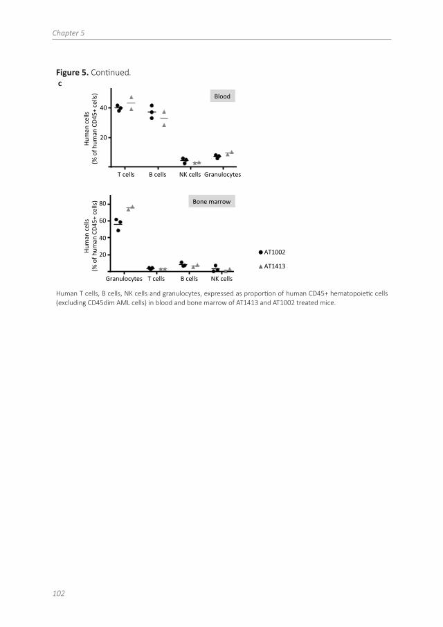
To more specifically identify the binding epitope of AT1413, we generated 10 FLAG-tagged truncated variants of CD43 that were expressed in THP1 cells (Figure S3A). Immunoprecipi-tation with AT1413 of lysates of THP1 cells transduced with truncation variants A-K revealed strong binding to variants A-F, weak binding to variant G, and no binding to variants H-K as shown in an immunoblot with a FLAG-specific antibody (Figure 2F). Full length endogenous and truncated CD43 immunoprecipitation was confirmed in a CD43 immunoblot (Figure S3B). Thus, the epitope of AT1413 is located between amino acids 133 and 165 of the CD43 protein (Figure S4). Mem59 and DF-T1 demonstrated strong binding to truncated variants A-C and no binding to variants D-K, identifying the epitope of these CD43 antibodies to be at a different location, between amino acids 59-82 (Figure S5). Of note, CD43s is a conserved epitope that is also expressed on murine AML cells (Figure 2G). Immunoprecipitation of the murine AML cell line WEHI-3b with AT1413 confirmed CD43s as the target (Figure 2H).
Chapter 5
90
Figure 2. The target of AT1413 is a novel sialylated CD43 epitope (CD43s).
B
A
Figure 2 The target of AT1413 is a novel sialylated CD43 epitope (CD43s)
Input
IP elute
THP1 Jurkat
250 kDa-‐
150 kDa-‐
100 kDa-‐
IP elute THP Molm
<
Input
250 kDa-‐
150 kDa-‐
100 kDa-‐
Immunoprecipitation (IP) with biotin-labeled, sortase-tagged AT1413 of THP-1 or Jurkat lysate. Imperial Coomassie stained gel.
B
A
Figure 2 The target of AT1413 is a novel sialylated CD43 epitope (CD43s)
Input
IP elute
THP1 Jurkat
250 kDa-‐
150 kDa-‐
100 kDa-‐
IP elute THP Molm
<
Input
250 kDa-‐
150 kDa-‐
100 kDa-‐
Western blot analysis of the AT1413 and AT1002 immunoprecipitates of THP-1 and Molm13 lysates with Mem59 mouse-anti-human CD43 antibody.C
Neuraminidase
0
0.5
1.0
1.5
No treatment Neu 1:20 Neu 1:2000.0
0.5
1.0
1.5
MFI A
PC
mAb binding
rAT14-13MEM59DF-T184-3C1L10
No treatment Neu 1:20 Neu 1:2000.0
0.5
1.0
1.5
mAb binding
rAT14-13MEM59DF-T1
MFI
AP
C 84-3C1L10
No treatment Neu 1:20 Neu 1:2000.0
0.5
1.0
1.5
MFI A
PC
mAb binding
rAT14-13MEM59DF-T184-3C1L10
No treatment Neu 1:20 Neu 1:2000.0
0.5
1.0
1.5
MFI A
PC
mAb binding
rAT14-13MEM59DF-T184-3C1L10AT1413
Mem59 DF-‐T1 84-‐3C1 L10
1:200 1:20 -‐
An>b
ody
bind
ing
(fo
ld in
crea
se)
Deglycosylation of THP-1 cells with neuraminidase (sialydase) abrogated binding of antibodies AT1413, Mem59, DF-T1 and 84-3C1 in a dose-dependent manner. Clone L10 does not target a sialylated epitope of CD43.36
Patient derived antibody recognizes unique CD43s on AML
91
5
Figure 2. ContinuedD
C
Neuraminidase 1:200 1:20
0
0.5
1.0
1.5
No treatment Neu 1:20 Neu 1:2000.0
0.5
1.0
1.5
MFI A
PC
mAb binding
rAT14-13MEM59DF-T184-3C1L10
No treatment Neu 1:20 Neu 1:2000.0
0.5
1.0
1.5
mAb binding
rAT14-13MEM59DF-T1
MFI
AP
C 84-3C1L10
No treatment Neu 1:20 Neu 1:2000.0
0.5
1.0
1.5
MFI A
PC
mAb binding
rAT14-13MEM59DF-T184-3C1L10
No treatment Neu 1:20 Neu 1:2000.0
0.5
1.0
1.5
MFI A
PC
mAb binding
rAT14-13MEM59DF-T184-3C1L10
-‐
AT1413 Mem59 DF-‐T1 84-‐3C1 L10
AT1413 L10 MEM59 DFT1
THP-‐1
Jurkat
AF647
Figure 2 conSnued
AnSb
ody binding
(fold increase)
Isotype control CD43 binding anSbody
Staining of THP-1 and Jurkat cells with AT1413 and with the commercially available CD43 specific antibodies DF-T1, L10 and Mem59.
Competition AT14-13 Biotin1 ug/ml
20,40,080,0160,00320,0006
40
60
80
100
ug/ml
% B
indi
ng
Mem59
DF0T1
8403C1
L10
AT14013
AT100002
E
Bind
ing (%
)
40
60
80
100
0.0006 0.0032 0.016 0.08 0.4 2.0
Compe7ng an7body concentra7on (μg/ml)
AT1413 AT1002 Mem59 DF-‐T1 84-‐3C1 L10
Competition AT14-13 Biotin1 ug/ml
20,40,080,0160,00320,0006
40
60
80
100
ug/ml
% B
indi
ng
Mem59
DF0T1
8403C1
L10
AT14013
AT100002
Compe7ng abs:
Competition experiment with AT1413 and commercially available CD43 specific antibodies. THP-1 cells were incu-bated with indicated antibodies, biotinylated AT1413 and streptavidin PECy7. AT1413 binding to THP-1 target cells was not affected by pre-incubation of the cells with commercially available CD43 antibodies, but was inhibited in a dose-dependent manner when THP-1 cells were pre-incubated with unlabeled AT1413.
Chapter 5
92
Figure 2. Continued
Competition AT14-13 Biotin1 ug/ml
20,40,080,0160,00320,0006
40
60
80
100
ug/ml%
Bin
ding
Mem59
DF0T1
8403C1
L10
AT14013
AT100002
F
E
A B C E D F G H I J K ctr
Figure 2 conSnued
Binding (%)
40
60
80
100
0.0006 0.0032 0.016 0.08 0.4 2.0
CompeSng anSbody concentraSon (μg/ml)
AT1413 AT1002 Mem59 DF-‐T1 84-‐3C1 L10
Competition AT14-13 Biotin1 ug/ml
20,40,080,0160,00320,0006
40
60
80
100
ug/ml
% B
indi
ng
Mem59
DF0T1
8403C1
L10
AT14013
AT100002
CompeSng abs:
Immunoprecipitation with AT1413 of truncated variants of THP-1 expressed FLAG-tagged CD43. Immunoblotting with FLAG antibody revealed binding of AT1413 to CD43 mutants A-F and no binding to mutants H-K. Truncations were performed as indicated in Figures S3 and S4. Ctr: control with GFP-transduced THP-1 cells.
Western blot analysis of the AT1413 and AT1002 immunoprecipitates of the mouse AML cell line WEHI-3b lysate with anti mouse CD43 antibody.
Patient derived antibody recognizes unique CD43s on AML
93
5
CD43s is overexpressed on AML and MDS patient derived leukemic blasts
Thus, AT1413 is an antibody that targets a unique, sialylated epitope on CD43 that is expressed on malignant myeloid cell lines. To further evaluate the breadth and specificity of AT1413, we tested binding of this antibody to bone marrow and peripheral blood samples obtained from patients with AML and MDS. The first patient to be tested was patient 101, the allogeneic HSCT recipient of whom AT1413 was obtained. Viable AML blasts of this patient had been frozen and stored at diagnosis and AT1413 showed clear binding to these leukemic blasts. (Figure 3A). We then tested binding of AT1413 to leukemic blasts obtained from 60 randomly selected, newly diagnosed patients with MDS and AML, and found that AT1413 bound to all samples tested (Figure 3B; Table 2). All WHO 2008 AML subtypes32 were represented in this patient cohort (although due to the relatively small sample size not in the same proportions as published33), including patients with high-risk MDS and patients with extramedullary AML (myeloid sarcoma, Figure 3C). Interestingly, in a patient diagnosed with therapy-related AML several years after intensive chemotherapy for multiple myeloma, AT1413 clearly distinguished myeloid leukemic blasts from multiple myeloma cells (Figure 3D). In a direct comparison with bone marrow obtained from six newly diagnosed patients with AML we confirmed that binding of AT1413 to leukemic blasts was stronger than binding to non-leukemic granulocytes, monocytes and lymphocytes of the same patient (Figure 3E).
Figure 3. CD43s is overexpressed by myeloid malignancies. A
Figure 3 CD43s is overexpressed on AML and MDS pa5ent derived leukemic blasts
CD34+CD38-‐ CD34+CD38+ CD34-‐CD38+
AF647
AT1002
AT1413
CD45 AF647
SSC
0 102 103 104 105
PE-A
0
102
103
104
105
PE
-Cy7
-A
16.3
CD34
CD38
AA
BB
C
C
AT1413 binding to CD34+ and CD38+ CD45dim AML blasts of patient 101. Bone marrow cells of this patient were isolated using a ficoll gradient and stored at diagnosis, precluding analysis of AT1413 interaction with non-malig-nant granulocytes.
Chapter 5
94
Figure 3. Continued
AML with recurrent gene5c abnormali5es inv (16) NPM1+ t(8;21)
BL-‐025 BL-‐010 BL-‐039
B
AML with myelodysplasia-‐related changes
BL-‐052 BL-‐054 BL-‐055
Therapy-‐related myeloid neoplasms
BL-‐047 BL-‐028 BL-‐074
AML not otherwise specified minimal diff. w/matura5on myelomonocy5c
BL-‐053 Pt 80 Pt 87
MDS RAEBII
BL-‐058 Pt 81
RAEBII BL-‐022
RAEBII
AF-‐647
AT1002 AT1413
Figure 3 con5nued
Representative examples of AT1413 binding to AML blasts obtained from newly diagnosed patients with AML or MDS (see also Table 2).
Patient derived antibody recognizes unique CD43s on AML
AT1413 binding to extramedullary AML of 2 patients (myeloid sarcoma (chloroma) of inguinal node (1) and skin (2)). Paraffin embedded THP-1 and Jurkat cells were used as a positive and negative control, respectively.
Bone marrow of a patient with concomitant multiple myeloma and therapy-related AML. Left panel: H&E stain-ing. Asterisk: malignant double nucleated plasma cell; arrowheads: AML blasts. Right panels: AT1413 staining of CD45dim AML blasts; CD138+ multiple myeloma plasma cells do not interact with AT1413.
Chapter 5
96
Figure 3. Continued
E
Figure 3 con5nued
AF647
CD45 + CD45 dim granulocytes lymphocytes AML blasts
Pa5ent BL-‐079
5.2 1.1 2.8 2.3
Pa5ent BL-‐092
1.0 1.8 3.2 2.7
monocytes
AT1002 AT1413
Pa5ent BL-‐091
1.5 5.2
Pa5ent BL-‐095
3.2 10.8 11.9
1.5 Pa5ent BL-‐096
3.3 1.7 Pa5ent BL-‐099
4.5 1.2 Pa5ent BL-‐106
74
9.2
3.6
5.2
AT1413 binding to CD45dim blasts of AML patients, to a lesser extent to CD45+ granulocytes and monocytes and absence of binding to CD45+ lymphocytes. In grey is indicated the fold increase MFI of AT1413 compared to the negative control (AT1002, filled grey histogram). Bone marrow (BL-079, BL-092, BL-095, BL-096, BL-099) or blood (BL-091, BL-106) of AML patients was freshly obtained and red blood cells lysed before FACs analysis.
Patient derived antibody recognizes unique CD43s on AML
97
5
Table 2. Expression of CD43s by AML and MDSPatient ID WHO 2008 classification AT1413 (MFI FI)
AML with recurrent genetic abnormalities
BL-039 AML with t(8;21) 3.2
BL-045 AML with t(8;21) 3.9
BL-065 AML with t(8;21) 15.8
BL-066 AML with t(8;21) 2.9
BL-069 AML with t(8;21) 7.1
BL-025 AML with inv(16) 21.3
BL-038 AML with inv (16) 4.2
BL-043 AML with inv (16) 5.4
BL-070 AML with inv (16) 5.1
BL-037 APL; T(15;17)(q22;q12) 5.0
BL-031 AML with t(6;9) 6.3
BL-068 AML with t(6;9) 4.0
BL-010 AML with mutated NPM1 9.2
BL-051 AML with mutated NPM1 15.9
BL-061 AML with mutated NPM1 5.5
Pt 78 AML with mutated NPM1 2.2
BL-057 EVI1 17p del 9.6
BL-059 RUNX1+ FLT3/ITD+ 12.4
Acute leukemia of ambiguous lineage
BL-060 Acute undifferentiated leukemia 12.9
Therapy-related myeloid neoplasms
BL-047 t-AML 7.5
BL-074 t-AML 3.8
BL-028 t-AML 2.2
AML, not otherwise specified
Pt 77 AML without maturation 13.8
Pt 86 AML without maturation 12.9
BL-064 AML without maturation 2.9
Pt 80 AML with minimal differentiation 48.9
BL-007 AML with minimal differentiation 21.3
BL-009 AML with minimal differentiation 10.9
BL-030 AML with minimal differentiation 29.0
Chapter 5
98
Table 2. ContinuedPatient ID WHO 2008 classification AT1413 (MFI FI)
AML with recurrent genetic abnormalities
BL-063 AML with minimal differentiation 8.4
Pt 87 AML with maturation 38.6
BL-071 AML with maturation 2.3
BL-046 Acute myelomonocytic leukemia 3.3
BL-053 Acute myelomonocytic leukemia 4.2
BL-034* Acute monoblastic/monocytic leukemia 2.5
Pt 101** Acute monoblastic/monocytic leukemia 6.4
AML with myelodysplasia-related changes
BL-014 History of MDS 3.1
BL-055 History of MDS 2.4
BL-052 Multilineage dysplasia 22.4
BL-054 Multilineage dysplasia 6.4
Myelodysplastic syndromes
BL-032 RCUD 2.2
BL-011 RCMD 5.7
Pt 81 RAEB-II 3.3
BL-022 RAEB-II 9.0
BL-033 RAEB-II 3.4
BL-042 RAEB-II 2.0
BL-058 RAEB-II 1.9
BL-062 RAEB-I 7.6
MFI fold increase (FI) for AT1413 binding to CD45dim leukemic blasts isolated from newly diagnosed AML and MDS patients. Depicted is MFI for AT1413 divided by the MFI of the negative control antibody (AT1002). *Patient deferred further treatment and cytogenetic and molecular analyses were not performed; **from this patient AT1413 was retrieved.
Patient derived antibody recognizes unique CD43s on AML
99
5
AT1413 induces ADCC and CDC of AML blasts but not non-malignant cells in vitro
We then tested the in vitro capacity of this antibody to induce target cell killing by antibody dependent cellular cytotoxicity (ADCC) and complement dependent cytotoxicity (CDC), as described previously.34 Incubation of the AML cell line SH2 with AT1413 and human peripheral blood mononuclear cells (PBMC) or rabbit complement induced ADCC and CDC, respectively (Figure 4A). Endothelial cells (HAEC or HUVEC) and granulocytes, that bound AT1413 albeit to a lesser extent than AML cells, were not killed when incubated with AT1413 and PBMC (Figure 4B). Moreover, when we incubated SH2 cells with AT1413 and whole blood from a healthy individual, SH2 cells were killed but healthy peripheral blood polymorphonuclear cells (PMN) were not affected (Figure 4C).
Figure 4. AT1413 induces ADCC and CDC of malignant myeloid cells in vitro.
0.0001 0.001 0.01 0.1 1 10 1000
20
40
60
80
100
ug/ml
% c
ell k
illin
g
Transform of ADCC Antibody titration
rAT10-002rAT14-013
A
Figure 4 AT1413 induces ADCC and CDC of AML but not non-‐malignant cells in vitro
AT1413 (open squares) induced antibody dependent cell mediated cytotoxicity (ADCC) and complement dependent cytotoxicity (CDC) of the AML cell line SH2 with EC50s of 1,1nM (0,16ug/ml) and 12,4 nM (1,86 ug/ml), respectively. Control antibody: AT1002 (black dots).
0.0001 0.001 0.01 0.1 1 10 1000
20
40
60
80
100
ug/ml
% c
ell k
illin
g
Transform of ADCC Antibody titration
rAT10-002rAT14-013
A
Figure 4 AT1413 induces ADCC and CDC of AML but not non-‐malignant cells in vitro
AT1413 (grey bars) induced antibody dependent cell mediated cytotoxicity (ADCC) of AML cells (SH2), but not of HAEC, HUVEC and granulocytes. Control antibody: AT1002 (black bars).
Labeled SH2 cells were incubated with whole blood from a healthy individual and with AT1413 or rituximab. AML cells but not mononuclear cells were killed. As a control experiment, CD20+ Ramos cells were incubated with healthy whole blood and AT1413 or rituximab. PMN: polymorphonuclear cells.
AT1413 specifically eliminates human AML blasts in vivo
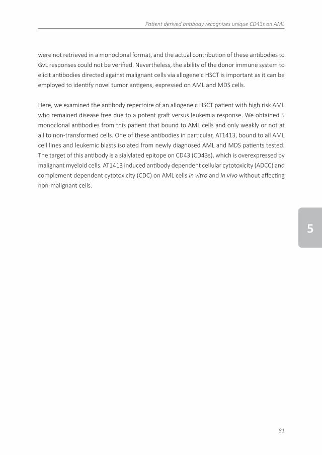
To test whether AT1413 affects tumor growth in vivo, we set up a mouse model for human AML in mice bearing human effector cells. Newborn female NSG mice were reconstituted with human hematopoietic stem cells (human immune system (HIS) mice27) and, after confirma-tion of engraftment, inoculated 5 months later with 1x10⁷ luciferase-GFP transduced human AML cell line SH2 cells via tail vein injection. Starting on day 19 after SH2 inoculation, mice were treated biweekly with AT1413 (15 mg/kg i.v.) or control antibody (AT1002, 15 mg/kg i.v.), and sacrificed at day 39. Tumor growth was strongly inhibited by AT1413, as measured by whole-body bioluminescence of the mice (Figure 5A). After mice were sacrificed, mouse organs (bone, liver, gut, lung and spleen) were analyzed individually for the presence of AML cells. Whereas the organs of control-antibody treated mice were heavily infiltrated with AML, no AML infiltration was observed in the organs of AT1413 treated mice (Figure 5B). Impor-tantly, AT1413 treatment did not affect human, non-malignant myeloid cells present in the tumor-bearing HIS mice, consistent with the observation that healthy myeloid cells bound by AT1413 were not killed in an ADCC assay in vitro. Proportions of human CD45+ cells, that
Patient derived antibody recognizes unique CD43s on AML
101
5
includes human granulocytes, T cells, B cells and NK cells but not AML cells, were similar between AT1413 and control antibody treated mice (Figure 5C). AT1413 bound weakly to mouse liver cells and mouse endothelial cells in FACs analysis (Figure S6) similar to what we observed for human cells (Figure 1D). However, injection of AT1413 did not induce significant side effects except for a transient reduction in food intake and weight loss of mice after the first injection only (Figure S7). We confirmed the efficacy of AT1413 in non-humanized NSG mice inoculated with luciferase labeled SH2 cells (Figure S8).
Figure 5. Anti-AML effect of AT1413 in vivo. A
Figure 5 AT1413 specifically eliminates human AML blasts in vivo
AT1002 AT1413
d14
d22
d29
d39
1.0
1.5
2.0
counts per minute (x10
6 )
0.5
10 20 30 40
Time (days aEer AML inoculaFon)
BM Liver PB Spleen0
1×1012×1013×1014×1015×1016×1017×1018×101
% o
f hC
D45
+ ce
lls
"Granulocytes"
AT1002
AT1413
Females
AT1002
AT1413
Human immune system (HIS) mice with human AML (luciferase-GFP transduced SH2) received biweekly treatment with AT1413 or control antibody AT1002 (15 mg/kg i.v.; indicated by asterix). AML progression was measured by bioluminescence (CPM) after luciferase injection.
bone liver guts lung spleen0
1×10⁴
2×10⁴
3×10⁴
4×10⁴
5×10⁴
cpm
organs bioluminescence
AT1002
AT1413
B
C
Figure 5 ConFnued
3
4
5
counts per minute (x10
4 )
2
AT1002
AT1413
bone liver gut lung spleen
1
40
Human cells
(% of hum
an CD4
5+ cells)
20
PB
T c
e l l s
PB
B c
e l l s
PB
NK
ce l l s
PB
- g
r a nu
l oc y t e
s
0
2 0
4 0
6 0
% o
f h
CD
45
+ c
ell
s
H e m a t o p o i e t i c c e l l s P B o n l y
0 3 9 - A T 1 0 0 2
0 3 9 - A T 1 4 1 3
T cells B cells NK cells Granulocytes
Blood
gr a n
ul o
c y t es
T c
e l l s
B c
e l l s
NK
ce l l s
0
2 0
4 0
6 0
8 0
1 0 0
% o
f h
CD
45
+ c
ell
s
H e m a t o p o i e t i c c e l l s B M o n l y
0 3 9 - A T 1 0 0 2
0 3 9 - A T 1 4 1 3
40
20
80
60
Human cells
(% of hum
an CD4
5+ cells)
T cells B cells NK cells Granulocytes
Bone marrow
BM Liver PB Spleen0
1×1012×1013×1014×1015×1016×1017×1018×101
% o
f hC
D45
+ ce
lls
"Granulocytes"
AT1002
AT1413
Females
AT1002
AT1413
Bioluminescence of individual organs harvested after mice were sacrificed.
Chapter 5
102
Figure 5. Continued.
bone liver guts lung spleen0
1×10⁴
2×10⁴
3×10⁴
4×10⁴
5×10⁴
cpm
organs bioluminescence
AT1002
AT1413
B
C
Figure 5 ConFnued
3
4
5
counts per minute (x10
4 )
2
AT1002
AT1413
bone liver gut lung spleen
1
40
Human cells
(% of hum
an CD4
5+ cells)
20
PB
T c
e l l s
PB
B c
e l l s
PB
NK
ce l l s
PB
- g
r a nu
l oc y t e
s
0
2 0
4 0
6 0
% o
f h
CD
45
+ c
ell
s
H e m a t o p o i e t i c c e l l s P B o n l y
0 3 9 - A T 1 0 0 2
0 3 9 - A T 1 4 1 3
T cells B cells NK cells Granulocytes
Blood
gr a n
ul o
c y t es
T c
e l l s
B c
e l l s
NK
ce l l s
0
2 0
4 0
6 0
8 0
1 0 0
% o
f h
CD
45
+ c
ell
s
H e m a t o p o i e t i c c e l l s B M o n l y
0 3 9 - A T 1 0 0 2
0 3 9 - A T 1 4 1 3
40
20
80
60
Human cells
(% of hum
an CD4
5+ cells)
T cells B cells NK cells Granulocytes
Bone marrow
BM Liver PB Spleen0
1×1012×1013×1014×1015×1016×1017×1018×101
% o
f hC
D45
+ ce
lls
"Granulocytes"
AT1002
AT1413
Females
AT1002
AT1413
Human T cells, B cells, NK cells and granulocytes, expressed as proportion of human CD45+ hematopoietic cells (excluding CD45dim AML cells) in blood and bone marrow of AT1413 and AT1002 treated mice.
Patient derived antibody recognizes unique CD43s on AML
103
5
Discussion
The development novel forms of immunotherapy - other than allogeneic HSCT - for the treat-ment of AML would be a significant therapeutic advance. In search for novel AML/MDS specific antigens that can be employed as targets for immunotherapy we screened the B cell reper-toire of a patient with a durable remission after receiving an allogeneic HSCT for relapsed AML, for antibodies that react with cell surface antigens expressed on AML cells. We isolated 5 B cell clones producing such antibodies and one of these antibodies, AT1413, recognized a novel sialylated epitope on CD43 (CD43s). CD43 is a heavily O-glycosylated mucin-like type I transmembrane protein that is present on the surface of most hematopoietic cells inclu- ding hematopoietic stem cells, but not on resting B cells and erythrocytes.35 Different CD43 glycoforms that can be co-expressed on one cell have been described, each with specific func-tions including roles in activation, proliferation, migration and apoptosis.36 While in healthy individuals CD43 is exclusively expressed on hematopoietic cells, certain CD43 glycoforms are expressed by a number of non-hematologic tumors, including colon, lung and breast carcinoma, where it affects growth, migration, metastasis and interaction with the immune system, and hence prognosis.28 For example the UN1/CD43 epitope is a specific CD43 glyco-form expressed by T lymphocytes, thymocytes and certain leukemic T cell lines, and by certain solid tumor types.37,38
We identified CD43s, a novel CD43 glycoform that is overexpressed on AML cell lines and blasts of AML patients. In fact, while the extent of expression is variable, CD43s is expressed on all WHO 2008 classification types of AML, including MDS-RAEB I and MDS-RAEB II.
CD43s is also expressed by myeloid progenitor cells, monocytes and granulocytes and weakly expressed by endothelial cells. CD43s is a conserved tumor epitope, as it is also expressed by murine AML cells. Engagement of AT1413 induced in vitro death of AML cells but not of healthy cells, via ADCC and CDC. Strikingly, AT1413 was highly efficacious against malignant cells in non-humanized NSG mice (Figure S8) and in a humanized HIS mouse AML model. The non-malignant human myeloid cells in the latter mice were however not affected. In addition no adverse side effects were observed in the treated mice with the exception of a transient weight loss after the firs dosing. These data suggest that this antibody may have therapeutic potential for treatment of AML patients. The reactivity of AT1413 with human endothelial cells may raise safety concerns, however, the binding is weak and AT1413 does not mediate ADCC or CDC against endothelial cells. In this respect it is noteworthy that trastuzumab, a HER2/neu specific antibody that is widely used in the treatment of HER2/neu over-expres-
Chapter 5
104
sing breast cancer interacts with cardiac endothelial cells but is safely applied in humans.39 Another important point with respect to safety is that although AT1413 is a highly mutated, antigen-experienced antibody, the patient of whom this antibody was derived did not expe-rience vascular complications, cardiac issues, neutropenia or pancytopenia after allogeneic HSCT. He did develop bronchiolitis obliterans syndrome and pulmonary dysfunction after syn-cytial respiratory (RS) virus infection, but is no longer on immunosuppressants.
In conclusion, we identified CD43s as an immunotherapeutic target that is overexpressed by all WHO 2008 types of AML and MDS. AT1413 induced ADCC and CDC of AML blasts in vitro and in vivo. While the efficacy of AT1413 in vivo needs to be confirmed in other models, these data suggest that AT1413 has the potential to be employed as a naked antibody, alone or in com-bination with standard AML chemotherapeutic regimens including cytarabine, anthracyclines or hypomethylating agents. Alternatively, AT1413 may be developed into an antibody-drug conjugate, a bispecific T cell engager (BiTE), CAR- T cells, or CD43s may be used in a vaccine, to generate autologous immune responses to the tumor.40 Such novel approaches to treat AML and MDS are highly anticipated, given the poor outcome of the majority of patients with AML or high-risk MDS.
Acknowledgements
The authors are greatly indebted to the patients that participated in this study. They thank Ludo Evers for chimerism analysis, their colleagues at the Department of Hematology Trial Office for the collection of AML samples and the Department of Pathology for supplying tissue micro arrays. This study was financially supported by an intramural grant of the AMC (M.A.G.), the Netherlands Organization for Scientific Research (M.D.H.) and the Dutch Cancer Founda-tion (M.D.H. and H.S.).
Patient derived antibody recognizes unique CD43s on AML
105
5
References
1. Weiden PL, Flournoy N, Thomas ED, et al. Antileukemic Effect of Graft-versus-Host Disease in Human Recipients of Allogeneic-Marrow Grafts. N Engl J Med. 1979;300(19):1068–1073.
2. Ferrara F, Schiffer CA. Acute myeloid leukaemia in adults. Lancet. 2013;381(9865):484–495. 3. Horowitz MM, Gale RP, Sondel PM, et al. Graft-Versus-Leukemia Reactions After Bone Marrow Transplantation.
Blood. 1990(75):555–562.4. Kantarjian H, O’Brien S, Cortes J, et al. Therapeutic advances in leukemia and myelodysplastic syndrome over
the past 40 years. Cancer. 2008;113(S7):1933–1952. 5. Shah A, Andersson TML, Rachet B, Björkholm M, Lambert PC. Survival and cure of acute myeloid leukaemia in
England, 1971-2006: a population-based study. Br J Haematol. 2013;162(4):509–516.6. Gooley TA, Chien JW, Pergam SA, et al. Reduced mortality after allogeneic hematopoietic-cell transplantation.
N Engl J Med. 2010;363(22):2091–2101. 7. Lokhorst HM, Plesner T, Laubach JP, et al. Targeting CD38 with Daratumumab Monotherapy in Multiple Myeloma.
N Engl J Med. August 2015;373(13):1207-1219.8. Maloney DG, Grillo-López AJ, White CA, Bodkin D. IDEC-C2B8 (Rituximab) anti-CD20 monoclonal antibody ther-
apy in patients with relapsed low-grade non-Hodgkin’s lymphoma. Blood. 1997;90(6):2188–2195.9. Younes A, Bartlett NL, Leonard JP, et al. Brentuximab vedotin (SGN-35) for relapsed CD30-positive lymphomas.
N Engl J Med. 2010;363(19):1812–1821. 10. Topp MS, Gokbuget N, Zugmaier G, et al. Phase II Trial of the Anti-CD19 Bispecific T Cell-Engager Blinatumomab
Shows Hematologic and Molecular Remissions in Patients With Relapsed or Refractory B-Precursor Acute Lym-phoblastic Leukemia. Journal of Clinical Oncology. 2014;32(36):4134–4140.
11. Eshhar Z, Waks T, Gross G, Schindler DG. Specific activation and targeting of cytotoxic lymphocytes through chimeric single chains consisting of antibody-binding domains and the gamma or zeta subunits of the immuno-globulin and T-cell receptors. Proc Natl Acad Sci USA. 1993;90(2):720–724.
12. Grupp SA, Kalos M, Barrett D, et al. Chimeric antigen receptor-modified T cells for acute lymphoid leukemia. N Engl J Med. 2013;368(16):1509–1518.
13. Brudno JN, Somerville RPT, Shi V, et al. Allogeneic T Cells That Express an Anti-CD19 Chimeric Antigen Receptor Induce Remissions of B-Cell Malignancies That Progress After Allogeneic Hematopoietic Stem-Cell Transplanta-tion Without Causing Graft-Versus-Host Disease. Journal of Clinical Oncology. 2016;34(10):1112–1121.
14. Kalos M, Levine BL, Porter DL, et al. T Cells with Chimeric Antigen Receptors Have Potent Antitumor Effects and Can Establish Memory in Patients with Advanced Leukemia. Science Translational Medicine. 2011;3(95):95ra73.
15. Tettamanti S, Magnani CF, Biondi A, Biagi E. Acute myeloid leukemia and novel biological treatments: Monoclonal antibodies and cell-based gene-modified immune effectors. Immunology Letters. 2013;155:43–46.
16. Hofmann M, e-Hovest LGS, bling TNU, et al. Generation, selection and preclinical characterization ofan Fc-op-timized FLT3 antibody for the treatment ofmyeloid leukemia. Leukemia. 2012;26(6):1228–1237.
17. Walter RB, Appelbaum FR, Estey EH, Bernstein ID. Acute myeloid leukemia stem cells and CD33-targeted immu-notherapy. Blood. 2012;119(26):6198–6208.
18. van Rhenen A, van Dongen GAMS, Kelder A, et al. The novel AML stem cell associated antigen CLL-1 aids in discrimination between normal and leukemic stem cells. Blood. 2007;110(7):2659–2666.
19. Jenq RR, van den Brink MRM. Allogeneic haematopoietic stem cell transplantation: individualized stem cell and immune therapy of cancer. Nat Rev Cancer. 2010;10(3):213–221.
20. Wu CJ, Yang XF, McLaughlin S, et al. Detection of a potent humoral response associated with immune-induced remission of chronic myelogenous leukemia. J Clin Invest. 2000;106(5):705–714.
21. Biernacki MA, Marina O, Zhang W, et al. Efficacious immune therapy in chronic myelogenous leukemia (CML) recognizes antigens that are expressed on CML progenitor cells. Cancer research. 2010;70(3):906–915.
22. Marina O, Hainz U, Biernacki MA, et al. Serologic Markers of Effective Tumor Immunity against Chronic Lympho-cytic Leukemia Include Nonmutated B-Cell Antigens. Cancer research. 2010;70(4):1344–1355.
23. Wadia PP, Coram M, Armstrong RJ, Mindrinos M, Butte AJ, Miklos DB. Antibodies specifically target AML antigen NuSAP1 after allogeneic bone marrow transplantation. Blood. 2010;115(10):2077–2087.
24. Grubman SA, Perrone RD, Lee DW, et al. Regulation of intracellular pH by immortalized human intrahepatic biliary epithelial cell lines. Am J Physiol. 2002;266(6 Pt 1):G1060–G1070.
Chapter 5
106
25. Kwakkenbos MJ, Diehl SA, Yasuda E, et al. Generation of stable monoclonal antibody–producing B cell recep-tor–positive human memory B cells by genetic programming. Nat Med. 2010;16(1):123–128.
26. Wagner K, Kwakkenbos MJ, Claassen YB, et al. Bispecific antibody generated with sortase and click chemistry has broad antiinfluenza virus activity. Proceedings of the National Academy of Sciences. 2014;111(47):16820–16825.
27. Legrand N, Weijer K, Spits H. Experimental model for the study of the human immune system: production and monitoring of “human immune system” Rag2-/-gamma c-/- mice. Methods Mol Biol. 2008;415(10):65–82.
28. Tuccillo FM, de Laurentiis A, Palmieri C, et al. Aberrant Glycosylation as Biomarker for Cancer: Focus on CD43. BioMed Research International. 2014;(2014):1–13.
29. Alvarado M, Klassen C, Cerny J, Horeji V, Schmidt RE. MEM-59 monoclonal antibody detects a CD43 epitope involved in lymphocyte activation. Eur J Immunol. 1995;25(4):1051–1055.
30. Stross WP, Warnke RA, Flavell DJ, et al. Molecule detected in formalin fixed tissue by antibodies MT1, DF-T1, and L60 (Leu-22) corresponds to CD43 antigen. Journal of clinical pathology. 1989;42(9):953–961.
31. Borche L, Lozano F, Vilella R, Vives J. CD43 Monoclonal Antibodies Recognize the Large Sialoglycoprotein of Human Leukocytes. European Journal of Immunology. 1987;17(10):1523–1526.
32. Vardiman JW, Thiele J, Arber DA, et al. The 2008 revision of the World Health Organization (WHO) classification of myeloid neoplasms and acute leukemia: rationale and important changes. Blood. 2009;114(5):937–951.
33. Döhner H, Estey E, Grimwade D, et al. Diagnosis and management of AML in adults: 2017 ELN recommendations from an international expert panel. Blood. 2017;129(4):424–447.
34. Gillissen MA, Yasuda E, de Jong G, et al. The modified FACS calcein AM retention assay: A high throughput flow cytometer based method to measure cytotoxicity. Journal of Immunological Methods. 2016;434(2016):16-23.
35. Schmid K, Hediger MA, Brossmer R, et al. Amino acid sequence of human plasma galactoglycoprotein: identity with the extracellular region of CD43 (sialophorin). Proc Natl Acad Sci USA. 1992;89(2):663–667.
36. Brown TJ, Shuford WW, Wang WC, et al. Characterization of a CD43/leukosialin-mediated pathway for inducing apoptosis in human T-lymphoblastoid cells. Journal of Biological Chemistry. 1996;271(44):27686–27695.
37. Tuccillo FM, de Laurentiis A, Palmieri C, et al. Partial purification and MALDI-TOF MS analysis of UN1, a tumor antigen membrane glycoprotein. International Journal of Biological Macromolecules. 2006;2014(1-3):122–126.
38. Tuccillo FM, Palmieri C, Fiume G, et al. Cancer-Associated CD43 Glycoforms as Target of Immunotherapy. Molec-ular Cancer Therapeutics. 2014;13(3):752–762. d
39. Tocchetti CG, Ragone G, Coppola C, et al. Detection, monitoring, and management of trastuzumab-induced left ventricular dysfunction: an actual challenge. European Journal of Heart Failure. 2014;14(2):130–137.
Patient derived antibody recognizes unique CD43s on AML
107
5
Supplemental Material
Supplemental Figure 1
Supplemental Figure 1 Iden%fica%on of AML specific clone 2K23
Fibroblasts Molm13 Caco2 THP-‐1
1.160.65601.77 AT1002
(control) 1.86
2.3899.35.0869.1 MC 2K23 75
MonoMac6
IgG -‐ AF647 75 69
1 2 0
99 2 5
2 1
FSC
Identification of AML specific clone 2K23. Representative examples of binding of the mini-culture 2K23 supernatant to the AML cell lines Molm13, MonoMac6 and THP-1. 2K23 supernatant did not bind to fibroblasts or the colon cell line Caco2.
Supplemental Figure 2 Mul$ple myeloma T-‐ALL
IgG -‐ AF647 / IgG -‐ PE
SKBR-‐3 Colo-‐205 LSTR DLD-‐1
MM1.s RPMI8226 U266 PA039
CA081 HE081-‐C HE081-‐I
Mamma Colon Ileum
Jurkat
Supplemental Figure 2 Absence of binding to non-‐myeloid malignancies
Absence of binding to non-myeloid malignancies. Representative examples of 2 experiments are shown. PA039 and CA081 are malignant cells obtained from newly diagnosed multiple myeloma or colon cancer patients in our clinic. HE081 are non-malignant cells isolated from colon and ileum resection material of a colon cancer patient. Grey filled histograms: AT1002.
Chapter 5
108
Supplemental Figure 3
CD43-‐Flag THP1 trunca4ons AT14-‐13 IP elute
A B C E D F G H I J K ctr
a-‐CD43 cyto
Supplemental Figure 3 Expression and pulldown confirma4on
CD43-‐Flag THP1 trunca4ons input lysates
A B C E D F G H I J K ctr
B
A
Expression and pulldown confirmation. A: Expression of CD43-Flag THP-1 truncations is confirmed with the input lysates in Westernblot. Staining: CD43 intracellular (Novus).
CD43-‐Flag THP1 trunca4ons AT14-‐13 IP elute
A B C E D F G H I J K ctr
a-‐CD43 cyto
Supplemental Figure 3 Expression and pulldown confirma4on
CD43-‐Flag THP1 trunca4ons input lysates
A B C E D F G H I J K ctr
B
A
B: Pulldown confirmation of the AT1413 IP. Immunoblot performed with anti CD43 (Novus) demonstrating pulldown of endogenous CD43 in all samples and truncated CD43 up to region F (G).
Supplemental Figure 4 Supplemental Figure 4 Epitope of AT1413
Epitope of AT1413. The amino acid sequence of the CD43 protein with the signaling peptide in bold and the starting amino acid of truncated versions A-K in red bold. The transmembrane part of the protein is in bold italics and the epitope of AT1413 is underlined.
Patient derived antibody recognizes unique CD43s on AML
109
5
Supplemental Figure 5
Supplemental Figure 5 CD43s is a novel epitope
CD43 immunoblot with Mem59
A B C E D F G H I J K ctr CD43 immunoblot wiht DFt1
A B C E D F G H I J K ctr
CD43 expression A B C E D F G H I J K ctr
CD43s is a novel epitope. The expression of CD43 is present in all CD43 mutants as is shown in the upper panel Staining: CD43 intracellular (Novus). Immunoblotting with Mem59 and DF-T1 shows that these antibodies bind an identical region on CD43, located between amino acids 59-82 and present on truncations A, B and C.
Chapter 5
110
Supplemental Figure 6 Supplemental Figure 6 Interac(on of AT1413 with mouse cells
2
4
MFI
(x10
4 )
AT1413 (ug/ml)
6
8
MAEC
HAEC
SH2
10000 5000 2500 1250 625 313 156 780
1000
2000
20000
40000
60000
80000
AT1413
HAEC
MAEC
SH2
ng/ml
MFI
APC
10000 5000 2500 1250 625 313 156 780
1000
2000
20000
40000
60000
80000
AT1413
HAEC
MAEC
SH2
ng/ml
MFI
APC
10000 5000 2500 1250 625 313 156 780
1000
2000
20000
40000
60000
80000
AT1413
HAEC
MAEC
SH2
ng/ml
MFI
APC
10 5 2,5 1,25 0,625 0,313 0,156 0,0780
2×10⁴
4×10⁴
6×10⁴
8×10⁴
ug/ml
MFI
APC
AT1413
HAEC
MAEC
SH2
0
Interaction of AT1413 with mouse cells. FACS staining of mouse aorta endothelial cells (MAEC), human aortic endothelial cells (HAEC) and SH2 cells with AT1413.
Supplemental Figure 7 Transient weight loss of mice treated with AT1413
Transient weight loss of mice treated with AT1413. Weight was set at 100% at the start of treatment (day 19 after AML inoculation).
Supplemental Figure 8
Supplemental Figure 8 AT1413 treatment of non-‐humanized NSG mouse with AML
0 1 0 2 0 3 0 4 0 5 0
0
1 ´ 1 0 5
2 ´ 1 0 5
3 ´ 1 0 5
4 ´ 1 0 5
B L I f e m a l e s n o n H I S m o u s e e x p
d a y s p o s t t u m o r e n g r a f t m e n t
cp
m
0 6 5 _ A T 1 0 0 2
0 6 5 _ A T 1 4 1 3
10 20 30 40
Time (days a@er AML inoculaCon)
2
3
4
coun
ts p
er m
inut
e (x
105 )
1
BM Liver PB Spleen0
1×1012×1013×1014×1015×1016×1017×1018×101
% o
f hC
D45
+ ce
lls
"Granulocytes"
AT1002
AT1413
Females
AT1002
AT1413
AT1413 treatment of non-humanized NSG mice with human AML. Female non-humanized NSG mice were inocu-lated with 107 luciferase labeled SH2 cells via tail vein injection. Mice were treated with the AML antibody AT1413 or with an influenza specific antibody (AT1002) twice a week, from day 19 to day 35.


![MKG - By Treister [ENG] - Oral Chronic GVHD - PPT](https://static.documents.pub/doc/80x56/5695cf301a28ab9b028cfad1/mkg-by-treister-eng-oral-chronic-gvhd-ppt.jpg)