UvA-DARE is a service provided by the library of the University of Amsterdam (http://dare.uva.nl) UvA-DARE (Digital Academic Repository) Diagnosis of intra-abdominal infections and management of catastrophic outcomes Atema, J.J. Link to publication Citation for published version (APA): Atema, J. J. (2015). Diagnosis of intra-abdominal infections and management of catastrophic outcomes. General rights It is not permitted to download or to forward/distribute the text or part of it without the consent of the author(s) and/or copyright holder(s), other than for strictly personal, individual use, unless the work is under an open content license (like Creative Commons). Disclaimer/Complaints regulations If you believe that digital publication of certain material infringes any of your rights or (privacy) interests, please let the Library know, stating your reasons. In case of a legitimate complaint, the Library will make the material inaccessible and/or remove it from the website. Please Ask the Library: https://uba.uva.nl/en/contact, or a letter to: Library of the University of Amsterdam, Secretariat, Singel 425, 1012 WP Amsterdam, The Netherlands. You will be contacted as soon as possible. Download date: 25 Dec 2019
Transcript
UvA-DARE is a service provided by the library of the University of Amsterdam (http://dare.uva.nl)
UvA-DARE (Digital Academic Repository)
Diagnosis of intra-abdominal infections and management of catastrophic outcomes
Atema, J.J.
Link to publication
Citation for published version (APA):Atema, J. J. (2015). Diagnosis of intra-abdominal infections and management of catastrophic outcomes.
General rightsIt is not permitted to download or to forward/distribute the text or part of it without the consent of the author(s) and/or copyright holder(s),other than for strictly personal, individual use, unless the work is under an open content license (like Creative Commons).
Disclaimer/Complaints regulationsIf you believe that digital publication of certain material infringes any of your rights or (privacy) interests, please let the Library know, statingyour reasons. In case of a legitimate complaint, the Library will make the material inaccessible and/or remove it from the website. Please Askthe Library: https://uba.uva.nl/en/contact, or a letter to: Library of the University of Amsterdam, Secretariat, Singel 425, 1012 WP Amsterdam,The Netherlands. You will be contacted as soon as possible.
The aim of this study was to evaluate whether contrast administration (oral, intravenous, rectal) increases accuracy of Computed Tomography (CT) as compared to non-enhanced CT (NECT) in patients presenting at the Emergency Department (ED) with acute abdominal pain.
Methods
A systematic literature search was performed of Medline, Embase and The Cochrane Library without any restrictions up to August 2014. An article was considered potentially relevant if the diagnostic accuracy of CT and any type of contrast agent was evaluated in unselected patients presenting at the ED with acute abdominal pain or if multiple types of contrast agents were evaluated head-to-head in unselected abdominal pain patients, or selected patients with the most common surgical causes of an acute abdomen. A bivariate random effects model was used to obtain summary estimates the diagnostic accuracy
Results
Eighteen studies (5890 patients) were eligible for inclusion. Eleven studies (4580 patients) investigated CT in acute abdominal pain and seven studies (1310 patients) performed a head-to- head comparison in selected patients. In unselected patients with acute abdominal pain, the pooled diagnostic accuracy of CT with intravenous contrast, 90.6 per cent (95 per cent confidence interval (CI) 83 to 95 per cent), was non-significantly higher than the pooled diagnostic accuracy of NECT, 73.4 per cent (95 per cent CI 44 to 91). In the head-to-head comparison none of the CT strategies resulted in significantly higher diagnostic accuracy.
Conclusion
Based on current available evidence no diagnostic advantage in terms of increased diagnostic accuracy could be established for any route of contrast administration in patients with acute abdominal pain presenting at the ED.
47
Impact of contrast media on the diagnostic accuracy of CT
INTRODUCTION
Abdominal pain is a frequent symptom of patients presenting at the emergency department (ED).1 The first step in the evaluation of patients with acute abdominal pain is clinical examination. However the diagnostic accuracy of history and physical examination is low. Several studies have demonstrated that in an unselected patient cohort the clinical diagnosis was correct in only 43-59 per cent.2 Treatment and management based solely on the clinical diagnosis can result in unnecessary interventions or delay.
Imaging modalities such as computed tomography (CT) and ultrasonography (US) are increasingly used to improve the diagnostic accuracy.3 Studies have demonstrated that CT results in the highest diagnostic accuracy, with the underlying cause being correctly diagnosed at CT in 62-96 per cent of patients.1–5 In current clinical practice wide variation exists in the CT protocol used, especially as controversy exists regarding the need for enteral (oral and rectal) and intravenous contrast administration.
The American college of Radiologists and the American college of Emergency Physicians are equivocal whether contrast agents are of additional value.6,7Their guidelines state that the administration of contrast differs based on institutional preferences. Different CT protocols are used, combining different types of contrast administration (intravenous, oral or rectal) or without contrast administration.8–13intravenous (IV This variation is most likely based on local preferences in relation to the suspected underlying cause. Each type of contrast agents has its advantages and disadvantages.
Intravenous contrast improves tissue contrast and highlights certain pathologic findings such as bowel wall inflammation. Downside of intravenous contrast is the cost and its potential to cause allergic reactions and contrast induced nephropathy. Oral contrast opacifies the bowel that can help differentiate the bowel from surrounding structures e.g. fluid collections and provides information regarding the bowel transit. Downsides of oral contrast are patient discomfort and prolonged time to scanning due to time needed before adequate transit of contrast.10,14–19 Rectal contrast can be given in case of suspected rectosigmoid perforation (e.g., complicated diverticulitis, perforated cancer) or anastomotic leakage but is associated with patient discomfort due to the administration and opacifies only the lower part of the bowel.
Whether contrast administration increases the diagnostic accuracy of CT remains under debate.6,7 Underlying medical conditions such as acute kidney failure, contrast allergies or patient care issues might hinder the administration of contrast medium in an Emergency Department setting. Increased time to scanning due to use of contrast increases time before a final diagnosis and subsequent management is established. The benefits of administration of contrast agents prior to CT should outweigh the downsides before administration of contrast medium can be advised in patients with acute abdominal pain.
48
Chapter 2
The aims of this study were two-fold: (1) to evaluate whether contrast administration, and if so which route of administration, increases accuracy in patients presenting at the Emergency Department (ED) for acute abdominal pain by means of a systematic review and meta-analysis, and (2) to evaluate whether contrast administration, and if so which route of administration, increases accuracy in head-to-head comparison studies in the same patient population concerning the most common surgical causes of acute abdominal pain namely acute appendicitis, diverticulitis, bowel obstruction and cholecystitis, also by means of a systematic review and meta-analysis.
METHODS
This systematic review and meta-analysis was performed according to the Preferred Reporting Items for Systematic Reviews and Meta-Analysis (PRISMA statement) guidelines.
Search strategy
Medline, Embase and The Cochrane Library were systematically searched without any restrictions up to August 2014. The search strategy included Mesh terms and free text words indexed for acute abdomen, abdominal pain, appendicitis, diverticulitis, bowel obstruction, cholecystitis combined with computed tomography and contrast agent.
Study selection
Two independent reviewers evaluated the titles and abstracts of all hits for eligibility (S.L.G. and J.J.A.). An article was considered potentially relevant if the diagnostic accuracy of CT and any type of contrast agent was evaluated in patients presenting at the Emergency Department (ED) for acute abdominal pain. Studies specifically evaluating the diagnostic accuracy of CT and multiple types of contrast agents head-to-head in the same patient with acute abdominal pain or in patients with the most common surgical causes for acute abdominal pain (acute appendicitis, diverticulitis, bowel obstruction or cholecystitis) were also included. Acute abdominal pain was defined as abdominal pain existing between 1 hour and 5 days. Exclusion criteria were insufficient data reported to extract the number of true positives, true negatives, false positives and false negatives. Studies evaluating acute abdominal pain of traumatic or haemorrhagic origin or acute abdominal pain in patients with known pregnancy were also excluded. Full text was obtained of all potentially relevant studies for further evaluation. Reference lists of key articles and reviews were manually searched to identify additional articles and pursued if relevant. In case of disagreement consensus was reached through consultation of a third independent reviewer (MB).
49
Impact of contrast media on the diagnostic accuracy of CT
Data extraction
Two reviewers extracted data from the included studies. In case of discrepancy consensus was reached by discussion. Quality of the included studies was assessed using The Quality Assessment of Diagnostic Accuracy Studies 2 (QUADAS-2) score.
Study design, patient characteristics and quality
The following study design characteristics were extracted: study period, department of the first author, country of origin, criteria for patient selection. Patient characteristics such as the number of patients included, the mean or median age and the age range and the male to female ratio were recorded. Technical characteristics of computed tomography and type of contrast medium administered were also extracted if available.
Reference standard
Information regarding the reference standard was extracted from studies. Details on number of patients undergoing surgery and or diagnostic laparoscopy as well as features recorded at surgery or pathology were extracted. The duration of follow up was also recorded.
Primary outcome
The diagnostic accuracy of CT and any protocol of administration of contrast agents was the outcome of interest. Contingency tables were extracted or reconstructed for each CT protocol reported in every study. When studies reported diagnostic accuracy compared between multiple observers only the data of the observer with the highest accuracy was included. Other diagnostic accuracy parameters such as sensitivity, specificity, negative predictive value and positive predictive value were calculated from the 2 x 2 contingency tables or extracted from the original studies if possible. Information regarding the negative side effects of CT such as time to scanning and incidence of contrast nephropathy were also recorded when available.
Meta-analysis
Meta-analysis was performed with the studies that provided sufficient qualitative information to calculate a contingency table for any protocol of CT scanning. An univariate random effects model was used to obtain summary estimates the diagnostic accuracy (correct diagnosis) and their corresponding 95 per cent confidence intervals (CI) for overall and subgroups on different types of contrast administration. A Z-test for unpaired data was used to compare the diagnostic accuracy estimates between types of contrast administration.
A bivariate random effects model was used to obtain summary estimates of sensitivity and specificity with their corresponding 95 per cent CI in head-to-head comparison studies. A Z-test for paired data was used in the model to compare sensitivity and specificity between
50
Chapter 2
types of contrast administration; non-enhanced CT (NECT) vs Contrast Enhanced CT (CECT)(any contrast agent), oral vs IV, and NECT vs IV.
All statistical analyses were performed using SPSS (version 20.0, IBM, Armonk, New York, USA), Excel (Microsoft Office 2007; Microsoft, Redmond, Washington) and SAS (version 9.3, SAS institute Inc., Cary North Carolina). P values of < 0.05 were considered to indicate statistical significance.
RESULTS
Search strategy and study selection
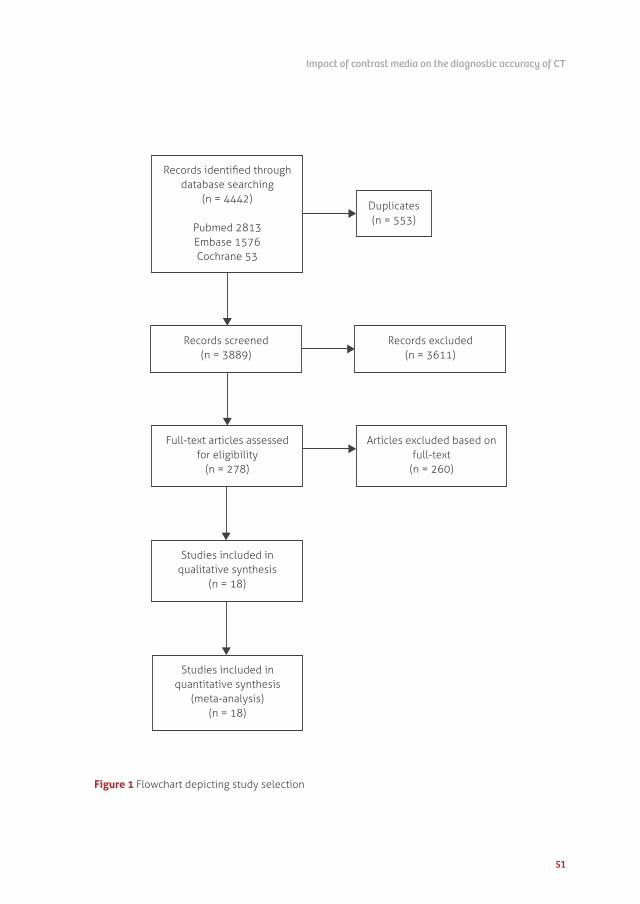
The search yielded a total of 4442 hits. After exclusion of duplicates 3889 titles and abstracts were screened for potential inclusion. The full text of 278 studies was retrieved for detailed examination. Inclusion criteria were not met in 260 studies, leaving 18 studies eligible for inclusion. Insufficient data to create a contingency table was the most common reason for exclusion. No additional studies were included after manual reference list searches. The complete study selection is depicted in figure 1. Eleven studies evaluated the diagnostic accuracy of computed tomography in unselected patients with acute abdominal pain1,20–29 and 7 studies were comparative studies on different contrast regimens for computed tomography in patients with acute abdominal pain or suspected acute appendicitis, acute pancreatitis, acute diverticulitis, bowel perforation or cholecystitis.9,12,13,30–33
Study characteristics
All studies were published between 1997 and 2014. Studies were initiated by the department of Radiology (n=15), the department of Surgery (n=2) or the department of Emergency Medicine (n=1). Eleven studies investigated the diagnostic accuracy of CT in unselected patients with acute abdominal pain, and seven studies made a head-to-head comparison of contrast entities in selected patients with a clinical suspicion of either acute appendicitis (n=5), abdominal pain (n=1) or diverticulitis (n=1). Table 1 and 2 summarizes further details on study characteristics.
A total of 5890 patients were analysed in all 18 studies combined. The accuracy of CT in patients with acute abdominal pain was evaluated in 11 studies comprising 4580 patients. Six studies investigated the accuracy of intravenous contrast. Only one study investigated the combination of oral and intravenous contrast and four studies investigated non-enhanced CT. Studies on the diagnostic accuracy of rectal contrast were lacking.
51
Impact of contrast media on the diagnostic accuracy of CT
Articles excluded based on full-text
(n = 260)
Studies included in quantitative synthesis
(meta-analysis) (n = 18)
Studies included in qualitative synthesis
(n = 18)
Full-text articles assessed for eligibility
(n = 278)
Records excluded(n = 3611)
Records screened(n = 3889)
Records identified throughdatabase searching
(n = 4442)
Pubmed 2813Embase 1576Cochrane 53
Duplicates(n = 553)
Figure 1 Flowchart depicting study selection
52
Chapter 2
Table 1 Characteristics of studies describing the diagnostic accuracy of computed tomography in unselected patients with acute abdominal pain
Study
Department
of first author
Observer
experience
reported Reference standard Type of CT
Contrast
used
Chin 21
2012
UK
Radiology No Expert panel or
discharge diagnosis
Computed tomography
with oral and IV
contrast
IV, Omni-
paque; Oral,
gastrografin
Haller 22
2010
Sweden
Radiology No Final diagnosis in
medical report within
30 days of radiological
examination
Non enhanced standard
dose computed
tomography and
low dose computed
tomography
NS
Ham 20
2012
Canada
Medical
imaging
No Not specified IV contrast enhanced
portal venous phase
abdominal computed
tomography
NS
Lameris 1
2009
the Netherlands
Surgery Yes Expert panel based on
follow up of 6 months
Computed tomography
with IV contrast
NS
Mackersie 23
2005
USA
Radiology No Follow up of 6 months Unenhanced helical
computed tomography
NS
Millet 24
2013
France
Medical
imaging
Yes Surgery, pathology
discharge diagnosis or
clinical follow up
Computed tomography
with IV contrast or non-
enhanced computed
tomography
IV, Xenetix
Rosen 25
2000
USA
Radiology No Pathology, surgery or
clinical follow up
Computed tomography
with IV and selective
oral contrast
NS
Sala 26
2007
UK
Radiology No Follow up of 6 months Computed tomography
with IV contrast in
portal venous phase
IV, lopamidol
Stromberg 27
2007
Sweden
Surgery No Follow up of 1 month Computed tomography
with IV contrast
IV, ioversol
Tsushima 28
2002
Japan
Radiology No Surgery, pathology or
discharge diagnosis
Computed tomography
with IV contrast
IV, ioversol
Udayasankar 29
2008
USA
Radiology No Follow up for 6 months Non contrast ultra-low
dose abdomino pelvic
computed tomography
NS
NS= not specified
53
Impact of contrast media on the diagnostic accuracy of CT
Table 2 Characteristics of head-to-head comparative studies on different contrast regimens for computed tomography in patients with acute abdominal pain or suspected acute appendicitis, acute pancreatitis, acute diverticulitis, bowel perforation or cholecystitis.
Study
Department of
first author
Observer
experience
reported Reference standard Comparison
Suspected acute appendicitis
Chiu 30
2013
Taiwan
Emergency
medicine
No Follow up 6 months Non enhanced computed
tomography vs enhanced computed
tomography (IV only)
Jacobs 31
2001
USA
Radiology Yes Surgery or follow up Focused non-enhanced computed
tomography (with oral contrast) vs
non focused enhanced computed
tomography (IV contrast)
Platon 12
2009
Switzerland
Radiology Yes Final discharge
diagnosis or surgical
reports
Low dose computed tomography
with oral contrast vs standard dose
IV contrast computed tomography
Tamburrini 13
2007
Italy
Radiology No Clinical follow up Non-enhanced computed
tomography vs enhanced computed
tomography (oral, rectal or IV)
Wise 32
2001
USA
Radiology No Surgery, pathology
or 3 months clinical
follow up
Unenhanced focused appendiceal
computed tomography vs
abdomino pelvic computed
tomography with IV contrast vs
focused appendiceal computed
tomography with colonic contrast
Suspected acute diverticulitis
Tack 33
2005
Belgium
Radiology Yes Expert panel based on
follow up
Unenhanced low dose computed
tomography vs IV enhanced
standard dose computed
tomography
Acute abdominal pain
Yeung 9
1997
Taiwan
Radiology No Surgery or clinical
follow up
Non-enhanced computed
tomography vs enhanced computed
tomography (IV contrast)
54
Chapter 2
Table 3 Patient characteristics of included studies evaluating diagnostic accuracy of CT in patients with acute abdominal pain
Study
No of
patients
% of
women
(n)
Mean age
(y)
Patient
selection Exclusion criteria
Chin 21 114 69% (79) 55 Acute severe
abdominal pain
Abdominal pain of traumatic origin
and patients referred for non-
contrast CT of kidney, bladder or
ureter
Haller 22 222 51% (113) 66 Acute abdominal
pain
Abdominal pain of traumatic origin,
no operations or imaging the last 2
weeks before presentation and age
<18years old
Ham 20 127 59% (75) 58 (=TN)
55 (=FP)
Upper
abdominal pain
No IV contrast, no portal venous
phase, patients with diffuse flank
and lower abdominal pain, history
of trauma, abdominal surgery in
past 3 months, active malignancy,
pregnancy, age<18 and known
inflammatory bowel disease
Lameris 1 1021 NS 47 (range
19-94)
Acute abdominal
pain
Haemorrhagic shock from
gastrointestinal bleeding, ruptured
aortic aneurysm or pregnancy and
patients being considered to be
discharged from the ED without
imaging
Mackersie 23
91 52% (47) 48.5 (+/-
18.7)
Non-traumatic
acute abdominal
pain
Patients who were clinically
intoxicated, pregnant or vaginal blee-
ding/discharge as primary symptom,
dysuria, haematuria without flank
pain and men with non-hemorrhagic
penile discharge.
Millet 24 339 56% (191) 83.7 (SD
5.9)
Elderly patients
with acute
abdominal pain
Age <75years and missing data
Rosen 25 57 58% (33) 48 (range
15-90)
Acute abdominal
pain
NS
Sala 26 99 58% (57) 59(42-
73) *
Nonspecific
acute abdominal
pain
Age <18years, pregnancy, rectal
bleeding, suspected renal colic,
suspected gynaecologic disorders
and traumatic origin
55
Impact of contrast media on the diagnostic accuracy of CT
* median reported instead of mean investigated in one comparative study.
In the 7 studies comparative studies a total of 1310 patients were investigated. Most studies, 5 in total, included patients suspected of acute appendicitis. Patients suspected of acute diverticulitis were investigated in only one study and abdominal pain in general was alsoNone of the studies compared different contrast entities in patients suspected of acute cholecystitis or bowel obstruction. The age of included patients ranged between 2 and 98 year. A slight majority of included patients was female, 57 per cent. Patient characteristics are summarized in Tables 3 and 4.
Quality of the included studies
The quality of included studies was assessed using the Quadas-2 tool (Appendix 1). All studies described a representative spectrum of patients. The index test was performed without the reference test. The time between index and reference test varied between included studies. The reference standard was similar between patients with a positive and negative index test, however between studies discrepancy existed in reference standard used. Most studies incorporated the index test (CT) in their reference standard increasing the risk of bias. In none of the studies the outcome assessors were blinded for the index test (Appendix 2). Follow up or pathology reports were most commonly used as the reference standard.
Diagnostic accuracy of CT in patients with acute abdominal pain
The individual results of the 11 studies analysing the diagnostic accuracy of CT in patients with acute abdominal pain are summarized in Table 5. The diagnostic accuracy of CT varied between 38 and 96.8 per cent. The pooled diagnostic accuracy was 86.3 per cent (95 per cent CI 75 to 93). In 4.4-41 per cent of patients the diagnosis was incorrect or non-specific on CT.
Table 3 Continued
56
Chapter 2
Six studies evaluated the diagnostic accuracy of CT with intravenous contrast,1,20,21,26-28 of which two studies provided insufficient data for pooled analysis. The remaining four studies demonstrated that CT with intravenous contrast had a pooled diagnostic accuracy of 90.6 per cent (95 per cent CI 83 to 95).1,21,27,28 Three other studies evaluated the diagnostic accuracy of NECT.22,23,29 The pooled diagnostic accuracy of NECT was 73.4 per cent (95 per cent CI 44 to 91), being non-significantly lower than the pooled diagnostic accuracy of CT with intravenous contrast.
Head-to-head comparison NECT vs CECT (any contrast)
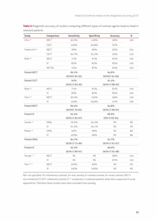
Three studies compared NECT with CECT in patients suspected of acute appendicitis (Table 6). The pooled sensitivity and specificity of NECT were 89.1 per cent (95 per cent CI 83 to 93) and 94.8 per cent (95 per cent CI 92 to 96), respectively. The pooled sensitivity and specificity of CECT were not significantly different, 91 per cent (95 per cent CI 83 to 95) and 92.4 per cent (95 per cent CI 88 to 95), respectively.13,30,32
Table 4 Patient characteristics of included studies for head-to-head comparison of contrast agents in selected patients t specified
Study
No of
patients
% of wo-
men (n)
Mean
age (y)
Patient
selection Exclusion criteria
Chiu 30 100 45% (45) 49.3
(range
18-90)
Suspicion
of acute
appendicitis
No appendicitis diagnosed intra
operatively, no iv contrast or oral
contrast agents administered
Jacobs 31 228 64% (145) 32
(range
13-87)
Suspicion
of acute
appendicitis
Crohn, inability to receive contrast or
previous appendectomy
Platon 12 86 52% (45) 45.6
(range
18-96)
Suspicion
of acute
appendicitis
Pregnancy
Tack 33 110 64% (70) 57
(range
30-82)
Suspicion of
diverticulitis
NS
Tamburrini 13 536 65% (316) 38
(range
18-86)
Suspicion
of acute
appendicitis
NS
Wise 32 100 74% (74) 38
(range
18-86)
Suspicion
of acute
appendicitis
NS
Yeung 9 150 43% (65) 52.4
(range
2-98)
Acute abdomen NS
57
Impact of contrast media on the diagnostic accuracy of CT
Head to head comparison NECT vs CECT with IV contrast
The diagnostic accuracy of NECT compared to CT with intravenous contrast was investigated in two studies in patients with suspected appendicitis.30,32 Pooled sensitivity and specificity of NECT were 89.4 per cent (95 per cent CI 79 to 95) and 94.8 per cent (95 per cent CI 89 to 97), respectively. CT with intravenous contrast resulted in a pooled sensitivity and specificity of 92.4 per cent (95 per cent CI 83 to 97) and 89.6 per cent (9 per cent CI 83 to 94), respectively. The diagnostic accuracy of CT with intravenous contrast was not significantly higher than that of NECT.
Head to head comparison CECT with Oral vs CECT with IV contrast
Two studies compared CT with oral contrast with CT enhanced by intravenous contrast, again in patients suspected of acute appendicitis.12,31 CT with oral contrast resulted in a pooled sensitivity of 84.1 per cent (95 per cent CI 75 to 90) and specificity of 94.7 per cent (95 per cent CI 91 to 97). CT with intravenous contrast resulted in a pooled sensitivity of 93.2 per cent (95 per cent CI 86 to 97) and specificity 96.0 per cent (95 per cent CI 93 to 98). There was no significant difference in diagnostic accuracy between both types of contrast enhancement.
Negative side effects
One study reported the patient discomfort on a scale ranging between 0 and 10.32 Mean patient discomfort whilst using unenhanced CT was significantly lower, 4.2, compared to 6.7 when using focused CT with colonic contrast material (p<0.001). None of the studies reported information regarding the incidence of contrast nephropathy or the difference in time to CT scanning regarding different routes of contrast administration. One study reported the difference in total duration of hospital stay when comparing their standard diagnostic work-up with CT in all patients.26 On average the hospital stay was almost 1 day shorter for patients in the CT group with a median stay of 4.2 days (IQR 1.1-7.6) compared to 5.3 days (IQR 2-9.5 days).
DISCUSSION
The administration of contrast agents regardless of the route of administration does not significantly increase the diagnostic accuracy in patients presenting with acute abdominal pain at the ED. Studies comparing CT with administration of intravenous contrast and NECT demonstrated a higher diagnostic accuracy of intravenous contrast although not statistically significant. The pooled diagnostic accuracy of CT with intravenous contrast was 90.6 per cent (95 per cent CI 83 to 95) compared with 73.4 per cent (95 per cent CI 44 to 91) for
58
Chapter 2
NECT. The diagnostic accuracy between different types of contrast was also comparable in studies performing a head-to-head comparison in selected patients, predominantly patients with suspected appendicitis. The pooled sensitivity and specificity were not significantly higher when comparing CECT and NECT. Furthermore, there was no significant difference when comparing CT with oral and intravenous contrast. In general, studies on the value of rectal contrast were lacking.
In current literature no consensus exists regarding the role of contrast agents and their perceived diagnostic advantage in patients with acute abdominal pain. Only a limited amount of studies evaluated the diagnostic accuracy of CT in patients with acute abdominal pain and only one study compared different CT strategies. These studies provide contrasting outcomes regarding the use of contrast agents in patients with acute abdominal pain. For selected patients the additional value of contrast agents also remains under debate. Several previous studies have examined the potential advantage of different routes of contrast administration in selected patients. These studies have reached conflicting conclusions whereas some studies demonstrated that NECT resulted in similar accuracy as contrast enhanced CT whilst other studies clearly demonstrated an advantage of contrast administration.8,10,14,18,34
Unfortunately only one study examined the diagnostic value of different contrast agents in patients with acute abdominal pain and acute diverticulitis in a head-to-head manner, and five studies investigated patients suspected of acute appendicitis in this way. A head-to-head comparison provides the best evidence and leads to the least risk of bias. This might be due to the fact that the comparison of different contrast entities for research purposes remains challenging. First of all due to ethical reasons, the additional exposure to ionizing radiation increases stochastic effects. Secondly once contrast is administered it will take time before the contrast is cleared and another CT strategy can be examined. In a crowded ED this will automatically lead to extended throughput of patients and possible delay of necessary treatment. The easiest and most ethical comparison in strategies is NECT versus any other type of contrast. Thus this is by far the most investigated comparison, possibly leading to a selection bias.
Only one study evaluated rectal contrast. This study compared the value of non-enhanced CT versus rectal contrast and intravenous contrast in patients suspected of acute appendicitis.32 The accuracy of rectal contrast was comparable with the diagnostic accuracy of NECT. The diagnostic accuracy of intravenous contrast was higher. The possible value of rectal contrast lies in its ability to depict intra-luminal pathology. Nevertheless in this study rectal contrast did not provide any added value. It is well established that around 10% of patients presenting with a presumed complicated diverticulitis on CT scan have an underlying colorectal carcinoma.34 A disadvantage of rectal contrast might be the logistical challenge for administration in an ED setting. Oral contrast administration was evaluated in only two
59
Impact of contrast media on the diagnostic accuracy of CT
studies including patients suspected of acute appendicitis.12,31 Oral contrast had no additional value when compared to intravenous contrast in these patients. Due to the downsides of oral contrast, such as patient discomfort and prolonged time due to ingestion of contrast, there seems little additional value of oral contrast.
Several limitations of this review need to be addressed. First, the outcomes of the individual studies were dependent on the choice of reference test and the definition of a positive index test. In most of the included studies the reference test consisted of the index test, CT, which could introduce bias. Ideally the final diagnosis should be based on an objective parameter such as pathology, however this would be impossible in daily practice since not all causes of acute abdominal pain necessitate surgical intervention. Even in studies investigating patients suspected of acute appendicitis, an entity that usually does necessitate surgery, it is impossible to provide pathology in all suspected patients. Within studies the reference test also differs. Some patients will have undergone surgery while in other patients follow up is the best available evidence of the final diagnosis. We aimed to minimize the risk of bias by only including studies with an acceptable reference standard.
The outcomes of the individual studies are also dependent on the observer experience. This differed between and within the included studies, which could introduce another risk of bias. In some studies the radiologists that judged the CT scans were residents or radiologists with limited experience during working hours. In other studies an experienced radiologist re-evaluated the CT scans performed outside of working hours. We tried to minimize this bias by performing a separate analysis on studies comparing contrast types head to head in the same patients. Ahead-to-head comparison in the same patient provides the most accurate and objective information regarding the additional diagnostic value. In the head to head comparison the same radiologist usually evaluated the different CT strategies in the same patient. Despite these differences our results reflect daily practice, as the experience of radiologists reading CT scans in daily practice may differ.
Based on current available evidence no diagnostic advantage in terms of increased diag-nostic accuracy could be proven for any type and route of contrast administration in patients with acute abdominal pain presenting at the ED. No studies have demonstrated additional advantages of intravenous, oral or rectal contrast over non-enhanced CT, although available evidence is limited. When balanced with the disadvantages such as patient discomfort and the risk of contrast-nephropathy, the administration of any contrast agent cannot be supported based on current literature. Future head-to-head studies in unselected patients with acute abdo-minal pain and in subgroups suspected of common surgical causes of acute abdominal pain are needed that compare different CT strategies assessing the additional value of intravenous, rectal and oral contrast or combinations of contrast agents over non-enhanced CT.
60
Chapter 2
Table 5 Diagnostic accuracy of CT in unselected patients with acute abdominal pain
Study Contrast Diagnosis correct (%)
Diagnosis incorrect or
a-specific (%)
CT of diagnostic
value (%)
Chin 21 IV 105/120 (87.5%) 15/120 (12.5%) NS
Ham 20 IV True negative
81/127 (63.7%)
False negative
46/127 (36.2%)
NS
Lameris 1 IV 867/1021 (84.9%) 154/1021 (15.1%) 147/1021 (14.3%)
Stromberg 27 IV 2151/2222 (96.8%) 71/2222 (3.2%) NS
Tsushima 28 IV 116/125 (92.8%) 9/125 (7.2%) 27/125 (21.6%)
Sala 26 IV NS NS 20/99 (20%)
Pooled diagnostic accuracy IV CECT 90.6% (95% CI 83-95%)
Mackersie 23 NECT 87/91 (95.6%) 4/91 (4.4%) NS
Haller 22 NECT 23/60 (38%) a
45/76 (59%) b
37/60 (62%) a
31/76 (41%) b
33/60 (49%) a
58/76 (72%) b
Udayasankar 29
NECT 127/163 (77.9%) 36/163 (22%) NS
Pooled diagnostic accuracy NECT 73.4% (95% CI 44-91%)
non enhanced CT, ECT= enhanced contrast CT. * comparison in selected patients other than suspected of acute
appendicitis. Therefore these studies have been excluded from pooling.
62
Chapter 2
REFERENCES1. Lameris W, Van Randen A, Van Es HW, et
al. Imaging strategies for detection of urgent conditions in patients with acute abdominal pain: diagnostic accuracy study. BMJ. 2009;339(jun26 2):b2431. doi:10.1136/bmj.b2431.
2. Gans SL, Pols M a., Stoker J, Boermeester M a. Guideline for the Diagnostic Pathway in Patients with Acute Abdominal Pain. Dig Surg. 2015:23–31. doi:10.1159/000371583.
3. Hastings RS, Powers RD. Abdominal pain in the ED: a 35 year retrospective. Am J Emerg Med. 2011;29(7):711–6. doi:10.1016/j.ajem.2010.01.045.
4. Laurell H, Hansson L-E, Gunnarsson U. Diagnostic pitfalls and accuracy of diagnosis in acute abdominal pain. Scand J Gastroenterol. 2006;41(10):1126–31. doi:10.1080/00365520600587485.
5. Toorenvliet BR, Bakker RFR, Flu HC, Merkus JWS, Hamming JF, Breslau PJ. Standard outpatient re-evaluation for patients not admitted to the hospital after emergency department evaluation for acute abdominal pain. World J Surg. 2010;34(3):480–6. doi:10.1007/s00268-009-0334-6.
6. Yaghmai V, Rosen M, Lalani T, et al. ACR Appropriateness Criteria - Acute (Nonlocalized) Abdominal Pain and Fever or Suspected Abdominal Abscess. Am Coll Radiol. 2012:1–10. doi:10.1016/j.jacr.2008.02.026.
7. Kulstad E. Current guidelines for diagnosis and management of abdominal pain in the emergency department. EM Pract Guidel. 2010;2(5):1–3. doi:10.1016/j.acpain.2005.03.003.
8. Hill BC, Johnson SC, Owens EK, Gerber JL, Senagore AJ. CT scan for suspected acute abdominal process: impact of combinations of IV, oral, and rectal contrast. World J Surg. 2010;34(4):699–703. doi:10.1007/s00268-009-0379-6.
9. Yeung KW, Sheu R, Chen C, Kuo Y, Liu G. Evaluation of Significance of Intravenous Contrast Administration for Diagnostic Accuracy during Emergent Abdominal Computed Tomographic Examination. Emerg Radiol. 1997;(8):276–282.
10. Hershko DD, Awad N, Fischer D, et al. Focused helical CT using rectal contrast material only as the preferred technique for the diagnosis of suspected acute appendicitis: a prospective, randomized, controlled study comparing three different techniques. Dis Colon Rectum. 2007;50(8):1223–9. doi:10.1007/s10350-007-0272-z.
11. Latifi A, Labruto F, Kaiser S, Ullberg U, Sundin A, Torkzad MR. Does enteral contrast increase the accuracy of appendicitis diagnosis? Radiol Technol. 2011;82(4):294–9.
12. Platon A, Jlassi H, Rutschmann OT, et al. Evaluation of a low-dose CT protocol with oral contrast for assessment of acute appendicitis. Eur Radiol. 2009;19(2):446–54. doi:10.1007/s00330-008-1164-x.
13. Tamburrini S, Brunetti A, Brown M, Sirlin C, Casola G. Acute appendicitis: diagnostic value of nonenhanced CT with selective use of contrast in routine clinical settings. Eur Radiol. 2007;17(8):2055–61. doi:10.1007/s00330-006-0527-4.
14. Hlibczuk V, Dattaro J a, Jin Z, Falzon L, Brown MD. Diagnostic accuracy of noncontrast computed tomography for appendicitis in adults: a systematic review. Ann Emerg Med. 2010;55(1):51–59.e1. doi:10.1016/j.annemergmed.2009.06.509.
15. Huynh V, Lalezarzadeh F. Abdominal computed tomography in the evaluation of acute and perforated appendicitis in the community setting. Am Surg. 2007.
63
Impact of contrast media on the diagnostic accuracy of CT
16. Dearing D, Recabaren J, Alexander M. Can computed tomography scan be performed effectively in the diagnosis of acute appendicitis without the added morbidity of rectal contrast? Am Surg. 2008.
17. Drake FT, Alfonso R, Bhargava P, et al. Enteral Contrast in the Computed Tomography Diagnosis of Appendicitis: Comparative Effectiveness in a Prospective Surgical Cohort. Ann Surg. 2013;260(2):311–316. doi:10.1097/SLA.0000000000000272.
18. Kepner AM, Bacasnot J V, Stahlman B a. Intravenous contrast alone vs intravenous and oral contrast computed tomography for the diagnosis of appendicitis in adult ED patients. Am J Emerg Med. 2012;30(9):1765–73. doi:10.1016/j.ajem.2012.02.011.
19. Keyzer C, Tack D, Maertelaer V De. Acute appendicitis: Comparison of Low-Dose and standard dose unenhanced multi detector row ct. Radiology. 2004.
20. Ham H, McInnes MDF, Woo M, Lemonde S. Negative predictive value of intravenous contrast-enhanced CT of the abdomen for patients presenting to the emergency department with undifferentiated upper abdominal pain. Emerg Radiol. 2012;19(1):19–26. doi:10.1007/s10140-011-0996-x.
21. Chin JY, Goldstraw E, Lunniss P, Patel K. Evaluation of the utility of abdominal CT scans in the diagnosis, management, outcome and information given at discharge of patients with non-traumatic acute abdominal pain. Br J Radiol. 2012;85(1017):e596–602. doi:10.1259/bjr/95400367.
22. Haller O, Karlsson L, Nyman R. Can low-dose abdominal CT replace abdominal plain film in evaluation of acute abdominal pain? Ups J Med Sci. 2010;115(2):113–20. doi:10.3109/03009730903294871.
23. MacKersie A, Lane M, Gerhardt R. nontraumatic acute abdominal pain:unenhanced helical ct compared with three view acute abdominal series. Radiology. 2005:126.
24. Millet I, Alili C, Bouic-Pages E, Curros-Doyon F, Nagot N, Taourel P. Journal club: Acute abdominal pain in elderly patients: effect of radiologist awareness of clinicobiologic information on CT accuracy. AJR Am J Roentgenol. 2013;201(6):1171–8; quiz 1179. doi:10.2214/AJR.12.10287.
25. Rosen MP, Sands DZ, Longmaid HE, Reynolds KF, Wagner M, Raptopoulos V. Impact of abdominal CT on the management of patients presenting to the emergency department with acute abdominal pain. AJR Am J Roentgenol. 2000;174(5):1391–6.
26. Sala E, Watson CJE, Beadsmoore C, Groot-wassink T, Shaw A, Dixon AK. A randomized , controlled trial of routine early abdominal computed tomography in patients presenting with non-specific acute abdominal pain. Acad Radiol. 2007:961–969. doi:10.1016/j.crad.2007.01.030.
27. Strömberg C, Johansson G, Adolfsson A. Acute abdominal pain: diagnostic impact of immediate CT scanning. World J Surg. 2007;31(12):2347–54; discussion 2355–8. doi:10.1007/s00268-007-9233-x.
28. Tsushima Y, Yamada S, Aoki J, Motojima T, Endo K. Effect of contrast-enhanced computed tomography on diagnosis and management of acute abdomen in adults. Clin Radiol. 2002;57(6):507–13. doi:10.1053/crad.2001.0925.
29. Udayasankar UK, Li J, Baumgarten DA, Small WC, Kalra MK. Acute abdominal pain: Value of non-contrast nhanced ultra-low-dose multi-detector row CT as a substitute for abdominal radiographs. Emerg Radiol. 2009;16:61–70. doi:10.1007/s10140-008-0743-0.
64
Chapter 2
30. Chiu Y-H, Chen J-D, Wang S-H, et al. Whether intravenous contrast is necessary for CT diagnosis of acute appendicitis in adult ED patients? Acad Radiol. 2013;20(1):73–8. doi:10.1016/j.acra.2012.07.007.
31. Jacobs JE, Birnbaum B a, Macari M, et al. Acute appendicitis: comparison of helical CT diagnosis focused technique with oral contrast material versus nonfocused technique with oral and intravenous contrast material. Radiology. 2001;220(3):683–90. doi:10.1148/radiol.2202001557.
32. Wise SW, Labuski MR, Kasales CJ, et al. Comparative assessment of CT and sonographic techniques for appendiceal imaging. Am J Roentgenol. 2001;176(April):933–941. doi:10.2214/ajr.176.4.1760933.
33. Tack D, Bohy P, Perlot I, Maertelaer V De, Sourtzis S, Gevenois PA. Suspected Acute Colon Diverticulitis : Imaging with Multi – Detector Row CT. Radiology. 2005;(9):189–196.
34. Funaki B, Grosskreutz SR, Funaki CN. Using unenhanced helical CT with enteric contrast material for suspected appendicitis in patients treated at a community hospital. AJR Am J Roentgenol. 1998;171(4):997–1001. doi:10.2214/ajr.171.4.9762983.
35. Sharma PV, Eglinton T, Hider P, Frizell F. Systematic review and meta-analysis of the role of routine colonic evaluation after radiologically confirmed acute diverticulitis. Ann Surg. 2014 Feb;259(2):263-72.
Appendix 1 Quality of the included studies assessed using the QUADAS-2 tool.
Patient Selection
Index Test
Reference Standard
Flow and Timing
0% 25% 50% 75% 100% 0% 25% 50% 75% 100%
High Unclear Low
Appendix 2 Risk of bias of the individual studies assessed by the QUADAS-2 tool


![Original Article - Endourology/Urolithiasis · follow-up of urolithiasis [1]. Prior to the use of computed tomography (CT), intravenous urography (IVU) was the Radiation dosing in](https://static.documents.pub/doc/80x56/5c8ae9ae09d3f232478d1cdb/original-article-endourologyurolithiasis-follow-up-of-urolithiasis-1-prior.jpg)


![Enteral versus intravenous approach for the sedation of …...ICU patients “calm, conscious, and cooperative” [11–13]. However, even if unjustified [8], a large proportion of](https://static.documents.pub/doc/80x56/608a3deaee8be61bb55baf1a/enteral-versus-intravenous-approach-for-the-sedation-of-icu-patients-aoecalm.jpg)


