29
Shelby Reinstein, MS, DVM Ophthalmology Resident Matthew J. Ryan Veterinary Hospital University of Pennsylvania Uveitis in Bartonella-infected cats: a case series
Shelby Reinstein, MS, DVM O p h t h a l m o l ogy R e s i d e n t
M a t t h e w J . R y a n V e t e r i n a r y H o s p i t a l U n i v e r s i t y o f P e n n s y l v a n i a
Uveitis in Bartonella-infected cats: a case series
Case Series
Sept 2010 – Dec 2011
6 cats (5 castrated males, 1 spayed female) 4 Domestic Shorthairs, 1 Russian Blue, 1 Maine Coon Age range: 4 – 17 years (average 8.6 y)
Diagnosed with uveitis, tested 4+ on Bartonella WB
Feline Bartonellosis
Gram negative bacteria Vector transmitted (flea excrement) Cats are the reservoir for Bartonella henselae Zoonotic potential – “cat scratch disease”
Correlation of infection with disease is unclear Most infected cats remain clinically normal Experimental infection no ocular disease
Ocular disease: Lappin 1999: Bartonella as a cause for uveitis in a cat Lappin 2000: Bartonella antibodies & DNA in AH of cat
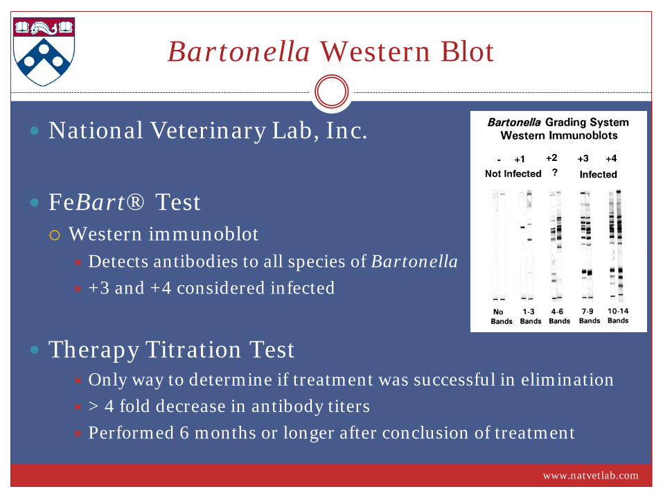
Bartonella Western Blot
National Veterinary Lab, Inc.
FeBart® Test Western immunoblot
Detects antibodies to all species of Bartonella +3 and +4 considered infected
Therapy Titration Test Only way to determine if treatment was successful in elimination > 4 fold decrease in antibody titers Performed 6 months or longer after conclusion of treatment
www.natvetlab.com
Uveitis Recommended Diagnostics
Complete Blood Count Chemistry Panel Urinalysis FeLV/FIV Toxoplasma titers Bartonella Western Blot +/- Crypto antigen Thoracic radiographs Abdominal ultrasound
Uveitis Recommended Treatment
Azithromycin 10 mg/kg PO q24 x 21 days 6 mg/kg PO q24 x 6 wks
Macrolide resistance
Doxycycline
10 mg/kg PO q12 x 2-6 wks Caution with pills Compounded suspension
Case #1: “Spartacus” 17y FS Maine Coon
Presented to ES on 9/20/10 CC: dilated pupil OS noted that morning Recent cecal adenocarcinoma resection w/ dirty margins
Exam Findings Visual OS, IOP 34 mmHg
1+ flare, KPs, diffuse iridal swelling Wide-spread MF to coalescing active chorioretinal lesions
OD normal
Diagnosis: Panuveitis with secondary glaucoma OS
Case #1: “Spartacus” 17y FS Maine Coon
Diagnostics CBC: Mild anemia (30%) Chem: NSF UA (cysto): NSF AUS: Scant effusion, healing ileocolic anastamosis Toxoplasma titers: IgG 1:512, IgM negative (chronic infection) Bartonella western blot: 4+
Treatment Clindamycin initially, added Azithromycin Pred acetate Tim/Dor Adriamycin chemotherapy
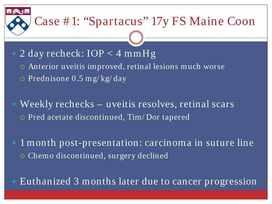
Case #1: “Spartacus” 17y FS Maine Coon
2 day recheck: IOP < 4 mmHg Anterior uveitis improved, retinal lesions much worse Prednisone 0.5 mg/kg/day
Weekly rechecks – uveitis resolves, retinal scars Pred acetate discontinued, Tim/Dor tapered
1 month post-presentation: carcinoma in suture line Chemo discontinued, surgery declined
Euthanized 3 months later due to cancer progression
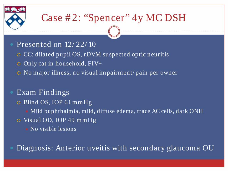
Case #2: “Spencer” 4y MC DSH
Presented on 12/22/10 CC: dilated pupil OS, rDVM suspected optic neuritis Only cat in household, FIV+ No major illness, no visual impairment/pain per owner
Exam Findings Blind OS, IOP 61 mmHg
Mild buphthalmia, mild, diffuse edema, trace AC cells, dark ONH Visual OD, IOP 49 mmHg
No visible lesions
Diagnosis: Anterior uveitis with secondary glaucoma OU

Case #2: “Spencer” 4y MC DSH
Diagnostics CBC: Normal Chem: Mild hyperglobulinemia 5.2 g/dL (3.1-5.0) Toxoplasma titers: IgG negative, IgM negative Bartonella western blot: +4
Treatment Timolol/Dorzolamide OU q8 Azithromycin 6 mg/kg PO q24 x 6 weeks
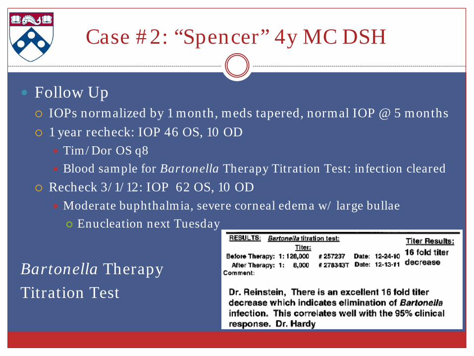
Case #2: “Spencer” 4y MC DSH
Follow Up IOPs normalized by 1 month, meds tapered, normal IOP @ 5 months 1 year recheck: IOP 46 OS, 10 OD
Tim/Dor OS q8 Blood sample for Bartonella Therapy Titration Test: infection cleared
Recheck 3/1/12: IOP 62 OS, 10 OD Moderate buphthalmia, severe corneal edema w/ large bullae Enucleation next Tuesday
Bartonella Therapy Titration Test
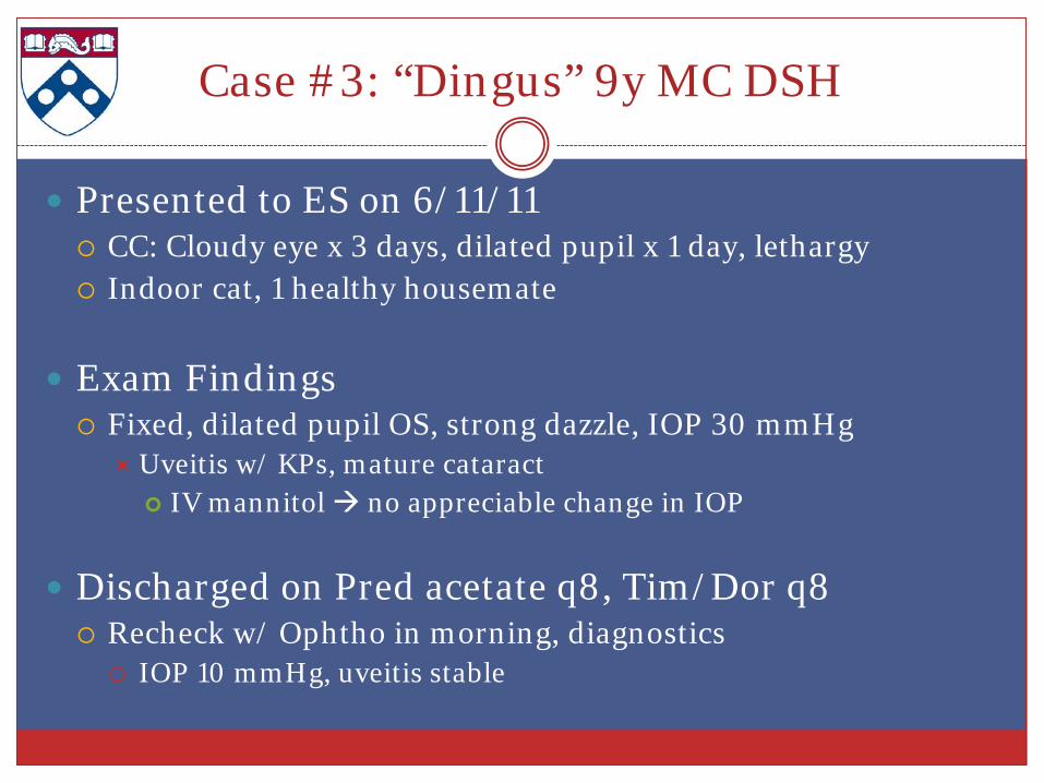
Case #3: “Dingus” 9y MC DSH
Presented to ES on 6/11/11 CC: Cloudy eye x 3 days, dilated pupil x 1 day, lethargy Indoor cat, 1 healthy housemate
Exam Findings Fixed, dilated pupil OS, strong dazzle, IOP 30 mmHg
Uveitis w/ KPs, mature cataract IV mannitol no appreciable change in IOP
Discharged on Pred acetate q8, Tim/Dor q8 Recheck w/ Ophtho in morning, diagnostics IOP 10 mmHg, uveitis stable
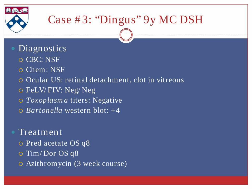
Case #3: “Dingus” 9y MC DSH
Diagnostics CBC: NSF Chem: NSF Ocular US: retinal detachment, clot in vitreous FeLV/FIV: Neg/Neg Toxoplasma titers: Negative Bartonella western blot: +4
Treatment Pred acetate OS q8 Tim/Dor OS q8 Azithromycin (3 week course)
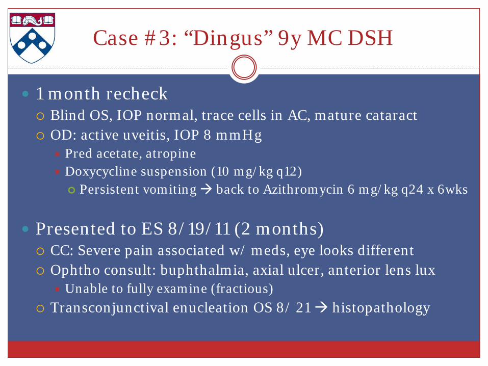
Case #3: “Dingus” 9y MC DSH
1 month recheck Blind OS, IOP normal, trace cells in AC, mature cataract OD: active uveitis, IOP 8 mmHg
Pred acetate, atropine Doxycycline suspension (10 mg/kg q12) Persistent vomiting back to Azithromycin 6 mg/kg q24 x 6wks
Presented to ES 8/19/11 (2 months) CC: Severe pain associated w/ meds, eye looks different Ophtho consult: buphthalmia, axial ulcer, anterior lens lux
Unable to fully examine (fractious) Transconjunctival enucleation OS 8/ 21 histopathology
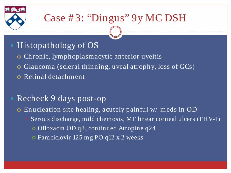
Case #3: “Dingus” 9y MC DSH
Histopathology of OS Chronic, lymphoplasmacytic anterior uveitis Glaucoma (scleral thinning, uveal atrophy, loss of GCs) Retinal detachment
Recheck 9 days post-op Enucleation site healing, acutely painful w/ meds in OD
Serous discharge, mild chemosis, MF linear corneal ulcers (FHV-1) Ofloxacin OD q8, continued Atropine q24 Famciclovir 125 mg PO q12 x 2 weeks
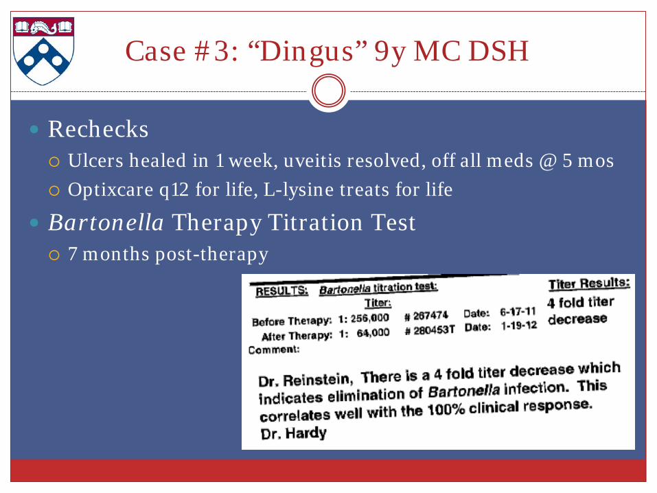
Case #3: “Dingus” 9y MC DSH
Rechecks Ulcers healed in 1 week, uveitis resolved, off all meds @ 5 mos Optixcare q12 for life, L-lysine treats for life
Bartonella Therapy Titration Test 7 months post-therapy
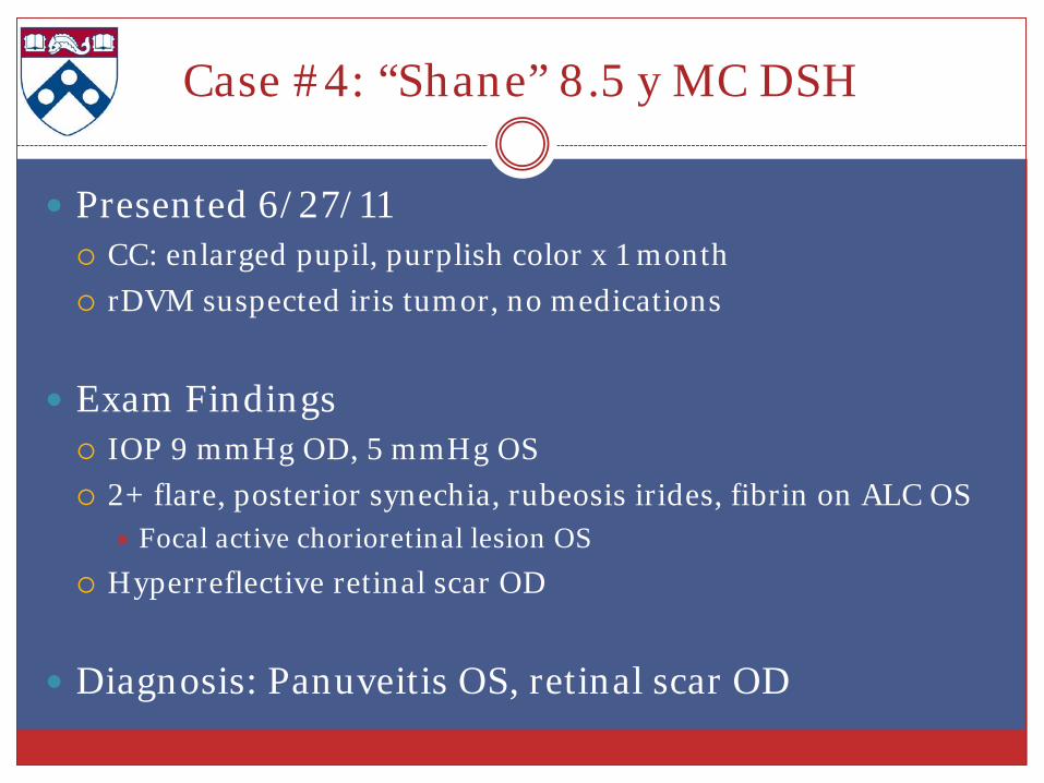
Case #4: “Shane” 8.5 y MC DSH
Presented 6/27/11 CC: enlarged pupil, purplish color x 1 month rDVM suspected iris tumor, no medications
Exam Findings IOP 9 mmHg OD, 5 mmHg OS 2+ flare, posterior synechia, rubeosis irides, fibrin on ALC OS
Focal active chorioretinal lesion OS Hyperreflective retinal scar OD
Diagnosis: Panuveitis OS, retinal scar OD
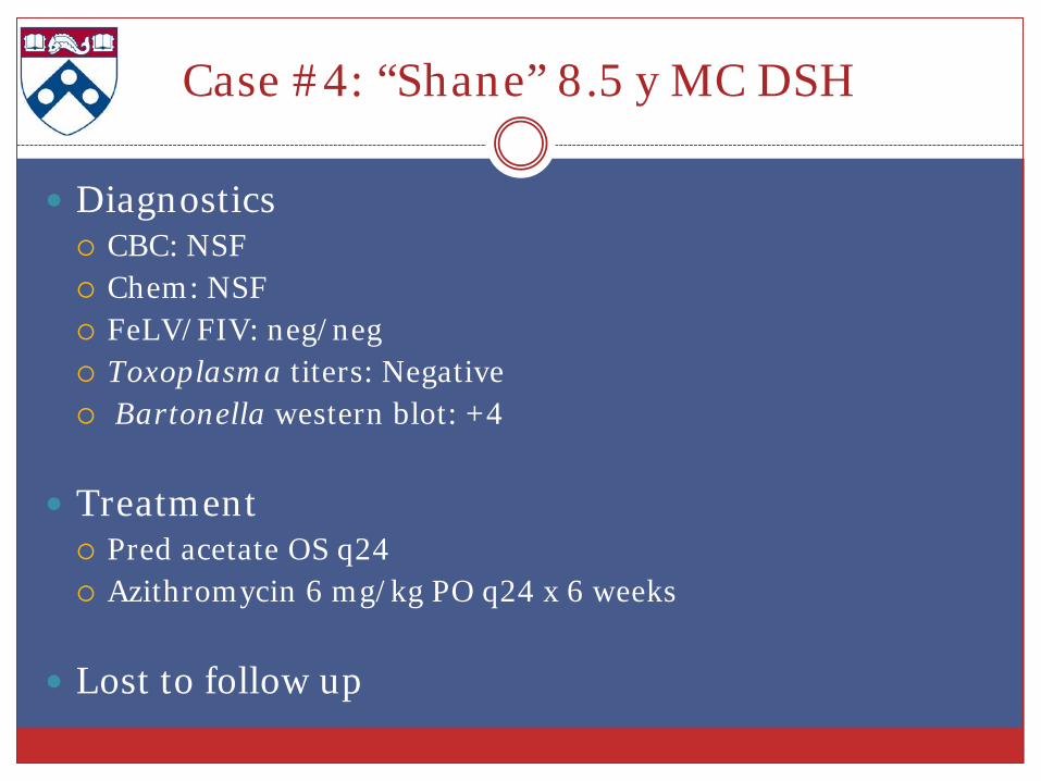
Case #4: “Shane” 8.5 y MC DSH
Diagnostics CBC: NSF Chem: NSF FeLV/FIV: neg/neg Toxoplasma titers: Negative Bartonella western blot: +4
Treatment Pred acetate OS q24 Azithromycin 6 mg/kg PO q24 x 6 weeks
Lost to follow up
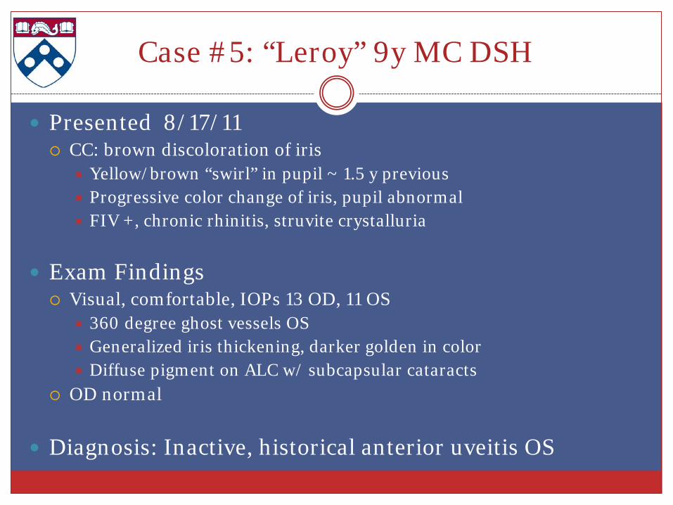
Case #5: “Leroy” 9y MC DSH
Presented 8/17/11 CC: brown discoloration of iris
Yellow/brown “swirl” in pupil ~ 1.5 y previous Progressive color change of iris, pupil abnormal FIV +, chronic rhinitis, struvite crystalluria
Exam Findings Visual, comfortable, IOPs 13 OD, 11 OS
360 degree ghost vessels OS Generalized iris thickening, darker golden in color Diffuse pigment on ALC w/ subcapsular cataracts
OD normal
Diagnosis: Inactive, historical anterior uveitis OS
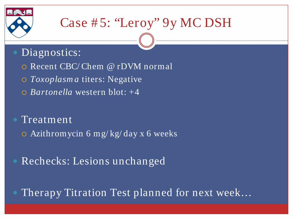
Case #5: “Leroy” 9y MC DSH
Diagnostics: Recent CBC/Chem @ rDVM normal Toxoplasma titers: Negative Bartonella western blot: +4
Treatment Azithromycin 6 mg/kg/day x 6 weeks
Rechecks: Lesions unchanged
Therapy Titration Test planned for next week…
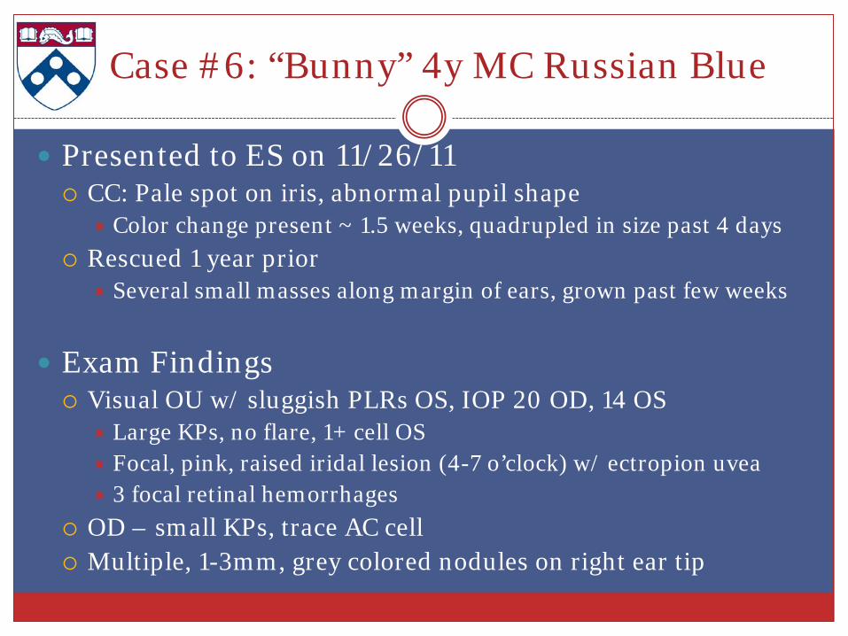
Case #6: “Bunny” 4y MC Russian Blue
Presented to ES on 11/26/11 CC: Pale spot on iris, abnormal pupil shape
Color change present ~ 1.5 weeks, quadrupled in size past 4 days Rescued 1 year prior
Several small masses along margin of ears, grown past few weeks
Exam Findings Visual OU w/ sluggish PLRs OS, IOP 20 OD, 14 OS
Large KPs, no flare, 1+ cell OS Focal, pink, raised iridal lesion (4-7 o’clock) w/ ectropion uvea 3 focal retinal hemorrhages
OD – small KPs, trace AC cell Multiple, 1-3mm, grey colored nodules on right ear tip
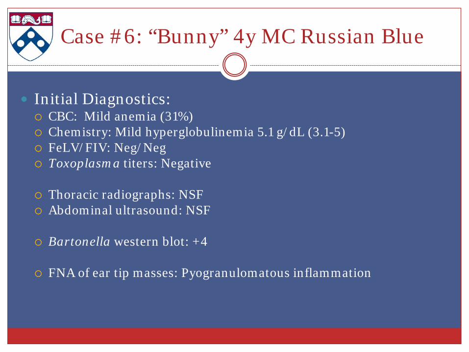
Case #6: “Bunny” 4y MC Russian Blue
Initial Diagnostics: CBC: Mild anemia (31%) Chemistry: Mild hyperglobulinemia 5.1 g/dL (3.1-5) FeLV/FIV: Neg/Neg Toxoplasma titers: Negative
Thoracic radiographs: NSF Abdominal ultrasound: NSF
Bartonella western blot: +4
FNA of ear tip masses: Pyogranulomatous inflammation
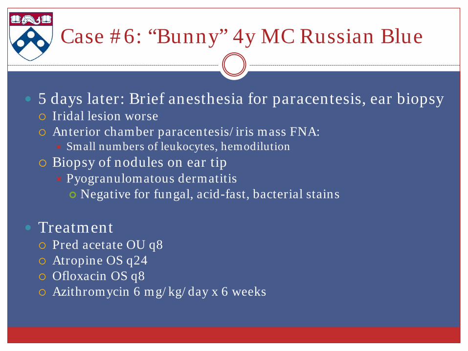
5 days later: Brief anesthesia for paracentesis, ear biopsy Iridal lesion worse Anterior chamber paracentesis/iris mass FNA:
Small numbers of leukocytes, hemodilution Biopsy of nodules on ear tip
Pyogranulomatous dermatitis Negative for fungal, acid-fast, bacterial stains
Treatment Pred acetate OU q8 Atropine OS q24 Ofloxacin OS q8 Azithromycin 6 mg/kg/day x 6 weeks
Case #6: “Bunny” 4y MC Russian Blue

Case #6: “Bunny” 4y MC Russian Blue
1 month recheck Owner reports recent difficulty administering topical meds Continues to squint, left eye still looks red
IOP 10 OD, 9 OS Axial, vascularized corneal ulcer with loose epithelial lip OS Dry debridement
Iridal swelling had progressed 1 o’clock – 7 o’clock OS OD iris still normal
Pred discontinued, topical Cidofovir OS q12 Recommended recheck in 2 wks…
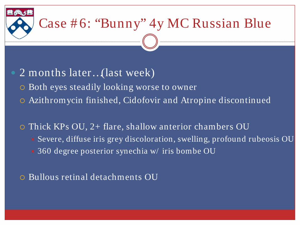
Case #6: “Bunny” 4y MC Russian Blue
2 months later…(last week) Both eyes steadily looking worse to owner Azithromycin finished, Cidofovir and Atropine discontinued
Thick KPs OU, 2+ flare, shallow anterior chambers OU
Severe, diffuse iris grey discoloration, swelling, profound rubeosis OU 360 degree posterior synechia w/ iris bombe OU
Bullous retinal detachments OU

Case #6: “Bunny” 4y MC Russian Blue
Additional diagnostics: Repeat CBC/Chem: NSF Cryptococcus antigen: Negative Declined for now: subretinal aspirates, repeat aqueocentesis, enucleation
for histopathology, repeat body imaging
Treatment Pred acetate OU q6 Cidofovir OU q12 Doxycycline suspension (10mg/kg q12)
Phone update: Eyes “much better”, vomited a few times…
Discussion
Trends Male cats over represented Anterior uveitis predominated over retinal pathology
Results of 2 titration tests indicate successful treatment Resolution of clinical disease, decrease in titers
Causal relationship remains unclear Indication to test for Bartonella in cats w/ uveitis Treatment is recommended when infection is documented in sick
cats
Audience Participation (thanks!)
Thoughts?
Experiences?
Recommendations?


