Page 1
International Journal of Obstetrics and Gynaecology Research (IJOGR)
Vol. 2 (2015) No.3, pp. 170-187
http://www.ijogr.com/
170 Sokkary et. al., Vaginal Fluid Lactate: A Method for the Diagnosis of Premature Rupture of Membrane
Abstract
To assess whether lactate determination in vaginal fluids " Lac-test ", can be used as a
diagnostic test for premature rupture of membranes (PROM) and to derive the best cutoff value
for a positive test. It was a cross-sectional study performed at Al Azhar Bab El Sheria and Ain
Shams Maternity Hospitals. 120 women were subdivided into 80 cases with sure or suspected
history of PROM and 40 cases with no history of PROM as a control group for determination of
lactate concentrations in vaginal fluid, nitrazine paper test and AFI were analysed. : A lactate
concentration ≥ 4.3 mmol/L was found to be the best cut-off value for a positive test. The "Lac-
test" had a sensitivity of 89.06%, specificity 85.71%, positive and negative predictive values of
87.7% and 87.3%, respectively. Corresponding values for the nitrazine paper test were 75%,
75%, 77.4% and 72.4%. Likelihood ratio for a positive "Lac-test" was 6.2 and for a negative test
0.1. Corresponding values for the nitrazine paper test were 3 and 0.3 respectively. "Lac-test" is a
valid test in cases with suspected PROM and may even be a better predictor than the nitrazine
test.
Keywords
Lactate Determination - Premature Rupture of Membranes
I. Introduction
Premature rupture of membranes (PROM) is
defined as rupture of fetal membranes before
the onset of labour at any time during the
Vaginal Fluid Lactate: A Method for the
Diagnosis of Premature Rupture of Membrane
El-Sokkary M.(MD)*1, Anbar A. (MD)2, Wahba K. (MD)1, , El-Mandouh M. (MD)1.
El-Shahawy Y (MD
1 Department of Obstetrics and Gynecology – Ain Shams University
2 Department of Obstetrics and Gynecology – Al- Azhar Maternity Hospital, cairo.
* Correspondence: Mohammed El-Sokkary – assistant professor of Obstetrics and Gynecology –
Faculty of Medicine – Ain Shams University – Abbasyia – Cairo
E-mail: [email protected]
Page 2
International Journal of Obstetrics and Gynaecology Research (IJOGR)
Vol. 2 (2015) No.3, pp. 170-187
http://www.ijogr.com/
171 Sokkary et. al., Vaginal Fluid Lactate: A Method for the Diagnosis of Premature Rupture of Membrane
gestational period. It occurs in 4.5-7.6% of
pregnant women [1]. Premature rupture of
membranes is associated with infectious
morbidity in mother and fetus, cord accidents,
imminent term or preterm labour. Diagnosis of
PROM is easy when the rupture is obvious but
difficult or indeed impossible when the rupture
is slight [2]. Failure to identify patients with
membrane rupture can result in failure to
implement obstetric measures. Conversely, the
false diagnosis of membrane rupture can lead to
inappropriate interventions such as
hospitalization or induction of labour.
Traditionally, the diagnosis of PROM has
relied on a combination of factors, including
the patient's history, identification of gross
pooling of amniotic fluid in the vagina, ferning
pattern, and a positive nitrazine test [3].
However, in equivocal cases of PROM, the
traditional methods have been associated with
both false-positive and false-negative results
[4].
The absence of a noninvasive "gold
standard" test for the diagnosis of membrane
rupture has led to the search for the alternative
biochemical markers. Any biological test used
to establish a correct diagnosis must be reliable,
simple and rapid [2].
Biochemical substances, which have high
amniotic concentration, e.g. prolactin [5],
alphafetoprotein (AFP) [6], insulin like growth
factor binding protein-1 (IGFBP-1) [7], fetal
fibronectin [8], diamino-oxidase9, β-HCG [10],
had all been previously studied. The nitrazine
test is a pH indicator. Vaginal pH is normally
between 4.5 and 5.5, but the presence of
amniotic fluid in the vagina increases the pH
value [11]. Other tests have been used as
markers for rupture of the membranes.
Arborization or ferning of vaginal fluid
suggests amniotic rather than cervical fluid. If
present, amniotic fluid crystallizes to form a
fern-like pattern due to the relative
concentrations of sodium chloride, proteins,
and carbohydrates in that fluid [12].
Over the past decades, a number of
ultrasound methods have been used to measure
the amount of amniotic fluid. Phelan and
colleagues (1987)[13] described the clinical
utility of quantification using the amniotic fluid
index (AFI). This is calculated by adding the
vertical depths of the largest pocket in each of
four equal uterine quadrants.
Lactate has also been reported to occur in
high concentration in amniotic fluid, (7-9
mmol/l)[14], which is four to six times higher
than in maternal or fetal blood [15]. The source
of the amniotic fluid lactate has been suggested
to be the fetus, mainly through urine and lung
fluid excretion [15].
II. Methods
A cross-sectional study was conducted on 120
pregnant women who attended the casualty
department at Al Azhar University Bab El
Sheria ana Ain Shams Maternity Hospitals with
history of PROM without uterine contractions
in between August 2011 and Mars 2015. The
study was approved by the local research ethics
committee in Al-Azhar university faculty of
medicine. After giving informed consent, all
the patients underwent ultrasonographic
examination for determination of gestational
age and calculation of amniotic fluid index. The
amniotic fluid index (AFI) was assessed in four
quadrants. According to the method of Phelan
et al.,(1987) an AFI of < 8cm was considered
as having oligohydroamnios. Pooling of
amniotic fluid in the posterior vaginal fornix
Page 3
International Journal of Obstetrics and Gynaecology Research (IJOGR)
Vol. 2 (2015) No.3, pp. 170-187
http://www.ijogr.com/
172 Sokkary et. al., Vaginal Fluid Lactate: A Method for the Diagnosis of Premature Rupture of Membrane
during speculum examination was used as the
gold standard in order to be able to calculate
the sensitivity, specificity, positive predictive
value and negative predictive value of each test.
After that the patients were divided into two
groups:
Group I :(n= 80) patients with sure or
suspected history of PROM (study group)
which were subdivided into:
Group Ia:(n=40) patients with sure history of
PROM confirmed by gush of fluid,
ultrasonographic amniotic fluid index (AFI).
Group Ib:(n=40) patients with suspected
history of PROM as they gave history of
leakage of fluid , with or without decrease in
AFI.
Group II: (n=40) patients with no history of
PROM, normal AFI (control group).
• Inclusion criteria:
1- Suspected or sure history of PROM.
2- Absence of uterine contractions, fetal
distress or cord prolapse.
3- Absence of vaginal discharge or previous
recent vaginal treatment.
• Exclusion criteria:
1- Presence of uterine contractions, fetal
distress or cord prolapse.
2- Fever.
3- Infected vaginal discharge or previous recent
vaginal treatment.
At the time of speculum examination, two
samples of vaginal fluid were aspirated. One
sample was used for lactate determination and
the other sample for the nitrazine paper test
which is a pH indicator. The vaginal pH is
normally between 4.5 and 5.5, so the presence
of amniotic fluid in the vagina will increase the
pH value. The test was interpreted as positive
when the test paper turns blue.
For lactate determination, the commercially
available Lactate Pro. (an electrochemical test
strip method) was used an (accutrend® lactate)
type 3012522 an electrochemical equipment of
Roche company and we used (BM–Lactate) as
test strips for the quantitative determination of
lactate in amniotic fluid. The test needed only
5 μl of fluid for analysis. It was carried out at
the bedside and results were available after 60
seconds.
Reaction Principle:
Each test strip has a test area containing
detection reagents. When amniotic fluid
is applied, a chemical reaction takes
place and the test area changes colour.
The (Accutrend® Lactate) records this
change in colour and converts the
measured signal to the displayed result
using the data previously entered by
means of the code strip.
The applied amniotic fluid seeps
through the yellow protective mesh into
a glass fibre fleece. Lactate is
determined by reflectance photometry at
a wave length of 657 nm via a
colorimetric lactate-oxidase mediator
reaction. L-lactate + mediator form І —
LOD → pyruvate + mediator reduced.
Mediator reduced +2,18-
phosphomolybdate → molybdane blue
+ mediator form ІІ
Components per test: lactate oxidase
(rec.Aerococcus viridans) 1,9 U;N,N-
bis-(2-hydroxyethyl)-4-hydroximino-
cyclohexa-2,5-dienylidene)ammonium-
chloride 7.2 μg; phosphomolybdate 11.4
μg.
Performance characteristics: The data
for BM-Lactate were determined in
series of tests during evaluation. The
Page 4
International Journal of Obstetrics and Gynaecology Research (IJOGR)
Vol. 2 (2015) No.3, pp. 170-187
http://www.ijogr.com/
173 Sokkary et. al., Vaginal Fluid Lactate: A Method for the Diagnosis of Premature Rupture of Membrane
majority of the data for the test were
within the given ranges. Repeatability
(within-series imprecision): CV
(coefficient of variation) 5.5 % in the
normal range, 5 % in the higher range.
Reproducibility (day-to-day
imprecision). CV 4.8 % in the low
range, 3.3 % in the pathological range;
sample material: control solutions.
Accuracy (methods comparisons,
mmol/l: regression equations, n
samples, correlation coefficients r) y =
0.957 x - 0.042 and 1.039 x + 0.325,
respectively, (n = 77-147, r = 0.970),
reference method x: Test Combination
Lactate, Roche Diagnostics. Detection
limit (lowest value detected) 0.8 mmol/l
and 0.7 mmol/l. respectively.
III. Results
The clinic-epidemiological characteristics of
patients in the three study groups were studied
in table 1. There was no statistically significant
difference among the three groups as regards
the gestational age (p-value, 0.870), parity (p-
value, 0.933), number of previous abortions (p-
value, 0.626) or the mode of delivery (p-value,
0.074)
Table 2 and Figure 1 show the results of
receiver-operating characteristic (ROC) curve
analysis for the diagnosis of ruptured
membranes using vaginal lactate. Vaginal
lactate had very good diagnostic value as
evidenced by an area under the ROC curve
(AUC) of 0.856 (95% CI, 0.780 - 0.913; p-
value, <0.0001). The best cut-off criterion was
a vaginal lactate level of >4.3 mmol/l (Youden
index, 0.748; 95% CI 0.609 - 0.851). This had a
sensitivity of 89.1% (95% CI, 78.8% - 95.5%),
a specificity of 85.7% (95% CI, 73.8% -
93.6%), a +LR of 6.2 (95% CI, 3.3 - 11.9), a -
LR of 0.1 (95% CI, 0.1 - 0.3), a +PV of 87.7%
(95% CI, 77.2% - 94.5%), and a -PV of 87.3%
(95% CI, 75.4% - 94.8%).
The prevalence of a vaginal lactate level of
>4.3 mmol/l in the three groups was studied.
34 women (85%) in the visible ROM group had
a vaginal lactate level of >4.3 mmol/l compared
with 23 (57.5%) and 8 (20%) patients in the
suspected ROM group and control group,
respectively with statistically significant
difference (p-value <0.001) as shown in figure
2.
The results of receiver-operating characteristic
(ROC) curve analysis for the diagnosis of
ruptured membranes using the AFI were shown
in table 3and figure 3. The AFI had excellent
diagnostic value as evidenced by an area under
the ROC curve (AUC) of 0.951 (95% CI, 0.896
- 0.982; p-value, <0.0001). The best cut-off
criterion was an AFI of ≤8 (Youden index,
0.746; 95% CI 0.609 - 0.819). This had a
sensitivity of 78.1% (95% CI, 66.0% - 87.5%),
a specificity of 96.4% (95% CI, 87.7% -
99.6%), a +LR of 21.9 (95% CI, 5.6 - 85.8), a -
LR of 0.2 (95% CI, 0.1 - 0.4), a +PV of 96.2%
(95% CI, 86.7% - 99.5%), and a -PV of 79.4%
(95% CI, 67.9% - 88.3%). The prevalence of
an AFI of ≤8 in the three study groups was
described in figure 4. Twenty-eight (70%)
patients in the Visible ROM group had an AFI
of ≤8 compared with 23 (57.5%) patients and 1
(2.5%) patient in the Suspected ROM group
and Control group, respectively. These
differences were statistically significant (p-
value <0.001).
The analysis of the ROC curve derived from
the predicted probability for ROM as estimated
Page 5
International Journal of Obstetrics and Gynaecology Research (IJOGR)
Vol. 2 (2015) No.3, pp. 170-187
http://www.ijogr.com/
174 Sokkary et. al., Vaginal Fluid Lactate: A Method for the Diagnosis of Premature Rupture of Membrane
from the simple logistic regression model using
a positive nitrazine test as a marker was
described in table 4 and figure 5. A positive
nitrazine test had a good diagnostic value as
evidenced by an area under the ROC curve
(AUC) of 0.75 (95% CI, 0.663 - 0.825; p-value,
<0.0001). The best cut-off criterion was a
predicted probability of >0.276 (Youden index,
0.5; 95% CI, 0.335 - 0.648). This had a
sensitivity of 75% (95% CI, 62.6% - 85.0%), a
specificity of 75% (95% CI, 61.6% - 85.6%), a
+LR of 3 (95% CI, 1.9 - 4.8), a -LR of 0.3
(95% CI, 0.2 - 0.5), a +PV of 77.4% (95% CI,
65.0% - 87.1%), and a -PV of 72.4% (95% CI,
59.1% - 83.3%). The prevalence of a positive
nitrazine test in the study groups was presented
in figure 6. 34 women (82.5%) in the Visible
ROM group had a positive nitrazine test
compared with 17 (42.5%) and 12 (30%)
women in the suspected ROM group and
control group, respectively. These differences
were statistically significant (p-value <0.001).
The analysis of the ROC curve derived from
the predicted probability for ROM as estimated
from the multivariable binary logistic
regression model using a vaginal lactate level
of >4.3 mmol/l, a positive nitrazine test, and an
AFI of ≤8 combined was presented in table 5
and figure 7. The model had an excellent
diagnostic value as evidenced by an area under
the ROC curve (AUC) of 0.979 (95% CI, 0.934
- 0.996; p-value, <0.0001). The best cut-off
criterion was a predicted probability of >0.366
(Youden index, 0.871; 95% CI, 0.775 - 0.951).
This had a sensitivity of 90.6% (95% CI, 80.7%
- 96.5%), a specificity of 96.4% (95% CI,
87.7% - 99.6%), a +LR of 25.4 (95% CI, 6.5 -
99.2), a -LR of 0.1 (95% CI, 0.1 - 0.2), a +PV
of 96.7% (95% CI, 88.4% - 99.6%), and a -PV
of 90% (95% CI, 79.5% - 96.2%). Figure 8
shows a comparison of the areas under the
ROC curves (AUCs) for vaginal lactate, AFI,
nitrazine test, or all three markers combined.
There was no statistically significant difference
between the AUC associated with the
combination of the three markers and the AUC
associated with the AFI (p-value, 0.055).
However, the AUC associated with the
combination of the three markers was
significantly larger than that associated with
vaginal lactate (p-value, <0.001) and that
associated with a positive nitrazine test (p-
value, <0.0001). The AUCs associated with
vaginal lactate and the AFI were significantly
larger than that associated with a positive
nitrazine test (p-value, 0.024 and p-value
<0.0001, respectively). The difference between
the AUC associated with vaginal lactate and
that associated with the AFI was not
statistically significant (p-value, 0.067).
Table 6 and Figure 9 show the results of the
Kaplan-Meier analysis for the time to onset of
labor. The median time to onset of labor was 25
h in women with vaginal lactate of ≤4.3 mmol/l
compared with 10 h in those with vaginal
lactate of >4.3 mmol/l with a hazard ratio of
3.9 (95% CI, 2.1 to 7.0; p-value, <0.0001).
IV. Discussion
The diagnosis of premature rupture of
membranes in premature pregnancy allows
estimating the dangers which threaten both the
fetus and the pregnant woman and helps to put
into practice the most accurate therapeutic
procedures [17]. Traditionally, the diagnosis
of membrane rupture has relied on patient's
report of fluid leakage, confirmed by the
presence of gross pooling of amniotic fluid in
Page 6
International Journal of Obstetrics and Gynaecology Research (IJOGR)
Vol. 2 (2015) No.3, pp. 170-187
http://www.ijogr.com/
175 Sokkary et. al., Vaginal Fluid Lactate: A Method for the Diagnosis of Premature Rupture of Membrane
vagina with speculum examination and alkaline
vaginal pH detected by nitrazine paper test or
the presence of characteristic ferning pattern
after microscopic examination of dried vaginal
secretions [2].
Cytological staining techniques for
identification of fetal lanugo, fat globules and
squamous cells are diagnostic tests that are no
longer used because they take time, technically
difficult and they are not found in all units and
their false-negative rate is high [18]. AFI
volume measurement might be used in the
diagnosis of PROM as well as having a
prognostic value. AFI is decreased if a large
volume of amnion has leaked [19].
This study is a cross sectional study which
was done to assess whether lactate
determination in vaginal fluids 'lac-test' can be
used as a diagnostic test for premature rupture
of membranes (PROM) and to drive the best
cut-off value for a positive test.
The study was carried out at Al Azhar
University Bab El Sheria and Ain Shams
Maternity Hospitals. 120 consenting pregnant
women after 37 wks, attending the hospital
were recruited in the current study, of whom
40 (30%) met the criteria for sure rupture of
membranes as a PROM group (group la), 40
(30%) with suspected PROM (group Ib) and 40
(30%) without rupture of membranes as a
control group (group II). Pooling of amniotic
fluid in the posterior vaginal fornix during
speculum examination was used as the gold
standard in order to be able to calculate the
sensitivity, specificity, positive predictive value
and negative predictive value of each test.
There was no statistically significant
differences in maternal age, gestational age at
membranes rupture, and the numbers of
deliveries and abortions.
In this study we compared three markers:
lactic acid, nitrazine test, and amniotic fluid
index (AF1) for the diagnosis of PROM. In all
patients, amniotic fluid index (AFI) was
assessed according to the method of Phelan et
al., (1987) [13]. Comparison between group la,
Ib and group II showed that there was a
statistically significant difference between the
three groups as regards the mean value of AFI,
group la and group Ib showed a statistically
significant lower AFI more than group II.
Amniotic fluid index (AFI) was statistically
lower among PROM group (group la)
compared to control group (group II).
Erdemoglu and Mungan (2004) [11]
demonstrated that AFI values of less than 5 cm
and 5-8 cm is usually accepted as definite
oligohydramnios and borderline
oligohydramnios, respectively. They found that
AFI less than 8 cm had 94% sensitivity, 91%
specificity.
Martinez et al., (2006) [20] observed that a
reduced amount of amniotic fluid might
represent other pregnancy complications, such
as placental insufficiency. They found that AFI
less than 5 cm had 19% diagnostic sensitivity,
100%diagnostic specificity, 100% PPV and
61% NPV. An AFI < 5cm at admission has
been found to be a useful prognostic variable in
the management of third trimester pregnancies
affected by PPROM.
Ultrasonographic AFI determination is
helpful but not reliable, because
oligohydramnios for any reason cannot be
distinguished easily from decreased amniotic
fluid volume as a result of PROM. Also, in
cases of minor membrane rupture and amniotic
fluid drainage, amniotic fluid volume may be
normal. Therefore, false-positive and false-
negative rates are high [21].
Page 7
International Journal of Obstetrics and Gynaecology Research (IJOGR)
Vol. 2 (2015) No.3, pp. 170-187
http://www.ijogr.com/
176 Sokkary et. al., Vaginal Fluid Lactate: A Method for the Diagnosis of Premature Rupture of Membrane
The mean lactate concentration in the vaginal
fluids of women with sure PROM, (group la)
and those with suspected PROM (group Ib)
were significantly higher than that of the
control group (group II), while there was no
statistically significant difference between the
means of lactate concentration in the vaginal
fluids of group la and group Ib in agreement
with previous studies [22].
Although, group la showed significantly
higher percentage of +ve results of nitrazine
test than group Ib and both groups showed a
significantly higher percentage of +ve test than
the control group (group II).These findings
support the data reported in the previous studies
[23]. But the reliability of nitrazine paper test
is poor after 48 hrs. Moreover, cervicitis,
vaginitis, contamination of vagina with alkaline
urine, semen, blood and antiseptics is
associated with false (+ve) nitrazine paper test
[24]. The use of an acidity indicator, such as
nitrazine sticks (Amnicator, Corsham), is not
reliable, as this indicates only that the vagina is
no longer acidic, an effect that can be produced
by urine or bath water.
In the present study, lactate level of
4.3mmol/L or greater provided a sensitivity of
89.06%, a specificity of 85.71% and a positive
predictive value of 87.7%, negative predictive
value of 87.3% was the best cut off point to
diagnose premature rupture of membranes.
The main difference between the "Lac test" and
the nitrazine paper test was the specificity:
when there was visible amniotic fluid, the
specificity of the "Lac test" was 85.71%, while
the specificity for the nitrazine test was 75%.
The sensitivity of "Lac test" (i.e. the prevalence
of a positive "Lac test") with actual PROM was
89.06%, while the sensitivity for the nitrazine
test was 75%.
Our findings support those of Wiberg-Itzel et
al., (2005) [25] who found that lactate test had
a sensetivity of 86%, specificity of 92%,
positive predictive value of 92% and negative
predictive value of 87% and a lactate
concentration > 4.5 mmol/L as the best cut-off
value for +ve test.
For lactate determination, the commercially
available Lactate Pro, was used in this study.
The test needs only 5 µl of amniotic fluid for
the analysis. It is carried out at the bedside and
the result will be available after 60 seconds.
Furthermore; our study supports those of
Wiberg-Itzel et al., (2006) [26], that lactate in
amniotic fluid can be used in the prediction of
spontaneous onset of labour for women with
suspected PROM. High lactate concentration >
4.3 mmol/L in the vaginal fluids can be used to
predict whether a woman with suspected
PROM will commence spontaneous onset of
labour within 4h to 48h
V. Conclusion
The detection of lactic acid in the vaginal fluid
is a rapid, reliable and noninvasive method for
diagnosis of premature rupture of membranes.
Unlike other tests, the test is not affected by
semen, vaginal discharge or the length of time
from membranes rupture to the application of
the test. The simplicity, the accuracy of the
lactate pro, being a quick bedside method,
makes it suitable in clinical practice for the
diagnosis of PROM.
. Competing interest
No competing interests to declare about this
work
Page 8
International Journal of Obstetrics and Gynaecology Research (IJOGR)
Vol. 2 (2015) No.3, pp. 170-187
http://www.ijogr.com/
177 Sokkary et. al., Vaginal Fluid Lactate: A Method for the Diagnosis of Premature Rupture of Membrane
Author contributions
All authors were included in conception,
design, acquisition of data, analysis and
interpretation of data, drafting of the
manuscript, critical revision of the manuscript
for important intellectual content, statistical
analysis and supervision
VI. References
[1]. Gibbs RS. Blanco JD (1982): Premature
rupture of the membranes. Obstet
Gynecol;60:671-9.
[2]. Esim E, Turan C, Unal O, Dansuk R,
Cengilglu B (2003): Diagnosis of
premature rupture of membranes by
identification of β-HCG in vaginal
washing fluid. Eur J Obstet Gynecol
Reprod Biol; 107(1): 37-40.
[3]. Garite TJ (1985): Premature rupture of the
membranes: the enigma of the obstetrician
. Am J Obstet Gynecol ;151:1001-5.
[4]. Kim YH, Park YW, Kwon HS, Kwon JY,
Kim BJ (2005b): Vaginal fluid beta-
human chorionic gonadotropin level in the
diagnosis of premature rupture of
membranes. Acta Obstet Gynecol Scand;
84(8): 802-805.
[5]. Phocas I, Sarandakou A, Kontoravdis A,
Chryssicopoulos A, Zourlas PA (1989):
Vaginal fluid prolactin: a reliable marker
for the diagnosis of prematurely ruptured
membranes. Comparison with vaginal
fluid α-fetoprotein and placental lactogen.
Eur J Obstet Gynecol Reprod Biol; 31:
133-141.
[6]. Gaucherand P, Gu baud S (1994):
Diagnosis of PROM by identification of
AFP in vaginal secretions. Acta Obstet
Gynecol Scand;73:456-9.
[7]. Lockwood CJ, Wein R, Chien D, Ghidini
A, Alvarez M, Berkowitz RL (1994):
Fetal membrane rupture is associated with
the presence of insulin-like growth factor
binding protein-1 in vaginal secretions.
Am J Obstet Gynecol; 171: 146-150.
[8]. Ericksen NL, Parisi VM, Daoust S,
Flamm B, Garite TJ, Cox SM (1992):
Fetal fibronectin: a method for detecting
the presence of amniotic fluid. Obstet
Gynecol; 80: 451-454.
[9]. Gaucherand P, Guibaud S(1995):
Comparative study of three amniotic fluid
markers in PROM. Acta Obstet Gynecol
Scand;74:118-21.
[10]. Kletzky OA, Rossman F(1995):
Dynamics of human chorionic
gonadotropin. prolactin and growth
hormone in serum and amniotic
throughout normal human pregnancy . Am
J Obstet Gynecol; 15 1:878-84.
[11]. Erdemoglu E, Mungan T (2004):
Significance of detecting insulin-like
growth factor binding protein-1 in
cervicovaginal secretions: comparison
with nitrazine test and amniotic fluid
volume assessment. Acta Obstet Gynecol
Scand; 83(7): 622-626.
[12]. Cunningham FG, Leveno KJ, Bloom
SL, Hauth JC, Gilstrap III LC,
WenstromKD (2005): Fetal Growth and
Development. In: Williams Obstetrics;
22nd Ed., New York, McGraw-Hill
Companies, Inc., V. 1, Ch. 4, pp. 102.
[13]. Phelan JP, Ahn MO, Smith CV,
Rutherford SE, Anderson E (1987):
Amniotic fluid index measurements
during pregnancy. J Reprod Med; 32: 601-
604.
Page 9
International Journal of Obstetrics and Gynaecology Research (IJOGR)
Vol. 2 (2015) No.3, pp. 170-187
http://www.ijogr.com/
178 Sokkary et. al., Vaginal Fluid Lactate: A Method for the Diagnosis of Premature Rupture of Membrane
[14]. Sims CJ, Fujito DT , Burholt DR,
Dadok J, Giles HR,Wilkinson
DA(1993):Quantification of human
amniotic fluid constituents by high
resolution proton nuclear magnetic
resonance (NMR) spectroscopy. Prenat
Diagn; 13:473-480.
[15]. Nordström L, Achanna S, Naka K,
Arulkumaran S (2001): Fetal and maternal
lactate increasing during active second
stage of labour. BJOG vol 108. pp 263-
268.
[16]. Liu KZ, Mantsch HH (1999):
Simultaneous quantitation from infrared
spectra of glucose concentrations, lactate
concentrations, and lecithin/
sphingomyelin ratios in amniotic fluid.
Am J Obstet Gynecol; 180: 696-702.
[17]. Woytoń J, Kłósek A, Zimmer M, Fuchs
T (1999): Insulin-like growth factor
binding protein 1 (IGFBP-1) in vaginal
secretion as a marker of premature rupture
of amniotic membranes. Ginekol Pol;
70(11): 809-814.
[18]. Friedman ML, McElin TW (1969):
Diagnosis of ruptured fetal membranes.
Am J Obstet Gynecol; 104: 544-550.
[19]. Stephen T, Vermillion MD, Austin M,
Kooba MD, David E (2000): Amniotic
fluid index values after preterm premature
rupture of the membranes and subsequent
perinatal infection. Am J Obstet Gynecol;
183: 271-276.
[20]. Martinez de Tejada B, Boulvain M,
Dumps P, Bischof P, Meisser A, Irion O
(2006): Can we improve the diagnosis of
rupture of membranes? The value of
insulin-like growth factor binding protein-
1. BJOG; 113: 1096-1099.
[21]. Buyukbayrak EE, Turan C, Unal O,
Dansuk R, Cengizoğlu B (2004):
Diagnostic power of the vaginal washing-
fluid prolactin assay as an alternative
method for the diagnosis of premature
rupture of membranes. The Journal of
Maternal-Fetal and Neonatal Medicine;
15: 120-125.
[22]. Wiberg-Itzel E, Cnattingius S,
Nordstrom L (2007): Association between
lactate in vaginal fluid and time to
spontaneous onset of labour for women
with suspected prelabour rupture of
membranes. BJOG; 114(5): 653-653.
[23]. Rutanen EM, Pekonen F, Karkkainen T
(1993): Measurement of insulin-like
growth factor binding protein-1 in
cervical/vaginal secretions: comparison
with the ROM-check membrane
immunoassay in the diagnosis of ruptured
fetal membranes. Clin Chim Acta; 214:
73-81.
[24]. Steer Ph, Flint C (1999): ABC of labour
care: Preterm labour and premature
rupture of membranes. BMJ; 318(7190):
1059-1062.
[25]. Wiberg-Itzel E, Cnattingius S,
Nordstrom L (2005): Lactate
determination in vaginal fluids: a new
method in the diagnosis of prelabour
rupture of membranes. BJOG; 112(6):
754-758.
[26]. Wiberg-Itzel E, Cnattingius S,
Nordstrom L (2006): Association between
lactate in vaginal fluid and time to
spontaneous onset of labour for women
with suspected prelabour rupture of
membranes. BJOG; 113(12): 1426-30.
Page 10
International Journal of Obstetrics and Gynaecology Research (IJOGR)
Vol. 2 (2015) No.3, pp. 170-187
http://www.ijogr.com/
179 Sokkary et. al., Vaginal Fluid Lactate: A Method for the Diagnosis of Premature Rupture of Membrane
Table 1. Patients’ characteristics of the three studied groups
Variable Control
(n=40)
Visible ROM
(n=40)
Suspected ROM
(n=40)
P value
Gestational age
37-40 wk 39 (97.5%) 37 (92.5%) 38 (95.0%) 0.870
>40 wk 1 (2.5%) 3 (7.5%) 2 (5.0%)
Parity
P0 16 (40.0%) 13 (32.5%) 14 (35.0%) 0.933
P1 9 (22.5%) 8 (20,0%) 13 (32.5%)
P2 8 (20.0%) 10 (25.0%) 7 (17.5%)
P3 5 (12.5%) 4 (10.0%) 5 (12.5%)
P4 2 (5.0%) 2 (5.0%) 0 (0.0%)
P5 0 (0.0%) 3 (7.5%) 1 (2.5%)
Previous
abortion
Nil 26 (65.0%) 29 (72.5%) 28 (70.0%) 0.626
One 9 (22.5%) 7 (17.5%) 9 (22.5%)
Two 2 (5.0%) 2 (5.0%) 1 (2.5%)
Three 2 (5.0%) 2 (5.0%) 0 (0.0%)
Four or more 1 (2.5%) 0 (0.0%) 2 (5.0%)
Mode of
delivery
Cesarean - 7 (17.5%) 9 (37.5%) 0.074
Vaginal
delivery
- 33 (82.5%) 15 (62.5%)
Data are presented as number (%).
Table 1 shows characteristics of patients in the three study groups. There was no statistically
significant difference among the three groups as regards the gestational age (p-value, 0.870),
parity (p-value, 0.933), number of previous abortions (p-value, 0.626) or the mode of delivery
(p-value, 0.074)
Page 11
International Journal of Obstetrics and Gynaecology Research (IJOGR)
Vol. 2 (2015) No.3, pp. 170-187
http://www.ijogr.com/
180 Sokkary et. al., Vaginal Fluid Lactate: A Method for the Diagnosis of Premature Rupture of Membrane
Table 2
Estimate 95% CI P value
AUC 0.856 0.780 -
0.913
<0.0001
Youden index J 0.748 0.609 -
0.851
Associated criterion >4.3 3.4 - 4.3
Sensitivity, % 89.1 78.8 - 95.5
Specificity, % 85.7 73.8 - 93.6
+LR 6.2 3.3 - 11.9
-LR 0.1 0.1 - 0.3
+PV, % 87.7 77.2 - 94.5
-PV, % 87.3 75.4 - 94.8
Page 12
International Journal of Obstetrics and Gynaecology Research (IJOGR)
Vol. 2 (2015) No.3, pp. 170-187
http://www.ijogr.com/
181 Sokkary et. al., Vaginal Fluid Lactate: A Method for the Diagnosis of Premature Rupture of Membrane
Figure (1)ب
Table 2 and Figure 1 show the results of receiver-operating characteristic (ROC) curve analysis
for the diagnosis of ruptured membranes using vaginal lactate. Vaginal lactate had very good
diagnostic value as evidenced by an area under the ROC curve (AUC) of 0.856 (95% CI, 0.780 -
0.913; p-value, <0.0001). The best cut-off criterion was a vaginal lactate level of >4.3 mmol/l
(Youden index, 0.748; 95% CI 0.609 - 0.851). This had a sensitivity of 89.1% (95% CI, 78.8% -
95.5%), a specificity of 85.7% (95% CI, 73.8% - 93.6%), a +LR of 6.2 (95% CI, 3.3 - 11.9), a -
LR of 0.1 (95% CI, 0.1 - 0.3), a +PV of 87.7% (95% CI, 77.2% - 94.5%), and a -PV of 87.3%
(95% CI, 75.4% - 94.8%).
Page 13
International Journal of Obstetrics and Gynaecology Research (IJOGR)
Vol. 2 (2015) No.3, pp. 170-187
http://www.ijogr.com/
182 Sokkary et. al., Vaginal Fluid Lactate: A Method for the Diagnosis of Premature Rupture of Membrane
Figure (2)
Figure 2 shows the prevalence of a vaginal lactate level of >4.3 mmol/l in the three groups. 34
women (85%) in the visible ROM group had a vaginal lactate level of >4.3 mmol/l compared
with 23 (57.5%) and 8 (20%) patients in the suspected ROM group and control group,
respectively with statistically significant difference (p-value <0.001).
Page 14
International Journal of Obstetrics and Gynaecology Research (IJOGR)
Vol. 2 (2015) No.3, pp. 170-187
http://www.ijogr.com/
183 Sokkary et. al., Vaginal Fluid Lactate: A Method for the Diagnosis of Premature Rupture of Membrane
Estimate 95% CI P value
AUC 0.951 0.896 - 0.982 <0.0001
Youden index J 0.746 0.609 - 0.819
Associated criterion
(probability)
≤8 6.461 - 9.0
Sensitivity, % 78.1 66.0 - 87.5
Specificity, % 96.4 87.7 - 99.6
+LR 21.9 5.6 - 85.8
-LR 0.2 0.1 - 0.4
+PV, % 96.2 86.7 - 99.5
-PV, % 79.4 67.9 - 88.3
Table 3 Figure (3)
Table 3 and Figure 3 show the results of receiver-operating characteristic (ROC) curve analysis
for the diagnosis of ruptured membranes using the AFI. The AFI had excellent diagnostic value
as evidenced by an area under the ROC curve (AUC) of 0.951 (95% CI, 0.896 - 0.982; p-value,
<0.0001). The best cut-off criterion was an AFI of ≤8 (Youden index, 0.746; 95% CI 0.609 -
0.819). This had a sensitivity of 78.1% (95% CI, 66.0% - 87.5%), a specificity of 96.4% (95%
CI, 87.7% - 99.6%), a +LR of 21.9 (95% CI, 5.6 - 85.8), a -LR of 0.2 (95% CI, 0.1 - 0.4), a +PV
of 96.2% (95% CI, 86.7% - 99.5%), and a -PV of 79.4% (95% CI, 67.9% - 88.3%).
Figure (4)
Page 15
International Journal of Obstetrics and Gynaecology Research (IJOGR)
Vol. 2 (2015) No.3, pp. 170-187
http://www.ijogr.com/
184 Sokkary et. al., Vaginal Fluid Lactate: A Method for the Diagnosis of Premature Rupture of Membrane
Figure 4 shows the prevalence of an AFI of ≤8 in the three study groups. Twenty-eight (70%)
patients in the Visible ROM group had an AFI of ≤8 compared with 23 (57.5%) patients and 1
(2.5%) patient in the Suspected ROM group and Control group, respectively. These differences
were statistically significant (p-value <0.001).
Estimate 95% CI P value
AUC 0.75 0.663 - 0.825 <0.0001
Youden index J 0.5 0.335 - 0.648
Associated criterion >0.276 0.276 - 0.276
Sensitivity, % 75 62.6 - 85.0
Specificity, % 75 61.6 - 85.6
+LR 3 1.9 - 4.8
-LR 0.3 0.2 - 0.5
+PV, % 77.4 65.0 - 87.1
-PV, % 72.4 59.1 - 83.3
Table 4 Figure (5)
Table 4 and Figure 5 show the analysis of the ROC curve derived from the predicted probability
for ROM as estimated from the simple logistic regression model using a positive nitrazine test as
a marker. A positive nitrazine test had a good diagnostic value as evidenced by an area under the
ROC curve (AUC) of 0.75 (95% CI, 0.663 - 0.825; p-value, <0.0001). The best cut-off criterion
was a predicted probability of >0.276 (Youden index, 0.5; 95% CI, 0.335 - 0.648). This had a
sensitivity of 75% (95% CI, 62.6% - 85.0%), a specificity of 75% (95% CI, 61.6% - 85.6%), a
+LR of 3 (95% CI, 1.9 - 4.8), a -LR of 0.3 (95% CI, 0.2 - 0.5), a +PV of 77.4% (95% CI,
65.0% - 87.1%), and a -PV of 72.4% (95% CI, 59.1% - 83.3%).
Page 16
International Journal of Obstetrics and Gynaecology Research (IJOGR)
Vol. 2 (2015) No.3, pp. 170-187
http://www.ijogr.com/
185 Sokkary et. al., Vaginal Fluid Lactate: A Method for the Diagnosis of Premature Rupture of Membrane
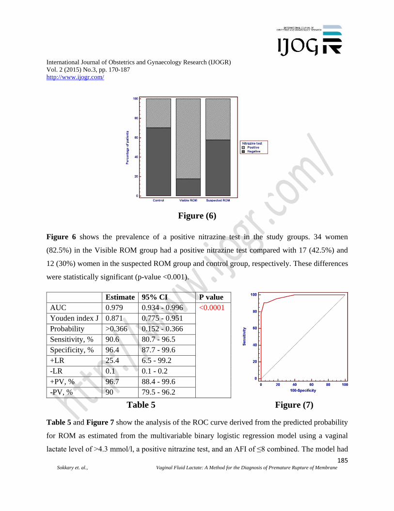
Figure (6)
Figure 6 shows the prevalence of a positive nitrazine test in the study groups. 34 women
(82.5%) in the Visible ROM group had a positive nitrazine test compared with 17 (42.5%) and
12 (30%) women in the suspected ROM group and control group, respectively. These differences
were statistically significant (p-value <0.001).
Estimate 95% CI P value
AUC 0.979 0.934 - 0.996 <0.0001
Youden index J 0.871 0.775 - 0.951
Probability >0.366 0.152 - 0.366
Sensitivity, % 90.6 80.7 - 96.5
Specificity, % 96.4 87.7 - 99.6
+LR 25.4 6.5 - 99.2
-LR 0.1 0.1 - 0.2
+PV, % 96.7 88.4 - 99.6
-PV, % 90 79.5 - 96.2
Table 5 Figure (7)
Table 5 and Figure 7 show the analysis of the ROC curve derived from the predicted probability
for ROM as estimated from the multivariable binary logistic regression model using a vaginal
lactate level of >4.3 mmol/l, a positive nitrazine test, and an AFI of ≤8 combined. The model had
Page 17
International Journal of Obstetrics and Gynaecology Research (IJOGR)
Vol. 2 (2015) No.3, pp. 170-187
http://www.ijogr.com/
186 Sokkary et. al., Vaginal Fluid Lactate: A Method for the Diagnosis of Premature Rupture of Membrane
an excellent diagnostic value as evidenced by an area under the ROC curve (AUC) of 0.979
(95% CI, 0.934 - 0.996; p-value, <0.0001). The best cut-off criterion was a predicted probability
of >0.366 (Youden index, 0.871; 95% CI, 0.775 - 0.951). This had a sensitivity of 90.6% (95%
CI, 80.7% - 96.5%), a specificity of 96.4% (95% CI, 87.7% - 99.6%), a +LR of 25.4 (95% CI,
6.5 - 99.2), a -LR of 0.1 (95% CI, 0.1 - 0.2), a +PV of 96.7% (95% CI, 88.4% - 99.6%), and a -
PV of 90% (95% CI, 79.5% - 96.2%).
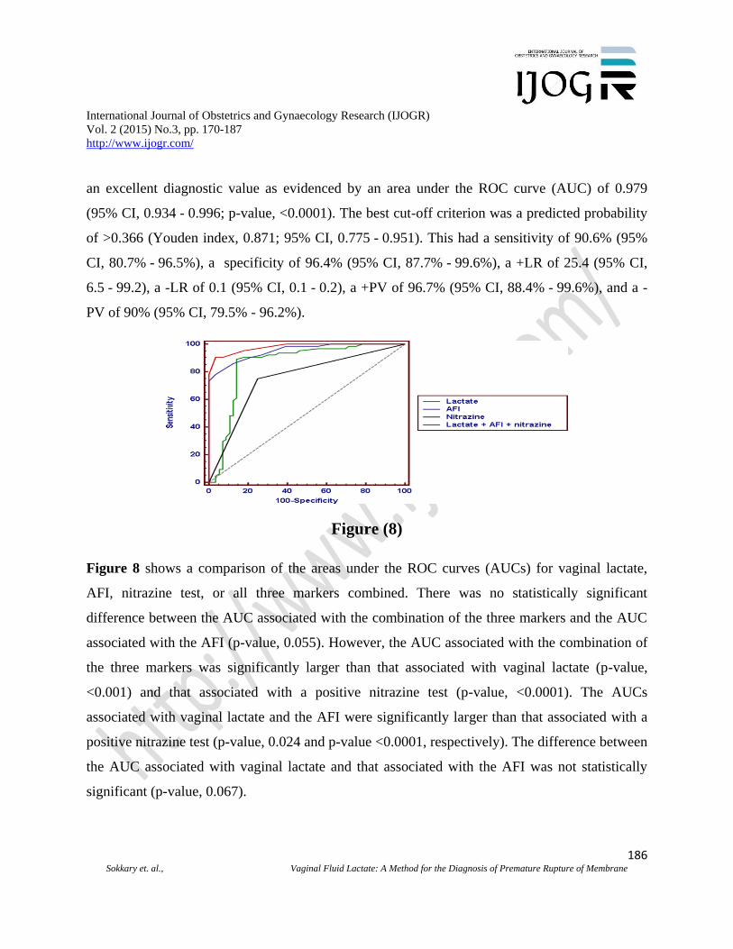
Figure (8)
Figure 8 shows a comparison of the areas under the ROC curves (AUCs) for vaginal lactate,
AFI, nitrazine test, or all three markers combined. There was no statistically significant
difference between the AUC associated with the combination of the three markers and the AUC
associated with the AFI (p-value, 0.055). However, the AUC associated with the combination of
the three markers was significantly larger than that associated with vaginal lactate (p-value,
<0.001) and that associated with a positive nitrazine test (p-value, <0.0001). The AUCs
associated with vaginal lactate and the AFI were significantly larger than that associated with a
positive nitrazine test (p-value, 0.024 and p-value <0.0001, respectively). The difference between
the AUC associated with vaginal lactate and that associated with the AFI was not statistically
significant (p-value, 0.067).
Page 18
International Journal of Obstetrics and Gynaecology Research (IJOGR)
Vol. 2 (2015) No.3, pp. 170-187
http://www.ijogr.com/
187 Sokkary et. al., Vaginal Fluid Lactate: A Method for the Diagnosis of Premature Rupture of Membrane
Vaginal lactate
≤4.3 mmol/l
(n=55)
Vaginal lactate
>4.3 mmol/l
(n=65)
Logrank test
Observed number with
onset of labor
Expected number with
onset of labor
6
17.1
42
30.9
Chi-square 19.723
DF 1
P value < 0.0001
Median time to onset of
labor (hr)
25.5 10
Hazard ratio 3.9 (95% CI,
2.1 to 7.0)
Table 6 Figure(9)
Table 6 and Figure 9 show the results of the Kaplan-Meier analysis for the time to onset of
labor. The median time to onset of labor was 25 h in women with vaginal lactate of ≤4.3 mmol/l
compared with 10 h in those with vaginal lactate of >4.3 mmol/l with a hazard ratio of 3.9 (95%
CI, 2.1 to 7.0; p-value, <0.0001).