NSH Symposium Seattle, WA Validation and Quality Control for IHC September, 2010 1 Validation and Quality Control for Immunohistochemistry Tim Morken, B.A. HTL(ASCP) Univ. California San Francisco Medical Center [email protected]Patsy Ruegg, HT(ASCP)QIHC IHCtech, LLC., Aurora, CO [email protected]NSH Symposium, Sept 2010 Seattle, Washington 2 Presentation Goals 1. Explain the term "Quality Control" 2. Discuss QC in the context of a "Quality System“ 3. Explain how proper validation leads to quality 4. Outline an IHC Quality System 5. Institute Quality Improvement using QC data. Upon completion of this presentation, participants will be able to: 3 Presentation Outline Quality Management System CAP / CLIA Requirements Assay Design Specifications Assay Validation Quality Control BREAK ER / PR / Her2 Validation Open discussion
Transcript
NSH SymposiumSeattle, WAValidation and Quality Control for IHC
September, 2010
1
Validation and Quality Control for Immunohistochemistry
Tim Morken, B.A. HTL(ASCP)Univ. California San Francisco Medical Center
Quality Assurance For Immuncytochemistry: Approved Guideline• Clinical Laboratory Standards Institute (formerly NCCLS) publication MM4-
A, Vol. 19, No. 26, 1999
Quality Assurance in Anatomic Pathology (book)• Nakhleh and Fitzgibbons, Ed., College of American Pathologists, 2005
College of American Pathologists (CAP)• General and Anatomic Pathology Accreditation Checklists• Current edition is dated June 15, 2009 (as of August 2010)
Reference handout
9
Quality Management
Quality Management
• Definitions
• Interaction
NSH SymposiumSeattle, WAValidation and Quality Control for IHC
September, 2010
4
10
CAP Checklists: Quality Management
General Laboratory Checklist:• GEN.13806 - 20369
• Quality Management Program
• Quality Monitoring
Anatomic Pathology Checklist• ANP.10000
• Is Quality Management Program Defined and Documented?
11
College of American Pathologists
Three Definitions concerning Quality Management:• 1) Quality Assurance (QA)• 2) Quality Control (QC)• 3) Quality Improvement (QI)
Together these constitute a "Quality System"• Interaction of these elements leads to better quality• System must be "worked" to succeed.
12
CAP Quality Assurance Definition
1) Quality Assurance• The practice of assessing performance in
all steps of the laboratory testing cycle including pre-analytic, analytic and post-analytic phases to promote excellent outcomes in medical care.
NSH SymposiumSeattle, WAValidation and Quality Control for IHC
September, 2010
5
13
… Deconstructed
"…assessing performance…"• Requires a set of performance standards• Requires comparison of results against the
standards
"…to promote excellent outcomes…"• Requires action if standards are not met
14
CAP Quality Control Definition
2) Quality Control• An integral component of quality assurance
consisting of the aggregate of processes and techniques to detect, reduce, and correct deficiencies in an analytical process.
15
… Deconstructed
"…processes and techniques to detect, reduce, and correct deficiencies…"
• "…Detect…": • Requires procedures that anticipate possible non-conformances.
• "…Reduce and Correct…": • Leads to procedures that reduce or eliminate confounding results.
NSH SymposiumSeattle, WAValidation and Quality Control for IHC
September, 2010
6
16
CAP Quality Improvement Definition
3) Quality Improvement
• The practice of continuously assessing and
adjusting performance using statistically and
scientifically accepted procedures.
17
… Deconstructed
"…continuously assessing and adjusting…"• Requires regular review of results data• Investigation of, and correction of root cause
"..statistically and scientifically…"• Data trends over a time period• Hypothesis of root cause• Eliminating variables• Testing solutions
18
Quality Management System Summary
Quality Assurance• Describe the quality process• Set standards
Quality Control• Institute methods to identify problems• Record and report results
Quality Improvement• Track problem trends identified by QC results• Use trend information to correct problems
NSH SymposiumSeattle, WAValidation and Quality Control for IHC
Quality Control• Controls identified by validation• Standardized procedures• Identification of process deficiencies
21
Validation – Optimization - Standardization
Verification
Optimization
Standardization
NSH SymposiumSeattle, WAValidation and Quality Control for IHC
September, 2010
8
22
CAP IHC Checklist
ANP.• 12425 ASR disclaimer for report• 21850 Positive and Negative controls for immunofluorescence• 22250 Procedure for each antibody• 22300 Documented modifications for fixation other than formalin• 22500 Buffer pH monitoring• 22550 Positive control use• 22570 Negative control use (tissue, reagent)• 22615 Avidin/Biotin blocking/controls• 22660 Review/Recording of IHC results• 22750 Validation of new antibody (except ER, PR, Her2)• 22760 New lot validation for Antibody and Detection system• 22800 Automated IHC staining instrument maintenance records• 22900 Quality of IHC stains• 22997 Her2 validation (IHC and ISH)
23
Validation Definition (1)
Establishing documented evidence which provides a high degree of assurance that a specific process will consistently produce a result or product meeting it's predetermined specifications and quality attributes
24
Validation Definition (2)
"Establishing documented evidence…"• Documentation of validation testing is readily available
“…which Provides a high degree of assurance…"• Studying an adequate number of samples to give
confidence that the new or changed process will work in your laboratory
NSH SymposiumSeattle, WAValidation and Quality Control for IHC
September, 2010
9
25
Validation Definition (3)
"…that a specific process will consistently produce…"• Identifying areas of actual or potential weaknesses so
improvements can be made prior to implementation.
"…a result or product meeting it's predeterminedspecifications and quality attributes."
• Establishing acceptance criteria before initiating the validation study.
26
Introducing a New Antibody
We want to introduce a new antibody…• Who wants it?
• What is it to be used for?
• When will it be used?
• Where will it be used?
• Why do we need it?• How will it be implemented?
27
Initial Assay Design Specification
Write a Specification:• The need for the assay
• The "why's"• Identify special requirements• Identify suppliers• Determine the expected results• Determine the validation procedure• Optimize the assay• Develop the standard protocol
NSH SymposiumSeattle, WAValidation and Quality Control for IHC
• Determine best controls• Range of expression, similar to expected cases (normal or disease?)• Preferably acquired and processed in your institution
Determine Precision (Reproducibility)• Intra-run: 10 slides in one run• Inter-run: 10 slides, Ten different runs with one slide each• Should have similar staining pattern and intensity on all slides
33
Validation Analytic Terms Applied to IHC
Accuracy:• Compare results with New antibody to a previously validated antibody
Precision:• Test samples with varying antigen expression• Intra-run, Inter-run tests, 10 slides each
Sensitivity:• True Positive vs False Negative (higher % FN = less sensitive)
Interferences [Specificity]:• True Negative vs False Positive (Higher % FP = less specific)• What could interfere to give a false positive or negative result?
Reportable Range• Establish a scoring system• Definition of a positive result• Criteria for rejection of the test
NSH SymposiumSeattle, WAValidation and Quality Control for IHC
September, 2010
12
34
Sensitivity
Analytic Sensitivity: • Lowest amount of substance detectable by the test
• Can only be done with controls of known concentration
Diagnostic Sensitivity:• Ability of the test to determine true diagnostic positive verses
false negative (higher % FN = less sensitive)• Requires comparison to a previously validated antibody
IHC Sensitivity:• Extent to which an antibody can be diluted and still achieve
target recognition.• NOTE: This is determined by antibody AND detection system!
(adapted from: Theoretical and Practical Aspects of Test Performance, in Immunomicroscopy, Taylor & Cote, 2005)
35
Dilution series for Sensitivity (1)
Dilution series to determine sensitivity
1:25 1:50
1:100 1:200
36
Dilution series for Sensitivity (2)
Dilution series to determine sensitivity
1:400
1:800
NEG CONTL
NSH SymposiumSeattle, WAValidation and Quality Control for IHC
September, 2010
13
37
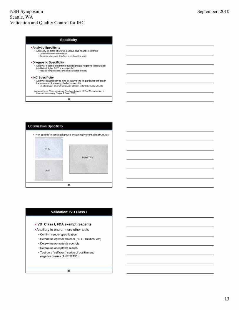
Specificity
Analytic Specificity• Accuracy on tests of known positive and negative controls
• Controls of known concentration• Determine what could “Interfere" to confound the result
Diagnostic Specificity• Ability of a test to determine true diagnostic negative verses false
positives (Higher % FP = less specific)• Requires comparison to a previously validated antibody
IHC Specificity• Ability of an antibody to bind exclusively to its particular antigen in
the absence of staining of other molecules• Or, staining of other structures in addition to target structures/cells
(adapted from: Theoretical and Practical Aspects of Test Performance, inImmunomicroscopy, Taylor & Cote, 2005)
38
Optimization Specificity
“Non-specific” means background or staining irrelvant cells/structures
1:400
1:800
NEGATIVE
39
Validation: IVD Class I
IVD Class I, FDA exempt reagentsAncillary to one or more other tests
• Confirm vendor specification• Determine optimal protocol (HIER, Dilution, etc)• Determine acceptable controls• Determine acceptable results• Test on a “sufficient” series of positive and
negative tissues (ANP.22750)
NSH SymposiumSeattle, WAValidation and Quality Control for IHC
September, 2010
14
40
Validation: Class II
IVD Class II FDA reagentsPredictive markers (ER, PR, Her2)
• Confirm vendor specification• Determine optimal protocol• Determine acceptable controls• Determine acceptable results• Validate on mix of 20-40 (ER, PR) or 25 to 100
(Her2) or more known positive and negative cases• Compare to previously validated tissue samples
(Her2: adapted from Arch Pathol Lab Med. 2007; 131:18-43)(ER, PR: adapted from Arch Pathol Lab Med. 2010; 134(6):930-935)
41
Validation: ASR
ASR: Analyte Specific Reagent (ANP.12425)“Active ingredient” of a testNot validated by vendor
• Vendor cannot indicate protocol to customer• Vendor cannot specify expected results• Laboratory is responsible for entire validation
• Expected results• Statistically valid test cohort (number, type of cases)• Comparison to similar test (i.e., another antibody to same target)• Documented results
42
Validation: RUO
RUO: Research Use Only
FDA Says: Laboratories should not use RUO reagents.
CLIA says: CLIA-certificated laboratories may use any reagent as long as CLIA validation procedures are followed (CAP is the deemed accrediting agency for Anatomic Pathology under CLIA)
CAP says: Nothing in current checklist about RUO’s.
If your laboratory decides to use an RUO reagent:
• Document unsuccessful search for IVD/ASR reagent• Follow ASR validation procedures: Comprehensive validation.• CAP Method Validation checklist: Gen.42020 - 42160
NSH SymposiumSeattle, WAValidation and Quality Control for IHC
September, 2010
15
43
Positive Control Tissue ANP.22550
Ideally, same specimen type as case tested• Not always possible, so validation documents acceptable tissue
• Remember, some antigens are decreased in tumor, not elevated
• Normal tissue should have consistent level of antigen • ( However, is “normal” tissue really normal?)
Fixed / processed with same procedures as sampleBest practice: put on same slide as sample
• However, one control per run, per antibody is acceptable.
Internal positive controls may be used,• Document for which cases/antibodies it is acceptable
Low expressers are ideal to avoid false negativesMulti-tissue blocks with positive and negative tissues are ideal
44
Negative Control Tissue ANP.22570
Tissue that is known to lack the antigen• Multi-tissue blocks, may have positive and negative
tissues
• Tissue elements within the positive control or test samples that should be negative
• Separate single negative tissue slide.
45
Optimization Overview
Verify Vendor Specifications• “to ascertain the truth or correctness of, as by examination,
research, or comparison”Vendor literature is a starting point
• Review the datasheet• Review references given by vendor • Literature search adds to knowledge• Compare with other vendors, users
Verify/Optimize:• Antigen Retrieval• Dilution• Control tissue• Expected results
NSH SymposiumSeattle, WAValidation and Quality Control for IHC
September, 2010
16
46
Optimization Method
Optimization• Vendor-supplied protocols are recommendations• Ideally work it into your regular protocol• Antigen retrieval type (Try several)
• None• Digestion (two or more types)• HIER (range of pH, temperature, time)
• Dilution series (bracket vendor recommendation)• Detection efficiency• Chromogen efficiency• Staining method (Manual or Automated)
47
Multi-expression tissue
LOW
MED
HIGH
48
Optimization Result
Write an optimized procedure for each antibody, including:
• .22250: Test Procedure • reagents, dilution, HIER, etc
NSH SymposiumSeattle, WAValidation and Quality Control for IHC
September, 2010
17
49
Standardization
Inter-Laboratory
Intra-Laboratory
50
Inter-Laboratory Standardization
Effort to standardize procedures for IHC• Studies show large variations in procedures and results between
laboratories (CAP, UK-NEQAS, NordiQC)
• Long standing effort to promote standardization• Largely failed in US due to lack of consequences
• UK-NEQAS has had success - took 20 years to achieve
• Oncologists, due to variable Her2 results, are driving current efforts
• Recommendations for Improved Standardization of Immunohistochemistry• Appl Immunohistochem Mol Morph 2007 15(2);124-133
• Recommendations for Human Epidermal Growth Factor Receptor 2 Testing in Breast Cancer, American Society of Clinical Oncology/College of American Pathologists Guideline,
• Arch Pathol Lab Med V.131 Jan 2007, pp.18-43.
51
Standardization within the Lab
Standardization• Research current literature for acceptable protocol
and "best" results (also UK-NEQAS, NordiQC)• Once validated and optimized the procedures must
be followed:• Every time!!!• By everyone!!!
• Record and report deviations from procedure• Helps with troubleshooting• Train your staff to accept that mistakes or variations will happen,
and admitting them is the first step of good troubleshooting.
NSH SymposiumSeattle, WAValidation and Quality Control for IHC
September, 2010
18
52
Validation: Post-Analytical
Controls• Define correct Scoring / Interpretation of staining• Define rejection criteria• Define reporting parameters• Document pathologist interpretation and performance• Determine variations from standard over time
• Reagent performance• Interpretation
53
Quality Control
Processes and techniques to detect, reduce and correct deficiencies in an
analytic process.
54
Quality Control Interpretation
Detect Deficiencies• Positive Control Tissue
• Negative Control Tissue
• Positive Control reagent (primary antibody)
• Negative Control Reagent
NSH SymposiumSeattle, WAValidation and Quality Control for IHC
September, 2010
19
55
QC: Pos / Neg Tissue Controls
Control Tissue• Positive Tissue Control
• If True Positive: Proves the system worksTests specific reactivity of primary antibodyRange of expression levels help determine sensitivityTests detection system
• If Negative: indicates some part of the system did not work No indication of what failed!
• Negative Tissue Control (negative for antibody)• Indicates specificity of primary antibody (cross-reactivity)• Indication of detection system problems (background, etc)
56
QC: Positive Reagent Controls
Positive Reagent Control: Primary Antibody• On Positive Control tissue
• Must be positive on the positive control tissueControls with range of expression will indicate sensitivityInternal control on patient tissue help determine tissue reactivity
• Negative control tissue must be negative (ANP.22570)Internal negative elements (Positive control or sample)Separate tissue block known to be negative for antibodyAt least one negative tissue control per antibody
Pre-immune serum from same animal (very rarely available)Isotype-specific negative control antibodyIrrelevant primary antibody from same species (expensive)Non-immune whole serum from same species (most common)Antibody diluent onlyWash buffer only
• Result: Must be negative on positive and negative tissuesIndicates specificity (cross reaction)Indicates problems with detection system (non-specific binding)Indicates problems with blocking reagents (not working?)Indicates patient tissue problems (fixation, processing, etc)
NSH SymposiumSeattle, WAValidation and Quality Control for IHC
September, 2010
20
58
Negative Control Reagent Exception
Special exception (comment to ANP.22570) :• If running:
• Two or more blocks from the same specimen,
• Received at the same time,
• Processed at the same time,
• For the same antibody:
• Only need to run a negative control reagent slide on one block of that specimen.
59
Quality Improvement
Quality Control Feeds Quality Improvement QC Results:
• Record results
• Track trends in deficiencies
• Open a Quality Improvement investigation for deficiencies identified.
• Determine a course of action to reduce or correct deficiency identified.
60
Quality Improvement: Reduce Deficiencies
Pre-Analytic• Standardized Acquisition, fixation, processing• Criteria for rejection of a specimen or sample
Analytical• Procedures detail optimal test protocol
• Must be followed by everyone to be effective
Post Analytical• Interpretation guidelines
• Acceptance criteria• Rejection criteria
NSH SymposiumSeattle, WAValidation and Quality Control for IHC
September, 2010
21
61
Quality Improvement: Correct Deficiencies
"Correction" entails (for example)• Do not perform test with inadequate specimen• Repeating the test• Determining what caused the failure• Testing different processes• Testing different reagents
Review trends at "defined" intervals• Monthly? Quarterly? (GEN.20262)
Identify most common and most serious issuesDetermine plan of action to identify root causeDetermine action to resolveTest solution(s)Determine if issue is resolved
Review Quality Assurance System (GEN.20369)• Review annually and determine if system works• Determine if improvements are needed
NSH SymposiumSeattle, WAValidation and Quality Control for IHC
September, 2010
22
64
Key Indicators
GEN:20316:Does the QM program include monitoring key indicators of quality?
• Some are defined in the CAP checklist• Patient/specimen ID accuracy
• Specimen Acceptability
• Some identified by trend analysis.• i.e. Failure/Repeat rate of a particular test
65
Break
Break!
66
Estrogen and Progesterone Receptor
ASCO/CAP Recommendations, ER, PR• VALIDATION of ER, PR:• Fitzgibbons, et. al., Arch Pathol Lab Med, June 2010
NSH SymposiumSeattle, WAValidation and Quality Control for IHC
September, 2010
23
67
Estrogen and Progesterone Receptor Validation (1)
ASCO/CAP Recommendations• Validate against previously validated tissue samples
• Another laboratory that has validated its assay against clinical outcomes• Validated tissue samples from another lab that uses an FDA-approved assay
and has validated the assay using ASCO/CAP testing requirements• Tissue samples validated using a separate assay (genetic, ligand binding
assay)• Tissues used in a proficiency testing program• Validated tissues provided by an established program
• ≥90% concordance with Positive validated samples• ≥95% concordance with Negative validated samples
68
Estrogen and Progesterone Receptor Validation (2)
ASCO/CAP Recommendations• Initial test validation of FDA-cleared assays
• ≥20 positive specimens (≥ 5 must be weakly positive)• ≥20 negative specimens• ≤ samples tested in one run (test samples in multiple runs, mutliple operators)
• Or Follow verification procedures in the Assay insert • Test must be used unmodified from manufacturers instructions
• Initial test validation of Laboratory Modified Assays (LMA)• If the lab modifies the test in ANY way a more thorough validation is called for• ≥40 Positive specimens ( ≥10 must be weakly postive)• ≥40 Negative specimens
69
CAP Her2 Validation (1)
ANP.22997- Her2 test validation• Wolff AC, et. al., Arch Pathol Lab Med 2007;131:18-43 • 25 - 100 cases, mix of:
• Variety of expression levels• Negative cases
• Compare to a validated alternative method (one or more)• Other antibody• FISH
• Fixation validation, if non-formalin• Validation of each change in methodology
NSH SymposiumSeattle, WAValidation and Quality Control for IHC
September, 2010
24
70
CAP Her2 Validation (2)
ANP.22998 Documented procedure for length of fixation
• 10% Neutral-buffered formalin (NBF)
• Minimum 6 hours, maximum 48 hours (under review)
• Record duration of fixation
• Qualify negative results of specimens fixed over 48 hours, consider confirmatory FISH testing
• Outside referral documentation of fixation duration
ANP.22999 Does lab use ASCO/CAP her2 scoring criteria?
71
Other Controls for IHC
Tissue arraysCell Culture slidesPeptide spots
72
Tissue Arrays as Controls
Tissue arrays allow extensive testing on one slide• Small arrays for particular antibodies
Brain, tonsil, colon, lung, thyroid, uterus, prostate, breast ca, placenta, melanoma, thymus/thymoma, skeletal muscle
• Larger arrays for validation• "FDA" array with 33 normal tissues, 40 tumors
Biochain, Pantomics, others• 10's or 100's of Ca-types / mix for pos / neg validation
NSH SymposiumSeattle, WAValidation and Quality Control for IHC
September, 2010
25
73
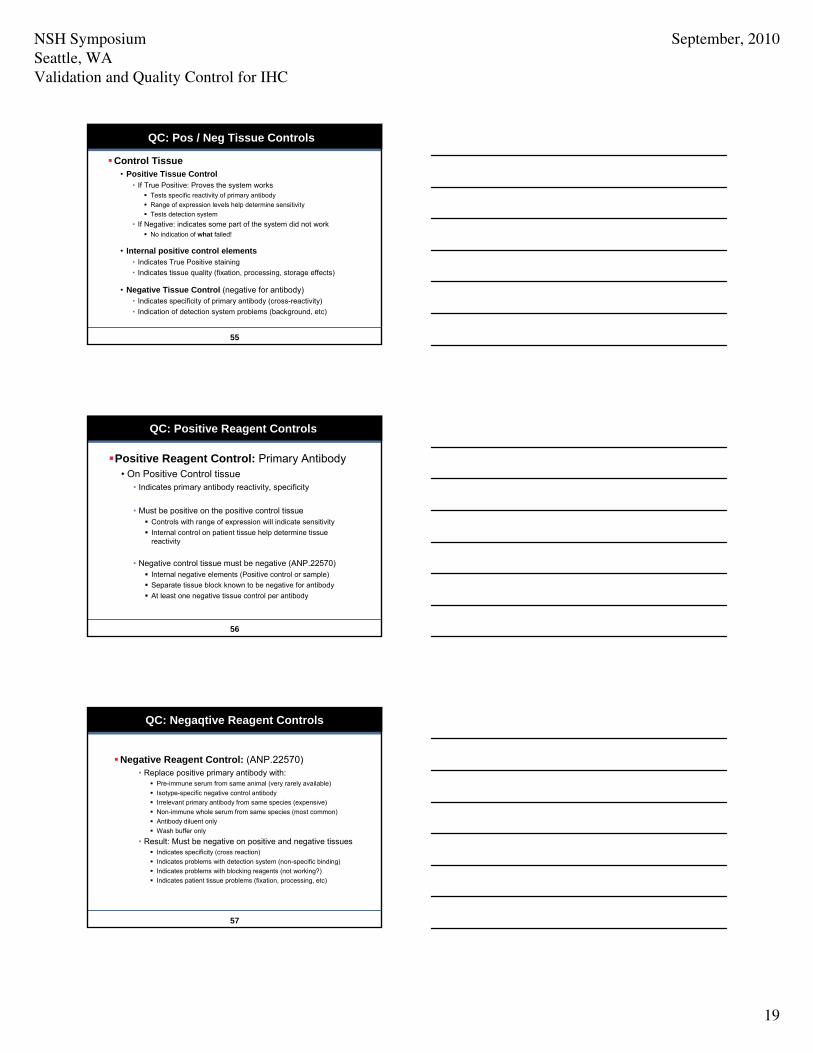
Array Size variety
Variety of arrays sizes/composition
24
6595
150
74
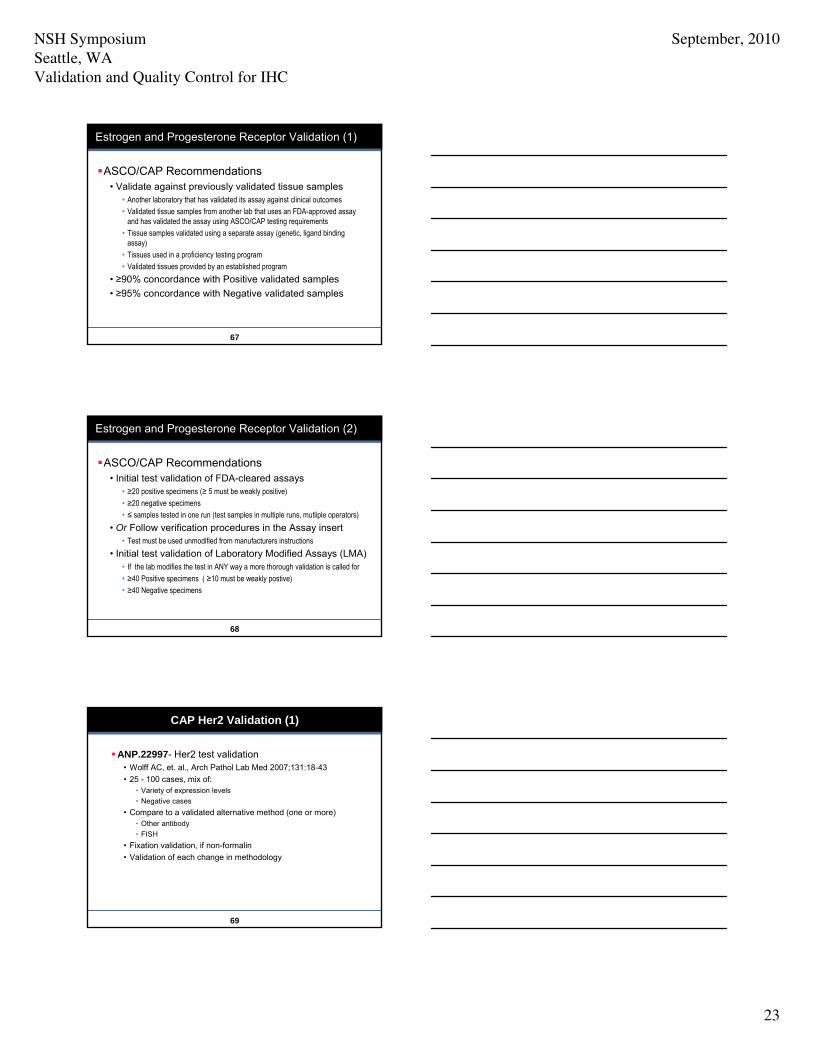
Predictive Test Validated Arrays
Her2, ER, PR, Validated arrays: 3+, 2+, 1+, Neg
Courtesy: Pantomics, Ltd, www.pantomics.com
75
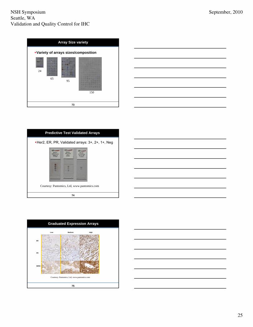
Graduated Expression Arrays
Low Medium High
ER
PR
HER2
Courtesy: Pantomics, Ltd, www.pantomics.com
NSH SymposiumSeattle, WAValidation and Quality Control for IHC
September, 2010
26
76
Cell Culture Controls
Genetically engineered expression level• Her2
• Estrogen / Progesterone Receptor
• Human papilloma virus
Fixed, processed in formalin/paraffin
Excellent for validation of tissue controls
77
Cell Culture Control Stain
Positive control cells using no AR has more background in serum than when AR used
POSITIVE HER2 CELLS W/O AR POSITIVE HER2 CELLS W/ AR
78
Peptide Spots
Peptide Spots (concept)• Peptide simulates epitope of native antigen• Peptides can be blended with several epitopes of different
antibodies
• Peptides do not degrade during deparaffinization and HIER
• Peptides can be produced synthetically in infinite amounts, withidentical quality
• Can help detect changes in antibody dilution and HIER
NSH SymposiumSeattle, WAValidation and Quality Control for IHC
September, 2010
27
79
Peptide Spots image
Peptide spots are positive or negative
NEG AR NEG NO AR
+ AR + NO AR
80
Peptide Spots vs Tissue Array
Top: Peptide spots (2 rows)
Middle: Cultured Cells
Bottom: Multi-tissue arrays with range of epressions
81
Peptide Controls in Practice
Peptide controls included with CAP 2006 Her2-B proficiency testing survey
• 18.3% sub optimal staining as judged by peptide control• 35% due to HIER errors• 20% due to antibody or staining protocol• 45% due to combination of the two
NSH SymposiumSeattle, WAValidation and Quality Control for IHC
September, 2010
28
82
Review
Quality Control is only part of the picture• QC is part of the Quality Management System
Quality Control is dependent on proper Validation• Validation determines protocols and controls
Quality Control feeds data to Quality Improvement• Trend analysis identifies problems
Questions?
Thank you.
Validation of Primary Antibody
Antibody Validation 1-1-2010 Page 1 of 6
Date: __________________ Project Inputs and Overall Design(s)
Name of Reagent:
Clone: Labeling* IVD ASR RUO
Proposed by Approved by
IVD = in vitro Diagnostic Device, FDA Approved; ASR = Anylate Specific Reagent, FDA regulated, RUO = Research Use Only, not FDA approved or regulated
Intended Use Diagnostic (IVD, ASR required) Research Immunohistochemistry Immunofluorescence In situ hybridization Others:
This product is intended for : Description of reagent:
Expected Staining Pattern:
Positive Control: Others: Others:
Sources of Input References Date
Company: Labeled: IVD ASR RUO Market Information Clone/Animal host: Company: Labeled: IVD ASR RUO Market Information Clone/Animal host: Company: Labeled: IVD ASR RUO Market Information Clone/Animal host: Title: Reference: Scientific Literature Conclusion: Title: Reference: Scientific Literature Conclusion: Title: Reference: Scientific Literature Conclusion:
Validation of Primary Antibody
Antibody Validation 1-1-2010 Page 2 of 6
Validation Design Input Describe the validation requirements
Platform (circle one) Dako Autostainer Leica Bond Ventana Ultra Manual Antibody Antigen Retrieval method Blocking regime Primary Dilution recommendation (initial trial) Primary antibody incubation time Detection system Chromogen Reproducibility testing None Inter-run (# slides_____) Intra-run (# slides_____) Control tissues: Tissue Case Number Positive element Additional testing required:
Approved by IHC Lead Technologist Date Medical Director, Immunohistochemistry Date
Design Output: First Trial Evaluation of Antibody
Reagent Source Catalog number Lot Number Date
Test (IHC, ISH, IF) Date Pass Fail Comments
See attached test records Validation/Verification Results Reagent does / does not match criteria detailed in design specification
Describe results:
Approved by IHC Lead Technologist Date Date Medical Director, Immunohistochemistry Date
Validation of Primary Antibody
Antibody Validation 1-1-2010 Page 3 of 6
Optimization Instructions (First Pass) Step Modification Note Date
Optimizaton Results
Test (IHC, ISH, IF) Modification Date Pass Fail Comments
Optimization Instructions (Second Pass)
Step Modification Note Date
Optimizaton Results
Test (IHC, ISH, IF) Modification Date Pass Fail Comments
Optimization Instructions (Third Pass)
Step Modification Note Date
Optimizaton Results
Test (IHC, ISH, IF) Modification Date Pass Fail Comments
Validation of Primary Antibody
Antibody Validation 1-1-2010 Page 4 of 6
Optimized Procedure
Platform (circle one) Dako Autostainer Leica Bond Ventana Ultra Manual Antibody Antigen Retrieval method Blocking regime Primary Dilution recommendation (initial trial) Primary antibody incubation time Detection system Chromogen Control tissues: Tissue Case Number Positive element(s) Attach list if extra control tissue necessary
Approved by Medical Director, Immunohistochemistry Date IHC Lead Technologist Date
Validation of Primary Antibody
Antibody Validation 1-1-2010 Page 5 of 6
Reproducibility Intra-Run reproducibility: 5 to 10 identical slides within one run
Test Date Pass Fail Comments IHC IHC IHC
See attached test records Inter-run reproducibility: 5 to 10 identical slides on 5 to 10 separate runs
Test Date Pass Fail Comments IHC IHC IHC
See attached test records Reproducibility approval Reagent does / does not meet reproducibility criteria
Approved by IHC Lead Technologist Date Medical Director, Immunohistochemistry Date
Validation of Primary Antibody
Antibody Validation 1-1-2010 Page 6 of 6
Design Validation Validation criteria Y N N/A Do results of internal and / or external (consultants / pathologist) testing meet the requirements and specifications of the reagent?
Are test results on panel of normal and tumor tissues acceptable?
Are reproducibility tests acceptable? Validation report:
Does reagent meet specification criteria?
Positive staining criteria:
Rejection criteria:
Comments:
Final Approval Medical Director, Immunohistochemistry Date Date
CoPath Entry
Name Abbreviation Search terms Description Label Text Entered in Copath Date: By:
Validation and Quality Control for Immunohistochemistry NSH Symposium
Seattle, WA September, 2010
References
1 of 7
Guidelines and Accreditation Checklists Quality Assurance For Immuncytochemistry: Approved Guideline, Clinical Laboratory Standards Institute (formerly NCCLS), Wayne PA, USA, publication MM4-A, Vol. 19, No. 26, 1999. www.clsi.org College of American Pathologists, Commission on Laboratory Accreditation. College of American Pathologists, Northfield, IL, USA, www.cap.org
September 27, 2007 Edition is the current edition in use (as of April, 2009) General Laboratory Checklist
Anatomic Pathology Checklist Clinical Laboratory Improvement Amendments (CLIA), Centers for Medicare and Medicaid Services, CMS-2226-F: 42 CFR 493 Interpretive Guidelines for Laboratories, Appendix C, Subpart K (Quality systems). www.cms.hs.gov/CLIA/03_interpretive_guidelines_for_laboratories.asp Books Quality Management in Immunohistochemistry, Brown RW, in Quality Management in Anatomic Pathology, Nakhleh RE, and Fitzgibbons PL, Eds. College of American Pathologists, Northfield, IL, USA, 2005. www.cap.org. Theoretical and Practical Aspects of Test Performance, in Immunohistology: A Diagnostic Tool for the Surgical Pathologist. 3rd. Ed., Volume 19 in Major Problems in Pathology, Taylor CR and Cote RJ, Eds., W.B Saunders, Philadelphia, 2005 Immunohistochemistry Quality Control, Hladik, CL and White, CL, in Theory and Practice of Histological Techniques, 4th Ed., Bancroft JD and Gamble M, Eds., Churchill Livingstone, 2007 Techniques of Immunohistochemistry: Principles, Pitfalls and Standardization, Taylor CR, et.al., in Diagnostic Immunohistochemistry, Dabbs, DJ, Ed., 2nd Edition, Churchill Livingstone, Selected Literature Validation and Standardization Tissue Preparation for Immunocytochemistry, Williams, JH, et.al., J Clin Pathol 1997;50:422-428 Conditional Epitopes: Is Your Antibody Always Specific?, Willingham, MC, J Histochem Cytochem, Vol. 47(10):1233-1235, 1999 Recommended Policies for Uses of Human Tissue in Research, Education, and Quality Control, Grizzle, W, et.al., Arch Pathol Lab Med, 1999; 123: 296-300
Validation and Quality Control for Immunohistochemistry NSH Symposium
Seattle, WA September, 2010
References
2 of 7
Specificity Controls for Immunocytochemical Methods, Burry, RW, J Histochem Cytochem, Vol. 48(2): 163-165, 2000 A Practical Approach for Evaluating New Antibodies in the Clinical Immunohistochemistry Laboratory, Hsi, ED, Arc Pathol Lab Med. 2001; 125: 289-294 Immunohistochemistry for the Age of Molecular Morphology, Taylor, CR, editorial, Appl Immunohist Mol Morph, 9(1): 1-2, 2001 Standardization in Immunohistochemistry, O’Leary, TJ, Appl Immunohistochem Mol Morph, 9(1): 3-8, 2001 Immunohistochemical Detection of Interferon-γ: Fake of Fact?, Van der Loos, CM, et.al., J Histochem Cytochem 2001;49(6):699-709 Progesterone receptor by immunohistochemistry and clinical outcome in breast cancer: a validation study, Mohsin, SK, et.al., Mod Path (2004) 17, 1545-1554 Can a More Selective Application of Antigen Retrieval Facilitate Standardization in Immunohistochemistry?, Boenisch, T, Appl Immunohistochem Mol Morph, Vol. 12, No. 2, 172-176, June 2004 Technical Aspects of Immunohistochemistry, Ramos-Vara, JA, Vet Pathol 42:405-426 (20050 A Comparison of Immunohistochemical Stain Quality in Conventional and Rapid Microwave Processed Tissues, Emerson, LL, et.al., Am J Clin Pathol 2006; 125:176-183 Recommendations for Improved Standardization of Immunohistochemistry, Goldstein, NS, et.al., and members of Ad-Hoc Committee on Immunohistochemical Standardization, Appl Immunohistochem Mol Morph, 2007 15(2): 124-133 The use of positive controls in immunohistochemistry – Some indicators of what is appropriate and what is not, Immunocytochemistry 2007, vol. 6, Issue 1: 11-12, UK NEQAS for Immuncytochemistry and FISH, 2007 Suggested guidelines for Immunohistochemical techniques in veterinary diagnostic laboratories, Ramos-Vara, JA, et.al. J Vet Diagn Invest 20:393-413 (2008) Controls for IHC, NordiQC, www.nordiQC.org/techniques/controls_sections.htm, 2008 Sense and Sensitivity, Wikipedia, www.wikipedia.org/wiki/sensitivity_(tests) A good overview of the subject. Antibody Validation (Review). Bordeaux J, et. al., BioTechniques 48:197-209 (March 2010) Test Validation: A Brave new World for Anatomic Pathology, Brown, RW and Sharkey, FE, May 19, 2010, 2010 Laboratory Accreditation Program, College of American Pathologists (www.cap.org) Quality Assurance / Quality Control Interlaboratory quality assurance of Immunohistochemical procedures. Recommended practices for daily application, Rickert RR, Maliniak, RM, Arch Pathol Lab Med 1989 Jun;113(6):673-9. Comparative quality assessment in immunocytochemistry: pilot study of CD15 staining in paraffin wax embedded tissue in Hodgkin’s disease, Angel CA, et.al., J Clin Pathol 1989;42:1096-1100.
Validation and Quality Control for Immunohistochemistry NSH Symposium
Seattle, WA September, 2010
References
3 of 7
Quality control in immunohistochemistry. Report of a workshop sponsored by the Biological Stain Commission, Elias JM, et.al., Am J Clin Pathol 1989 Dec;92(6): 836-43. The Taming of Immunohistochemistry: The New Era of Quality Control, Herman GE and Elfont EA, Biotech & Histochem, 1991;66(4):194-9 Quality control in immunohistochemistry: Experiences with the estrogen receptor assay, Bosmon FT, et.al., J Clin Pathol 1992;45:120-124 Audit and internal quality control in immunohistochemistry, Maxwell P and McCluggage WG, J Clin Pathol 2000; 53:929-932 The Total Test Approach to Standardization of Immunohistochemistry, Editorial, Taylor CR, Arch Pathol Lab Med – 2000;124:945-951 Recommendations for Quality Assurance and Improvement in Surgical Pathology and Autopsy Pathology, Association of Directors of Anatomic and Surgical Pathology, Am J Clin Path 2006;126:337-340 Technical variations in prostatic immunohistochemistry: need for standardization and stringent quality assurance in PSA and PSAP immunostaining, Varma M, et.al., J Clin Pathol 2004;57:687-690 Controls for Immunohistochemistry: Is “Brown” Good Enough?, Editorial, Ward JM, Toxicologic Pathology, 32:273-274, 2004. The Quality Systems Approach, Nevalainen DE, Arch Pathol Lab Med, 1999;123: 566-568 Evaluating Laboratory Performance on Quality Indicators With the Six Sigma Scale, Nevalainen DE, et.al., Arch Pathol Lab Med April 2000;124:516-519 Digital Image Documentation for Quality Assessment, Cruz D, et.al., Arch Pathol Lab Med Nov 2001; 125:1430-1435 Blinded Review as a Method for Quality Improvement in Surgical Pathology, Renshaw AA, et.al., Arch Pathol Lab Med, 2002;126:961-963 Quality Assurance in Immunohistochemistry, Results of an Interlaboratory Trial Involving 172 Pathologists, Rudigerf T, et.al., Am J Surg Pathol 26(7):873-882, 2002 Source of Error in Laboratory Medicine, Matlow AG, et.al, Lab Med June 2004, Vol. 35, No. 6: 331-334 Quality Systems – the Role of People, Passiment E., LabMed 2005;36(10): 630-632 Reducing Errors in the Practices of Pathology and Laboratory Medicine, Novis DA and Konstantakos G, Am J Clin Pathol 2006;126(Suppl):S30-S35 What is quality in surgical pathology?, Nakhleh RE, J Clin Pathol 2006;59:669-672 My approach to internal quality control in a clinical immunology laboratory, Lock RJ, J Clin Pathol 2006;59:681-684 Diagnostic Immunohistochemistry: What can Go Wrong?, Yaziji, H and Barry T, Adv Anat Pathol, Vol. 13, No. 5: 238-246, Sept 2006
Validation and Quality Control for Immunohistochemistry NSH Symposium
Seattle, WA September, 2010
References
4 of 7
The Henry Ford Production System, Measures of Process Defects and Waste in Surgical Pathology as a Basis for Quality Improvement Initiatives, D’Angelo, R, et.al., Am J Clin Pathol 2007;128:423-429 Quality Management in Immunohistochemistry, Eisen RN, Diag Histopath 14(7):299-307 2008 External Quality Assessment in Immunohistochemistry – Is It the Solution to a Complex Problem?, Editorial, Reiner-Concin A, Breast Care 2008;3:78-79 Quality control in molecular immunohistochemistry, True LD, Histochem Cell Biol (2008) 130:473-480 Breast Carcinoma-specific QC Estrogen Receptor Reliability of Immunohistochemical demonstration of oestrogen receptors in routine practice: interlaboratory variance in the sensitivity of detection and evaluation of scoring systems, Rhodes A, et.al., J Clin Pathol 2000;53:125-30 The effect of fixation and processing on the sensitivity of oestrogen receptor assay by immunohistochemistry in breast carcinoma, Lee H, et.al., J Clin Pathol 2002;55:236-238 Minimum Formalin Fixation Time for Consistent Estrogen Receptor Immunohistochemical Staining of Invasive Breast Carcinoma. Goldstein NS, et. al., Am J Clin Pathol 2003;120:86-92 Estrogen Receptor Testing of Breast Cancer in Current Practice: What is the Question?, Schnitt SJ, J Clin Oncol, 2006;24(12):1797-1799 Frequency and reliability of oestrogen receptor, progesterone receptor and HER2 in breast carcinoma determined by immunohistochemistry in Australasia: results of the RCPA Quality Assurance Program, Francis GD, et.al., J Clin Pathol 2007;60:1277-1283 Effects of Fixation, Processing and Evaluation Criteria on Immunohistochemical Detection of Hormone Receptors in Breast Cancer, Oyama, T, et. al., Breast Cancer 14:182-188, 2007 Prognostic and Predictive Value of Centrally Reviewed Expression of Estrogen and Progesterone Receptors in a Randomized Trial by Comparing Letrozole and Tamoxifin Adjuvant Therapy for Postmenopausal Early Breast Cancer: BIG 1-98, Viale G, et.al., J Clin Oncol 25:3846-3852. 2007 Comparison of Evaluations for Hormone Receptors in Breast Carcinoma Using Two Manual and Three Automated Immunohistochemical Assays, Arihiro, K, et.al., Am J Clin Pathol 2007;127:356-365. Bad Cancer Tests Draw Scrutiny, Anne Wilde Mathews, Wall Street Journal, January 4, 2008. Commentary: Hormone Receptor Testing in Breast Cancer: A Distress Signal from Canada, Allred, DC, The Oncologist, 13; 1134-1136, Nov 2008 Consensus Recommendations on Estrogen Receptor Testing in Breast Cancer By Immunohistochemistry, Yaziji, H, et.al., including Members of the Standardization Ad-Hoc Consensus Committee, Appl Immunohistochem Mol Morphol, Vol. 16, No. 6, Dec 2008
Validation and Quality Control for Immunohistochemistry NSH Symposium
Seattle, WA September, 2010
References
5 of 7
American Society of Clinical Oncology/College of American Pathologists Guideline Recommendations for Immunohistological Testing of Estrogen and Progesterone Receptors in Breast Cancer. = Recommendations for Validating Estrogen and Progesterone Receptor Immunohistochemistry Assays, Fitzgibbons PL, et. al., Arch Pathol Lab Med. 2010; 134(6): 930-935 Her2 Specific QC HER2 testing in breast cancer: NCCN Task Force report and recommendations, J Natl Compr Canc Netw. 2006 Jul;4 Suppl 3:S1-S22 Concordance between central and local laboratory HER2 testing from a community-based clinical study, Reddy JC, et.al., Clin Breast Cancer 2006 Jun;7(2):153-7 Effects of Fixative and Fixation protocols on Assessment of Her2/neu Oncogene Amplification Status by Fluorescence In Situ Hybridization, Willmore-Payne, C, et.al., Appl Immunohistochem Mol Morph 2007;15:84-87 American Society of Clinical Oncology / College of American Pathologists Guideline Recommendations for Human Epidermal Growth Factor Receptor 2 Testing in Breast Cancer, Wolff AC, et.al., Arch Pathol Lab Med 2007;131:18-43 The Need for a Quality Control or the Whole Process of Immunohistochemistry Human Epidermal Growth Factor Receptor 2/neu Determination: A United Kingdom National External Quality Network for Quality Assessment Service / Italian Network for Quality Assessment of Tumor Biomarkers Pilot Experience, Paradiso A, et.al., Letter to Editor, J Clin Pathol 2007;25(22) HER2 Testing in A Population-Based Study of Patients with Metastatic Breast Cancer Treated with Trastusumab, O’Malley FP, et.al., Arch Pathol Lab Med 2008;132:61-65 Implementation of American Society of Clinical Oncology/College of American Pathologists HER2 Guideline Recommendations in a Tertiary Care Facility Increases HER2 Immunohistochemistry and Fluorescence In Situ Hybridization Concordance and Decreases the Number of Inconclusive Cases. Middleton LP, et. al., Arch Pathol Lab Med. Vol. 133:775-780. Controls Tissue Array Controls The multi-tumor (sausage) tissue block. Novel method for immunohistochemical antibody testing. Battifora H, Lab Invest 1986;55:244-8 Tissue Array Technology for Testing Interlaboratory and Interobserver Reproduciblity of Immunohistochemical Estrogen Receptor Analysis in a Large Multicenter Trial, von Wasielewski R, et.al., Am J Clin Pathol 118(5):675-682 Assessment of Interlaboratory variation in the Immunohistochemical determination of estrogen receptor status using a breast cancer tissue microarray, Parker RL, et.al., Am J Clin Pathol 2002 May;117(5):723-8 Tissue microarrays for predictive molecular pathology, G Sauter and M Mirlacher, J Clin Pathol 2002;55:575-576
Validation and Quality Control for Immunohistochemistry NSH Symposium
Seattle, WA September, 2010
References
6 of 7
Tissue microarrays: a new approach for quality control in immunohistochemistry, Packeisen J, et.al., J Clin Pathol 2002;55:613-615 Demystified: Tissue microarray technology, Packeisen, J, et.al., J Clin Pathol: Mol Pathol 2003;56:198-204 Miniature tissue microarrays for HercepTest® standardization and analysis, Gulmann C, et.al., J Clin Pathol 2004;57:1229-1231 The CD117 Immunohistochemistry Tissue Microarray Survey for Quality Assurance and Intralaboratory Comparison, Dorfman DM, et.al., A College of American Pathologists Cell Markers Committee Study, Arch Pathol Lab Med 2006;130:779-782 Clinical Validation of Breast Cancer Biomarkers Using Tissue Microarray Technology, Quraishi I, et.al., Appl Immunohistochem Mol Morph 2007;15:45-49
Validation and Quality Control for Immunohistochemistry NSH Symposium
Seattle, WA September, 2010
References
7 of 7
Cell Culture Controls Her2 A formalin-fixed, paraffin-processed cell line standard for quality control of Immunohistochemical assay of HER-2/neu expression in breast cancer, Rhodes A, et.al., Am J Clin Pathol 2002 Jan;117(1):7-8 Available from Dako www.dakousa.com, slides (available in kits only?) Ventana Medical www.ventanamed.com, slides Invitrogen www.invitrogen.com, slides or block available QC Sciences www.qcsciences.com slides Estrogen and Progesterone Receptor Dako (available in kits only?) Invitrogen Human papilloma virus QC Sciences, slides Invitrogen, slides or block Peptide Controls Synthetic Peptides Identified from Phage-displayed Combinatorial Libraries as Immunodiagnostic Assay Surrogate Quality-Control Targets, Sompuram SR, et.al., Clin Chem 2002;48(3):410-420 A Novel Quality Control Slide for Quantitative Immunohistochemistry Testing, Sompuram SR, et.al., J Histochem Cytochem 2002; 50(11):1425-1433 National HER2 Proficiency Test Results Using Standardized Quantitative Controls, Vani, K, et.al., Arch Pathol Lab Med 2008;132:211-216 Quality Assessment Organizations College of American Pathologists (USA). www.cap.org, Several quality management tools including Q-probes, Anatomic Pathology Surveys/IHC, Anatomic Pathology Education programs with IHC, HistoQIP program. United Kingdom National External Quality Assessment Scheme (UK-NEQAS), www.ukneqasicc.ucl.as.uk, Of great interest are the downloadable pdf’s of the UK-NEQAS Immunocytochemistry Journal, which discusses in detail the results of the IHC surveys the UK-NEQAS conducts each year. www.ukneqasicc.ucl.as.uk/neqasicc.shtml Nordic Quality Control. www.nordiqc.org. A quality control survey program of the Nordic countries. Extensive reviews of antibodies, procedures and suggestions for IHC.