+ All Categories
Home > Documents > Variants in nuclear factor I genes influence growth and ...

Variants in nuclear factor I genes influence growth and ...

Date post: 08-Dec-2021
Category:
Upload: others
View: 2 times
Download: 0 times
Share this document with a friend
50
Bunt Jens (Orcid ID: 0000-0003-0397-2019) Zenker Martin (Orcid ID: 0000-0003-1618-9269) Variants in Nuclear Factor I Genes Influence Growth and Development Martin Zenker 1,10 , Jens Bunt 2,10 , Ina Schanze 1 , Denny Schanze 1 , Michael Piper 2,3 , Manuela Priolo 4 , Erica H. Gerkes 5 , Richard M. Gronostajski 6 , Linda J. Richards 2,3 , Julie Vogt 7 , Marja W. Wessels 8 , Raoul C. Hennekam 9 1 Institute of Human Genetics, University Hospital Otto-von-Guericke-University, Magdeburg, Germany 2 Queensland Brain Institute, The University of Queensland, Brisbane 4072, Australia 3 School of Biomedical Sciences, The University of Queensland, Brisbane 4072, Australia 4 Operative Unite of Medical Genetics, Great Metropolitan Hospital Bianchi-Melacrino- Morelli, Reggio Calabria, Italy 5 Department of Genetics, University of Groningen, University Medical Center Groningen, Groningen, the Netherlands 6 Department of Biochemistry, Program in Genetics, Genomics and Bioinformatics, Center of Excellence in Bioinformatics and Life Sciences, State University of New York, Buffalo, NY 14203, USA 7 West Midlands Regional Clinical Genetics Service and Birmingham Health Partners, Birmingham Women's and Children's Hospitals NHS Foundation Trust, Birmingham, UK 8 Department of Clinical Genetics, Erasmus MC, University Medical Center Rotterdam, Rotterdam, the Netherlands 9 Department of Pediatrics, Amsterdam UMC – location AMC, University of Amsterdam, Amsterdam, the Netherlands 10 Martin Zenker and Jens Bunt should be considered joint first author Correspondence: This article is protected by copyright. All rights reserved. This is the author manuscript accepted for publication and has undergone full peer review but has not been through the copyediting, typesetting, pagination and proofreading process, which may lead to differences between this version and the Version of Record. Please cite this article as doi: 10.1002/ajmg.c.31747
Transcript

Bunt Jens (Orcid ID: 0000-0003-0397-2019) Zenker Martin (Orcid ID: 0000-0003-1618-9269) Variants in Nuclear Factor I Genes Influence Growth and Development

Martin Zenker1,10, Jens Bunt2,10, Ina Schanze1, Denny Schanze1, Michael Piper2,3, Manuela

Priolo4, Erica H. Gerkes5, Richard M. Gronostajski6, Linda J. Richards2,3, Julie Vogt7, Marja W.

Wessels8, Raoul C. Hennekam9

1 Institute of Human Genetics, University Hospital Otto-von-Guericke-University,

Magdeburg, Germany

2 Queensland Brain Institute, The University of Queensland, Brisbane 4072, Australia

3 School of Biomedical Sciences, The University of Queensland, Brisbane 4072,

Australia

4 Operative Unite of Medical Genetics, Great Metropolitan Hospital Bianchi-Melacrino-

Morelli, Reggio Calabria, Italy

5 Department of Genetics, University of Groningen, University Medical Center

Groningen, Groningen, the Netherlands

6 Department of Biochemistry, Program in Genetics, Genomics and Bioinformatics,

Center of Excellence in Bioinformatics and Life Sciences, State University of New York,

Buffalo, NY 14203, USA

7 West Midlands Regional Clinical Genetics Service and Birmingham Health Partners,

Birmingham Women's and Children's Hospitals NHS Foundation Trust, Birmingham, UK

8 Department of Clinical Genetics, Erasmus MC, University Medical Center Rotterdam,

Rotterdam, the Netherlands

9 Department of Pediatrics, Amsterdam UMC – location AMC, University of

Amsterdam, Amsterdam, the Netherlands

10 Martin Zenker and Jens Bunt should be considered joint first author

Correspondence:

This article is protected by copyright. All rights reserved.

This is the author manuscript accepted for publication and has undergone full peer review buthas not been through the copyediting, typesetting, pagination and proofreading process, whichmay lead to differences between this version and the Version of Record. Please cite this articleas doi: 10.1002/ajmg.c.31747

Martin Zenker, Institute of Human Genetics, University Hospital Magdeburg, Otto-von-

Guericke University, Leipziger Str. 44, 39120 Magdeburg, Germany. E-Mail:

[email protected]

or

Jens Bunt, Brain Development and Disorders Laboratory, Queensland Brain Institute, The

University of Queensland, Brisbane, Queensland 4072, Australia. E-Mail: [email protected]

Funding information:

National Health and Medical Research Council Australia, Grant/Award Number:

GNT1100443 and GNT1120615; German Ministry of Education and Research (BMBF),

Grant/Award Number: GeNeRARe 01GM1519A

This article is protected by copyright. All rights reserved.

Abstract

The nuclear factor one (NFI) site-specific DNA-binding proteins represent a family of

transcription factors that are important for the development of multiple organ systems,

including the brain. During brain development in mice, the expression patterns of Nfia, Nfib,

and Nfix overlap, and knockout mice for each of these exhibit overlapping brain defects,

including megalencephaly, dysgenesis of the corpus callosum and enlarged ventricles, which

implies a common but not redundant function in brain development. In line with these

models, human phenotypes caused by haploinsufficiency of NFIA, NFIB and NFIX display

significant overlap, sharing neurodevelopmental deficits, macrocephaly, brain anomalies and

variable somatic overgrowth. Other anomalies may be present depending on the NFI gene

involved. The possibility of variants in NFI genes should therefore be considered in

individuals with intellectual disability and brain overgrowth, with individual NFI-related

conditions being differentiated from one another by additional signs and symptoms. The

exception is provided by specific NFIX variants that act in a dominant negative manner, as

these cause a recognizable entity with more severe cognitive impairment and marked bone

dysplasia, Marshall-Smith syndrome. NFIX duplications are associated with a phenotype

opposite to that of haploinsufficiency, characterized by short stature, small head

circumference, and delayed bone age. The spectrum of NFI-related disorders will likely be

further expanded, as larger cohorts are assessed.

Keywords:

Nuclear factor one; NFIA; NFIB; NFIX; Marshall-Smith syndrome; Malan syndrome;

microdeletion 1p32-p31; macrocephaly; overgrowth; intellectual disability

This article is protected by copyright. All rights reserved.

1 INTRODUCTION

The nuclear factor one (NFI) family of DNA binding proteins was first identified as a regulator

of viral replication (Nagata, Guggenheimer, Enomoto, Lichy, & Hurwitz, 1982), but its

members were subsequently identified as important transcription factors (K. S. Chen, Lim,

Richards, & Bunt, 2017; Harris, Genovesi, Gronostajski, Wainwright, & Piper, 2015; Murtagh,

Martin, & Gronostajski, 2003). The proteins, previously also known as CCAAT box-binding

transcription factors (CTF) or TGGCA-binding proteins (Borgmeyer, Nowock, & Sippel, 1984),

have four members in vertebrates: NFIA, NFIB, NFIC and NFIX. They share an N-terminal DNA

binding and dimerization domain coded by exons 2 and 3, which is highly conserved

between family members as well as between species (Kruse & Sippel, 1994; Rupp et al.,

1990; Santoro, Mermod, Andrews, & Tjian, 1988). Consequently, all four members bind to

the same palindromic DNA binding motif as hetero- and homodimers (Gronostajski, Adhya,

Nagata, Guggenheimer, & Hurwitz, 1985; Hennighausen et al., 1985; Jolma et al., 2013;

Kruse & Sippel, 1994; Leegwater, van Driel, & van der Vliet, 1985). The NFI proteins can also

bind to half sites, but with lower affinity (Meisterernst, Gander, Rogge, & Winnacker, 1988).

In contrast, the C-terminal transactivation and repression domains differ between the

members, potentially providing differential binding to other protein partners. Moreover,

each member has multiple isoforms due to alternative splicing (Fletcher, Jenkins, Copeland,

Chaudhry, & Gronostajski, 1999). Together, these features of the NFI genes mean that they

are promiscuous DNA binding proteins with many different isoforms and identifying specific

functions for a given family member or isoform is therefore complex.

2 MOUSE STUDIES

This article is protected by copyright. All rights reserved.

Expression of the NFI genes has been studied in various tissues and cell types (reviewed by

Gronostajski and co-workers (Gronostajski, 2000), with most of our knowledge being derived

from analyses in mice. Based on in situ hybridization on mouse embryos, each family

member has a unique but overlapping expression pattern during neural development

(Chaudhry, Lyons, & Gronostajski, 1997), with Nfia, Nfib and Nfix expression being most

prominent within in the dorsal telencephalon and cerebellum. In the dorsal telencephalon,

Nfia, Nfib and Nfix are first expressed at embryonal day (E)11 within radial glia (Campbell et

al., 2008; Mason, Piper, Gronostajski, & Richards, 2009; Plachez et al., 2008). They continue

to be expressed in radial glia and ependymal cells, which arise from last radial glia (K. S.

Chen, Harris, et al., 2017; Vidovic, Davila, Gronostajski, Harvey, & Piper, 2018). The

expression patterns of the NFIA and NFIB proteins are initially similar, producing a high

caudo-medial to low rostro-lateral gradient in the developing dorsal telencephalon in mice

(Bunt, Lim, Zhao, Mason, & Richards, 2015). During neurogenesis, intermediate progenitors

show reduced expression of NFI proteins, whereas neurons of the deeper cortical layers

exhibit higher expression (Bunt et al., 2015; Plachez et al., 2008). This neuronal expression

persists into adulthood (K. S. Chen, Harris, et al., 2017). Similarly, NFIA and NFIB are

expressed in most astrocytes, while NFIA is the main NFI expressed in oligodendrocytes (K. S.

Chen, Harris, et al., 2017). In contrast, NFIX expression is mainly restricted to neurons and is

greater in the more superficial layer of the cortex, whereas glial expression is more limited

(K. S. Chen, Harris, et al., 2017). During development, NFIX expression is higher in

hippocampal intermediate progenitor cells, although it is also expressed by radial glial cells

(Harris et al., 2018).

These three genes are essential for brain development, as knockout mice for any of

them display severe brain phenotypes (Supporting Information Table S1)(Bunt et al., 2015;

Campbell et al., 2008; K. S. Chen, Harris, et al., 2017; das Neves et al., 1999; Driller et al.,

2007; Fraser et al., 2016; Lu et al., 2007; Plachez et al., 2012; Plachez et al., 2008; Shu, Butz,

Plachez, Gronostajski, & Richards, 2003; Steele-Perkins et al., 2005). No apparent brain

phenotype has been observed in the only Nfic knockout mouse model currently available

This article is protected by copyright. All rights reserved.

(Steele-Perkins et al., 2003), although this gene is expressed at low levels within the

developing nervous system (Chaudhry, Vitullo, & Gronostajski, 1998). The main brain

phenotypes shared between mice lacking Nfia, Nfib or Nfix include megalencephaly,

enlarged ventricles and/or hydrocephaly, malformation of the hippocampus and dysgenesis

of the corpus callosum (Figure 1). In addition to the brain phenotypes, each mouse model

also displays other defects unique to that family member (Figure 1a): Nfia knockout mice

demonstrate urinary tract and kidney anomalies (das Neves et al., 1999; Lu et al., 2007), Nfib

knockout mice exhibit lung defects (Steele-Perkins et al., 2005), Nfic knockout mice have

abnormal teeth (Steele-Perkins et al., 2003), and Nfix have bone abnormalities (Driller et al.,

2007). Given these additional organ system defects, Nfia, Nfib and Nfix knockout mice have

a very limited perinatal or postnatal viability, as a result of which the long-term

consequences of knocking-out of the genes cannot be reliably studied in these models. The

few studies of surviving animals have reported defects such as kidney failure (Nfia) and

feeding impairment (Nfic and Nfix), which markedly influence growth. Heterozygous animals

have not been studied extensively, although they seem to consistently display an

intermediate phenotype (Driller et al., 2007; Harris et al., 2013; Steele-Perkins et al., 2005).

Recently, however, conditional deletion mouse models have been generated to overcome

such lethality and allow the study of the role of NFI proteins in specific organs and cells in

isolation (Chang et al., 2013; Fraser et al., 2016; I. Schanze et al., 2018).

Analyses of knockout mice have demonstrated that the shared cortical defects of Nfi-

deficient mice originate from the delayed differentiation of radial glial cells (Figure 1b and c).

Compared to wildtype embryos, each knockout strain exhibits delayed neurogenesis and

gliogenesis, as a consequence of delayed radial glia differentiation (Barry et al., 2008; Bunt

et al., 2017; Harris et al., 2016; Piper et al., 2014; Piper et al., 2009). Hence, the radial glia

remain self-renewing for an extended period of time, before switching to asymmetric cell

divisions to generate differentiated progeny. This extended self-renewal results in an

increase in the total number of neural progenitors in the developing dorsal telencephalon

(Barry et al., 2008; Betancourt, Katzman, & Chen, 2014; Heng et al., 2014). This could

This article is protected by copyright. All rights reserved.

contribute to the structural enlargement of the lateral ventricles to accommodate the

additional progenitors, as well as brain overgrowth (Bunt et al., 2015; das Neves et al., 1999;

Lu et al., 2007; Piper et al., 2014). Although Nfia and Nfib knockout mice die at birth, Nfix

knockout mice can survive until postnatal day 20 on a C57Bl6 background. These knockout

mice present with enlargement of the cingulate cortex and elevated numbers of neurons

and glia within the dorsal telencephalon. In line with a delay in differentiation, cortical radial

glia eventually differentiate in the absence of Nfix and produce more postmitotic progeny.

Nfix knockout mice also exhibit dysmorphic hippocampal development, and commonly

develop hydrocephaly around postnatal day 10, likely due to abnormal differentiation of

radial glia into ependymal cells (Driller et al., 2007; Shu et al., 2003; Vidovic et al., 2018;

Vidovic et al., 2015). Due to the lethality, feeding difficulties and frequent hydrocephaly,

overgrowth is hard to determine in any of the Nfi homozygous knockout strains. However,

cerebral overgrowth is observed in mice with heterozygous knockout of Nfix as well as in

those with cortical-specific deletion of Nfib (Oishi et al., 2019; I. Schanze et al., 2018).

Independent of the cerebral defects, complete or partial agenesis of the corpus

callosum occurs in both Nfia and Nfib knockout mice (das Neves et al., 1999; Gobius et al.,

2016; Steele-Perkins et al., 2005). This defect originates from a delay in interhemispheric

remodeling due to defects in midline glia development (Gobius et al., 2016). As a result,

callosal axons are unable to cross the midline and instead form Probst bundles (Shu et al.,

2003; Steele-Perkins et al., 2005). Hence, with the presence of Nfi in these specific radial glia,

the midline and the corpus callosum form normally (I. Schanze et al., 2018). In line with their

function in the forebrain, the three Nfi genes are also important in cerebellar and spinal cord

development, with knockout animals displaying delayed differentiation of progenitor cells in

these regions (Deneen et al., 2006; Fraser et al., 2019; Kang et al., 2012; Kilpatrick, Wang,

Gronostajski, & Litwack, 2012; Kumbasar, Plachez, Gronostajski, Richards, & Litwack, 2009;

Matuzelski et al., 2017; Wang, Crandall, Litwack, Gronostajski, & Kilpatrick, 2010; Wang et

al., 2004).

This article is protected by copyright. All rights reserved.

Currently, little is known about the functional consequences of abnormal brain

development evident in Nfi-deficient mice. At this point, behavioral studies have only been

performed on adult Nfix heterozygous and conditional knockout animals (Harris et al., 2013;

Harris et al., 2018; Oishi et al., 2019; Zalucki et al., 2018). In Nfix heterozygous mice, the

overall brain size is increased by approximately 15%, with all structures being enlarged,

including the lateral ventricles (Oishi et al., 2019). In scale with the neocortex, the corpus

callosum is also larger and all forebrain commissures display altered properties and

connectivity based on diffusion tensor MRI analyses (Oishi et al., 2019). These animals breed

normally and display normal motor function, suggesting limited cerebellar defects despite

the altered size. Although their anxiety-related behavior is also normal, their spatial learning

and memory are impaired. This can be at least partly attributed to impaired neurogenesis in

the dysmorphic hippocampus, as similar behavioral changes are observed in conditional

mice with postnatal deletion of Nfix from hippocampal neural stem cells (Harris et al., 2018;

Zalucki et al., 2018).

Outside the nervous system, the role of NFIs in timing and regulating cellular

differentiation appears conserved. For instance, NFIB is essential for the differentiation and

maturation of the lung epithelium, as evidenced by the immature lungs and respiratory

defects reported in Nfib knockout mice (Hsu et al., 2011; Steele-Perkins et al., 2005).

Although the role of NFI proteins has also been investigated in other cell and tissue types on

a molecular or cellular level, including hematopoiesis and hair follicles, it remains to be

determined whether and how they translate to clear phenotypes within the mouse models.

Although studied since the 1980s, the NFI regulatory pathways still require further

elucidation. Of note is our limited of knowledge concerning the upstream regulation of the

NFI genes. Although transcription factors including SOX9, BRN2, NOTCH, NFkB and LHX2

have been implicated (Deneen et al., 2006; M. E. Fane et al., 2017; Glasgow et al., 2017;

Kang et al., 2012), understanding the precise regulation is complicated by post-

transcriptional regulation by various microRNAs and RNA binding proteins such as DROSHA

(Glasgow et al., 2013; Rolando et al., 2016; Tsuyama et al., 2015). As expected based on their

This article is protected by copyright. All rights reserved.

shared DNA binding motifs and similar brain phenotypes, the NFI-regulated genes in

knockout models overlap in the developing cortex (Bunt et al., 2017; Harris et al., 2016). The

NFI proteins function in combination, as the number of deleted alleles seems to determine

the severity of the phenotype (Bunt et al., 2017; Harris et al., 2016). It remains to be

determined whether this holds true within later stages of development or within other

tissues. Although some in vitro studies have reported similar abilities of NFI proteins to

activate promoters (Bachurski, Yang, Currier, Gronostajski, & Hong, 2003; Brun et al., 2009;

Gobius et al., 2016), others have reported differences between family members and splicing

isoforms (Mukhopadhyay, Wyszomierski, Gronostajski, & Rosen, 2001; Perez-Casellas et al.,

2009; Singh et al., 2011).

Traditionally, NFI research has focused on NFI binding to target sites in the promoters

of genes. More recent studies in development and cancer have revealed that NFI proteins

can also act as epigenetic regulators and that binding is associated with open, active

chromatin and active enhancers (Denny et al., 2016; Fane, Harris, Smith, & Piper, 2017;

Hiraike et al., 2017; Martynoga et al., 2013; Shin et al., 2016; Willi, Yoo, Wang, Trajanoski, &

Hennighausen, 2016). This more global regulation of gene expression might be important for

the multiple functions that NFI proteins play in the development of various tissues.

This article is protected by copyright. All rights reserved.

3 HUMAN DISORDERS CAUSED BY NFI VARIANTS

3.1 NFIA

The human NFIA gene (MIM 600727) has its cytogenetic location on 1p31.3. Lu and co-

workers were the first to propose that NFIA haploinsufficiency was responsible for a

developmental syndrome including brain anomalies (malformed corpus callosum,

ventriculomegaly and other abnormalities) and urinary tract defects based on five

individuals with translocations or interstitial deletions disrupting the NFIA locus (Lu et al.,

2007). However, in all these cases a contribution of position effects or other genes affected

at translocation breakpoints or by microdeletions could not be excluded. Subsequently, a

254 kb intragenic deletion encompassing exons 4-11 of NFIA was reported in a young adult

with intellectual disability, bipolar disorder, macrocephaly, small corpus callosum,

hydrocephalus, and scoliosis (Mikhail et al., 2011). while the first case with a de novo NFIA

point mutation was reported in 2012 as part of a series of children with autism spectrum

disorder (Iossifov et al., 2012). To date, 15 individuals from nine unrelated families with

genetic variants affecting solely NFIA have been reported with sufficient clinical data, to

which we add two unreported individuals (Figure2; Supporting Information Table S2)(Revah-

Politi et al., 2017). In six cases, the variant was found to have occurred de novo, four were

familial with transmission of the variant from an affected parent, and in one case the

segregation remained unclear (Mikhail et al., 2011). All NFIA changes have been detected by

either microarray (deletions) or exome sequencing (point mutations). The observed genetic

changes included three nonsense variants, two frameshift variants, one missense change,

five small deletions affecting solely NFIA, and one translocation disrupting NFIA (Figure 3,

Supporting Information Figure S1). The nature and distribution of point mutations and

deletions described thus far strongly suggest haploinsufficiency of NFIA as the common

pathogenic mechanism in all of these genetic variants.

This article is protected by copyright. All rights reserved.

Several individuals have been reported with microdeletions involving NFIA and a

variable number of flanking genes (C. P. Chen et al., 2011; Ji, Salamon, & Quintero-Rivera,

2014; Koehler et al., 2010; Labonne et al., 2016; Schirwani, Smith, & Balasubramanian,

2018), or with translocations in which additional genes on the translocated chromosome

were disrupted or with position effects on other genes on either sides of the breakpoints of

both chromosomes involved may have contributed to the phenotype (Lu et al., 2007;

Shanske, Edelmann, Kardon, Gosset, & Levy, 2004). In other patients a single additional

gene, such as PTEN or RBFOX1 (Revah-Politi et al., 2017; Zhao, 2013), was found to harbor a

variant, which may have influenced the phenotype. Furthermore, four reported patients and

two unpublished ones are known to us for whom insufficient clinical data were available to

include them here (T. Attie-Bitach, G. Battista Ferrero and K. Devriendt, personal

communications 2019)(Hollenbeck et al., 2017; Krumm et al., 2015). We did not include the

existing data on these individuals in determining the phenotype overview (Table 1). One

three-generation family is known to us (J.V.) with a 3.07 kb intronic microdeletion

(61,789,065-61,792,138) of NFIA. In this family the child has a phenotype fitting others with

an NFIA variant, whereas the parent and grandparent do not. As it remains uncertain

whether this variant alters gene splicing or expression we have not included this family in

the clinical overview presented here.

The phenotype of isolated NFIA haploinsufficiency in the 17 patients with point

mutations and deletions affecting NFIA only (core NFIA phenotype) is summarized in Table 1.

Developmental delay and intellectual disability (mild to moderate) and brain anomalies

represent the most common clinical features. Affected individuals display a variable and

wide spectrum of abnormal brain functions, including autism spectrum disorder, behavioral

abnormalities, psychiatric symptoms, and seizures. Among the abnormal findings observed

with brain imaging, small or absent corpus callosum as well as ventriculomegaly are present

in a majority of individuals. Chiari malformation, likely as a reflection of cerebral overgrowth,

has been observed in several instances. Pre- and postnatal macrocephaly is very common,

whereas macrosomia and tall stature has only been observed only in a minority of

This article is protected by copyright. All rights reserved.

individuals. Urinary tract anomalies, which are also found in Nfia-deficient mice, were

initially suggested to be a key symptom of NFIA haploinsufficiency (Revah-Politi et al., 2017),

but significant urinary tract abnormalities were present less than 30% of cases included in

our review. Craniosynostosis has been reported in four cases, but three individuals affected

by craniosynostosis were members of one family (Nyboe, Kreiborg, Kirchhoff, & Hove, 2015),

raising the possibility of additional genetic modifiers in this family. Although there is not a

recognizable craniofacial gestalt, many affected individuals do have minor morphologic signs

(Figure 2). A prominent forehead is the most commonly observed anomaly (corresponding

to the frequent finding of macrocephaly). While neurodevelopmental defects, macrocephaly

with ventriculomegaly and callosal hypoplasia or dysgenesis have clearly emerged as the key

features of NFIA haploinsufficiency, the limited number of clinically well-described cases still

leaves considerable uncertainty regarding the full clinical spectrum and reporting of

additional cases is strongly supported.

At least 20 variably sized microdeletions (≤5Mb), including in the NFIA locus as well as

additional genes (1p32p31 deletions; MIM 613735), have recently been described and

reviewed (Prontera et al., 2017; Revah-Politi et al., 2017). Next to variable

neurodevelopmental deficits, the majority of affected individuals demonstrate

macrocephaly, corpus callosum anomalies, and wide ventricles, thereby providing evidence

that NFIA is the critical gene for the brain phenotype of 1p32p31 deletions. Chiari

malformation, urinary tract abnormalities, and craniofacial anomalies occur in

microdeletions at similar frequencies to intragenic variants (Revah-Politi et al., 2017).

However, genes flanking NFIA are also likely to play a role in the 1p32p31 deletion syndrome

phenotype, as individuals with larger deletions tend to have more severe

neurodevelopmental phenotypes (Revah-Politi et al., 2017). No specific additional signs or

symptoms are known to occur regularly in individuals with deletions involving other genes.

With larger deletions (>5Mb), other anomalies have been reported such as craniosynostosis

and Moyamoya angiopathy (Prontera et al., 2017) and ambiguous genitalia (C. P. Chen et al.,

2011), but it generally remains difficult to relate individual features to one gene in such

This article is protected by copyright. All rights reserved.

deletions, and the possible contributions of flanking genes to the 1p32p31 microdeletion

syndrome currently remain speculative.

3.2 NFIB

The human NFIB gene (MIM 600728) has its cytogenetic location on 9p23p22.3. NFIB has

only recently been related to human developmental disorders, when 13 individuals from 10

unrelated families with variants affecting solely NFIB were reported (I. Schanze et al., 2018).

There is one report on disruption of NFIB by a balanced translocation (Aristidou et al., 2018),

but the clinical data on this case are too limited to indicate clear phenotype. The observed

genetic changes included three nonsense variants, three missense variants, one frameshift

variant, and three small deletions affecting only NFIB (Figure 3). All cases have been

detected by either microarray (deletions) or exome sequencing (point mutations).

Haploinsufficiency as the underlying mechanism was evidenced by the nature and

distribution of point mutations and deletions. The missense changes that affected the highly

conserved DNA-binding domain of the protein were demonstrated to confer loss of

transcriptional activity in an in vitro reporter assay (I. Schanze et al., 2018). Moreover, for

two of these variants, pathogenic missense changes in the corresponding codons of NFIX

have been observed in individuals with Malan syndrome (Supporting Information Figure S1),

which is the phenotype associated with haploinsufficiency of NFIX (vide infra). Pathogenic

NFIB variants occurred de novo in seven affected individuals, and were inherited from a

similarly affected parent in two. In three families the segregation remained unclear.

The phenotype of isolated NFIB haploinsufficiency in patients with point mutations

and deletions affecting only NFIB is summarized in Table 1. The data suggest a core NFIB

phenotype consisting of developmental delay and mild to moderate intellectual disability,

macrocephaly and non-specific craniofacial anomalies (Figure 2). Many affected individuals

exhibit behavioral abnormalities and psychiatric disorders. Structural brain anomalies are

This article is protected by copyright. All rights reserved.

less consistent but resemble those seen in NFIA haploinsufficiency, especially in the

underdevelopment of the corpus callosum. Postnatal macrocephaly is a very common

finding, but prenatal skull growth can also be normal with other growth parameters being

elevated in only a small minority of individuals. Pulmonary defects have not yet been

observed in patients with NFIB haploinsufficiency. As for NFIA, the number of reported

individuals with NFIB variants is still too small to delineate the clinical spectrum completely.

Several 9p23p22.2 microdeletions encompassing NFIB and neighboring genes have

been reported (Sajan et al., 2013; I. Schanze et al., 2018). Despite the varying size (1.5Mb -

4.9Mb) and difference in the position of the deletions the degree of cognitive impairment of

individuals did not significantly differ from that observed in intragenic variants (I. Schanze et

al., 2018; Vissers et al., 2011). A small or absent corpus callosum is a recurrent but

inconsistent finding in 9p23p22.2 microdeletions (Sajan et al., 2013; I. Schanze et al., 2018).

It is remarkable that three individuals with larger deletions displayed a similar facial gestalt

but had no significant macrocephaly (I. Schanze et al., 2018). 9p22.3 deletions have

previously been reported in individuals with metopic craniosynostosis and

neurodevelopmental deficits, and it has been suggested that the deletion of FREM1

(adjacent to NFIB) was the major cause of this phenotype (Vissers et al., 2011). However, it

seems unlikely that a recessively acting gene that causes no phenotype in carriers of loss-of-

function variants is sufficient to explain the phenotype. This suggests a contribution by other

dosage-sensitive genes to the phenotype in individuals with larger deletions, mitigating the

macrocephaly. Hence, NFIB appears to be a critical gene for the phenotype of 9p23p22.2

microdeletions, but deletion of additional genes likely influences the phenotype. Further

work is therefore needed to uncover the mechanism involved.

3.3 NFIC

This article is protected by copyright. All rights reserved.

To date, no individuals have been reported with an intragenic NFIC variant or with a

microdeletion or microduplication of chromosome 19p13.3 involving only NFIC (MIM

600729). A series of 27 individuals with microdeletions or microduplications of this region

have been reported, and in 19 of these NFIC was involved (Nevado et al., 2015). Patients

typically demonstrate intellectual disability, macrocephaly and short stature. Other features

can be variable facial characteristics, gastroesophageal reflux, syndactylies and congenital

heart malformations. However, this phenotype was found not only in those in whom NFIC

was included in the deleted or duplicated region, but also in those in whom NFIC was not

included. Dental signs, which form part of the phenotype in the Nfic knockout mouse model,

have not been reported (Lee et al., 2009; Steele-Perkins et al., 2003). It has been suggested

that PIAS4 in particularis responsible for the phenotype but individuals with imbalances in

which PIAS4 was not deleted showed a similar phenotype (Nevado et al., 2015). It therefore

seems likely that both the deleted PIAS4 and NFIC are involved in the phenotype (personal

communication, Pablo Lapunzina, December 2018). Based on our current knowledge the

role of NFIC dosage sensitivity in humans with 19p13.3 microdeletions and microduplications

remains uncertain.

3.3 NFIX: Malan Syndrome

NFIX (MIM 164005) is located at chromosome 19p13.13. Microdeletions of NFIX lead to

Malan syndrome, and variants in NFIX may lead to two different disorders, Malan syndrome

and Marshall-Smith syndrome (vide infra). Malan syndrome (MIM 614753) was initially

described in 2010 when NFIX variants were first reported in a series of individuals with

Marshall-Smith syndrome; three individuals with intellectual disability and overgrowth were

also found to have an NFIX variant (Malan et al., 2010). Numerous subsequent reports

confirmed that this constitutes a separate entity, with a recent review describing 45 newly

recognized affected individuals and 35 who had been reported in literature, with either

This article is protected by copyright. All rights reserved.

point mutations or 19p13.13 deletions, including the NFIX locus (Priolo et al., 2018). The

majority of NFIX variants were ascertained either by microarray (deletions) or by WES (point

mutations), but in a few instances mutations were also detected by targeted sequencing of

NFIX in individuals who resembled Sotos syndrome and previously tested negative for NSD1

mutations (Priolo et al., 2018). Malan syndrome-associated NFIX point mutations detected in

56 affected individuals comprised 51 different variants. More than half of these were

nonsense and frameshift variants predicting premature stop codons mostly in the 5′ part of

the mRNA, presumably leading to nonsense-mediated mRNA decay. Twenty-three different

missense variants were reported, most of which affect highly conserved residues in the DNA

binding and dimerization domain (Figure 3, Supporting Information Figure S1). Intragenic

deletions of one or more exons have not yet been reported in Malan syndrome, but specific

exon deletions are associated with Marshall-Smith syndrome (vide infra). Malan syndrome

typically occurs sporadically and is caused by a de novo mutational event. Exceptionally,

familial occurrence has been observed, including two instances of transmission by a mildly

affected parent with a proven mosaic status in one case (Priolo et al., 2018; Yoneda et al.,

2012), as well as one pair of affected siblings due to presumed parental germline mosaicism

(Nimmakayalu et al., 2013).

The main clinical characteristics are the overgrowth, most marked in prenatal and

postnatal skull growth, but also frequently evident in increased height and weight (Table 1).

The face can be characteristically: long, triangular, with a prominent forehead, everted lower

lip and a prominent chin (Figure 2), but variation is marked. A slender habitus and long

hands occur in over 50% of cases, with infrequent pectus formation. An advanced bone age

is very common. Recently it was suggested that a widened aorta occurs with an increased

frequency (Priolo et al., 2018), but abnormalities of the internal organs are otherwise

infrequent. Neuro-imaging yields normal results in most patients. Infrequent findings are

cortical dysplasia and periventricular heterotopia. Intellectual disability, sometimes mild, but

typically moderate to severe, has been observed in all described individuals. Behavioral

This article is protected by copyright. All rights reserved.

problems are frequent and are dominated by anxieties, and less frequently aggression

towards themselves or others. However, detailed studies are lacking.

A comparison between 24 individuals with microdeletions that included the complete

gene and 56 individuals with intragenic NFIX variants and a Malan phenotype, failed to show

differences in growth pattern, cognitive impairment , and facial or skeletal characteristics

(Priolo et al., 2018). This indicates that NFIX is the critical gene for the phenotype of the

19p13.2 microdeletion syndrome. The exception occurs when patients exhibit seizures and

EEG abnormalities; patients with intragenic variants or deletions, which contain only NFIX

and no other genes, have only occassional seizures, whereas 10 out of 14 individuals with a

microdeletion that included CACNA1A developed seizures (Priolo et al., 2018). This indicates

that a microdeletion involving both NFIX and CACNA1A should be considered a contiguous

gene syndrome.

3.4 NFIX: Marshall-Smith Syndrome

Marshall-Smith syndrome (MIM 602535) was first described in 1971 by Marshall, Smith, and

colleagues in two infants with an unusual face, delayed growth and development, and

abnormal osseous maturation (Marshall, Graham, Scott, & Smith, 1971). Subsequently, a

study of 58 affected individuals demonstrated that the most common manifestations are a

marked developmental delay, typical face (high forehead, proptosis, underdeveloped

midface, anteverted nares and retrognathia), respiratory problems especially in infancy and

early childhood, hypertrichosis, and the disturbed bone formation which leads to a

seemingly advanced bone age in childhood (this led to tagging the entity as an overgrowth

disorder despite the lack of overgrowth), decreased growth in height, progressive kyphosis

and scoliosis from early puberty, and osteoporosis which continues to progress in adulthood

(Figure 2; Table 1)(Shaw et al., 2010). The spinal anomalies can lead to cervical spine

compression and spastic tetraparesis. Brain neuro-imaging may show underdevelopment of

This article is protected by copyright. All rights reserved.

the corpus callosum, and sometimes wide ventricles, sometimes pachygyria, polymicrogyria

and septo-optic dysplasia dysplasia (Shaw et al., 2010). Seizures are uncommon. The

behavior of the affected individuals is characterized by a marked delay in cognition, motor

development and adaptive functioning, and a friendly and happy demeanor and enjoyment

of social interactions (van Balkom et al., 2011).

In 2010, variants in NFIX were reported (Malan et al., 2010), which were

subsequently found to be limited to exon deletions, indels and splice site variants that lead

to frameshift downstream of exon 5 (Figure 3)(Priolo et al., 2018; D. Schanze et al., 2014). All

variants occurred de novo. Functional studies have shown that the mutant NFIX proteins in

individuals with Marshall-Smith syndrome are not cleared by nonsense-mediated mRNA

decay, have a preserved DNA binding and dimerization domain, and therefore likely act in a

dominant negative manner (D. Schanze et al., 2014).

3.5 NFIX: Duplications

A small number of individuals have been reported who were found to have a duplication of

part of chromosome 19 that includes NFIX (Dolan et al., 2010; Trimouille et al., 2018) .The

duplications vary in size from to 3.1 Mb to 479 kb, with a 422 kb minimal region of overlap

which contains 16 genes. The phenotype is remarkable in that it shares intellectual disability

with Malan and Marshall-Smith, but the other signs could be summarized as the opposite of

Malan syndrome: short stature, small head circumference, and delayed bone age. The facial

characteristics are variable, but highly arched eyebrows, full cheeks, and a thick, everted

lower lip have been reported (Figure 2). Several affected individuals had brachydactyly and

gastrointestinal motility disorders, one had an underdeveloped aorta, another an atrial

septal defect and still another lagophthalmos (Table 1)(Trimouille et al., 2018). No results of

neuroimaging have been described. One cannot be sure that the phenotype is mainly caused

by duplication of NFIX and not of one of the other genes involved, but since the growth

This article is protected by copyright. All rights reserved.

pattern and bone development are opposite to those present in patients with NFIX

deletions, this NFIX duplications may be an example of a mirror phenotype (Lindstrand et al.,

2014).

4 DISCUSSION

The above overview indicates that variations in copy number or intragenic variants of NFI

genes are emerging as a new family of human (neuro)developmental disorders. The findings

document the importance of these genes in human development, particularly in the brain,

and their tight physiological regulation.

The phenotypes associated with haploinsufficiency of NFIA, NFIB or NFIX display a

significant overlap, particularly regarding the spectrum of structural and functional brain

defects: increased cerebral volume, corpus callosum anomalies, and neurodevelopmental

and behavioral impairment. Macrocephaly is consistently seen at high prevalence

throughout the NFI haploinsufficiency disorders, thereby making the entire group an

important differential diagnosis in an individual presenting with macrocephaly and

intellectual disability. Clinicians are inclined to consider Sotos syndrome (MIM #117550) in

every individual with macrocephaly and intellectual disability, whereas obviously a series of

entities can cause this (see this Issue). The NFIs should be included when considering causes

of this phenotype. If studied more carefully, typically also the clinical differences are usually

obvious (Priolo et al., 2018).

The similarities in the phenotype of the NFI haploinsufficiency disorders document

the overlapping but non-redundant functions of NFIs during brain development, as

suggested by the findings in mouse models. In all three Nfi knockout models, radial glia of

the forebrain display a similar delay in the switch from the generation of more progenitors

This article is protected by copyright. All rights reserved.

to neurogenesis and gliogenesis. As the NFI proteins regulate comparable biological

processes and the number of mutated Nfi alleles is directly indicative of the severity of the

radial glial phenotype (Bunt et al., 2017; Harris et al., 2018). the overall combined levels of

NFI proteins might impact the cerebral phenotype. The importance of NFI levels in human

brain growth and development is further supported by the mirror phenotype between NFIX

deletions and NFIX duplications. Due to the additive nature of NFI function in the brain,

further study is required to determine whether the variability in phenotypes observed

between individuals with haploinsufficiency for one of the NFI genes is in part also

determined by additional variants affecting function or regulation of the other NFI family

members.

Physical overgrowth is also a characteristic of NFI haploinsufficiency, but seems less

frequent and less significant compared to other entities showing overgrowth with

intellectual disability discussed in this Issue. Nonetheless, the NFI proteins show many

similarities and interactions with other overgrowth genes. NFI proteins also function as

epigenetic modifiers, and physically interact with modifying proteins, including EED, P300

and CREBBP, and NFIB has been reported to regulate the expression of Ezh2 (Cao et al.,

2014; M. E. Fane et al., 2017; Leahy, Crawford, Grossman, Gronostajski, & Hanson, 1999;

Piper et al., 2014). Furthermore, within the context of brain development, NFI proteins have

been shown to be essential downstream effectors of FGF-FGFR mediated signaling (Gobius

et al., 2016). Due to the lack of appropriate mouse models to investigate postnatal growth

and limited information about the NFI expression or function within this biological process, it

remains unclear whether the function of the NFI proteins is redundant. In the case of NFIX

haploinsufficiency (Malan syndrome), the association with tall stature and physical

overgrowth appears to be stronger than for the other NFI genes. This may reflect a distinct

biological function of NFIX, or NFIX might be the predominantly expressed family member in

this context.

The same could also hold true for other observed differences between the NFI

haploinsufficiencies. The higher prevalence of a small or absent corpus callosum in

This article is protected by copyright. All rights reserved.

individuals with NFIA haploinsufficiency compared to Malan syndrome, might be related to

differences in the abundance or spatial or temporal expression patterns during brain

development, particularly as NFIX is less expressed in astrocytes (K. S. Chen, Harris, et al.,

2017). The intellectual deficits appear to be more severe in NFIX haploinsufficiency than

NFIA and NFIB haploinsufficiency, which is also supported by the observation of parent-child

transmissions for the latter genes, but not for NFIX (the few reported familial cases have

been explained by mosaicism in the transmitting parent)(Priolo et al., 2018). This could be

indicative of a specific role for NFIX in neurons, as it is the predominantly expressed NFI in

these cells and heterozygous knockout mice have altered brain wiring (K. S. Chen, Harris, et

al., 2017; Oishi et al., 2019). Regarding the associated physical abnormalities unrelated to

brain development, the association of NFIA haploinsufficiency and renal and urinary tract

anomalies is notable. Although the first description of NFIA haploinsufficiency based on five

subjects with large genomic aberrations identified significant defects in all three subjects

with renal and urinary examination data available (Lu et al., 2007), similar anomalies were

only reported for three out of 15 individuals with isolated NFIA haploinsufficiency that are

reviewed here (Table 1). This association, and the taller stature observed in Malan

syndrome, reflect the associations with urinary tract abnormalities and more severe skeletal

involvement in Nfia and Nfix knockout mouse models, respectively (Driller et al., 2007; Lu et

al., 2007; Messina et al., 2010). Lung defects as an anomaly specific to Nfib knockout mice

have not been observed in humans with NFIB haploinsufficiency. This could be consistent

with the hypothesis that the impact of altered regulation caused by the loss of just one allele

of NFIB (and other NFIs) is greater in the development of the brain than other organs.

However, in the Nfib mouse model, abnormalities in lung development, although

asymptomatic in heterozygotes, are nevertheless detectable by microscopy. Similar

subclinical anomalies could also exist in humans with NFIB haploinsufficiency and might be

associated with health issues, possibly of later onset and of which we are currently unaware.

Although differences between the phenotypes of NFIA, NFIB and NFIX haploinsufficiencies

This article is protected by copyright. All rights reserved.

are emerging and seem to also be reflected in the mouse models, they need corroboration

by studies on larger cohorts, particularly in the case of NFIA and NFIB haploinsufficiency.

The skeletal involvement in Marshall-Smith syndrome is remarkably stronger than in

the other phenotypes. The specific N-terminal mutations in this syndrome are thought to

function as dominant negative mutations as their coded proteins will compete with wild-

type NFI proteins for binding, but are unable to regulate transcription. In this way, they

represent a more severe defect of NFIX than haploinsufficiency. Hence, the Marshall-Smith

syndrome skeletal phenotype might better match the Nfix homozygous knockout mouse

phenotype, as already indicated by Malan et al (Malan et al., 2010). This could point towards

a convergence of human NFI dominant-negative phenotypes with the respective

homozygous knockout mouse models. In analogy to NFIX, it is also possible that similar

mutations exist for NFIA and NFIB that lead to dominant negatively-acting proteins, with

intact DNA binding capacities, but impaired interaction with transcriptional regulators. As

both 5’ and 3’ terminal truncated isoforms of NFIB exist in human cells with a proven

dominant negative function in vitro (L. Chen et al., 2014; Liu, Bernard, & Apt, 1997), these

mutations could occur in introns to alter splicing, and therefore might not be revealed using

exome sequencing. If such dominant negative mutations in NFIA and NFIB exist, one might

expect them to be associated with stronger urinary tract and lung defects, respectively,

which could potentially be perinatally lethal.

In line with their general role in regulating progenitor cells differentiation, NFI family

members have been implicated in cancers in tissues matching their developmental

expression (K. S. Chen, Lim, et al., 2017). As would be expected, NFIA, NFIB and NFIX are

mainly implicated in brain tumors (Bleeker, Hopman, & Hennekam, 2014). Insertional

mutagenesis mouse models for high-grade glioma and medulloblastoma have all identified

insertions in the Nfi alleles, indicating that disruption of these genes is beneficial for tumor

development (Bender et al., 2010; Genovesi et al., 2013; Lastowska et al., 2013; Vyazunova

et al., 2014). Furthermore, in a medulloblastoma mouse model the tumor frequency

increased from 38% to 62% and tumor latency was reduced when one copy of Nfia was

This article is protected by copyright. All rights reserved.

deleted (Genovesi et al., 2013). Therefore, germline NFI haploinsufficiency might increase

the development or progression of brain tumors, although this increased risk is likely to be

marginal as brain tumors have not been observed in any of the heterozygous knockout mice

in almost two decades of breeding. The COSMIC database

(https://cancer.sanger.ac.uk/cosmic/) suggests that somatic sequence variants of NFIA,

NFIB, and NFIX occur at low frequency in a broad spectrum of cancers. The observed variant

distribution is compatible with loss of function and there is some overlap between the

variants in tumours and and the germline variants reported in the developmental syndromes

that are reviewed here. However, copy number variations and structural aberrations, such

as translocations, as well as overexpression of NFI genes have also been observed, thus

suggesting that their role in oncogenesis is complex and may include tumor suppressive and

oncogenic functions depending on the type of tumor (K. S. Chen, Lim, et al., 2017). Together,

these aspects raise the possibility that NFI-associated disorders, similar to many other

overgrowth conditions, might be associated with increased tumor risk. On the basis of

current knowledge, however, the tumor risk appears to be low, as only a single patient with

(molecularly unconfirmed) Marshall-Smith syndrome has been reported with a Wilms tumor

(Ng et al., 2007), with no instance of a malignant disorder being reported for any of the

diseases caused by the other NFIs. Based on current knowledge, routine preventive

measures in individuals affected by NFI-related disorders are not justified.

5 CONCLUSIONS

Pathogenic variants in the three NFI genes for which such variants have been reported cause

an overlapping phenotype characterized by intellectual disability and macrocephaly, except

in the case of specific NFIX variants which cause a more severe cognitive impairment and

marked bone dysplasia, known as Marshall-Smith syndrome. A spectrum of other anomalies

This article is protected by copyright. All rights reserved.

can be present which are NFI-gene specific. The human phenotypes are recapitulated in the

various existing mouse models.

The mutation mechanisms are similar in the various NFI genes: truncating variants

and whole gene deletions act through loss-of-function, and missense variants affect critical

residues in the DNA binding domains that cause loss-of-binding and, subsequently, loss-of-

function. Other variants that act in a dominant negative manner have only been described in

NFIX mutations and cause the different phenotype of Marshall-Smith syndrome.

Variants in NFI genes should be considered in every individual with intellectual

disability and brain overgrowth, and can be differentiated from one another by additional

signs and symptoms. While the diagnosis of Marshall-Smith syndrome can be made on a

clinical basis and confirmed by targeted genetic testing, the clinical diagnosis of disorders

caused by NFIA, NFIB and NFIX haploinsufficiency remains challenging due to the lack of high

specificity of the observed phenotypes and to the abundance of differential diagnoses (as

outlined in this issue). Hence, we recommend that any broad genetic testing strategy for

individuals with unspecified intellectual disability – based on multigene panel, whole exome

or whole genome analysis – should include sequence as well as copy number analysis of NFI

genes, especially in the presence of macrocephaly. Further studies are needed to determine

the influence of the combination of NFI protein functions on phenotypes and to delineate

the complete phenotype spectrum, as the presently known number of affected individuals is

limited, especially for NFIA and NFIB variants.

ACKNOWLEDGEMENTS

We thank Drs Jan Liebelt (Adelaide, Australia) and Aurélien Trimouille (Bordeaux,

France) for allowing us to publish clinical pictures of their patients. We are grateful to Rowan

Tweedale for her critical comments on the manuscript.

This article is protected by copyright. All rights reserved.

DECLARATION OF INTERESTS

The authors declare no competing interests.

This article is protected by copyright. All rights reserved.

References

Aristidou, C., Theodosiou, A., Bak, M., Mehrjouy, M.M., Constantinou, E., Alexandrou, A., . . . Sismani, C. (2018). Position effect, cryptic complexity, and direct gene disruption as disease mechanisms in de novo apparently balanced translocation cases. PLoS One, 13(10), e0205298. doi: 10.1371/journal.pone.0205298

Bachurski, C.J., Yang, G.H., Currier, T.A., Gronostajski, R.M., & Hong, D. (2003). Nuclear factor I/thyroid transcription factor 1 interactions modulate surfactant protein C transcription. Mol Cell Biol, 23(14645514), 9014-9024. doi: papers://B6389229-9CD4-4FBB-9AC7-11365527EA5E/Paper/p521

Barry, G., Piper, M., Lindwall, C., Moldrich, R., Mason, S., Little, E., . . . Richards, L.J. (2008). Specific glial populations regulate hippocampal morphogenesis. The Journal of neuroscience : the official journal of the Society for Neuroscience, 28(47), 12328-12340. doi: 10.1523/JNEUROSCI.4000-08.2008

Bender, A.M., Collier, L.S., Rodriguez, F.J., Tieu, C., Larson, J.D., Halder, C., . . . Jenkins, R.B. (2010). Sleeping beauty-mediated somatic mutagenesis implicates CSF1 in the formation of high-grade astrocytomas. Cancer Res, 70(9), 3557-3565. doi: 10.1158/0008-5472.CAN-09-4674

Betancourt, J., Katzman, S., & Chen, B. (2014). Nuclear factor one B regulates neural stem cell differentiation and axonal projection of corticofugal neurons. J Comp Neurol, 522(1), 6-35. doi: 10.1002/cne.23373

Bleeker, F.E., Hopman, S.M., & Hennekam, R.C. (2014). Co-occurrence in body site of malformations and cancer. Eur J Med Genet, 57(8), 480-485. doi: 10.1016/j.ejmg.2014.04.013

Borgmeyer, U., Nowock, J., & Sippel, A.E. (1984). The TGGCA-binding protein: a eukaryotic nuclear protein recognizing a symmetrical sequence on double-stranded linear DNA. Nucl. Acids Res., 12, 4295-4311.

Brun, M., Coles, J.E., Monckton, E.A., Glubrecht, D.D., Bisgrove, D., & Godbout, R. (2009). Nuclear factor I regulates brain fatty acid-binding protein and glial fibrillary acidic protein gene expression in malignant glioma cell lines. Journal of molecular biology, 391(2), 282-300. doi: 10.1016/j.jmb.2009.06.041

Bunt, J., Lim, J.W., Zhao, L., Mason, S., & Richards, L.J. (2015). PAX6 does not regulate Nfia and Nfib expression during neocortical development. Sci Rep, 5, 10668. doi: 10.1038/srep10668

Bunt, J., Osinki, J., Lim, J.W.C., Vidovic, D., Ye, Y., Zalucki, O., . . . Piper, M. (2017). Combined allelic dosage of Nfia and Nfib regulates cortical development. BNA, 1, 1–21. doi: 10.1177/2398212817739433

Campbell, C.E., Piper, M., Plachez, C., Yeh, Y.T., Baizer, J.S., Osinski, J.M., . . . Gronostajski, R.M. (2008). The transcription factor Nfix is essential for normal brain development. BMC developmental biology, 8, 52. doi: 10.1186/1471-213X-8-52

This article is protected by copyright. All rights reserved.

Cao, Q., Wang, X., Zhao, M., Yang, R., Malik, R., Qiao, Y., . . . Chinnaiyan, A.M. (2014). The central role of EED in the orchestration of polycomb group complexes. Nat Commun, 5, 3127. doi: 10.1038/ncomms4127

Chang, C.Y., Pasolli, H.A., Giannopoulou, E.G., Guasch, G., Gronostajski, R.M., Elemento, O., & Fuchs, E. (2013). NFIB is a governor of epithelial-melanocyte stem cell behaviour in a shared niche. Nature, 495(7439), 98-102. doi: 10.1038/nature11847

Chaudhry, A.Z., Lyons, G.E., & Gronostajski, R.M. (1997). Expression patterns of the four nuclear factor I genes during mouse embryogenesis indicate a potential role in development. Developmental dynamics : an official publication of the American Association of Anatomists, 208(3), 313-325. doi: 10.1002/(SICI)1097-0177(199703)208:3<313::AID-AJA3>3.0.CO;2-L

Chaudhry, A.Z., Vitullo, A.D., & Gronostajski, R.M. (1998). Nuclear factor I (NFI) isoforms differentially activate simple versus complex NFI-responsive promoters. The Journal of biological chemistry, 273(29), 18538-18546.

Chen, C.P., Su, Y.N., Chen, Y.Y., Chern, S.R., Liu, Y.P., Wu, P.C., . . . Wang, W. (2011). Chromosome 1p32-p31 deletion syndrome: prenatal diagnosis by array comparative genomic hybridization using uncultured amniocytes and association with NFIA haploinsufficiency, ventriculomegaly, corpus callosum hypogenesis, abnormal external genitalia, and intrauterine growth restriction. Taiwanese journal of obstetrics & gynecology, 50(3), 345-352. doi: 10.1016/j.tjog.2011.07.014

Chen, K.S., Harris, L., Lim, J.W.C., Harvey, T.J., Piper, M., Gronostajski, R.M., . . . Bunt, J. (2017). Differential neuronal and glial expression of nuclear factor I proteins in the cerebral cortex of adult mice. J Comp Neurol, 525(11), 2465-2483. doi: 10.1002/cne.24206

Chen, K.S., Lim, J.W.C., Richards, L.J., & Bunt, J. (2017). The convergent roles of the nuclear factor I transcription factors in development and cancer. Cancer Lett, 410, 124-138. doi: 10.1016/j.canlet.2017.09.015

Chen, L., Kostadima, M., Martens, J.H.A., Canu, G., Garcia, S.P., Turro, E., . . . Rendon, A. (2014). Transcriptional diversity during lineage commitment of human blood progenitors. Science, 345(6204), 1251033. doi: 10.1126/science.1251033

das Neves, L., Duchala, C.S., Tolentino-Silva, F., Haxhiu, M.A., Colmenares, C., Macklin, W.B., . . . Gronostajski, R.M. (1999). Disruption of the murine nuclear factor I-A gene (Nfia) results in perinatal lethality, hydrocephalus, and agenesis of the corpus callosum. Proc Natl Acad Sci U S A 96(21), 11946-11951.

Deneen, B., Ho, R., Lukaszewicz, A., Hochstim, C.J., Gronostajski, R.M., & Anderson, D.J. (2006). The transcription factor NFIA controls the onset of gliogenesis in the developing spinal cord. Neuron, 52(6), 953-968. doi: 10.1016/j.neuron.2006.11.019

Denny, S.K., Yang, D., Chuang, C.H., Brady, J.J., Lim, J.S., Gruner, B.M., . . . Winslow, M.M. (2016). Nfib promotes metastasis through a widespread increase in chromatin accessibility. Cell, 166(2), 328-342. doi: 10.1016/j.cell.2016.05.052

This article is protected by copyright. All rights reserved.

Dolan, M., Mendelsohn, N.J., Pierpont, M.E., Schimmenti, L.A., Berry, S.A., & Hirsch, B. (2010). A novel microdeletion/microduplication syndrome of 19p13.13. Genet Med, 12(8), 503-511. doi: 10.1097/GIM.0b013e3181e59291

Driller, K., Pagenstecher, A., Uhl, M., Omran, H., Berlis, A., Grunder, A., & Sippel, A.E. (2007). Nuclear factor I X deficiency causes brain malformation and severe skeletal defects. Mol Cell Biol, 27(10), 3855-3867. doi: 10.1128/MCB.02293-06

Fane, M., Harris, L., Smith, A.G., & Piper, M. (2017). Nuclear factor one transcription factors as epigenetic regulators in cancer. Int J Cancer, 140(12), 2634-2641. doi: 10.1002/ijc.30603

Fane, M.E., Chhabra, Y., Hollingsworth, D.E., Simmons, J.L., Spoerri, L., Oh, T.G., . . . Smith, A.G. (2017). NFIB mediates BRN2 driven melanoma cell migration and invasion through regulation of EZH2 and MITF. EBioMedicine, 16, 63-75. doi: 10.1016/j.ebiom.2017.01.013

Fletcher, C.F., Jenkins, N.A., Copeland, N.G., Chaudhry, A.Z., & Gronostajski, R.M. (1999). Exon structure of the nuclear factor I DNA-binding domain from C. elegans to mammals. Mammalian genome : official journal of the International Mammalian Genome Society, 10(4), 390-396.

Fraser, J., Essebier, A., Brown, A.S., Davila, R.A., Sengar, A.S., Tu, Y., . . . Piper, M. (2019). Granule neuron precursor cell proliferation is regulated by NFIX and intersectin 1 during postnatal cerebellar development. Brain Struct Funct, 224(2), 811-827. doi: 10.1007/s00429-018-1801-3

Fraser, J., Essebier, A., Gronostajski, R.M., Boden, M., Wainwright, B.J., Harvey, T.J., & Piper, M. (2016). Cell-type-specific expression of NFIX in the developing and adult cerebellum. Brain Struct Funct, 222(5), 2251-2270. doi: 10.1007/s00429-016-1340-8

Genovesi, L.A., Ng, C.G., Davis, M.J., Remke, M., Taylor, M.D., Adams, D.J., . . . Wainwright, B.J. (2013). Sleeping Beauty mutagenesis in a mouse medulloblastoma model defines networks that discriminate between human molecular subgroups. Proc Natl Acad Sci U S A, 110(46), E4325-4334. doi: 10.1073/pnas.1318639110

Glasgow, S.M., Carlson, J.C., Zhu, W., Chaboub, L.S., Kang, P., Lee, H.K., . . . Deneen, B. (2017). Glia-specific enhancers and chromatin structure regulate NFIA expression and glioma tumorigenesis. Nat Neurosci, 20(11), 1520-1528. doi: 10.1038/nn.4638

Glasgow, S.M., Laug, D., Brawley, V.S., Zhang, Z., Corder, A., Yin, Z., . . . Deneen, B. (2013). The miR-223/nuclear factor I-A axis regulates glial precursor proliferation and tumorigenesis in the CNS. J Neurosci, 33(33), 13560-13568. doi: 10.1523/JNEUROSCI.0321-13.2013

Gobius, I., Morcom, L., Suarez, R., Bunt, J., Bukshpun, P., Reardon, W., . . . Richards, L.J. (2016). Astroglial-mediated remodeling of the interhemispheric midline is required for the formation of the corpus callosum. Cell Rep, 17(3), 735-747. doi: 10.1016/j.celrep.2016.09.033

Gronostajski, R.M. (2000). Roles of the NFI/CTF gene family in transcription and development. Gene, 249(1-2), 31-45.

This article is protected by copyright. All rights reserved.

Gronostajski, R.M., Adhya, S., Nagata, K., Guggenheimer, R.A., & Hurwitz, J. (1985). Site-specific DNA binding of nuclear factor I: analyses of cellular binding sites. Mol Cell Biol, 5(5), 964-971.

Harris, L., Dixon, C., Cato, K., Heng, Y.H., Kurniawan, N.D., Ullmann, J.F., . . . Piper, M. (2013). Heterozygosity for nuclear factor one x affects hippocampal-dependent behaviour in mice. PloS one, 8(6), e65478. doi: 10.1371/journal.pone.0065478

Harris, L., Genovesi, L.A., Gronostajski, R.M., Wainwright, B.J., & Piper, M. (2015). Nuclear factor one transcription factors: Divergent functions in developmental versus adult stem cell populations. Dev Dyn, 244(3), 227-238. doi: 10.1002/dvdy.24182

Harris, L., Zalucki, O., Clement, O., Fraser, J., Matuzelski, E., Oishi, S., . . . Piper, M. (2018). Neurogenic differentiation by hippocampal neural stem and progenitor cells is biased by NFIX expression. Development, 145(3), dev155689. doi: 10.1242/dev.155689

Harris, L., Zalucki, O., Gobius, I., McDonald, H., Osinki, J., Harvey, T.J., . . . Piper, M. (2016). Transcriptional regulation of intermediate progenitor cell generation during hippocampal development. Development, 143(24), 4620-4630. doi: 10.1242/dev.140681

Heng, Y.H., McLeay, R.C., Harvey, T.J., Smith, A.G., Barry, G., Cato, K., . . . Piper, M. (2014). NFIX regulates neural progenitor cell differentiation during hippocampal morphogenesis. Cerebral cortex, 24(1), 261-279. doi: 10.1093/cercor/bhs307

Hennighausen, L., Siebenlist, U., Danner, D., Leder, P., Rawlins, D., Rosenfeld, P., & Kelly, T., Jr. (1985). High-affinity binding site for a specific nuclear protein in the human IgM gene. Nature, 314(6008), 289-292.

Hiraike, Y., Waki, H., Yu, J., Nakamura, M., Miyake, K., Nagano, G., . . . Kadowaki, T. (2017). NFIA co-localizes with PPARgamma and transcriptionally controls the brown fat gene program. Nat Cell Biol, 19(9), 1081-1092. doi: 10.1038/ncb3590

Hollenbeck, D., Williams, C.L., Drazba, K., Descartes, M., Korf, B.R., Rutledge, S.L., . . . Mikhail, F.M. (2017). Clinical relevance of small copy-number variants in chromosomal microarray clinical testing. Genet Med, 19(4), 377-385. doi: 10.1038/gim.2016.132

Hsu, Y.C., Osinski, J., Campbell, C.E., Litwack, E.D., Wang, D., Liu, S., . . . Gronostajski, R.M. (2011). Mesenchymal nuclear factor I B regulates cell proliferation and epithelial differentiation during lung maturation. Dev Biol, 354(2), 242-252. doi: 10.1016/j.ydbio.2011.04.002

Iossifov, I., Ronemus, M., Levy, D., Wang, Z., Hakker, I., Rosenbaum, J., . . . Wigler, M. (2012). De novo gene disruptions in children on the autistic spectrum. Neuron, 74(2), 285-299. doi: 10.1016/j.neuron.2012.04.009

Ji, J., Salamon, N., & Quintero-Rivera, F. (2014). Microdeletion of 1p32-p31 involving NFIA in a patient with hypoplastic corpus callosum, ventriculomegaly, seizures and urinary tract defects. European journal of medical genetics, 57(6), 267-268. doi: 10.1016/j.ejmg.2014.03.004

Jolma, A., Yan, J., Whitington, T., Toivonen, J., Nitta, K.R., Rastas, P., . . . Taipale, J. (2013). DNA-binding specificities of human transcription factors. Cell, 152(1-2), 327-339. doi: 10.1016/j.cell.2012.12.009

This article is protected by copyright. All rights reserved.

Kang, P., Lee, H.K., Glasgow, S.M., Finley, M., Donti, T., Gaber, Z.B., . . . Deneen, B. (2012). Sox9 and NFIA coordinate a transcriptional regulatory cascade during the initiation of gliogenesis. Neuron, 74(1), 79-94. doi: 10.1016/j.neuron.2012.01.024

Kilpatrick, D.L., Wang, W., Gronostajski, R., & Litwack, E.D. (2012). Nuclear factor I and cerebellar granule neuron development: an intrinsic-extrinsic interplay. Cerebellum, 11(1), 41-49.

Koehler, U., Holinski-Feder, E., Ertl-Wagner, B., Kunz, J., von Moers, A., von Voss, H., & Schell-Apacik, C. (2010). A novel 1p31.3p32.2 deletion involving the NFIA gene detected by array CGH in a patient with macrocephaly and hypoplasia of the corpus callosum. European journal of pediatrics, 169(4), 463-468. doi: 10.1007/s00431-009-1057-2

Krumm, N., Turner, T.N., Baker, C., Vives, L., Mohajeri, K., Witherspoon, K., . . . Eichler, E.E. (2015). Excess of rare, inherited truncating mutations in autism. Nat Genet, 47(6), 582-588. doi: 10.1038/ng.3303

Kruse, U., & Sippel, A.E. (1994). Transcription factor nuclear factor I proteins form stable homo- and heterodimers. FEBS letters, 348(1), 46-50.

Kumbasar, A., Plachez, C., Gronostajski, R.M., Richards, L.J., & Litwack, E.D. (2009). Absence of the transcription factor Nfib delays the formation of the basilar pontine and other mossy fiber nuclei. J Comp Neurol, 513(1), 98-112. doi: 10.1002/cne.21943

Labonne, J.D., Shen, Y., Kong, I.K., Diamond, M.P., Layman, L.C., & Kim, H.G. (2016). Comparative deletion mapping at 1p31.3-p32.2 implies NFIA responsible for intellectual disability coupled with macrocephaly and the presence of several other genes for syndromic intellectual disability. Mol Cytogenet, 9, 24. doi: 10.1186/s13039-016-0234-z

Lastowska, M., Al-Afghani, H., Al-Balool, H.H., Sheth, H., Mercer, E., Coxhead, J.M., . . . Jackson, M.S. (2013). Identification of a neuronal transcription factor network involved in medulloblastoma development. Acta neuropathologica communications, 1(1), 35. doi: 10.1186/2051-5960-1-35

Leahy, P., Crawford, D.R., Grossman, G., Gronostajski, R.M., & Hanson, R.W. (1999). CREB binding protein coordinates the function of multiple transcription factors including nuclear factor I to regulate phosphoenolpyruvate carboxykinase (GTP) gene transcription. J Biol Chem, 274(13), 8813-8822.

Lee, D.S., Park, J.T., Kim, H.M., Ko, J.S., Son, H.H., Gronostajski, R.M., . . . Park, J.C. (2009). Nuclear factor I-C is essential for odontogenic cell proliferation and odontoblast differentiation during tooth root development. J Biol Chem, 284(25), 17293-17303. doi: 10.1074/jbc.M109.009084

Leegwater, P.A., van Driel, W., & van der Vliet, P.C. (1985). Recognition site of nuclear factor I, a sequence-specific DNA-binding protein from HeLa cells that stimulates adenovirus DNA replication. EMBO J, 4(6), 1515-1521.

Lindstrand, A., Grigelioniene, G., Nilsson, D., Pettersson, M., Hofmeister, W., Anderlid, B.M., . . . Nordgren, A. (2014). Different mutations in PDE4D associated with developmental

This article is protected by copyright. All rights reserved.

disorders with mirror phenotypes. J Med Genet, 51(1), 45-54. doi: 10.1136/jmedgenet-2013-101937

Liu, Y., Bernard, H.U., & Apt, D. (1997). NFI-B3, a novel transcriptional repressor of the nuclear factor I family, is generated by alternative RNA processing. J Biol Chem, 272(16), 10739-10745.

Lu, W., Quintero-Rivera, F., Fan, Y., Alkuraya, F.S., Donovan, D.J., Xi, Q., . . . Maas, R.L. (2007). NFIA haploinsufficiency is associated with a CNS malformation syndrome and urinary tract defects. PLoS genetics, 3(5), e80. doi: 10.1371/journal.pgen.0030080

Malan, V., Rajan, D., Thomas, S., Shaw, A.C., Louis Dit Picard, H., Layet, V., . . . Cormier-Daire, V. (2010). Distinct effects of allelic NFIX mutations on nonsense-mediated mRNA decay engender either a Sotos-like or a Marshall-Smith syndrome. American journal of human genetics, 87(2), 189-198. doi: 10.1016/j.ajhg.2010.07.001

Marshall, R.E., Graham, C.B., Scott, C.R., & Smith, D.W. (1971). Syndrome of accelerated skeletal maturation and relative failure to thrive: a newly recognized clinical growth disorder. J Pediatr, 78(1), 95-101.

Martynoga, B., Mateo, J.L., Zhou, B., Andersen, J., Achimastou, A., Urban, N., . . . Guillemot, F. (2013). Epigenomic enhancer annotation reveals a key role for NFIX in neural stem cell quiescence. Genes Dev, 27(16), 1769-1786. doi: 10.1101/gad.216804.113

Mason, S., Piper, M., Gronostajski, R.M., & Richards, L.J. (2009). Nuclear factor one transcription factors in CNS development. Molecular neurobiology, 39(1), 10-23. doi: 10.1007/s12035-008-8048-6

Matuzelski, E., Bunt, J., Harkins, D., Lim, J.W.C., Gronostajski, R.M., Richards, L.J., . . . Piper, M. (2017). Transcriptional regulation of Nfix by NFIB drives astrocytic maturation within the developing spinal cord. Dev Biol, 432(2), 286-297. doi: 10.1016/j.ydbio.2017.10.019

Meisterernst, M., Gander, I., Rogge, L., & Winnacker, E.L. (1988). A quantitative analysis of nuclear factor I/DNA interactions. Nucleic Acids Res, 16(10), 4419-4435.

Messina, G., Biressi, S., Monteverde, S., Magli, A., Cassano, M., Perani, L., . . . Cossu, G. (2010). Nfix regulates fetal-specific transcription in developing skeletal muscle. Cell, 140(4), 554-566. doi: 10.1016/j.cell.2010.01.027

Mikhail, F.M., Lose, E.J., Robin, N.H., Descartes, M.D., Rutledge, K.D., Rutledge, S.L., . . . Carroll, A.J. (2011). Clinically relevant single gene or intragenic deletions encompassing critical neurodevelopmental genes in patients with developmental delay, mental retardation, and/or autism spectrum disorders. Am J Med Genet A, 155A(10), 2386-2396. doi: 10.1002/ajmg.a.34177

Mukhopadhyay, S.S., Wyszomierski, S.L., Gronostajski, R.M., & Rosen, J.M. (2001). Differential interactions of specific nuclear factor I isoforms with the glucocorticoid receptor and STAT5 in the cooperative regulation of WAP gene transcription. Mol Cell Biol, 21(20), 6859-6869. doi: 10.1128/MCB.21.20.6859-6869.2001

Murtagh, J., Martin, F., & Gronostajski, R.M. (2003). The Nuclear Factor I (NFI) gene family in mammary gland development and function. Journal of mammary gland biology and neoplasia, 8(2), 241-254.

This article is protected by copyright. All rights reserved.

Nagata, K., Guggenheimer, R.A., Enomoto, T., Lichy, J.H., & Hurwitz, J. (1982). Adenovirus DNA replication in vitro: identification of a host factor that stimulates synthesis of the preterminal protein-dCMP complex. Proc Natl Acad Sci U S A, 79(21), 6438-6442.

Nevado, J., Rosenfeld, J.A., Mena, R., Palomares-Bralo, M., Vallespin, E., Angeles Mori, M., . . . Lapunzina, P. (2015). PIAS4 is associated with macro/microcephaly in the novel interstitial 19p13.3 microdeletion/microduplication syndrome. Eur J Hum Genet, 23(12), 1615-1626. doi: 10.1038/ejhg.2015.51

Ng, A., Griffiths, A., Cole, T., Davison, V., Griffiths, M., Larkin, S., . . . Grundy, R.G. (2007). Congenital abnormalities and clinical features associated with Wilms' tumour: a comprehensive study from a centre serving a large population. Eur J Cancer, 43(9), 1422-1429. doi: 10.1016/j.ejca.2007.03.020

Nimmakayalu, M., Horton, V.K., Darbro, B., Patil, S.R., Alsayouf, H., Keppler-Noreuil, K., & Shchelochkov, O.A. (2013). Apparent germline mosaicism for a novel 19p13.13 deletion disrupting NFIX and CACNA1A. Am J Med Genet A, 161A(5), 1105-1109. doi: 10.1002/ajmg.a.35790

Nyboe, D., Kreiborg, S., Kirchhoff, M., & Hove, H.B. (2015). Familial craniosynostosis associated with a microdeletion involving the NFIA gene. Clinical dysmorphology. doi: 10.1097/MCD.0000000000000079

Oishi, S., Harkins, D., Kurniawan, N.D., Kasherman, M., Harris, L., Zalucki, O., . . . Piper, M. (2019). Heterozygosity for Nuclear Factor One X in mice models features of Malan syndrome. EBioMedicine, 39, 388-400. doi: 10.1016/j.ebiom.2018.11.044

Perez-Casellas, L.A., Wang, X., Howard, K.D., Rehage, M.W., Strong, D.D., & Linkhart, T.A. (2009). Nuclear factor I transcription factors regulate IGF binding protein 5 gene transcription in human osteoblasts. Biochim Biophys Acta, 1789(2), 78-87. doi: 10.1016/j.bbagrm.2008.08.013

Piper, M., Barry, G., Harvey, T.J., McLeay, R., Smith, A.G., Harris, L., . . . Richards, L.J. (2014). NFIB-mediated repression of the epigenetic factor Ezh2 regulates cortical development. The Journal of neuroscience : the official journal of the Society for Neuroscience, 34(8), 2921-2930. doi: 10.1523/JNEUROSCI.2319-13.2014

Piper, M., Moldrich, R.X., Lindwall, C., Little, E., Barry, G., Mason, S., . . . Richards, L.J. (2009). Multiple non-cell-autonomous defects underlie neocortical callosal dysgenesis in Nfib-deficient mice. Neural development, 4, 43. doi: 10.1186/1749-8104-4-43

Plachez, C., Cato, K., McLeay, R.C., Heng, Y.H., Bailey, T.L., Gronostasjki, R.M., . . . Piper, M. (2012). Expression of nuclear factor one A and -B in the olfactory bulb. J Comp Neurol 520(22886731), 3135-3149. doi: papers://B6389229-9CD4-4FBB-9AC7-11365527EA5E/Paper/p5

Plachez, C., Lindwall, C., Sunn, N., Piper, M., Moldrich, R.X., Campbell, C.E., . . . Richards, L.J. (2008). Nuclear factor I gene expression in the developing forebrain. The Journal of comparative neurology, 508(3), 385-401. doi: 10.1002/cne.21645

Priolo, M., Schanze, D., Tatton-Brown, K., Mulder, P.A., Tenorio, J., Kooblall, K., . . . Hennekam, R.C. (2018). Further delineation of Malan syndrome. Hum Mutat, 39(9), 1226-1237. doi: 10.1002/humu.23563

This article is protected by copyright. All rights reserved.

Prontera, P., Rogaia, D., Mencarelli, A., Ottaviani, V., Sallicandro, E., Guercini, G., . . . Stangoni, G. (2017). Juvenile Moyamoya and Craniosynostosis in a Child with Deletion 1p32p31: Expanding the Clinical Spectrum of 1p32p31 Deletion Syndrome and a Review of the Literature. Int J Mol Sci, 18(9), E1998. doi: 10.3390/ijms18091998

Revah-Politi, A., Ganapathi, M., Bier, L., Cho, M.T., Goldstein, D.B., Hemati, P., . . . Anyane-Yeboa, K. (2017). Loss-of-function variants in NFIA provide further support that NFIA is a critical gene in 1p32-p31 deletion syndrome: A four patient series. Am J Med Genet A, 173(12), 3158-3164. doi: 10.1002/ajmg.a.38460

Rolando, C., Erni, A., Grison, A., Beattie, R., Engler, A., Gokhale, P.J., . . . Taylor, V. (2016). Multipotency of adult hippocampal NSCs in vivo is restricted by Drosha/NFIB. Cell Stem Cell, 19(5), 653-662. doi: 10.1016/j.stem.2016.07.003

Rupp, R.A., Kruse, U., Multhaup, G., Gobel, U., Beyreuther, K., & Sippel, A.E. (1990). Chicken NFI/TGGCA proteins are encoded by at least three independent genes: NFI-A, NFI-B and NFI-C with homologues in mammalian genomes. Nucleic Acids Res, 18(9), 2607-2616.

Sajan, S.A., Fernandez, L., Nieh, S.E., Rider, E., Bukshpun, P., Wakahiro, M., . . . Sherr, E.H. (2013). Both rare and copy number variants are prevalent in agenesis of the corpus callosum but not in cerebellar hypoplasia or polymicrogyria. PLoS genetics, 9(10), e1003823. doi: 10.1371/journal.pgen.1003823

Santoro, C., Mermod, N., Andrews, P.C., & Tjian, R. (1988). A family of human CCAAT-box-binding proteins active in transcription and DNA replication: cloning and expression of multiple cDNAs. Nature, 334(6179), 218-224. doi: 10.1038/334218a0

Schanze, D., Neubauer, D., Cormier-Daire, V., Delrue, M.A., Dieux-Coeslier, A., Hasegawa, T., . . . Zenker, M. (2014). Deletions in the 3' part of the NFIX gene including a recurrent Alu-mediated deletion of exon 6 and 7 account for previously unexplained cases of Marshall-Smith syndrome. Hum Mutat, 35(9), 1092-1100. doi: 10.1002/humu.22603

Schanze, I., Bunt, J., Lim, J.W.C., Schanze, D., Dean, R.J., Alders, M., . . . Richards, L.J. (2018). NFIB haploinsufficiency is associated with intellectual disability and macrocephaly. Am J Hum Genet, 103(5), 752-768. doi: 10.1016/j.ajhg.2018.10.006

Schirwani, S., Smith, K., & Balasubramanian, M. (2018). Clinical and molecular characterization of the first familial report of 1p32 microdeletion. Clin Dysmorphol, 27(2), 36-41. doi: 10.1097/MCD.0000000000000209

Shanske, A.L., Edelmann, L., Kardon, N.B., Gosset, P., & Levy, B. (2004). Detection of an interstitial deletion of 2q21-22 by high resolution comparative genomic hybridization in a child with multiple congenital anomalies and an apparent balanced translocation. Am J Med Genet A, 131(1), 29-35. doi: 10.1002/ajmg.a.30311

Shaw, A.C., van Balkom, I.D., Bauer, M., Cole, T.R., Delrue, M.A., Van Haeringen, A., . . . Hennekam, R.C. (2010). Phenotype and natural history in Marshall-Smith syndrome. Am J Med Genet A, 152A(11), 2714-2726. doi: 10.1002/ajmg.a.33709

This article is protected by copyright. All rights reserved.

Shin, H.Y., Willi, M., Yoo, K.H., Zeng, X., Wang, C., Metser, G., & Hennighausen, L. (2016). Hierarchy within the mammary STAT5-driven Wap super-enhancer. Nat Genet, 48(8), 904-911. doi: 10.1038/ng.3606

Shu, T., Butz, K.G., Plachez, C., Gronostajski, R.M., & Richards, L.J. (2003). Abnormal development of forebrain midline glia and commissural projections in Nfia knock-out mice. J Neurosci, 23(1), 203-212.

Singh, S.K., Wilczynska, K.M., Grzybowski, A., Yester, J., Osrah, B., Bryan, L., . . . Kordula, T. (2011). The unique transcriptional activation domain of nuclear factor-I-X3 is critical to specifically induce marker gene expression in astrocytes. J Biol Chem, 286(9), 7315-7326. doi: 10.1074/jbc.M110.152421

Steele-Perkins, G., Butz, K.G., Lyons, G.E., Zeichner-David, M., Kim, H.J., Cho, M.I., & Gronostajski, R.M. (2003). Essential role for NFI-C/CTF transcription-replication factor in tooth root development. Mol Cell Biol, 23(3), 1075-1084.

Steele-Perkins, G., Plachez, C., Butz, K.G., Yang, G., Bachurski, C.J., Kinsman, S.L., . . . Gronostajski, R.M. (2005). The transcription factor gene Nfib is essential for both lung maturation and brain development. Mol Cell Biol, 25(2), 685-698. doi: 10.1128/MCB.25.2.685-698.2005

Trimouille, A., Houcinat, N., Vuillaume, M.L., Fergelot, P., Boucher, C., Toutain, J., . . . Moutton, S. (2018). 19p13 microduplications encompassing NFIX are responsible for intellectual disability, short stature and small head circumference. Eur J Hum Genet, 26(1), 85-93. doi: 10.1038/s41431-017-0037-7

Tsuyama, J., Bunt, J., Richards, L.J., Iwanari, H., Mochizuki, Y., Hamakubo, T., . . . Okano, H. (2015). MicroRNA-153 Regulates the Acquisition of Gliogenic Competence by Neural Stem Cells. Stem Cell Reports, 5(3), 365-377. doi: 10.1016/j.stemcr.2015.06.006

van Balkom, I.D., Shaw, A., Vuijk, P.J., Franssens, M., Hoek, H.W., & Hennekam, R.C. (2011). Development and behaviour in Marshall-Smith syndrome: an exploratory study of cognition, phenotype and autism. J Intellect Disabil Res, 55(10), 973-987. doi: 10.1111/j.1365-2788.2011.01451.x

Vidovic, D., Davila, R.A., Gronostajski, R.M., Harvey, T.J., & Piper, M. (2018). Transcriptional regulation of ependymal cell maturation within the postnatal brain. Neural Dev, 13(1), 2. doi: 10.1186/s13064-018-0099-4

Vidovic, D., Harris, L., Harvey, T.J., Evelyn Heng, Y.H., Smith, A.G., Osinski, J., . . . Piper, M. (2015). Expansion of the lateral ventricles and ependymal deficits underlie the hydrocephalus evident in mice lacking the transcription factor NFIX. Brain Res, 1616, 71-87. doi: 10.1016/j.brainres.2015.04.057

Vissers, L.E., Cox, T.C., Maga, A.M., Short, K.M., Wiradjaja, F., Janssen, I.M., . . . Roscioli, T. (2011). Heterozygous mutations of FREM1 are associated with an increased risk of isolated metopic craniosynostosis in humans and mice. PLoS Genet, 7(9), e1002278. doi: 10.1371/journal.pgen.1002278

Vyazunova, I., Maklakova, V.I., Berman, S., De, I., Steffen, M.D., Hong, W., . . . Collier, L.S. (2014). Sleeping beauty mouse models identify candidate genes involved in gliomagenesis. Plos One, 9(11), e113489. doi: Artn E113489

This article is protected by copyright. All rights reserved.

10.1371/Journal.Pone.0113489 Wang, W., Crandall, J.E., Litwack, E.D., Gronostajski, R.M., & Kilpatrick, D.L. (2010). Targets of

the nuclear factor I regulon involved in early and late development of postmitotic cerebellar granule neurons. Journal of neuroscience research, 88(2), 258-265. doi: 10.1002/jnr.22199

Wang, W., Stock, R.E., Gronostajski, R.M., Wong, Y.W., Schachner, M., & Kilpatrick, D.L. (2004). A role for nuclear factor I in the intrinsic control of cerebellar granule neuron gene expression. J Biol Chem, 279(51), 53491-53497. doi: 10.1074/jbc.M410370200

Willi, M., Yoo, K.H., Wang, C., Trajanoski, Z., & Hennighausen, L. (2016). Differential cytokine sensitivities of STAT5-dependent enhancers rely on Stat5 autoregulation. Nucleic Acids Res, 44(21), 10277-10291. doi: 10.1093/nar/gkw844

Yoneda, Y., Saitsu, H., Touyama, M., Makita, Y., Miyamoto, A., Hamada, K., . . . Matsumoto, N. (2012). Missense mutations in the DNA-binding/dimerization domain of NFIX cause Sotos-like features. Journal of human genetics, 57(3), 207-211. doi: 10.1038/jhg.2012.7

Zalucki, O., Harkins, D., Harris, L., Burne, T.H.J., Gronostajski, R.M., & Piper, M. (2018). Analysis of hippocampal-dependent learning and memory behaviour in mice lacking Nfix from adult neural stem cells. BMC Res Notes, 11(1), 564. doi: 10.1186/s13104-018-3652-7

Zhao, W.W. (2013). Intragenic deletion of RBFOX1 associated with neurodevelopmental/neuropsychiatric disorders and possibly other clinical presentations. Molecular cytogenetics, 6(1), 26. doi: 10.1186/1755-8166-6-26

This article is protected by copyright. All rights reserved.

LEGENDS

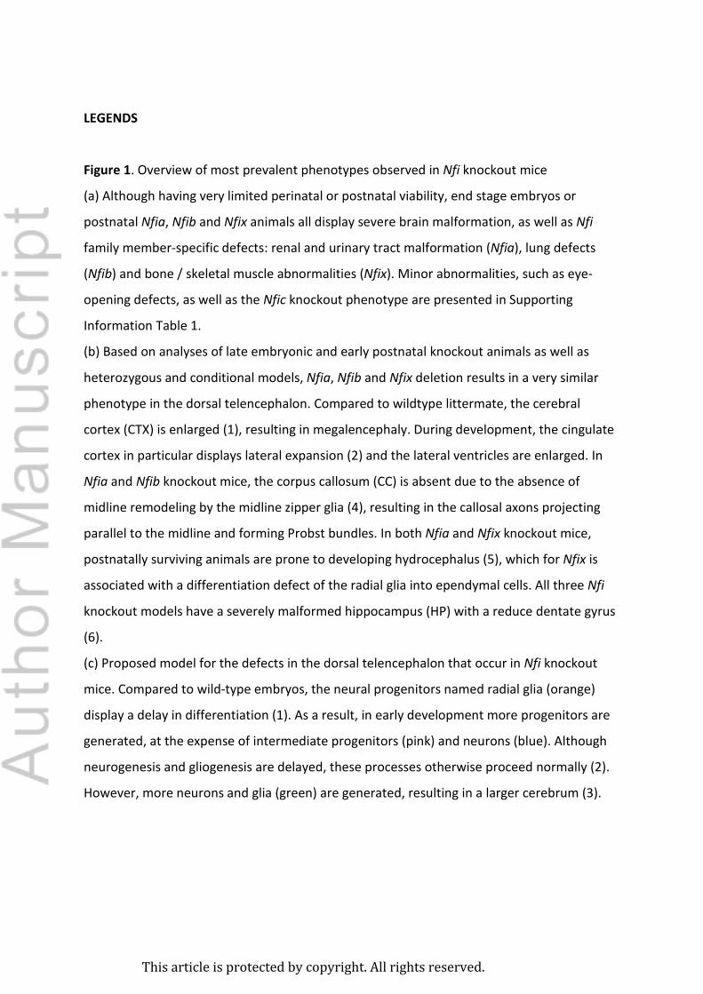
Figure 1. Overview of most prevalent phenotypes observed in Nfi knockout mice

(a) Although having very limited perinatal or postnatal viability, end stage embryos or

postnatal Nfia, Nfib and Nfix animals all display severe brain malformation, as well as Nfi

family member-specific defects: renal and urinary tract malformation (Nfia), lung defects

(Nfib) and bone / skeletal muscle abnormalities (Nfix). Minor abnormalities, such as eye-

opening defects, as well as the Nfic knockout phenotype are presented in Supporting

Information Table 1.

(b) Based on analyses of late embryonic and early postnatal knockout animals as well as

heterozygous and conditional models, Nfia, Nfib and Nfix deletion results in a very similar

phenotype in the dorsal telencephalon. Compared to wildtype littermate, the cerebral

cortex (CTX) is enlarged (1), resulting in megalencephaly. During development, the cingulate

cortex in particular displays lateral expansion (2) and the lateral ventricles are enlarged. In

Nfia and Nfib knockout mice, the corpus callosum (CC) is absent due to the absence of

midline remodeling by the midline zipper glia (4), resulting in the callosal axons projecting

parallel to the midline and forming Probst bundles. In both Nfia and Nfix knockout mice,

postnatally surviving animals are prone to developing hydrocephalus (5), which for Nfix is

associated with a differentiation defect of the radial glia into ependymal cells. All three Nfi

knockout models have a severely malformed hippocampus (HP) with a reduce dentate gyrus

(6).

(c) Proposed model for the defects in the dorsal telencephalon that occur in Nfi knockout

mice. Compared to wild-type embryos, the neural progenitors named radial glia (orange)

display a delay in differentiation (1). As a result, in early development more progenitors are

generated, at the expense of intermediate progenitors (pink) and neurons (blue). Although

neurogenesis and gliogenesis are delayed, these processes otherwise proceed normally (2).

However, more neurons and glia (green) are generated, resulting in a larger cerebrum (3).

This article is protected by copyright. All rights reserved.

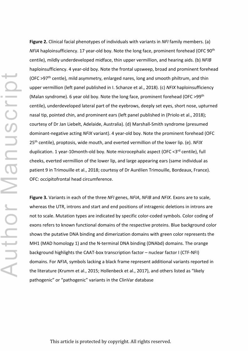
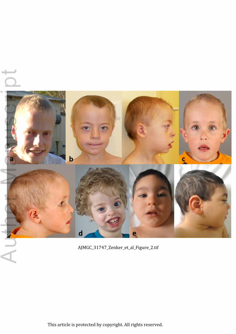
Figure 2. Clinical facial phenotypes of individuals with variants in NFI family members. (a)

NFIA haploinsufficiency. 17 year-old boy. Note the long face, prominent forehead (OFC 90th

centile), mildly underdeveloped midface, thin upper vermillion, and hearing aids. (b) NFIB

haploinsufficiency. 4 year-old boy. Note the frontal upsweep, broad and prominent forehead

(OFC >97th centle), mild asymmetry, enlarged nares, long and smooth philtrum, and thin

upper vermillion (left panel published in I. Schanze et al., 2018). (c) NFIX haploinsufficiency

(Malan syndrome). 6 year old boy. Note the long face, prominent forehead (OFC >99th

centile), underdeveloped lateral part of the eyebrows, deeply set eyes, short nose, upturned

nasal tip, pointed chin, and prominent ears (left panel published in (Priolo et al., 2018);

courtesy of Dr Jan Liebelt, Adelaide, Australia). (d) Marshall-Smith syndrome (presumed

dominant-negative acting NFIX variant). 4 year-old boy. Note the prominent forehead (OFC

25th centile), proptosis, wide mouth, and everted vermillion of the lower lip. (e). NFIX

duplication. 1 year-10month-old boy. Note microcephalic aspect (OFC <3rd centile), full

cheeks, everted vermillion of the lower lip, and large appearing ears (same individual as

patient 9 in Trimouille et al., 2018; courtesy of Dr Aurélien Trimouille, Bordeaux, France).

OFC: occipitofrontal head circumference.

Figure 3. Variants in each of the three NFI genes, NFIA, NFIB and NFIX. Exons are to scale,

whereas the UTR, introns and start and end positions of intragenic deletions in introns are

not to scale. Mutation types are indicated by specific color-coded symbols. Color coding of

exons refers to known functional domains of the respective proteins. Blue background color

shows the putative DNA binding and dimerization domains with green color represents the

MH1 (MAD homology 1) and the N-terminal DNA binding (DNAbd) domains. The orange

background highlights the CAAT-box transcription factor – nuclear factor I (CTF-NFI)

domains. For NFIA, symbols lacking a black frame represent additional variants reported in

the literature (Krumm et al., 2015; Hollenbeck et al., 2017), and others listed as “likely

pathogenic” or “pathogenic” variants in the ClinVar database

This article is protected by copyright. All rights reserved.

(https://www.ncbi.nlm.nih.gov/clinvar/). For these NFIA variants, no information about the

associated phenotype is available to verify differences or similarities to the NFIA-associated

phenotype described here. For NFIB and NFIX, the displayed variants refer to recently

published original and review papers, respectively (I. Schanze et al., 2018; (Priolo et al.,

2018)) Malan syndrome-associated variants are shown above and Marshall-Smith syndrome-

associated variants are shown below the schematic for the NFIX gene. Isoform references:

NFIA, ENST00000403491.7; NFIB, ENST00000380953.5; NFIX, ENST00000592199.5

This article is protected by copyright. All rights reserved.

Table 1. Major clinical characteristics of the phenotypes caused by variants in the individual

NFI genes.

NFIA NFIB NFIX Malan s.1 Marshall- Dup3

Smith s.2

Total included individuals 17 13 80 57 10 Prenatal: length increased 0/16 1/12 7/45 2/26 0/6 birth weight (mean)4 (3860g)6 3360g 3460g 3125g 3140g Postnatal: length/height increased7 5/11 2/11 44/79 2/36 0/10 adult height (mean)5 males (167.5cm)6 (182.8cm)6 (184.7cm)6 (140 cm)6 NA females (154.5cm)6 NA (158.0cm)6 (127cm)6,8 NA Prenatal: macrocephaly 6/6 2/5 19/46 1/56 0/4 birth OFC (mean)4 (39.6cm)6 37.1cm 36.2cm 35.7cm (33.5cm)6

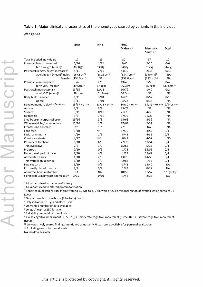
Postnatal: macrocephaly 13/15 12/12 60/79 1/40 0/2 adult OFC (mean)5 (59.1cm)6 (61.5cm)6 60.6cm NA NA Body build: slender 1/11 3/10 46/78 0/57 0/10 obese 3/11 1/10 3/78 0/36 NA Developmental delay9 +/++/+++ 15/17 + or ++ 13/13 + or ++ 80/80 + or ++ 39/39 ++or+++ 9/9+or +++ Autism 2/11 3/9 23/74 NA NA Seizures 3/11 0/11 21/79 4/38 NA Hypotonia 5/7 7/11 57/75 12/28 NA Small/absent corpus callosum 15/16 3/8 14/63 8/39 NA Wide ventricles/hydrocephaly 12/16 1/7 17/63 2/39 NA Frontal lobe anomaly 310 110 210 NA NA Long face 1/10 NA 67/79 3/57 0/4 Facial asymmetry 4/10 1/9 5/42 4/36 0/4 Craniosynostosis 4/17 NM 0/42 4/57 NM Prominent forehead 6/10 6/9 77/79 53/54 0/4 Thin eyebrows 2/6 1/9 15/66 5/35 0/4 Proptosis 0/10 0/9 1/78 55/56 0/4 Underdeveloped midface 2/10 4/9 1/79 38/42 0/4 Anteverted nares 1/10 2/9 43/76 44/53 0/4 Thin vermillion upper lip 6/10 3/9 42/63 1/35 0/4 Low-set ears 5/10 0/9 8/42 13/40 NA Proximally placed thumbs 4/7 0/9 1/42 0/27 NA Abnormal bone maturation NA NA 40/50 57/57 5/6 (delay) Significant urinary tract anomalies11 3/15 0/10 1/42 2/36 NA 1 All variants lead to haploinsufficiency 2 All variants lead to altered protein formation 3 Reported duplications vary in size from to 3.1 Mb to 479 kb, with a 422 kb minimal region of overlap which contains 16 genes 4 Only at term born newborns (38-42wks) used 5 Only individuals 16 yr and older used 6 Only small number of data available 7 Length/height ≥ 2SD for age 8 Reliability limited due to scoliosis 9 + mild cognitive impairment (IQ 50-70); ++ moderate cognitive impairment (IQ35-50); +++ severe cognitive impairment (IQ<35) 10 Only positively scored findings mentioned as not all MRI scan were available for personal evaluation 11 Excluding one or two small cysts NA, no data available

This article is protected by copyright. All rights reserved.

NM, not mentioned

This article is protected by copyright. All rights reserved.

This image cannot currently be displayed.

This article is protected by copyright. All rights reserved.

This article is protected by copyright. All rights reserved.

This article is protected by copyright. All rights reserved.

This article is protected by copyright. All rights reserved.

This article is protected by copyright. All rights reserved.

This article is protected by copyright. All rights reserved.

Permission for Publication

Herewith, undersigned, parents of Joas Laan dob ……..

grants permission to Dr Raoul C Hennekam (Amsterdam, the Netherlands) to use the medical

data and clinical pictures of their child for publication in a medical journal. We know the

target readership are scientists, doctors and researchers, but the general audience can view the

pictures as well as journals are nowadays often also available in an electronic manner and

accessible on the internet. The name of our child will not be mentioned in the publication. We

understand that when a paper is accepted for publication and thereafter published, a consent

cannot be revoked anymore. We know we will not receive any payment or royalties in

connection with the use and publication of the medical data and clinical pictures.

We warrant that we have the full right, power and authority to sign this consent form, and we

grant permission on behalf of our child shown in the pictures.

Date: 27 maart

Place: Den Haag.

Name(s): H.W. Laan

Signature(s): .................................................

This article is protected by copyright. All rights reserved.

ajmgc_31747_zenker_et_al_figure_1.eps

This article is protected by copyright. All rights reserved.

AJMGC_31747_Zenker_et_al_Figure_2.tif

This article is protected by copyright. All rights reserved.

AJMGC_31747_Zenker_et_al_Figure_3.png

This article is protected by copyright. All rights reserved.


Recommended