+ All Categories
Home > Documents > Vascular Integrin av13: A New Prognostic Indicator in...

Vascular Integrin av13: A New Prognostic Indicator in...

Date post: 15-Feb-2019
Category:
Upload: dangthuy
View: 215 times
Download: 0 times
Share this document with a friend
11
Vol. 4, 2625-2634, November 1998 Clinical Cancer Research 2625 Vascular Integrin av13: A New Prognostic Indicator in Breast Cancer1 Giampietro Gasparini,2 Peter C. Brooks, Elia Biganzoli, Peter B. Vermeulen, Emanuela Bonoldi, Luc Y. Dirix, Girolamo Ranieri, Rosalba Miceli, and David A. Cheresh Division of Medical Oncology, Azienda Ospedaliera “Bianchi- Melacrino-Morelli,” 89100 Reggio Calabria, Italy [G. G., 0. R.], Departments of Immunology and Vascular Biology, The Scripps Research Institute, La Jolla, California 92037 [P. C. B., D. A. C.]; Oncological Center and Angiogenesis Laboratory, St. Augustinus Hospital, Wilrijk, B-2610, Belgium [P. B. V., L. Y. D.]; Department of Medical Statistics and Biometry, National Cancer Institute, Milan, 20122 Milan, Italy [E. B., R. M.]; and Institute of Pathology, St. Bortolo Hospital 36100 Vicenza, Italy [E. B.] ABSTRACT Blood vessel density is a prognostic indicator of multi- plc tumor types. Recently, it has been established that tu- mor-associated blood vessels express elevated levels of inte- grin aj3. In fact, there is evidence that integrin avI3 identifies the most proliferative endothelial cells within hu- man breast carcinomas. Therefore, we evaluated breast can- cer tissue in terms of both blood vessel density and a33 expression. We found that the antibody LM609 to integrin aI3 preferentially stains the blood vessels of small caliber. Furthermore, comparative studies between LM609 and anti-CD31 antibodies on normal breast indicate that very low and weak expression of integrin aj33 was found on vessels within normal tissue, whereas CD31 antigen was expressed in almost all vasculature. Indeed, expression of integrin #{176}vI3 was significantly higher in tumors of patients with metastasis than in those without metastasis. In a series of 197 consecutive patients with invasive breast cancer and long follow-up, vascular expression of integrin a33 in tu- mor vascular “hot spots” was found to be the most signifi- cant prognostic factor predictive of relapse-free survival in both node-negative and node-positive patients. These find- ings support the contention that angiogenesis plays a critical role in breast cancer progression and suggest that integrin Received 7/10/98; accepted 8/10/98. The costs of publication of this article were defrayed in part by the payment of page charges. This article must therefore be hereby marked advertisement in accordance with 18 U.S.C. Section 1734 solely to indicate this fact. I This study was supported in part by grants from the Associazione Italiana per Ia Ricerca sul Cancro (Milan, Italy) and the Regione Veneto (Progetto Sanitario Finalizzato No. 675-01-96; Venice, Italy). D. A. C. was supported by NIH grants HL5444, CA50286, and CA45726. 2 To whom requests for reprints should be addressed, at Division of Medical Oncology, Ospedali Riuniti, Via Melacrino, 89100 Reggio Calabria, Italy. Phone: (39)-965-397331; Fax: (39)-965-397334; E-mail: [email protected]. avI3 5 an endothelial cell marker with significant prognos- tic value and potential usefulness as a target for specific antiangiogenic therapy. INTRODUCTION The formation and growth of new blood vessels from preexisting ones, i.e. , angiogenesis, is a pivotal biological proc- ess in mammalian organisms. Angiogenesis is necessary in a variety of physiological processes, such as embryonic develop- ment, chronic inflammation, and wound repair, in which it is highly regulated (reviewed in Refs. 1 and 2). Abnormal angio- genesis is involved in the pathogenesis of several diseases, such as chronic inflammatory disorders and degenerative ocular dis- eases, and it is necessary for tumor growth and progression (reviewed in Ref. 1). Tumor-induced angiogenesis is initiated by tumor and stromal cell release of angiogenic peptides and/or by down-regulation of endogenous angiogenesis inhibitors (re- viewed in Ref. 3) and involves modifications of the ECM.3 Several studies suggest that endothelial cell migration and in- vasion through ECM are regulated by cellular adhesion recep- tors (reviewed in Refs. 4 and 5). Previous studies (6-8) dem- onstrate that integrin a33 plays a key role in angiogenesis induced by a variety of stimuli in viva. Integrin ttv33 promotes vascular endothelial cell survival in viva (9). In fact, during angiogenesis, proliferative endothelial cells become apoptotic in response to antagonists of integrin ttv3i. which also causes regression of vascularization and tumor growth by a mecha- nisms involving p53. the cell cycle inhibitor p21WAF/CIPI and alterations of the bcl-2/bax ratio (5, 9). Initial studies (10) examined the functional role of integrin ttv13 in human breast cancer and provided evidence that this integrin is highly expressed on angiogenic vessels associated with invasive breast cancer and that systemic administration of LM609 antibody directed to integrin a3 is capable of disrupt- ing intratumoral neovessels, thereby inhibiting tumor formation in a severe combined immunodeficient mouse/human chimeric model. We undertook these studies to further define the expression of integrin ttv13 in human breast cancer. LM609 antibody preferentially immunostains the intratumoral microvessels of smaller caliber. This staining pattern was selective for invasive cancer and specific for tumor-induced neovascularization. In contrast, integrin a33 expression was not readily detected in nonangiogenic benign breast tissue. The degree of VECs stained by LM609 antibody was significantly higher in tumors of pa- tients with metastasis, and it was moderately associated with 3 The abbreviations used are: ECM, extracellular matrix; VEC. vascular endothelial cell; KW, Kruskal-Wallis; RFS, relapse-free survival; OS, overall survival; LRT, likelihood ratio statistic; Mab, monoclonal anti- body; KS, Kolmogorov-Smirnov; HR. hazard ratio. Research. on February 14, 2019. © 1998 American Association for Cancer clincancerres.aacrjournals.org Downloaded from
Transcript

Vol. 4, 2625-2634, November 1998 Clinical Cancer Research 2625

Vascular Integrin av1�3: A New Prognostic Indicator in

Breast Cancer1

Giampietro Gasparini,2 Peter C. Brooks,

Elia Biganzoli, Peter B. Vermeulen,

Emanuela Bonoldi, Luc Y. Dirix,

Girolamo Ranieri, Rosalba Miceli, and

David A. ChereshDivision of Medical Oncology, Azienda Ospedaliera “Bianchi-

Melacrino-Morelli,” 89100 Reggio Calabria, Italy [G. G., 0. R.],Departments of Immunology and Vascular Biology, The Scripps

Research Institute, La Jolla, California 92037 [P. C. B., D. A. C.];Oncological Center and Angiogenesis Laboratory, St. AugustinusHospital, Wilrijk, B-2610, Belgium [P. B. V., L. Y. D.]; Departmentof Medical Statistics and Biometry, National Cancer Institute, Milan,20122 Milan, Italy [E. B., R. M.]; and Institute of Pathology, St.

Bortolo Hospital 36100 Vicenza, Italy [E. B.]

ABSTRACTBlood vessel density is a prognostic indicator of multi-

plc tumor types. Recently, it has been established that tu-

mor-associated blood vessels express elevated levels of inte-

grin aj�3. In fact, there is evidence that integrin avI�3identifies the most proliferative endothelial cells within hu-

man breast carcinomas. Therefore, we evaluated breast can-

cer tissue in terms of both blood vessel density and a��33

expression. We found that the antibody LM609 to integrin

a�I�3 preferentially stains the blood vessels of small caliber.

Furthermore, comparative studies between LM609 and

anti-CD31 antibodies on normal breast indicate that very

low and weak expression of integrin aj33 was found on

vessels within normal tissue, whereas CD31 antigen was

expressed in almost all vasculature. Indeed, expression of

integrin #{176}�vI�3was significantly higher in tumors of patients

with metastasis than in those without metastasis. In a series

of 197 consecutive patients with invasive breast cancer and

long follow-up, vascular expression of integrin a��33 in tu-

mor vascular “hot spots” was found to be the most signifi-

cant prognostic factor predictive of relapse-free survival in

both node-negative and node-positive patients. These find-

ings support the contention that angiogenesis plays a critical

role in breast cancer progression and suggest that integrin

Received 7/10/98; accepted 8/10/98.

The costs of publication of this article were defrayed in part by the

payment of page charges. This article must therefore be hereby marked

advertisement in accordance with 18 U.S.C. Section 1734 solely to

indicate this fact.

I This study was supported in part by grants from the Associazione

Italiana per Ia Ricerca sul Cancro (Milan, Italy) and the Regione Veneto(Progetto Sanitario Finalizzato No. 675-01-96; Venice, Italy). D. A. C.was supported by NIH grants HL5444, CA50286, and CA45726.

2 To whom requests for reprints should be addressed, at Division ofMedical Oncology, Ospedali Riuniti, Via Melacrino, 89100 Reggio

Calabria, Italy. Phone: (39)-965-397331; Fax: (39)-965-397334; E-mail:[email protected].

avI�3 �5 an endothelial cell marker with significant prognos-

tic value and potential usefulness as a target for specific

antiangiogenic therapy.

INTRODUCTIONThe formation and growth of new blood vessels from

preexisting ones, i.e. , angiogenesis, is a pivotal biological proc-

ess in mammalian organisms. Angiogenesis is necessary in a

variety of physiological processes, such as embryonic develop-

ment, chronic inflammation, and wound repair, in which it is

highly regulated (reviewed in Refs. 1 and 2). Abnormal angio-

genesis is involved in the pathogenesis of several diseases, such

as chronic inflammatory disorders and degenerative ocular dis-

eases, and it is necessary for tumor growth and progression

(reviewed in Ref. 1). Tumor-induced angiogenesis is initiated by

tumor and stromal cell release of angiogenic peptides and/or by

down-regulation of endogenous angiogenesis inhibitors (re-

viewed in Ref. 3) and involves modifications of the ECM.3

Several studies suggest that endothelial cell migration and in-

vasion through ECM are regulated by cellular adhesion recep-

tors (reviewed in Refs. 4 and 5). Previous studies (6-8) dem-

onstrate that integrin a��33 plays a key role in angiogenesis

induced by a variety of stimuli in viva. Integrin ttv�33 promotes

vascular endothelial cell survival in viva (9). In fact, during

angiogenesis, proliferative endothelial cells become apoptotic in

response to antagonists of integrin ttv�3i. which also causes

regression of vascularization and tumor growth by a mecha-

nisms involving p53. the cell cycle inhibitor p21WAF/CIPI and

alterations of the bcl-2/bax ratio (5, 9).

Initial studies (10) examined the functional role of integrin

ttv1�3 in human breast cancer and provided evidence that this

integrin is highly expressed on angiogenic vessels associated

with invasive breast cancer and that systemic administration of

LM609 antibody directed to integrin a��3 is capable of disrupt-

ing intratumoral neovessels, thereby inhibiting tumor formation

in a severe combined immunodeficient mouse/human chimeric

model.

We undertook these studies to further define the expression

of integrin ttv1�3 in human breast cancer. LM609 antibody

preferentially immunostains the intratumoral microvessels of

smaller caliber. This staining pattern was selective for invasive

cancer and specific for tumor-induced neovascularization. In

contrast, integrin a��33 expression was not readily detected in

nonangiogenic benign breast tissue. The degree of VECs stained

by LM609 antibody was significantly higher in tumors of pa-

tients with metastasis, and it was moderately associated with

3 The abbreviations used are: ECM, extracellular matrix; VEC. vascularendothelial cell; KW, Kruskal-Wallis; RFS, relapse-free survival; OS,overall survival; LRT, likelihood ratio statistic; Mab, monoclonal anti-

body; KS, Kolmogorov-Smirnov; HR. hazard ratio.

Research. on February 14, 2019. © 1998 American Association for Cancerclincancerres.aacrjournals.org Downloaded from

2626 aJ3, as a Prognostic Indicator in Breast Cancer

endothelial cell but not tumor cell proliferative rate. In addition,

the LM609 antibody defined the vascular “hot spot” of each

cancer in a consecutive series of 197 breast cancer patients and

proved to be a significant predictor of clinical outcome of the

patients, both before and after adjustment for the conventional

prognostic indicators.

MATERIALS AND METHODSPatient Population. The first preliminary study was un-

dertaken on 46 patients with invasive breast carcinoma who

underwent surgery between 1990 and 1991 at the Vicenza

General Hospital. This series was made up of pairs of cases who

had disease recurrence (24 patients) and cases who developed

metastasis (22 patients), randomly selected from a larger series

of consecutive patients who underwent surgery during the same

period and who had available follow-up data. The main char-

acteristics of the patients were as follows. The median age was

59 years (range, 29-87 years). Sixteen patients were pre- or

perimenopausal, and 26 were postmenopausal patients. Histo-

logical types were ductal invasive (38 cases) and lobular or

other invasive types (8 cases). Tumor sizes were pT1 (24 cases),

pT2 ( 17 cases) and pT3 in 5 cases. There was axillary node

involvement (N+) in 28 cases, whereas 18 cases were

N- . Histological grades (Bloom and Richardson) were G�

(1 1 cases), 02 (15 cases), and G3 (20 cases). Sixteen patients did

not receive adjuvant therapy, whereas 10 were treated with

adjuvant chemotherapy and 20 were treated with adjuvant ta-

moxifen. The median follow-up of the series exceeded 5 years.

In addition, 15 samples of tissue from subjects with normal

breast were studied for comparison.

A second cohort of 197 consecutive patients with operable

invasive breast carcinoma who underwent surgery between

1987 and 1991 at the same institution and who had no evidence

of distant metastatic disease at diagnosis were studied for prog-

nostic purposes. Patients were followed until the date of their

death, the date they were last known to be alive, or the end of the

follow-up period, whichever came first. The criteria of admin-

istration of adjuvant treatments to node-positive patients were

reported in detail elsewhere (1 1).

Tissue Evaluation. Tumors were classified adopting the

1989 pathological staging system of the American Joint Corn-

mittee on Cancer (1992) regarding primary tumor size and

axillary lymph node involvement. Tumors were assigned a

histological grade from I (low) to III (high), according to the

Bloom and Richardson grading scheme.

Antibodies and Immunohistochemical Studies. Anti-

bodies used for the study included: previously characterized

affinity-purified anti-integrin a��33 LM609 (6, 10); monoclonal

anti-CD3 1 clone JC/70A (purchased from DAKO, Glostrup,

Denmark); anti-CD1O5 monoclonal E-9 (kindly supplied by

Prof. Shant Kumar, University of Manchester, Manchester,

United Kingdom); and monoclonal anti-Ki-67 (purchased from

DAKO). The dilutions used were: 1:1000; 1:400; 1:50, and 1:50,

respectively.

All the immunohistochemical determinations were per-

formed on representative samples snap-frozen in liquid nitrogen

and stored at -80#{176}C until sectioning. Cryostat sections, 4-6

�i.m thick, were fixed in cold acetone at 4#{176}Cfor 10 mm,

air-dried, and incubated at room temperature with the above

antibodies for 1 h. After being washed with PBS, bound anti-

bodies were visualized using antirnouse biotinylated antibody,

the streptavidin-peroxidase complex (DAKO), and 3-3’-diami-

nobenzidine. For the detection of the nuclear antigen Ki-67, as

a marker of cell proliferation, and of the membrane epitope E-9,

the latter being a marker of endothelial cells, a double immu-

nostaining technique was adopted, as reported elsewhere (12).

E-9-positive endothelial cells stained red, and proliferating

cell nuclei stained dark brown. We performed a systematic

comparison in the same fields of the samples tested between the

panendothelial marker anti-CD3 1 , as an internal positive con-

trol, and LM609 to test the sensitivity and specificity of the

latter antibody for microvessels. For negative controls, in all

instances, we used nonspecific IgG as the primary antibody.

Criteria of Evaluation. To quantitate and compare ex-

pression of integrin Uv�33 and CD3 1 antigen, microvessels ex-

pressing either antigen were counted in five fields for each

sample within a X 100 field that stained positive for both inte-

grin ttv�33 and CD3 1 . The primary field was chosen from areas

that combined a large number of total microvessels based on the

CD31-positive staining, and then the additional four fields were

derived by moving in four separate directions from that of the

original field.

Tumors were heterogeneous in their microvessel density,

but the areas of most intense neovascularization were identified

by scanning the tumor sections at low power (X 100), and then

the areas of invasive carcinoma with the highest number of

distinct microvessels stained with the above endothelial markers

were recorded. Once the single area of highest neovasculariza-

tion (vascular hot spot) was identified for each tumor, individual

microvessels were counted on a X200 field (i.e., X20 objective

lens and X 10 ocular lens; 0.738 mm2 per field) as reported

previously (10-13).

Because the most prominent integrin rtvf3i staining was

colocalized in the most highly vascularized area by CD31 stain-

ing, integrin tt�1�3 expression was quantitated by counting

LM609-positive microvessels in the same areas in which the

highest expression of CD3 1 was assessed. Three independent

observers (P. C. B., E. B., and G. R.) evaluated vascular vessel

counts.

The methodology of identifying endothelial cells and

counting cycling cells has been described previously (14). Five

adjacent fields were analyzed at X400, each field crossing the

entire tumor tissue available on the slide. In every field, the

cycling endothelial or tumor cells were counted (P. B. V.). The

mean of the fractions of cycling cells of the five fields was used

for evaluation. A minimum of 450 and a maximum of 2300

endothelial cells (median, 970 cells), and a minimum of 750 and

a maximum 3250 tumor cells (median, 1780 cells) were counted

per section, respectively.

Statistical Methods. In the preliminary study, the hy-

pothesis that integrin ttv�33 was identically distributed in the

three subgroups of patients was evaluated using the nonpara-

metric KW test (15). A multiple comparison procedure was

subsequently adopted that was suitable for protection from type

I statistical error. The association between the number of mi-

crovessels stained by LM609 with the other continuous van-

ables was evaluated by Spearman’s rank correlation coefficient.

Research. on February 14, 2019. © 1998 American Association for Cancerclincancerres.aacrjournals.org Downloaded from

Clinical Cancer Research 2627

. #{149}�

�..

#{149}4�� � � � #{149}�

‘�;:‘ �

� ..‘�:__ ‘�-

St. *_.‘

, *.r_� k, - .

,� .(�

‘ \� _�l�S*’ � � � S : B

, 3 �- �. $, .*%-. � � � � :.,,� �

%� S � �: : � �

:.“.‘li. ‘. .%,‘.. ,:,�.* � � � ,. :� �. . . : � ‘�:.:.‘�i -‘.;., � ... #{149}�:‘� � .

. � I #{149}‘ � , #{149}�‘.

.- . #{149}.‘\ :.‘ .,v:..�.:..*, a � � � � ,#{149} .

:� �‘ � � 4 �‘e� �

.#{149}. %, -* #{149}�‘:�

‘� - .‘ ‘�I*�’I, �F �y’

� ‘, k�, �

9;J��4��;JP

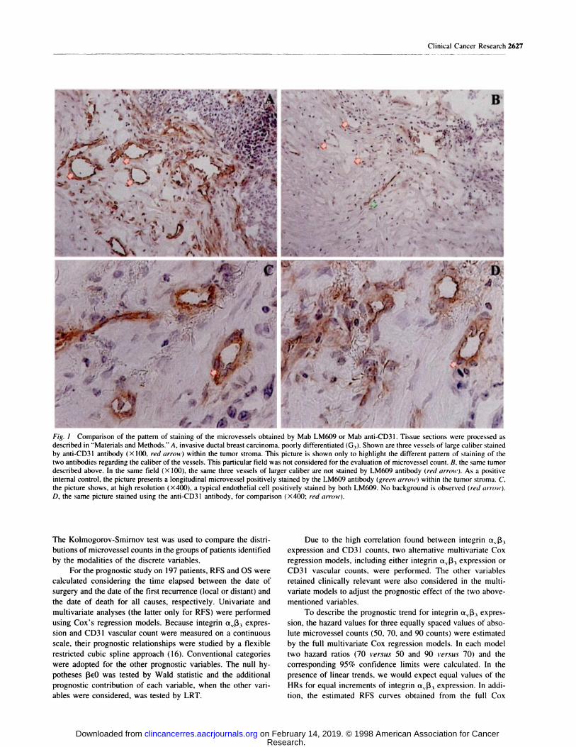
� . �.--.-. .� �r �Fig. I Comparison of the pattern of staining of the microvessels obtained by Mab LM609 or Mab anti-CD3 1 . Tissue sections were processed as

described in “Materials and Methods.” A, invasive ductal breast carcinoma, poorly differentiated (G3). Shown are three vessels of large caliber stained

by anti-CD31 antibody (X 100, red arrow) within the tumor stroma. This picture is shown only to highlight the different pattern of staining of the

two antibodies regarding the caliber of the vessels. This particular field was not considered for the evaluation of microvessel count. B. the same tumordescribed above. In the same field (X 100). the same three vessels of larger caliber are not stained by LM609 antibody (red arrmt-). As a positive

internal control, the picture presents a longitudinal microvessel positively stained by the LMflO9 antibody (green arrow) within the tumor strorna. C.the picture shows, at high resolution (X400), a typical endothelial cell positively stained by both LM609. No background is observed (red arrott’).

D, the same picture stained using the anti-CD3I antibody, for comparison (X400; red arrow).

The Kolmogorov-Smirnov test was used to compare the distri-

butions of microvessel counts in the groups of patients identified

by the modalities of the discrete variables.

For the prognostic study on 197 patients, RFS and OS were

calculated considering the time elapsed between the date of

surgery and the date of the first recurrence (local or distant) and

the date of death for all causes, respectively. Univariate and

multivanate analyses (the latter only for RFS) were performed

using Cox’s regression models. Because integrin a��33 expres-

sion and CD3 1 vascular count were measured on a continuous

scale, their prognostic relationships were studied by a flexible

restricted cubic spline approach (16). Conventional categories

were adopted for the other prognostic variables. The null hy-

potheses �3#{128}Owas tested by Wald statistic and the additional

prognostic contribution of each variable, when the other vari-

ables were considered, was tested by LRT.

Due to the high correlation found between integrin a��33

expression and CD3 1 counts, two alternative multivariate Cox

regression models, including either integrin a��33 expression or

CD3 I vascular counts, were performed. The other variables

retained clinically relevant were also considered in the multi-

variate models to adjust the prognostic effect of the two above-

mentioned variables.

To describe the prognostic trend for integrin aj3� expres-

sion, the hazard values for three equally spaced values of abso-

lute microvessel counts (50, 70, and 90 counts) were estimated

by the full multivariate Cox regression models. In each model

two hazard ratios (70 versus 50 and 90 versus- 70) and the

corresponding 95% confidence limits were calculated. In the

presence of linear trends, we would expect equal values of the

HRs for equal increments of integrin a��33 expression. In addi-

tion, the estimated RFS curves obtained from the full Cox

Research. on February 14, 2019. © 1998 American Association for Cancerclincancerres.aacrjournals.org Downloaded from

y#{149}�

- �.,i.. ._.*�_. �..-

I � � �

� � #{149}. -.... �,#{149} ,..� ., * PS

� ... � ,-�‘

p � � - . �_)‘, ,. /. ‘ l�

� � -I ‘ � “I :, . . -. - � � . � � � � -:�;�,

‘�‘B�

“Pc ,� � � �

� . , � � - t_ :� #{149}��‘j; ;i

., �;r” ‘P ,�--:. .-� � - � ‘ � �p�: � ?� - � ‘�#{149}. �

, � � .:“ #{149}:,� � . -

..?“� � ‘* � ‘ �-.; �

.#{149}4/’ � -,. . j..- ,,� � �.,::. . - . . 0�, � �

‘ # , - ‘ . - -�

‘�1�( � E� � �.‘ I ,.�“ ;4

14�* ‘�, � �1,t

�:�‘ � �- - � �

�-�:-( :-.��“: -

I �‘ ‘., ��:- �‘ -

� ; “- ,-.,�.‘.

.‘ . #{149}4fr;/.

‘F

r� ‘ � .�

-.� .,

. I � �

“4’ .,,�

#{149}).

,.� t

. I�

‘ ‘,i &%�

..,�.. : � :... DI.#{149}. ,--,‘., ��i#{188}’ �

- - --- /..- . . :�- %jr5 �

.- . .1 � #{149}�.

� : #{149}#{149}: �

o.,-._�;

-. 4� S #{149}-��- S

, 4_.� ‘ ,

t

. � �

as � � � ,�, �.. 4’.. #{149} .;

:., � ‘

2628 aj3� as a Prognostic Indicator in Breast Cancer

�.-:*‘. t t ‘�

� . � � #{149}�,:� � � � � . ,.�,. ;� ; ,

. \ () .� #{149}.. -‘�:,�

\..‘ � .� * p � :, ‘� � - � -�‘ ‘ ‘<.; �: , � � ��. . , � -p- _ , .� ‘:1�- �, .-. .-O -

I 4 :��4

V � I � ;� � :c�’ \I4�*�� � � �, a� �‘p!��;

:.‘�( , � .� #{149}1 “#{149}�.� � ‘ �

� , ‘ � ;, -.. �

. , � �, * t;.� � #{149}.�.: #{149}#{149}� ‘ .� -�‘ � �‘-

.,� r’,�. � �4’a’�- � � .�- �

� :,:[�,4 t\’�

Fig. 2 A, invasive ductal breast carcinoma, moderately differentiated (02). The picture shows the vascular hot spot selected as indicated in “Materials

and Methods.” A large number of microvessels stained by anti-CD3I antibody (X 100) can be identified (brown). B, the same tumor described above.In the same field ( X I 00), a large number of microvessels are stained by LM609 antibody at the vascular hot spot (brown). Double green arrow,

reference area between A and B. C, the same tumor described above. The picture shows a non-hot spot field (X 100) showing a small number of

microvessels stained by anti-CD3 I antibody (brown). Some background areas are indicated (yellow arrows). D, the same tumor described above. Thepicture shows a non-hot spot field (X 100), showing a small number of microvessels stained by LM609 antibody (brown). Double green arrow,

reference area between C and D.

regression models were provided corresponding to the three

above-considered integrin ttv�33 expression values in the patients

with N - or N + tumors separately.

The predictive capability of the models was investigated by

the Harrell c statistic ( 17). This statistic assumes values ranging

from 0.5 to 1 .00. If the model has no predictive capability, it is

expected to be 0.5, and it approaches 1.00 in the case of high

prognostic capability.

Because adjuvant treatments were not allocated in a ran-

dom fashion, the univariate analyses were performed separately

for the following groups of patients: N-, not treated; N+,

treated with adjuvant chemotherapy; and N+, treated with hor-

mone therapy. The multivariate analyses were performed sepa-

rately for N - and N + patients. We also performed an analysis

on all the series. However, we advise that the results of the latter

analysis be interpreted with caution because the nodal status

effect can be confused with the treatment effect. Consequently,

it is influenced by the criteria adopted to allocate the patients to

each adjuvant treatment (1 1).

RESULTSExpression of Integrin a��3 and CD31 Antigen in

Breast Disease. To establish the total tumor-associated blood

vessel count in both invasive cancer of the breast and normal

breast tissue, cryostat sections of 46 invasive cancers and 15

normal breast tissues were stained with the panendothelial cell

marker anti-CD3 1 antibody. In all tumors (Fig. 1A) and normal

tissue studied, CD3 1 antigen was shown to be consistently

associated with VECs, independently of their caliber. In adja-

cent sections of the same tissue frozen block, we also detected

integrin ttv1�3 antigen. However, expression of integrin ttv�33

was restricted to VECs lining tumor-associated microvessels

and was preferentially expressed on the vessels of minor caliber

Research. on February 14, 2019. © 1998 American Association for Cancerclincancerres.aacrjournals.org Downloaded from

1�I

4

1�

*5.- ---

- � � j*’

-� V

S

Relapsedtumors

Non relapsedtumors

#{149}[ I

.‘ F ii.#{149}#{149}

.

...

.

. � .

. S.

.

.

.

..

.

C

.

.

.

. .

S

. ,

.#{149}#{149}#{149}#{149}5C

C C

.

�, #{149} .

C

.

6 breasttlssue LEENonnal

0 50

0 1 2 3 4 5

100 150

Uv1&3 IfltS9IIfl expression

Clinical Cancer Research 2629

Fig. 3 Double immunostain-ing with the antibodies E-9 and

Ki-67 showing an invasive duc-

tal carcinoma of the breast withhigh tumor cell proliferation

rate (brown nuclei) and lowproliferating endothelial cells(brown nuclei and red vessel).

Arrow, a typical proliferating

endothelial cell.

C

E.9/Ki-67

Fig. 4 The degree of expression of integrin a�ft (LM609 antibody) on

blood endothelial cells in invasive breast cancer is moderately associ-

ated with high rate of proliferating endothelial cells as assessed bydouble staining techniques using E-9 and Ki-67 antibodies (a = 46).

(Fig. 1B). LM609 is highly specific for tumor-associated endo-

thelium, and it does not cross-react with unrelated structures or

normal vessels (Fig. lC). Among the 46 invasive cancers, the

median number of microvessels immunostained by LM609 at

the hot spot was 67 per field (range, 18-151), whereas little or

no ttvI�3 was observed in vessels of normal breast tissue. As

shown in Fig. 2, an invasive ductal breast carcinoma is repre-

sented by both an area of hot spot and non-hot spot stained with

either anti CD31 or LM609. LM609 detects primarily small

caliber hot spot vessels (Fig. 2B).

Correlation of LM609-stained Microvessels with Endo-

thelial Cell Proliferation Rate. To further characterize the

a��3P05iti’�� vessels at the hot spots, we double-stained the

tumor sections with Mab E-9, directed to the proliferation

marker CD1O5, and Ki-67 antibody (Fig. 3). There was an

association between these markers as suggested by the Spear-

man’s rank correlation p. value of 0.5 1 (P 0.001 ; Fig. 4).

I

Fig. 5 Box plot diagrams showing the distributions for the counts of

microvessels with integrin Uv�33 expression. The counts in tumors of

patients who developed metastasis (top bar; a = 22) are higher when

compared to those of the patients without metastasis (middle bar; �i =

24). Invasive breast carcinomas either with or without metastasis pre-sented a higher number of LM609-stained microvessels than normal

breast tissue (bottom bar. n = 15).

Conversely, integrin ttv1�3 expression on VECs was not corre-

lated with tumor size (KS test = 0.26; P = 0.33); axillary nodal

status (KS = 0.29; P = 0.24); histological grading (KS = 0.19;

P = 0.72) or tumor cell proliferative rate (Ki-67 labeling index;

Spearman’s p = 0.15).

Integrin a��3 and Metastasis. To establish whether

ttv1�3 expression on VECs was associated with tumor malig-

nancy, we examined LM609 staining within cryostat sections

from patients who developed metastasis (n = 22) and compared

it to staining in sections from patients that showed no disease

recurrence (ii = 24). The median number of LM609-stained

Research. on February 14, 2019. © 1998 American Association for Cancerclincancerres.aacrjournals.org Downloaded from

2630 � as a Prognostic Indicator in Breast Cancer

Table I Clinicopathological characteristics of the series of 197 breast cancer patient s stratified by nodal status and adjuvant therapy

Node-positive Node-positive

Node-negative patients treated with patients treated withpatients chemotherapy hormone therapy

Feature No. of patients % No. of patients % No. of patients %

No. of evaluable patients 100 42 55

Median age, yr (range) 54 (35-83) 46 (31-68) 62 (29-70)Menopausal statusPre/perimenopausal 37 37 31 74 5 9Postmenopausal 63 63 11 26 50 91Histological type

Ductal invasive 65 65 35 83 42 76Lobular invasive 19 19 7 17 11 20Other 16 16 0 0 2 4

Tumor sizepT1 62 62 17 40.5 20 36pT2_3 35 35 25 59.5 35 64pT� 3 3 0 0 0 0

American Joint Committee on Cancer stage (1992)I 62 0 0IIA

T0N1 0 0 0

TN, 0 18 20T,N0 36 0 0

IIBT2N1 0 15 27

T3N0 2 0 0

lIlAT0N, 0 0 0

T1N1 0 0 2T,N, 0 5 4

T3N1 0 4 2

T1N2 0 0 0

Histological grading”

G 9 9 6 14 9 16G, 55 55 15 36 26 48

G2 36 36 21 50 20 36Type of surgery

Conservative 47 47 14 33 16 29Radical mastectomy 53 53 28 67 39 71

Median no. of vascular microvessels with expression of 50.5 (18-140) 57.5 (15-180) 52 (19-152)integrin a433 (range)”

Median no. of vascular microvessels stained with anti-CD31 61 (22-181) 72.5 (31-215) 71 (31-180)antibody (range)”

“ Bloom and Richardson grading system.h Microvessel counts at the hot spot of each single tumor.

microvessels at the vascular hot spot in the tumors with metas-

tasis was significantly higher than that associated with tumors

from the nonmetastatic patients (KW test = 20.39). Neverthe-

less, both nonmetastatic tumors (KW = 20.25) and relapsed

tumors (KW = 40.64) had a significant higher number of

LM609-stained vessels than did normal breast tissue (experi-

mentwise error, <0.05; Fig. 5).

Prognostic Value of Integrin � Expression. To

evaluate the prognostic value of av�33 in breast cancer biopsies,

we examined a series of 197 consecutive patients with invasive

breast cancer for the presence of VEC-associated a��33. The

median follow-up of these patients was 86 months for both RFS

and OS (range, 1-1 12 months and 5-122 months, respectively).

The main clinicopathological characteristics of the cohort stud-

ied, stratified by axillary lymph node status and adjuvant ther-

apy, are listed in Table 1.

High integrin av1�3 expression on intratumoral microves-

sels was found to be a strong prognostic indicator of shorter RFS

and OS on univariate analysis in all the subgroups of patients,

including: N- patients (LRTRFS = 42.80, P < 0.0001 ; and

LRT05 = 17.60; P = 0.00015); N+ patients treated with

adjuvant chemotherapy (LRTRFS = 9.46, P = 0.0088; and

LRTOS 8.82, P = 0.012) and N+ patients treated with

adjuvant hormone therapy (LRTRFS = 20.80, P < 0.0001; and

LRTOS 10.20, P = 0.0059). The graphic representations of

the relationship between the degree of integrin aj33 expression

on VECs, as a continuous variable, with the logarithm of the

hazard ratio for RFS and OS, are shown in Fig. 6. On univariate

analysis, also microvessel counts by anti-CD3 I antibody stain-

ing was a highly significant prognostic factor in all the sub-

groups (N-, LRTRFS _ 47.10, P < 0.0001, and LRTOS =

19.30, P < 0.0001 ; N+ treated with chemotherapy, LRTRFS =

8.48, P = 0.014, and LRT05 = 10.50, P = 0.05; N+ treated

with hormone therapy, LRTRFS _ 19.50, P < 0.0001, and

Research. on February 14, 2019. © 1998 American Association for Cancerclincancerres.aacrjournals.org Downloaded from

C.”

0

cc,

A Nodenegative

�L�120 60 100 140

B Node-negative

�0�60�1O0�0

Node-positive- CMF

20 60 100 140

clVII.3 Integnn expression

Node-positiveCMF

20 60 100140

Node-positiveTAM

20 60 � 140

Node-posOveTAM

20 60 100 140

C’, -

0

cc,

Clinical Cancer Research 2631

a,113 Integrin expression

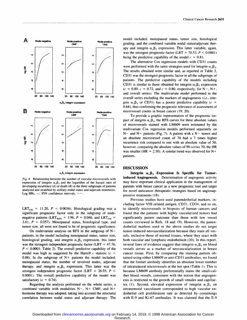
Fig. 6 Relationship between the number of vascular microvessels withexpression of integrin aJ33 and the logarithm of the hazard ratio ofdeveloping recurrence (A) or death (B) in the three subgroups of patients

analyzed and stratified by axillary nodal status and adjuvant treatments.Log HRs, -; 95% confidence intervals. . . ...

LRT05 = 1 1.20, P = 0.0036). Histological grading was a

significant prognostic factor only in the subgroup of node-

negative patients (LRTRFS 3.96, P 0.046, and LRT05

3.61, P = 0.057). Menopausal status, histological type, and

tumor size, all were not found to be of prognostic significance.

On multivariate analysis on RFS in the subgroup of N -

patients, in the model including menopausal status, tumor size,

histological grading, and integrin ttv�33 expression, this latter

was the strongest independent prognostic factor (LRT 47.76,

P < 0.0001 ; Table 2). The overall predictive capability of the

model was high as suggested by the Harrell c statistic (c

0.88). In the subgroup of N+ patients the model included:

menopausal status, the number of involved nodes, adjuvant

therapy, and integrin aj33 expression. This latter was the

strongest independent prognostic factor (LRT = 26.93, P <

0.0001). The overall predictive capability of the model was

satisfactory (c = 0.74).

Regarding the analysis performed on the whole series, a

combined variable with modalities N- , N+ CMF, and N+

hormone therapy was adopted, taking into account the structural

correlation between nodal status and adjuvant therapy. The

model included: menopausal status, tumor size, histological

grading, and the combined variable nodal status/adjuvant flier-

apy and integrin ttv1�3 expression. This latter variable, again,

was the strongest prognostic factor (LRT 70.53, P < 0.0001)

being the predictive capability of the model � 0.81.

The alternative Cox regression models with CD3 I counts

were performed with the same strategies used for integrin ttv�33.

The results obtained were similar and, as reported in Table 3,

CD3 I was the strongest prognostic factor in all the subgroups of

patients. The predictive capability of the models including

CD3 I is similar to those obtained for integrin a,j33 expression

(c 0.89, c 0.72, and c = 0.80, respectively, for N-, N+,

and overall series). The multivariate model performed in the

overall series excluding the markers of angiogenesis (i.e. , inte-

grin ttv1�3 or CD3 1 ) has a poorer predictive capability (c

0.68). thus confirming the prognostic relevance of assessment of

microvessel counts in breast cancer ( 1 9, 20).

To provide a graphic representation of the prognostic im-

pact of integrin ttv1�3� the RFS curves for three absolute values

of microvessels stained with LM609 were estimated by the

multivariate Cox regression models performed separately on

N- and N+ patients (Fig. 7). A patient with a N- tumor and

an absolute microvessel count of 70 had a 7 times higher

recurrence risk compared to one with an absolute value of 50;

however, comparing the absolute values of 90 versus 70, the HR

was smaller (HR = 2.50). A similar trend was observed for N+

patients.

DISCUSSIONIntegrin a�I.33 Expression Is Specific for Tumor-

induced Angiogenesis. Determination of angiogenic activity

may have important clinical application in the management of

patients with breast cancer as a new prognostic tool and target

for novel anticancer therapeutic strategies based on angiosup-

pressive treatments (18).

Previous studies have used panendothelial markers, in-

cluding factor VIII-related antigen, CD31, CD34, and so on,

to identify microvessels in biopsies of human cancers and

found that the patients with highly vascularized tumors had

significantly poorer outcome than those with low vessel

counts (reviewed in Refs. 18 and 19). However, the panen-

dothelial markers used in the above studies do not target

tumor-induced neovascularization because they stain all yes-

sels, inclusive those of normal tissues, where they react with

both vascular and lymphatic endothelium (20). In this report,

several lines of evidence suggest that integrin x��3 on blood

vessels serves as a marker of neovascularization in breast

cancer tissue. First, by comparing the staining pattern ob-

tamed using either LM609 or anti-CD3 I antibodies, we found

that the former antibody identifies an absolute lower number

of intratumoral microvessels at the hot spot (Table 1 ). This is

because LM609 antibody preferentially stains the small-cal-

iber blood vessels, consistent with the notion that angiogen-

esis is restricted to the growth of small venules and capillar-

ies (1). Second, elevated expression of integrin a�ft, on

intratumoral vasculature corresponded to high vascular en-

dothelial cell proliferation rate as detected by costainings

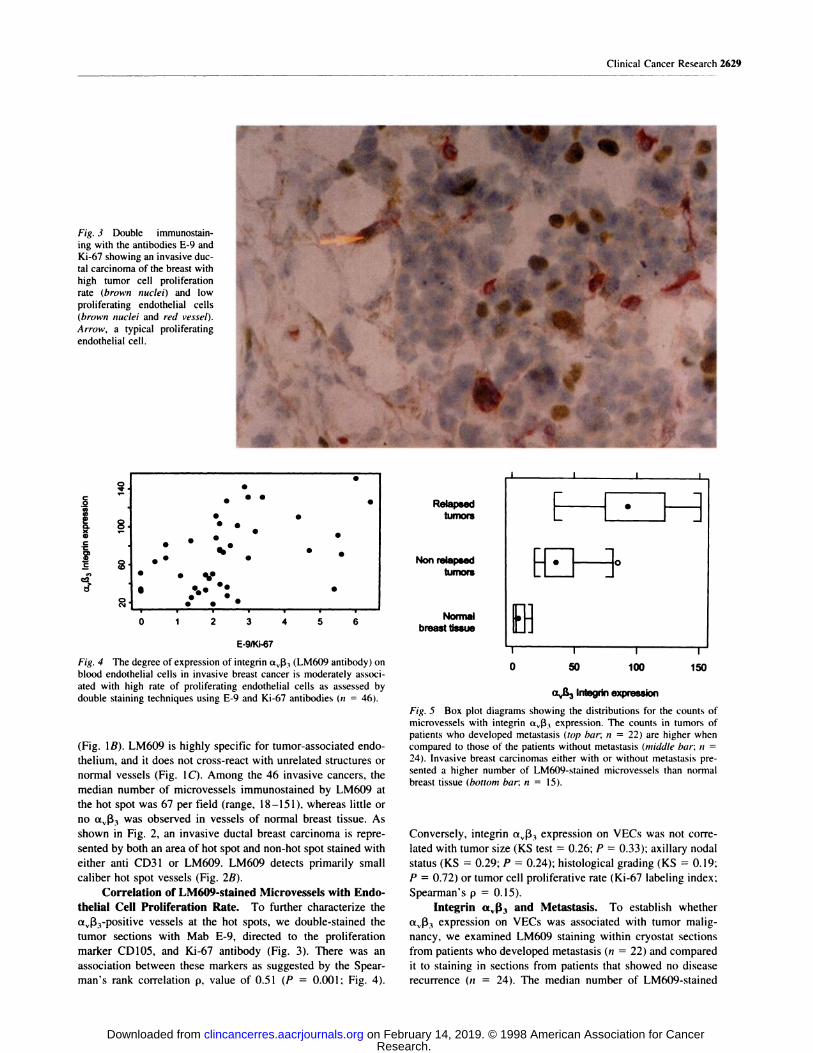
with E-9 and Ki-67 antibodies. It was claimed that the E-9

Research. on February 14, 2019. © 1998 American Association for Cancerclincancerres.aacrjournals.org Downloaded from

2632 � as a Prognostic Indicator in Breast Cancer

Table 2 Multivariate analysis on relapse-free survival

Model includes integrin av�33 expression.

Node-negative Node-positivepatients patients All patients

Wald Wald Wald

-- Variable statistic” P statistic” P statistic” P

Microvessel counts with integrin a.’f33 expression

Linear component I 1.10 0.0009 7.27 0.0070 17.80 <0.0001

Nonlinear component 7.46 0.0063 2.52 0.1 100 8.90 0.0029Menopausal status, post vs. pre/peri 7.77 0.0053 2.58 0.1000 5.26 0.0218

Tumor size. pT2� ts. pT, 3.34 0.0670 1.24 0.2660

Histological grading. G3 vs. G,_2 2.60 0.1070 5.19 0.0227No. of involved axillary nodes, �3 vs. <3 1.11 0.2900

Adjuvant therapy, hormone vs. chemotherapy 0.18 0.6600

Nodal status: adjuvant therapy

N+ CMF vs. N- 0.08 0.7830

N+ TAM i’s. N- 0.84 0.3590

“ One degree of freedom.

Table 3 Multivariate analysis on relapse-free survival

Alternative model including CD3 1 counts.

Node-negative Node-positive

patients patients

Wald Wald

All patients

Wald

- - Variable statistic” P statistic” P statistic#{176} P

Microvessel counts by anti-CD3 1

Linear component 1 1.20 0.0009 5.93 0.0149 16.90 <0.0001

Nonlinear component 7.09 0.0078 1.61 0.2050 8.60 0.0034Menopausal status, post vs. pre/peri 1.52 0.2180 2.53 0.1 1 10 2.71 0.0995

Tumor size, pT2, vs. pT, 1.62 0.2030 0.62 0.4320

Histological grading, G3 t’s. G1, 0.85 0.3580 4.20 0.0405

No. of involved axillary nodes. �3 vs. <3 1.03 0.3100Adjuvant therapy. hormone vs. chemotherapy 0.005 0.9450

Nodal status: adjuvant therapy

N+ CMF vs. N- 0.09 0.7690

N + TAM vs. N - 0. 12 0.7280

“ One degree of freedom.

antibody, specific against the CD1O5 antigen, was selectively

expressed on “activated” intratumoral vessels (21). There-

fore, the correlation found between the expression of integrin

a��33, CD1O5, and proliferative endothelial cell nuclei sug-

gests that integrin aJ33 is a marker of angiogenic blood

vessels. Finally, the comparative studies between LM609 and

anti-CD3 I antibodies on normal breast tissue indicate that

very low and weak expression of integrin ttv�33 was found on

quiescent normal vessels, whereas CD31 antigen was ex-

pressed in almost all vasculature, regardless of their size. We

found that the number of VECs within tumoral stroma was

significantly higher than that of normal tissue regarding

integrin a�133 expression, but not CD31 antigen (data not

shown). These findings confirm and extend the results pre-

viously reported by Brooks et al. (10) demonstrating the

presence of ttv1�3 �fl a small series of human breast cancers.

Vascular Integrin aj�3 Expression and Metastasis.

To verify whether the levels of expression of integrin a.,ft, are

predictive of metastasis, we compared the absolute highest

vascular counts with LM609 staining in a subgroup of primary

tumors without metastasis and in those of patients who devel-

oped metastasis. We found that the breast cancers that showed

evidence of metastasis had a significantly higher VEC integrin

ttv1�3 expression than did those without metastasis (Fig. 5).

These results may be explained by experimental evidence that

elevated levels of integnn ttv1�3 are stimulated by production of

angiogenic peptides, such as basic fibroblast growth factor, and

cytokines, such as tumor necrosis factor-ct (9). Alternatively,

activated endothelial cells produce various proteases that could

alter the structure of ECM and, thereby, facilitate tumor cells

invasiveness and metastasis (10).

Vascular Integrin a�f�3 Expression, Prognosis, and Po-

tential Therapeutic Implications. Expression of integrin

av1�3 was found to be a strong prognostic factor in a consecutive

series of breast cancer patients after surgery with long-term

follow-up. Of particular relevance, in all the patient subgroups

studied, tumors containing elevated microvessel counts with

LM609 staining had a significantly poorer prognosis relative to

those with low expression of integrin ttv�3i. Multivariate anal-

yses, including the conventional prognostic indicators, suggest

Research. on February 14, 2019. © 1998 American Association for Cancerclincancerres.aacrjournals.org Downloaded from

A Node-negative patients

.�. 0

.�e

.�

2

, .ti..

Cl) 0U-�

00 � , , , , , , � ,

0 12 24 36 48 60 72 84 96

Months

B Node-positive patients

� ‘k

0 12 24 36 48 60 72 84 96

Clinical Cancer Research 2633

Months

Fig. 7 RFS curves estimated from the Cox multivariate regressionmodel. A, node-negative patients. The curves are estimated for three

absolute values of the number of microvessels with integrin aj33

expression (50, 70, and 90 counts) adjusted by selected categories of the

other covariates: postmenopausal status, pT,, and grade I-Il. -, 50;,70; , 90. The corresponding HRs (95% confidence inter-

vals) are: 50 versus 70, HR 7.19 (2.71-19.06); and 70 versus 90.HR = 2.50 (1.72-3.64). B. node-positive patients. The curves are

estimated for the above reported absolute values of the number of

microvessels adjusted by the following covariates: postmenopausal sta-

tus and number of axillary lymph nodes <3. -, 50; , 70;

90. The corresponding HRs (95% confidence intervals) are: 50 versus

70; HR = 1.98 (1.37-2.86); and 70 versus 90, HR 1.58 (1.32-1.90).

that integrin ttv1�3 expression on tumoral vasculature was an

independent prognostic indicator of recurrence in the patients

with node-negative as well as in those with node-positive dis-

ease treated with adjuvant therapy.

The degree of integrin ttv(33 expression on VECs discrim-

mated between different prognostic groups when the impact of

this variable was adjusted for the other covariates included in

the multivariate model (Fig. 7). Integrin aj33 expression on

intratumoral vasculature had, in this series, a prognostic value

similar to that of the panendothelial marker CD3 1, as suggested

by the alternative model of multivariate analysis reported in

Table 3. Furthermore, the prognostic value of integrin aj33

compares favorably with the results of previous studies, which

assessed the prognostic significance of microvessels count de-

termined using various panendothelial markers in biopsies from

breast cancer (1 1, 13, 22, 23). Finally, the multivariate model,

which did not include a marker of angiogenesis, had a poorer

predictive value as compared to those of the models reported in

Tables 2 and 3 (as assessed using the Harrell c statistic). These

findings further support the clinical relevance of angiogenesis as

a marker of malignant breast cancer. In this context, LM609

antibody would be considered a more specific and sensitive

marker of angiogenesis. However, this can be further tested in

prospective studies.

The results of this study coupled with the previous reports

(10) that systemic administration of LM609 antibody blocked

angiogenesis and breast tumor growth, suggest that antagonists

of integrin ttv1�3 may provide an antiangiogenic approach wor-

thy of serious consideration to improve treatment of human

breast cancer. In fact, a humanized form of LM609 (Vitaxin) has

recently completed Phase I clinical trials in patients with ad-

vanced stage cancer. This antibody showed no toxic side effects

and appeared to provide clinical benefit in some of the patients

(24). Furthermore, recent evidence has been reported suggesting

that integrin �v1�3 �5 a target molecule for disruption of tumor

vasculature induced by administration of tumor necrosis factor

and IFN-y (25). Because inhibition of angiogenesis is presently

considered one of the more promising novel therapeutic ap-

proaches for cancer patients, the development of markers of

angiogenic activity is an integral part of proper clinical trial

designs in inhibitors of angiogenesis (26). In conclusion, it

seems likely that the use of the LM609 antibody to determine

integrin ttv1�3 vascular expression, which is detectable by a

single and reliable immunohistochemical assay, may become

part of breast cancer evaluation as a prognostic marker. Further-

more, LM609 staining may be a useful surrogate maker pre-

dictive of the efficacy of systemic administration of antagonists

of integrin ttv1�3 or of other inhibitors of angiogenesis capable of

blocking endothelial cell growth (reviewed in Ref. 26).

ACKNOWLEDGMENTS

We thank Prof. Ettore Marubini and Dr. Patrizia Boracchi of theInstitute of Biometry and Medical Statistics of the University of Milan.

(Milan, Italy) for their valuable suggestions regarding the statistical

analyses. We also thank Daniela Mazzocco for preparation of the

manuscript.

REFERENCES1 . Folkman, J. Angiogenesis in cancer, vascular. rheumatoid and other

disease. Nat. Med., 1: 27-31, 1995.

2. Polverini, P. J. Cellular adhesion molecules. Newly identified medi-ators of angiogenesis. Am. J. Pathol., 148: 1023-1029, 1996.

3. Hanahan, D., and Folkman J. Patterns of emerging mechanisms ofthe angiogenic switch during tumorigenesis. Cell. 86: 353-364. 1996.

4. Hynes, R. 0. Integrins: versatility. modulation, and signaling in cell

adhesion. Cell, 69: 1 1-25. 1992.

5. Str#{246}mblad,S., and Cheresh, D. A. Cell adhesion and angiogenesis.Trends Cell Biol., 6: 462-468, 1996.

6. Brooks. P. C., Clark, R. A. F., and Cheresh, D. A. Requirement ofvascular UvI�3 for angiogenesis. Science (Washington DC), 264: 569-

571, 1994.

7. Friedlander. M., Brooks, P. C., Schaffer, R. W.. Kincaid, C. M..Varner, J. A., and Cheresh, D. A. Definition of two angiogenic path-

Research. on February 14, 2019. © 1998 American Association for Cancerclincancerres.aacrjournals.org Downloaded from

2634 #{128}tv�33as a Prognostic Indicator in Breast Cancer

ways by distinct a� integrins. Science (Washington DC), 270: 1500-1502, 1995.

8. Drake, C. J., Cheresh, D. A., and Little, C. D. An antagonist of

integrin cs�l33 prevents maturation of blood vessels during embryonic

neovascularization. J. Cell. Sci., 108: 2655-2661, 1995.

9. Strdmblad, S., Becker, J. C., Yebra, M., Brooks, P. C., and Cheresh,D. A. Suppression of p53 activity and �21WAFI�h1� expression by

vascular cell integrin � during angiogenesis. J. Clin. Invest., 98:

426-433, 1996.

10. Brooks, P. C., Strdmblad, S., Klemke, R., Visscher, D.. Sarkar,

F. H., and Cheresh, D. A. Antiintegrin aj33 blocks human breast cancergrowth and angiogenesis in human skin. J. Clin. Invest., 96: 1815-1822,

1995.

1 1. Gasparini, G., Barbareschi. M., Boracchi, P.. Verderio, P., Caffo,

0., Meli, S., Della Palma, P. D., Marubini, E., and Bevilacqua. P. Tumorangiogenesis predicts clinical outcome of node-positive breast cancer

patients treated with adjuvant hormone therapy or chemotherapy. Can-

cerJ. Sci. Am., 1: 131-141, 1995.

12. Vermeulen, P. B.. Verhoeven, D., Hubens, G., Van Marck, E.,

Goovaerts, G., Huyghe, M., De Bruijn, E. A., Van Oosterom, A. T., andDirix, L. Y. Microvessel density, endothelial cell proliferation and tu-

mor cell proliferation in human colorectal adenocarcinomas. Ann. On-

col.. 6: 59-64, 1995.

13. Gasparini, G., Weidner, N., Bevilacqua. P., Maluta, S., Dalla Palma,

P., Caffo, 0., Barbareschi, M., Boracchi, P., Marubini, E., and Pozza, F.Tumor microvessel density. p53 expression. tumor size and peritumorallymphatic vessel invasion are relevant prognostic markers in node-

negative breast carcinoma. J. Clin. Oncol., 12: 454-466, 1994.

14. Vermeulen. P. B., Dirix, L. Y., Libura, J., Vanhoolst, I. F., VanMarck, E., and Van Oosterom, A. T. Correlation of the fractions of

proliferating tumor and endothelial cells in breast and colorectal adeno-

carcinomas is independent of tumor histotype and microvessel density.

Microvasc. Res., 54: 88-92, 1997.

15. Hollander, M., and Wolfe, D. A. Nonparametric Statistical Meth-

ods. New York: John Wiley & Sons, 1973.

16. Gasparini, G., Toi, M., Gion, M.. Verderio, P.. Dittadi, R.,Hanatani, M., Matsubara, I.. Vinante, 0., Bonoldi, E., Boracchi, P.,

Gatti, C.. Suzuki, N., and Tominaga. T. Prognostic significance of

vascular endothelial growth factor protein in node-negative breast car-

cinoma. J. NatI. Cancer Inst. (Bethesda), 89: 139-147, 1997.

17. Harrell, F. E., Lee, K. L., and Mark, D. B. Tutorial in biostatistics.

Multivariable prognostic models: issues in developing models, evaluat-

ing assumptions and adequacy, and measuring and reducing errors. Stat.

Med., 15: 361-387, 1996.

18. Gasparini, G., and Harris, A. L. Clinical importance of the deter-

mination of tumor angiogenesis in breast carcinoma: much more than a

new prognostic tool. J. Clin. Oncol., 13. 765-782, 1995.

19. Gasparini, G. Current controversies in cancer. Is determination ofangiogenic activity in human tumours clinically useful? Eur. J. Cancer,

34: 615-618, 1998.

20. Vermeulen, P. B., Gasparini, G., Fox, S. B., Toi, M., Martin, L.,

McCulloch. P., Pezzella, F., Viale. G., Weidner, N., Harris. A. L., andDirix, L. Y. Quantification of angiogenesis in solid human tumours: aninternational consensus on the methodology and criteria of evaluation.

Eur. J. Cancer. 32A: 2474-2484, 1996.

21. Wang, J. M., Kumar, S., Pye. D., Haboubi, N., and Al-Nakib, L.

Breast carcinoma: comparative study of tumor vasculature using twoendothelial cell markers. J. Natl. Cancer Inst. (Bethesda), 86: 386-388,

1994.

22. Weidner, N., Folkman, J., Pozza, F., Bevilacqua, P., Allred. E. N.,Moore, D. H., Meli, S., and Gasparini, G. Tumor angiogenesis: a newsignificant and independent prognostic indicator in early-stage breast

carcinoma. J. Nail Cancer Inst. (Bethesda), 84: 1875-1887, 1992.

23. Gasparini, G., Fox, S. B., Verderio, P., Bonoldi, E., Bevilacqua,

P.. Boracchi, P., Dante, S., Marubini, E., and Harris, A. L. Determi-nation of angiogenesis adds information to estrogen receptor status in

predicting the efficacy of adjuvant tamoxifen in node-positive breast

cancer patients. Clin. Cancer Res., 2: 1 191-I 198, 1996.

24. Gutheil, J. C., Campbell, T. N., Pierce, P. R., Watkins, J. D., Huse,

W. D., Bodkin, D. J., Hart, J., and Cheresh. D. A. Phase I study ofvitaxin, an anti-angiogenic humanized monoclonal antibody to vascular

integrin a�l�3 Proc. Am. Soc. Clin. Oncol. 17: 2lSa, 1998.

25. Ruegg, C., Yilmaz, A., Bieler, G., Bamat, J., Chaubert, P., andLejeune, F. J. Evidence for the involvement of endothelial cell integrin

av1�3 in the disruption of the tumor vasculature induced by TNF and

IFN--y. Nat. Med., 4: 408-414, 1998.

26. Gasparini, G. Antiangiogenic drugs as a novel anticancer therapeu-

tic strategy. Which are the more promising agents? What are the clinical

developments and indications? Crit. Rev. Oncol. Hematol., 26: 147-

162. 1997.

Research. on February 14, 2019. © 1998 American Association for Cancerclincancerres.aacrjournals.org Downloaded from

1998;4:2625-2634. Clin Cancer Res   G Gasparini, P C Brooks, E Biganzoli, et al.   breast cancer.Vascular integrin alpha(v)beta3: a new prognostic indicator in

  Updated version

  http://clincancerres.aacrjournals.org/content/4/11/2625

Access the most recent version of this article at:


  E-mail alerts related to this article or journal.Sign up to receive free email-alerts

  Subscriptions

Reprints and

  [email protected] at

To order reprints of this article or to subscribe to the journal, contact the AACR Publications

  Permissions

  Rightslink site. Click on "Request Permissions" which will take you to the Copyright Clearance Center's (CCC)

.http://clincancerres.aacrjournals.org/content/4/11/2625To request permission to re-use all or part of this article, use this link

Research. on February 14, 2019. © 1998 American Association for Cancerclincancerres.aacrjournals.org Downloaded from


Recommended