228
Lection: Venous and lymphatic vessels.

Lection:
Venous and lymphatic vessels.

ANATOM.UA

PART 1
Ch. 1 Anatomia generalis
PART 2 – SYSTEMATA MUSCULOSKELETALIA
Ch. 2 Ossa
Ch. 3 Juncturae
Ch. 4 Musculi
PART 3 – SYSTEMATA VISCERALIA
Ch. 5 Systema digestorium
Ch. 6 Systema respiratorium
Ch. 7 Cavitas thoracis
Ch. 8 Systema urinarium
Ch. 9 Systemata genitalia
Ch. 10 Cavitas abdominopelvica
PART 4 – SYSTEMATA INTEGRANTIA I
Ch. 11 Glandulae endocrinae
Ch. 12 Systema cardiovasculare
Ch. 13 Organa lymphoidea
PART 5 – SYSTEMATA INTEGRANTIA II
Ch. 14 Systema nervosum
Ch. 15 Organa sensuum
Ch. 16 Integumentum commune
ANATOM.UA
https://fipat.library.dal.ca/ta2/
ANATOM.UA
ANATOM.UA 5
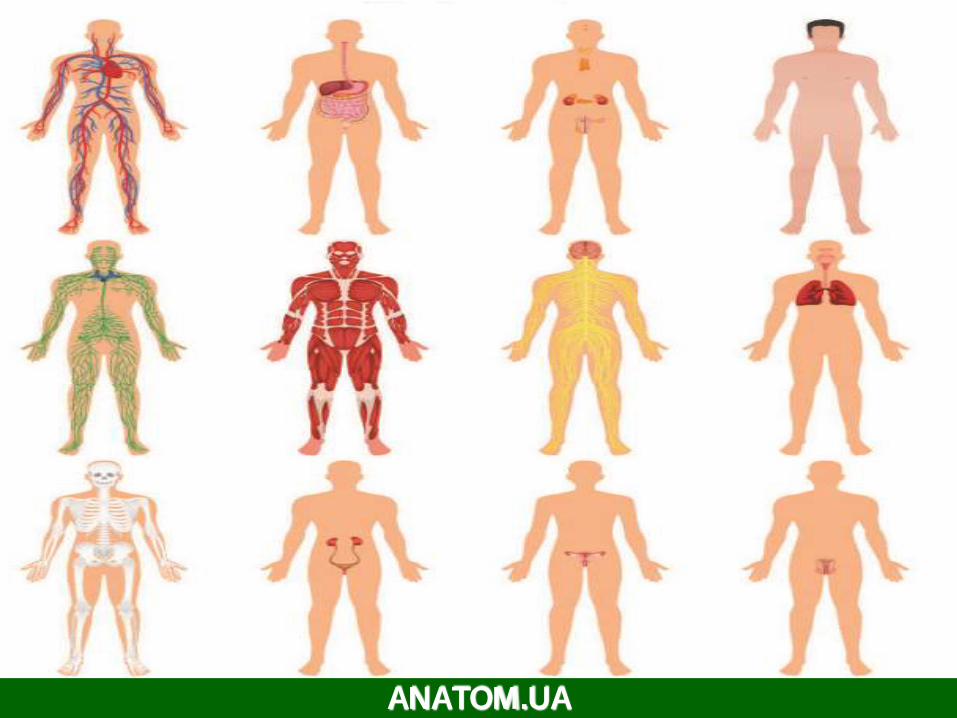
Cardiovascular system (systema
cardiovasculare) consists of the heart
and the tubes, that are used for
transporting the liquid with special
functions – the blood or lymph, that are
necessary for supplying the cells with
nutritional substances and the oxygen.

Veins
Veins are blood vessels that bring blood back to theheart.
All veins carry deoxygenatedblood with the exception of thepulmonary veins and umbilical veins
There are two types of veins: Superficial veins: close to the surface of thebody
NO corresponding arteries
Deep veins: found deeper in the body With corresponding arteries
Veins of the systemiccirculation: Superior and inferior vena cava with their tributaries
Veins of the portal circulation: Portal vein
ANATOM.UA


Superior Vena Cava
Formed by the union of the right and left Brachiocephalic veins. Brachiocephalic veins are formed by the union of internal jugular and subclavianveins.
Drains venous blood from: Head &neck
Thoracic wall Upper limbs
It Passes downward and enter the rightatrium.
Receives azygos vein on the posterior aspect just before it enters theheart.
ANATOM.UA

Veins of Head & Neck
Twodivisions:
SuperficialVeins
External Jugular veins
Anterior jugular veins
Deep Veins
Internal Jugularsveins
ANATOM.UA

Superficial Veins of Head & Neck
External JugularVeins: Lies superficial to the sternomastoidmuscle
It passes down the neck and it is the only
tributary of the subclavianvein.
It drains bloodfrom: Outside of the skull
Deep parts of the face.
ANATOM.UA

Superficial Veins of Head & Neck
Anterior jugularveins: It begins in the upper part of the neck
by the union of the submental veins.
It descends close to the median line of
the neck, medial to the sternomastoid
muscle.
At the lower part of the neck, it passes
laterally beneath that muscle to drain
into the external jugular vein.
Just above the sternum the two
anterior jugular veins communicate by
a transverse vein to form the jugular
arch.
ANATOM.UA
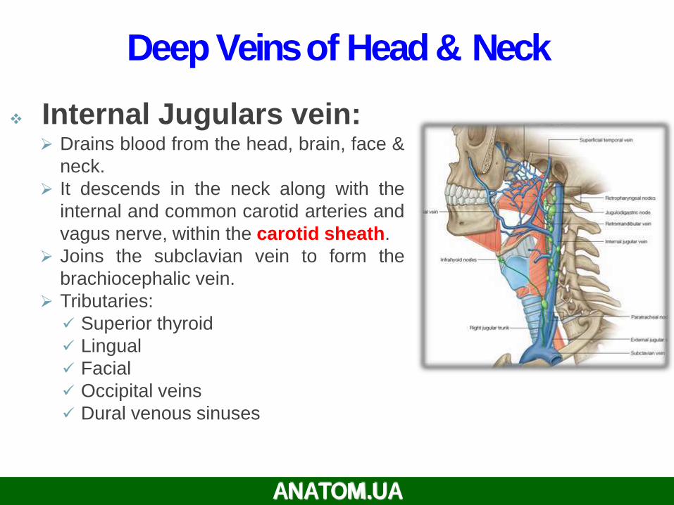
Deep Veins of Head & Neck
Internal Jugulars vein: Drains blood from the head, brain, face &
neck.
It descends in the neck along with the
internal and common carotid arteries and
vagus nerve, within the carotid sheath.
Joins the subclavian vein to form the
brachiocephalic vein.
Tributaries:
Superior thyroid
Lingual
Facial
Occipital veins
Dural venous sinuses
ANATOM.UA

ANATOM.UA

Veins of Upper Limbs
Two divisions:
Superficial Veins
Deep Veins
ANATOM.UA

Veins of Upper Limbs
Superficial Veins Cephalic vein
Ascends in the superficial fascia
on the lateral side of the biceps.
Drains into the Axillary vein.
Basilic vein Ascends in the superficial fascia
on the medial side of the
biceps.
Halfway up the arm, it pierces
the deep fascia
At the lower border of the teres
major it joins the venae
comitantes of the brachial artery
to form the Axillary vein.ANATOM.UA

Veins of Upper Limbs
Deep Veins Venaecommitantes
large Which accompany all the
arteries, usually in pairs.
Brachial Vein
Ulnar Vein
Radial Vein
Axillary vein Formed by the union of basilic
vein and the venae comitantes
of the brachial artery.
ANATOM.UA

Inferior VenaCava
Drains most of the blood from the
body below the diaphragm to the
right atrium.
Formed by the union of the 2
common iliac veins behind the
right common iliac artery at the
level of the 5th lumbar vertebra.
Ascend
s aorta
Pierces
on the right side of the
the central tendon ofdiaphragmat the level of the 8th
thoracic vertebra.
ANATOM.UA

Tributaries of Inferior Vena Cava
Two common iliac veins
Median sacral vein
Four paired lumbar veins
Right gonadal vein the left vein drains into the left renal
vein
Paired renal veins
Right suprarenal vein the left vein drains into the left renal
vein
Hepatic veins
Paired inferior phrenic vein
ANATOM.UA
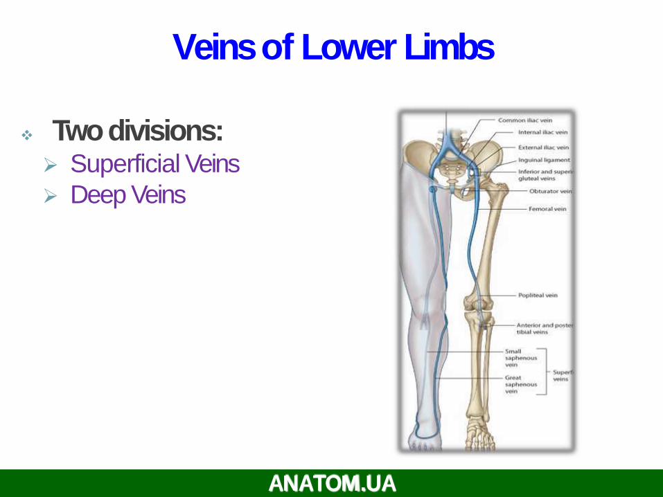
Veins of Lower Limbs
Twodivisions: Superficial Veins
Deep Veins
ANATOM.UA

Veins of Lower Limbs
SuperficialVeins Form a network in the subcutaneoustissue
Pattern is variable
They are the tributaries of the: Great (long) saphenous vein
Small (short) saphenous vein
ANATOM.UA

Great SaphenousVein
The longest vein
Begins from the medial end of the
dorsal
venous arch of the foot.
Passes upward in front
of the medial malleolus with the
saphenous nerve.
Then it ascends in accompany with the
saphenous nerve in the superficial
fascia over the medial side of the leg.
ANATOM.UA
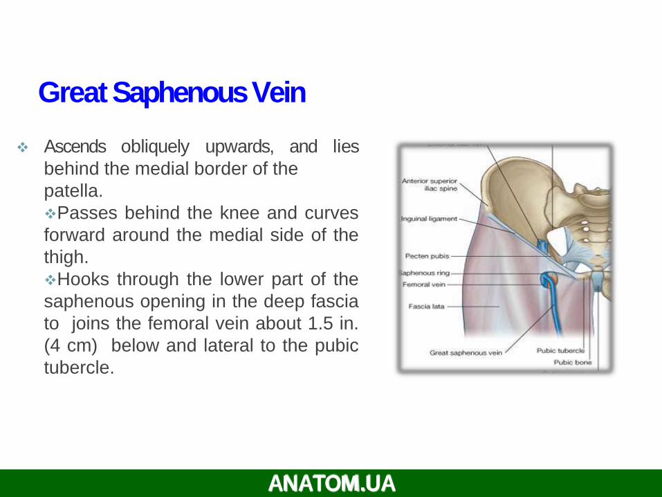
Ascends obliquely upwards, and lies
behind the medial border of the
patella.
Passes behind the knee and curves
forward around the medial side of the
thigh.
Hooks through the lower part of the
saphenous opening in the deep fascia
to joins the femoral vein about 1.5 in.
(4 cm) below and lateral to the pubic
tubercle.
Great SaphenousVein
ANATOM.UA

It is connected to the small saphenous vein
by one or two branches that pass behind
the knee.
Numerous perforating veins connect the
great saphenous vein with the deep veins.
The perforating veins have valves which
allow blood flow from superficial to deep
veins. The great saphenous vein is used in
venous grafting and saphenous cut down(take care of the saphenous nerve)
Great SaphenousVein
ANATOM.UA
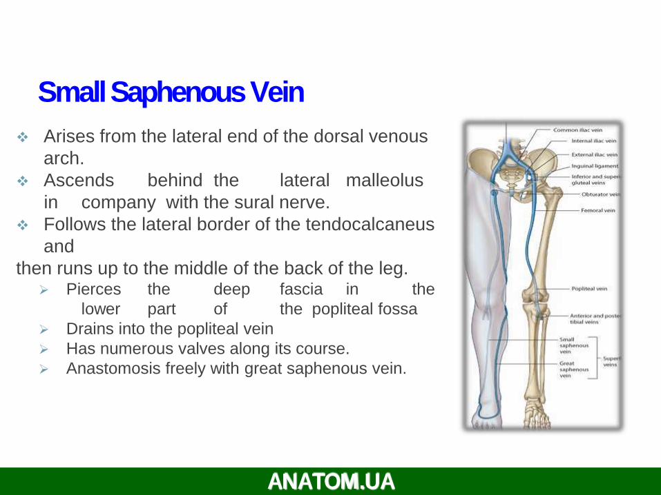
Arises from the lateral end of the dorsal venous
arch.
Ascends behind the lateral malleolus
in company with the sural nerve.
Follows the lateral border of the tendocalcaneus
and
then runs up to the middle of the back of the leg. Pierces the deep fascia in the
lower part of the popliteal fossa
Drains into the popliteal vein
Has numerous valves along its course.
Anastomosis freely with great saphenous vein.
Small SaphenousVein
ANATOM.UA

Veins of Lower Limbs
Deep Veins Comprise the venae comitantes,
which accompany all the large
arteries, usually in pairs.
Venae comitantes unite to form the
popliteal vein, which continues as the
femoral vein.
Receive blood from
superficial veins
through perforating veins.
Femoral Vein
Popliteal vein
Peroneal vein
Anterior tibial vein
Posterior tibial vein
ANATOM.UA

Mechanism of Venous
Return from Lower
Limb (FYI) Much of the saphenous blood passes
from superficial to deep veins through
the perforating veins
The blood is pumped upwards in the
deep veins by the contraction of the calf
muscles (calf pump).
This action of ‘calf pump’ is assisted by
the tight sleeve of deep fascia
surrounding these muscles.
Varicose veins: If the valves in the
perforating veins become incompetent,
the direction of blood flow is reversed
and the veins become varicosed. Most
common in posterior & medial parts of
the lower limb, particularly in old people.
ANATOM.UA
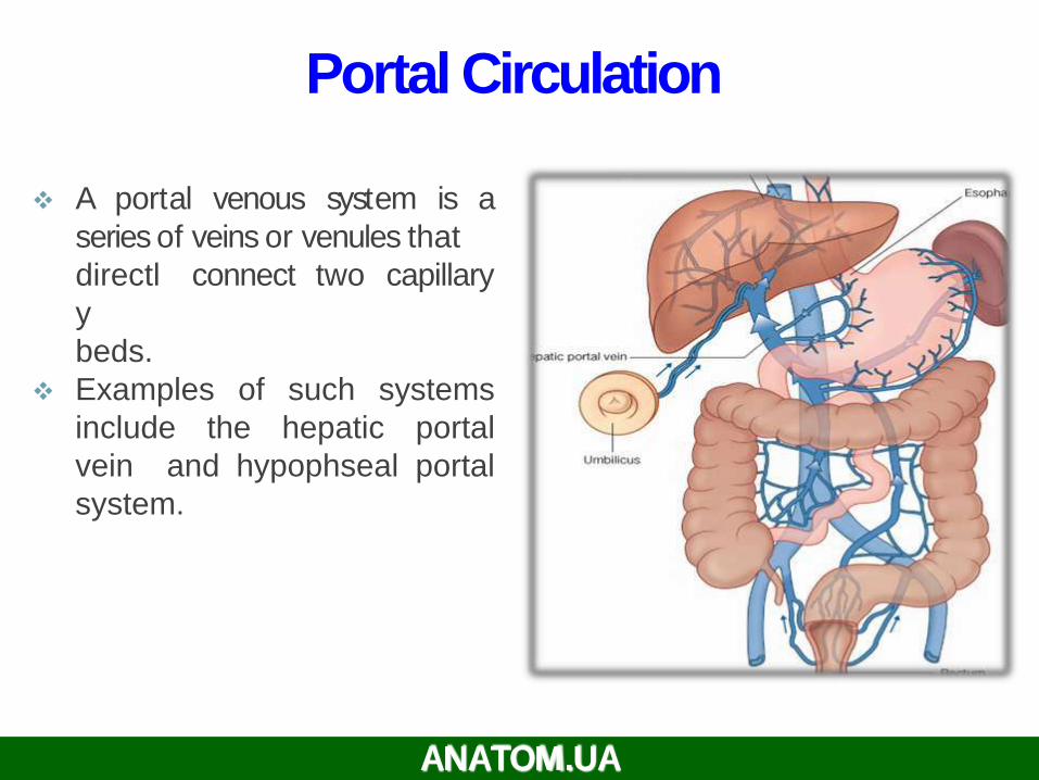
Portal Circulation
A portal venous system is a
series of veins or venules that
connect two capillarydirectl
y
beds.
Examples of such systems
include the hepatic portal
vein and hypophseal portal
system.
ANATOM.UA

Hepatic Portal Vein
Drains blood from the
gastrointestinal
tract and spleen
It is formed by the union of the
superior mesenteric and splenic
veins.
Immediately before reaching the
liver, the portal vein divides into right
and left that enter the liver.
Tributaries: Gastric and cystic veins
ANATOM.UA

Portocaval Anastomosis
A portacaval anastomosis (also known
as portal systemic anastomosis) is a
specific type of anastomosis that occurs
between the veins of portal circulation
and those of systemic circulation.
The anastomotic channels become
dilated
(varicosed) in case of portal hypertension.
ANATOM.UA

Sites of Portocaval Anastomosis
Lower end of esophagus: left gastric vein
&
azygos vein
Lower part of rectum: (Hemorrhoids)
superior and middle rectal veins &
inferior rectal vein
Para umbilical region: (Caput Medusae)
Para
umbilical veins & superficial epigastric vein
Retroperitoneal: Veins draining colon &
veins of the posterior abdominal wall
Patent ductus venosus:
Left branch of portal vein & inferior vena
cava.
ANATOM.UA


Lymphatic system (systemalymphoideum), is morphologically andfunctionally connected with thecardiovascular system. This term goes afterthe latin word lympha - the clean water, andthe greek word nympha - the bride.
Lymphatic system is the most developedof the all vessels that transport liquids
ANATOM.UA 34
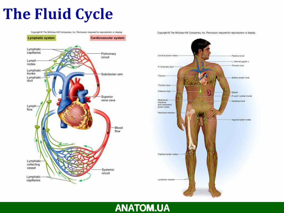
The Fluid Cycle
ANATOM.UA

36
ANATOM.UA
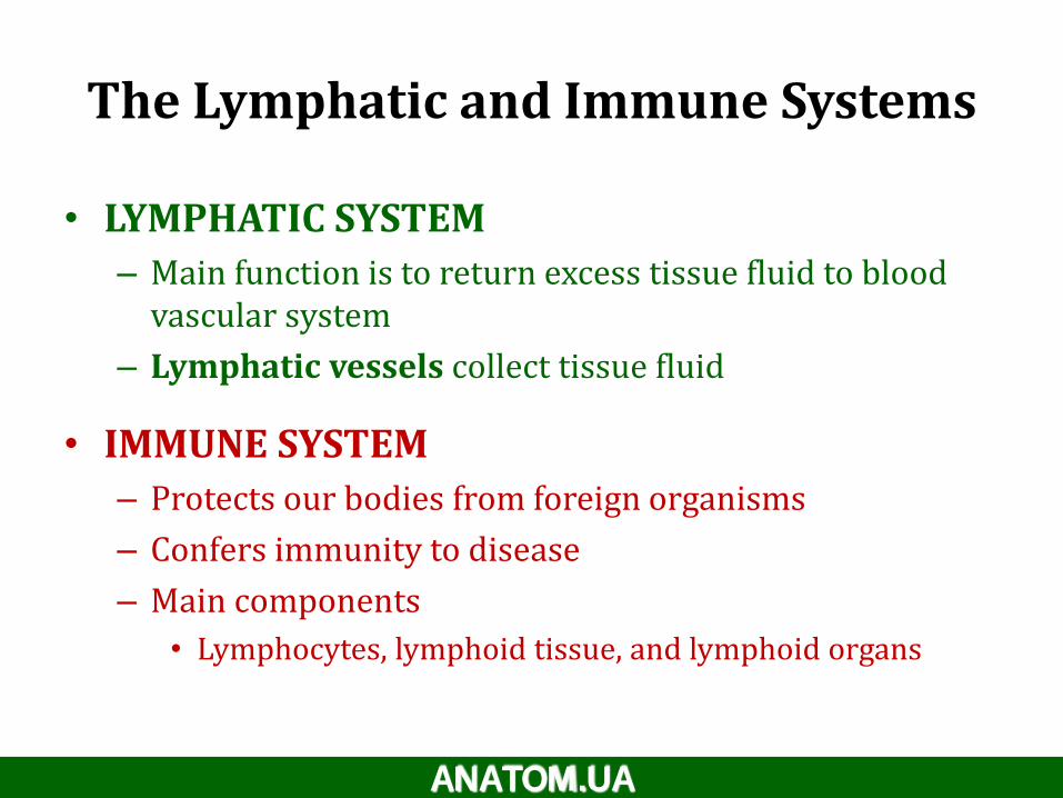
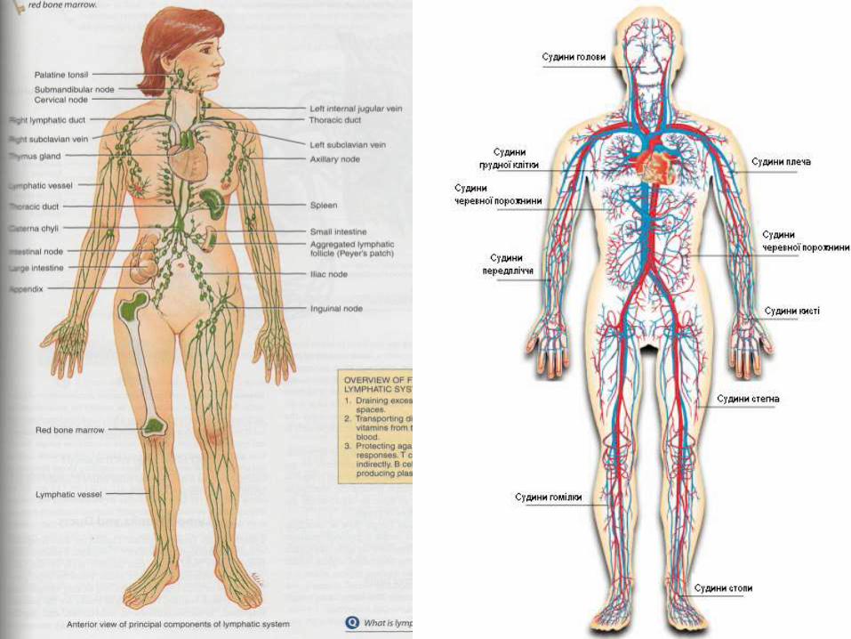
The Lymphatic and Immune Systems
• LYMPHATIC SYSTEM
– Main function is to return excess tissue fluid to blood vascular system
– Lymphatic vessels collect tissue fluid
• IMMUNE SYSTEM
– Protects our bodies from foreign organisms
– Confers immunity to disease
– Main components
• Lymphocytes, lymphoid tissue, and lymphoid organs
ANATOM.UA
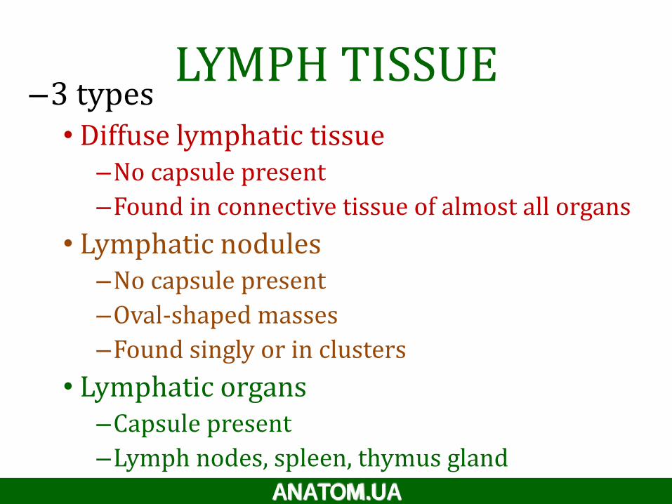
LYMPH TISSUE–3 types
• Diffuse lymphatic tissue–No capsule present
–Found in connective tissue of almost all organs
• Lymphatic nodules–No capsule present
–Oval-shaped masses
–Found singly or in clusters
• Lymphatic organs–Capsule present
–Lymph nodes, spleen, thymus gland
ANATOM.UA

21-39
• Maintain fluid balance
• Protect body from infection and disease
Lymphatic and Immune Systems

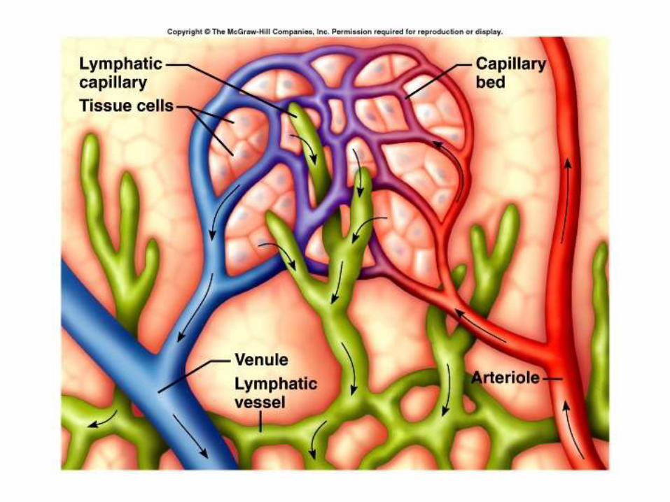
Lymph Capillaries

Lymphatic Vessels

42
ANATOM.UA


ANATOM.UA

• Lymphatic vessels collect tissue fluid from loose connective tissue
– Carry fluid to great veins in the neck
– Fluid flows only toward the heart
– Once tissue fluid is within lymphatic vessels it is termed lymph
• Functions of lymphatic vessels – collect excess tissue fluid and blood proteins
• Return tissue fluid and blood proteins to bloodstream
ANATOM.UA

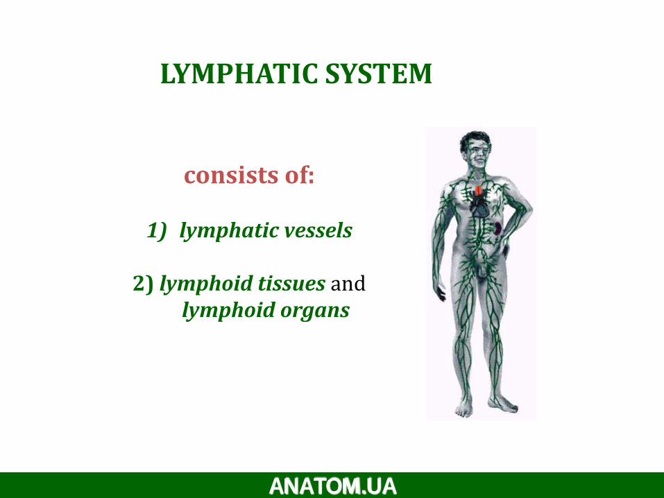
LYMPHATIC SYSTEM
consists of:
1) lymphatic vessels
2) lymphoid tissues and lymphoid organs
ANATOM.UA

travel along with blood
vessels.
1) lymphatic vessels
ANATOM.UA
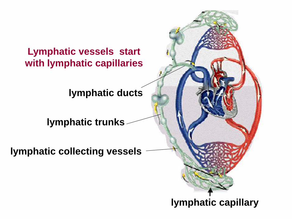
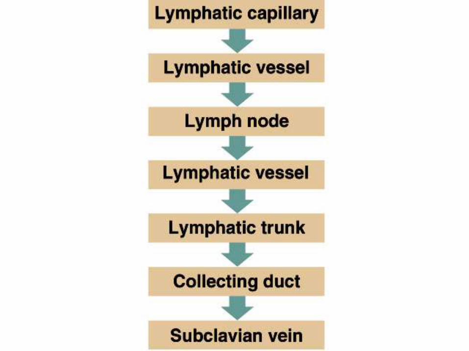
lymphatic capillary
lymphatic trunks
lymphatic collecting vessels
lymphatic ducts
Lymphatic vessels start
with lymphatic capillaries

- blind ended vessels
- permeable to proteins even cells
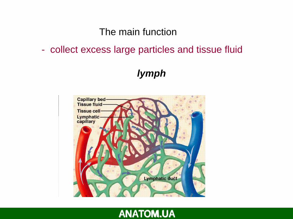
The main function
- collect excess large particles and tissue fluid
lymph
ANATOM.UA
LYMPH
• What is lymph ?
Tissue fluid (interstitial fluid) that enters the lymphatic vessels

FORMATION AND TRANSPORT OF TISSUE FLUID
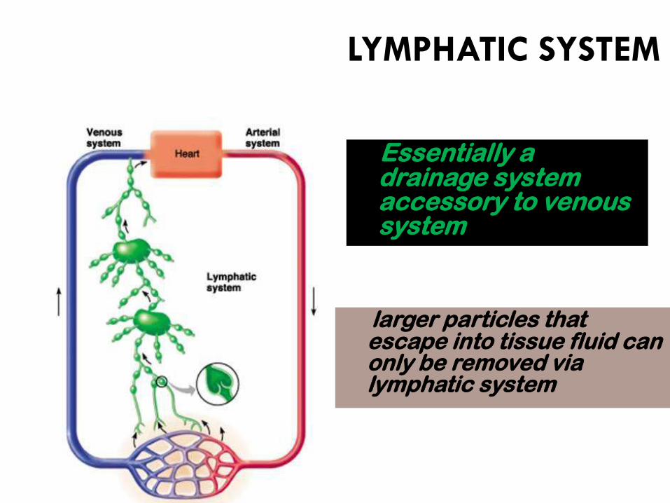
Essentially a drainage system accessory to venous system
larger particles that escape into tissue fluid can only be removed via lymphatic system
LYMPHATIC SYSTEM

Functions of the Lymphatic System
24
-
58
• Reabsorbs excess interstitial fluid:– returns it to the venous circulation– maintain blood volume levels– prevent interstitial fluid levels from rising out of control.
• Transport dietary lipids:– transported through lacteals– drain into larger lymphatic vessels– eventually into the bloodstream.
• lymphocyte development, and the immune response.
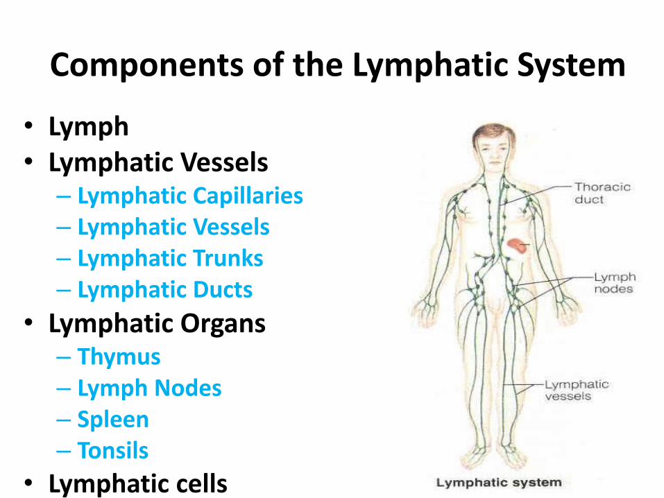
Components of the Lymphatic System24
-
59
• Lymph• Lymphatic Vessels
– Lymphatic Capillaries– Lymphatic Vessels– Lymphatic Trunks– Lymphatic Ducts
• Lymphatic Organs– Thymus– Lymph Nodes– Spleen– Tonsils
• Lymphatic cells

Lymph Vessels
• Lymphatic capillaries –
• Lymphatic collecting vessels
• Lymphatic trunks –
• Lymphatic ducts –
ANATOM.UA

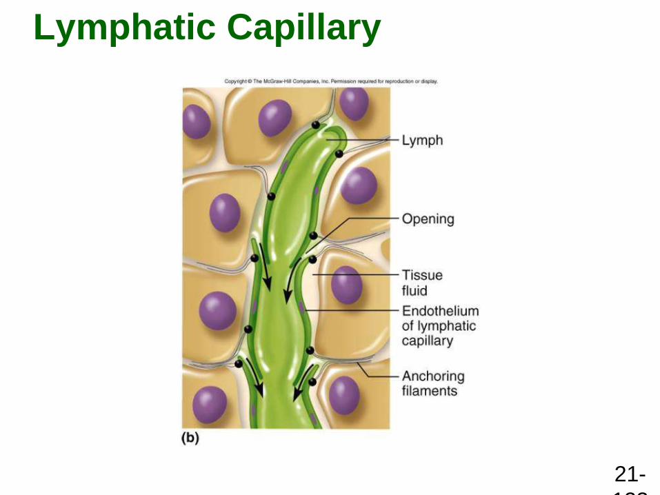
Lymphatic Capillaries
24
-
62
Features of structure:
• Blind end
• Single layer of overlapping endothelial cells
• More permeable than that of blood capillary
• Absent from avascular structures, brain, spinal cord splenic pulp and bone marrow
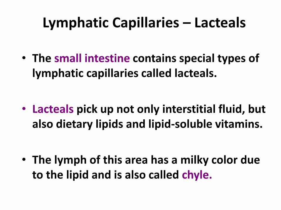
Lymphatic Capillaries – Lacteals
24
-
63• The small intestine contains special types of
lymphatic capillaries called lacteals.
• Lacteals pick up not only interstitial fluid, but also dietary lipids and lipid-soluble vitamins.
• The lymph of this area has a milky color due to the lipid and is also called chyle.

Lymphatic Vessels
24
-
64Features of structure
Three layered wall but thinner than vein,
More numerous valves than in vein
Interposed by lymph nodes at intervals
Arranged in superficial and deep sets
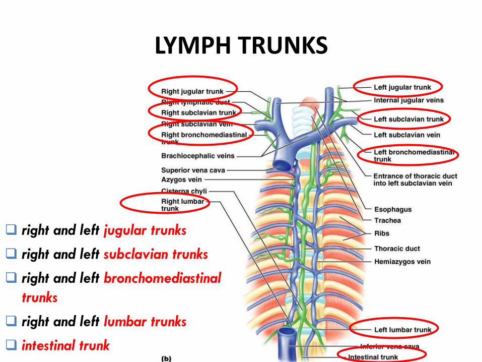
LYMPH TRUNKS
right and left jugular trunks
right and left subclavian trunks
right and left bronchomediastinal
trunks
right and left lumbar trunks
intestinal trunk
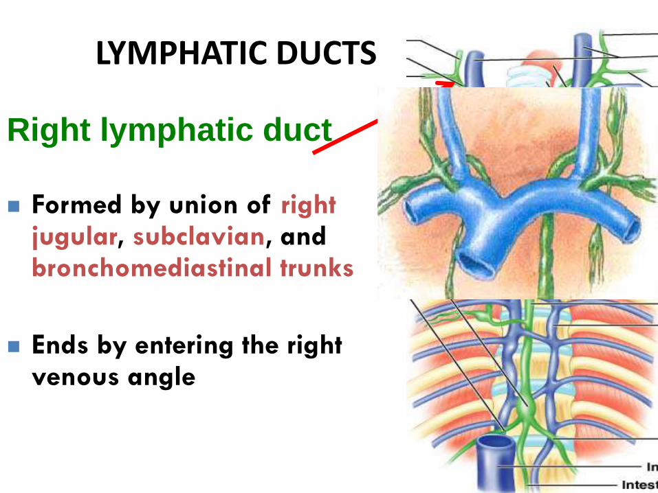
LYMPHATIC DUCTS
24
-
66
Right lymphatic duct
Formed by union of right jugular, subclavian, and bronchomediastinal trunks
Ends by entering the right venous angle

Thoracic duct• Begins in front of L1 as a
dilated sac, the cisterna chyli,
• formed by left and right lumbar trunks and intestinal trunk
• Enter thoracic cavity & ascends
• Travels upward, veering to the left at the level of T5
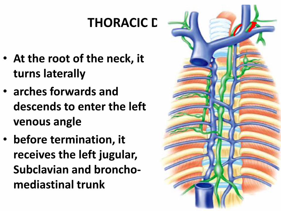
LYMPHATIC DUCTS
68
THORACIC DUCT…..
• At the root of the neck, it turns laterally
• arches forwards and descends to enter the left venous angle
• before termination, it receives the left jugular, Subclavian and broncho-mediastinal trunk

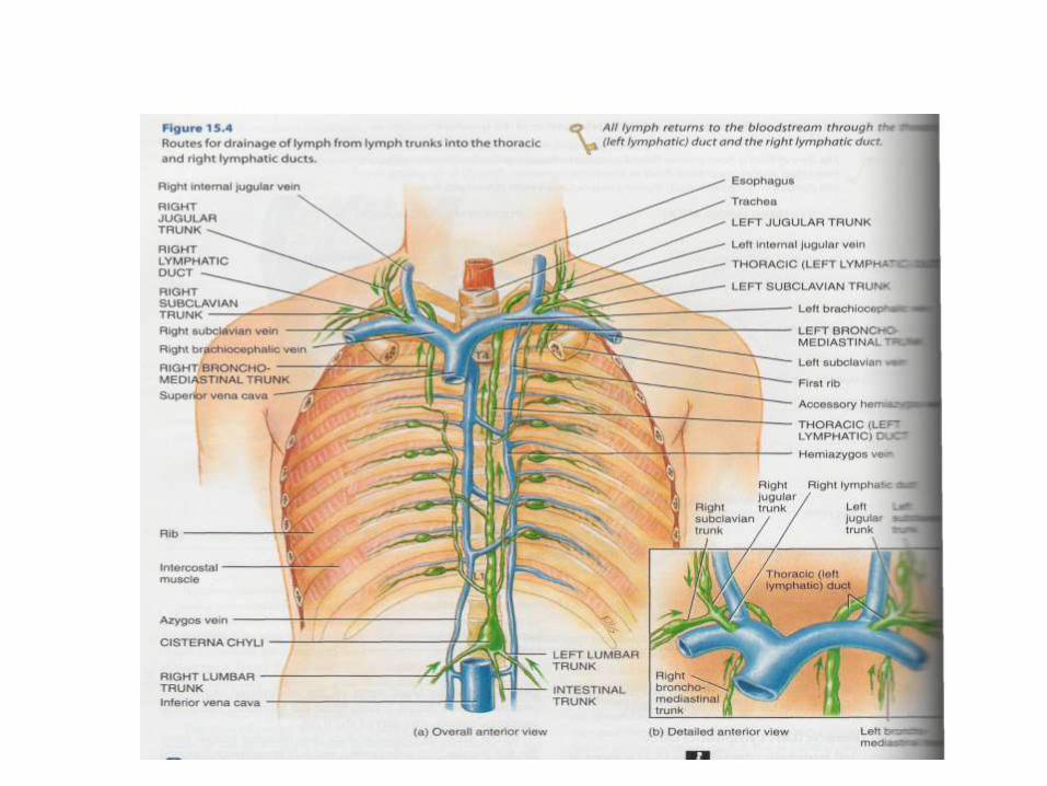
DRAINAGE PATTERN
THORACIC DUCT - Drains
lymph from lower limbs,
pelvic cavity, abdominal
cavity, left side of thorax,
and left side of the head,
neck and left upper limb
RIGHT LYMPHATIC DUCT -
Receives lymph from right
half of head, neck, thorax
and right upper limb, right
lung, right side of heart, right
surface of liver

Lymphatic Cells
24
-
70• Also called lymphoid cells.
• Located in both the lymphatic system and the cardiovascular system.
• Work together to elicit an immune response.
• Types of lymphatic cells are:
– macrophages
– epithelial cells
– dendritic cells
– lymphocytes
ANATOM.UA

LYMPHATIC ORGANS
Primary organs
– Red bone marrow
– Thymus gland
Secondary organs
– Lymph nodes
– Lymph nodules
– Spleen
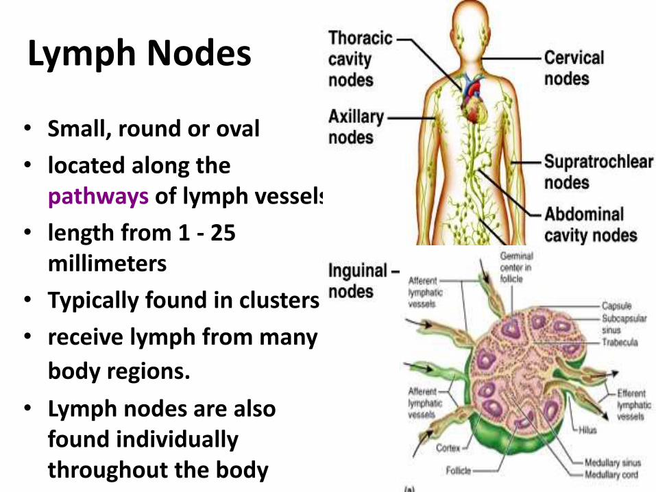
Lymph Nodes
24
-
72
• Small, round or oval
• located along the pathways of lymph vessels.
• length from 1 - 25 millimeters
• Typically found in clusters
• receive lymph from many
body regions.
• Lymph nodes are also found individually throughout the body
tissues.

Lymph node
Features
Bean-shaped bodies
With afferent vessels (entering
at the periphery) and efferent
lymph vessels(emerging at the
hilus)
Arranged in groups, along the
blood vessels or the flexural
side of the joint
Divided into superficial and
deep groups
74
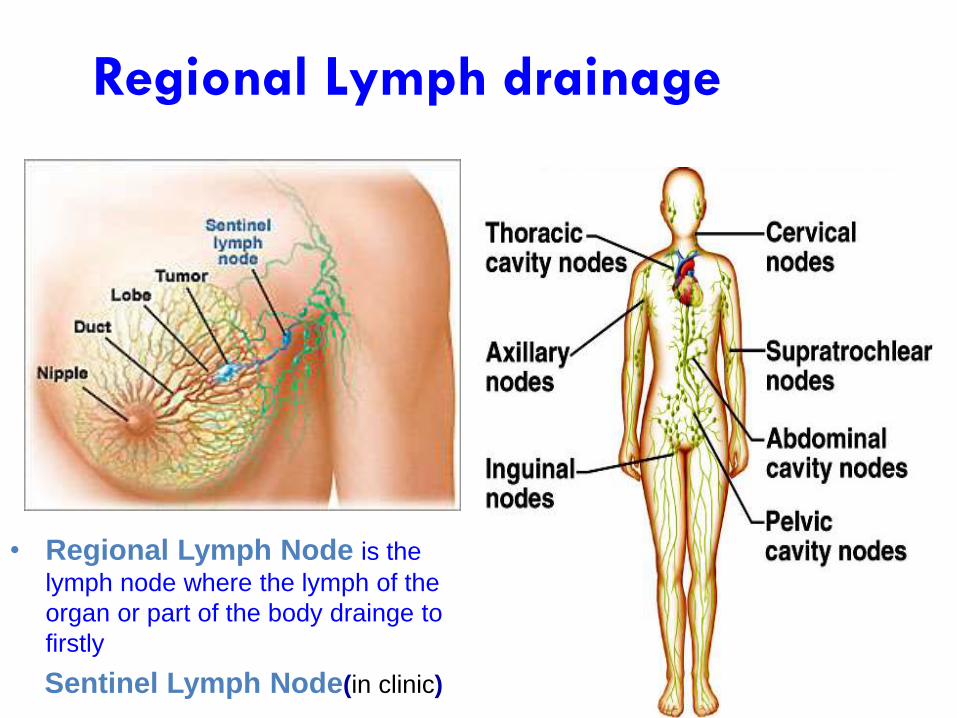
• Regional Lymph Node is the
lymph node where the lymph of the
organ or part of the body drainge to
firstly
Sentinel Lymph Node(in clinic)
Regional Lymph drainage

75
Spleen
Location Left epigastric regionbetween 9th-11th rib in line of 10th rib
• Largest lymphatic organ in the body.
• Can vary considerably in size and weight
Function
76
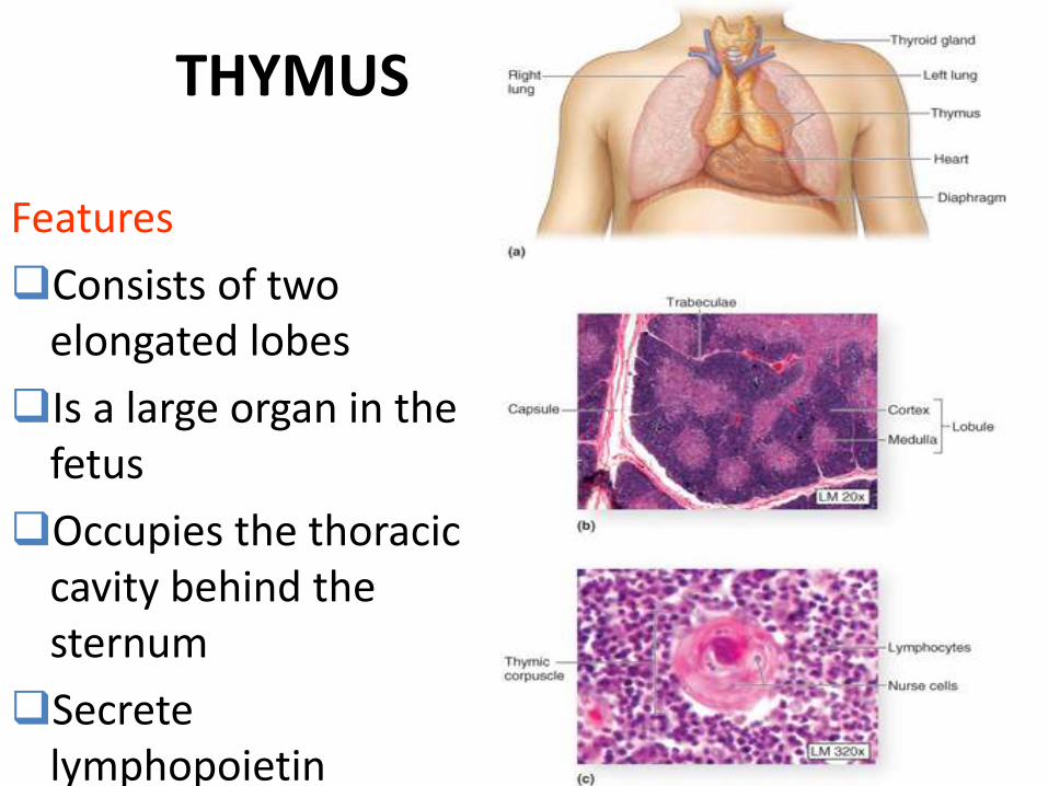
THYMUS
Features
Consists of two elongated lobes
Is a large organ in the fetus
Occupies the thoracic cavity behind the sternum
Secrete lymphopoietin

Lymphatic Nodules
24
-
77
• Oval clusters of lymphatic cells with some extracellular matrix that are not surrounded by a connective tissue capsule.
• Filter and attack antigens.
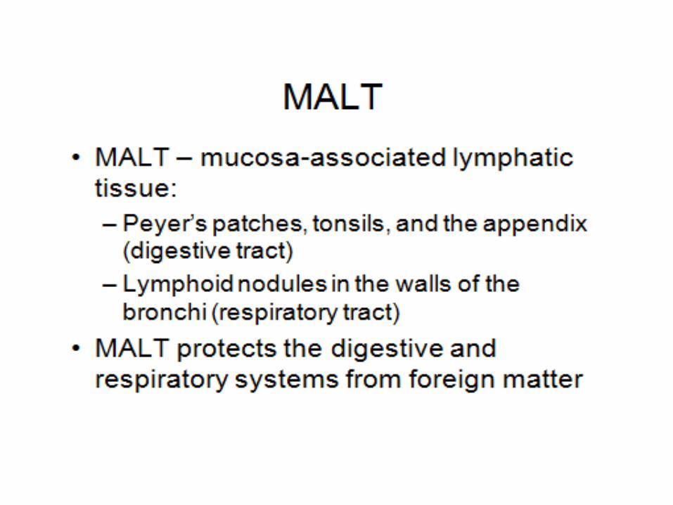
• In some areas of the body, many lymphatic nodules group together to form larger structures.– mucosa-associated lymphatic tissue (MALT) or tonsils– very prominent in the mucosa of the small intestine,
primarily in the ileum • Peyer patches
– also present in the appendix

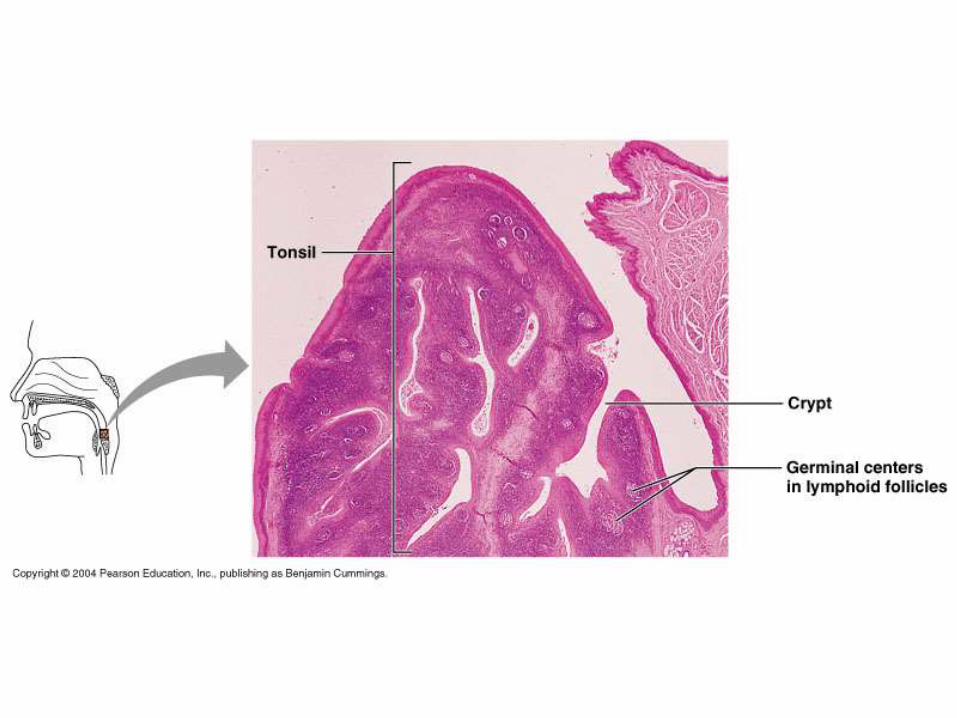
Tonsils
24
-
79
• clusters of lymphatic cells and extracellular matrix not completely surrounded by a connective tissue capsule.
• Consist of multiple germinal centers and crypts
• Several groups of tonsils form a protective ringaround the pharynx. – pharyngeal tonsils (or adenoids) in nasopharynx– palatine tonsils in oral cavity – lingual tonsils along posterior one-third of the
tongue
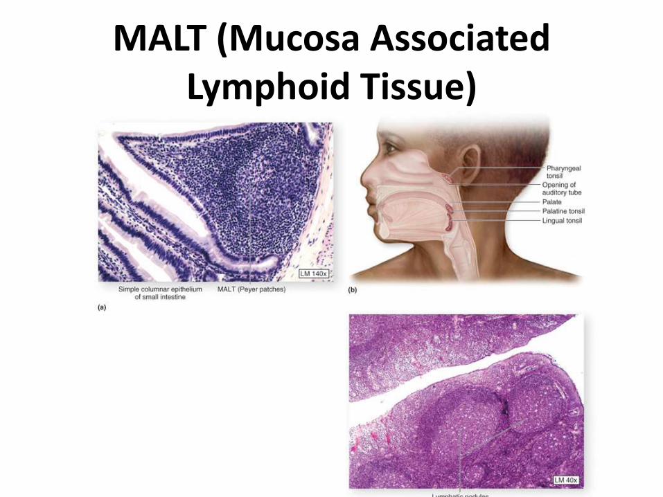
MALT (Mucosa Associated Lymphoid Tissue)
80

Special lymph capillaries --- Lacteals
- collect digested fats ( in chylomicrons)

Valves are present to
prevent backflow.

connection to the veins
ANATOM.UA
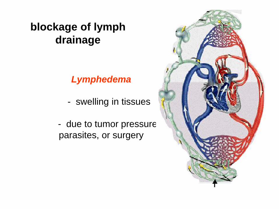
Lymphedema
- swelling in tissues
- due to tumor pressure,
parasites, or surgery
blockage of lymph
drainage
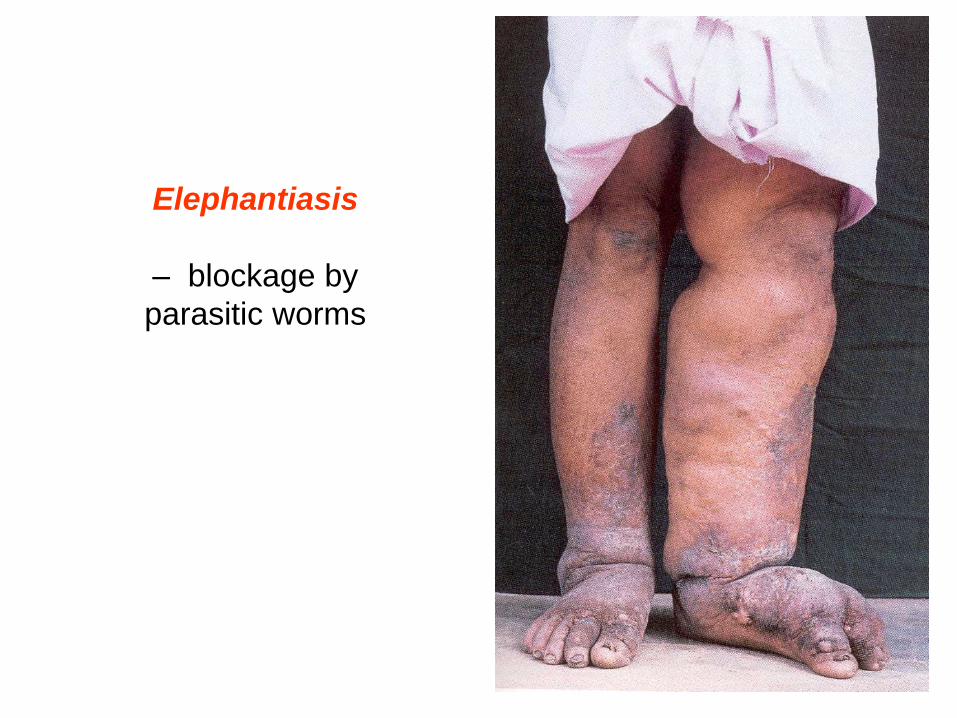
Elephantiasis
– blockage by
parasitic worms
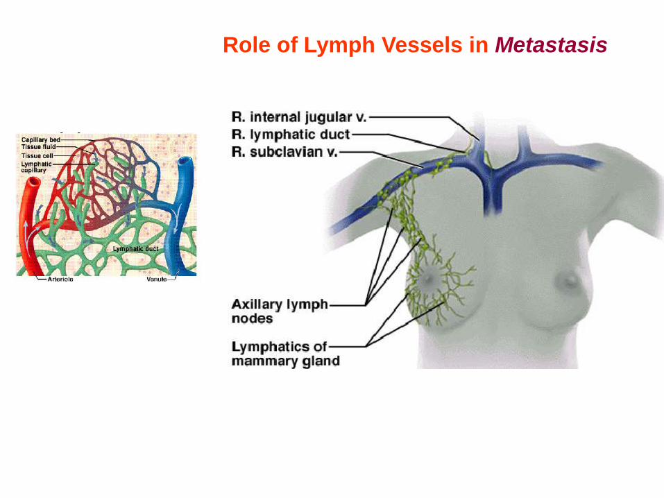
Role of Lymph Vessels in Metastasis

87
ANATOM.UA

Include:
Function:
host defense
eliminates abnormal (sick, aged, or cancerous)
cells and pathogens
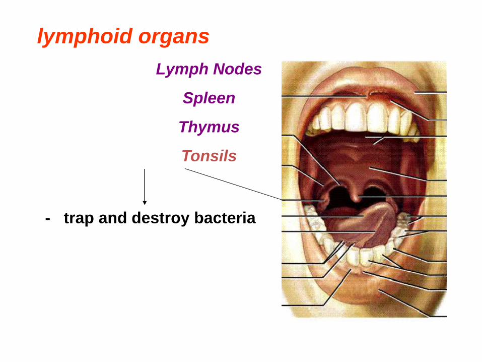
Lymph Nodes
Spleen
Thymus
Tonsils
lymphoid organs

Lymph Nodes
lymphoid organs
- Macrophages and lymphocytes
attack microorganisms
Swollen lymph nodes is caused by
expansion in the number of
lymphocytes

Lymph Nodes
Spleen
lymphoid organs
- site for immune
surveillance and response
- removes debris, foreign
matter, toxins, bacteria,
viruses, old blood cells
- readily subject to rupture
from mechanical trauma

Lymph Nodes
Spleen
Thymus
lymphoid organs - site of maturation of T
lymphocytes
- secretes hormones
(thymopoietin and
thymosins)
- critical role in
childhood
Lymph Nodes
Spleen
Thymus
Tonsils
lymphoid organs
- trap and destroy bacteria

ANATOM.UA

•The Tonsils
Figure 14-5

21-
95
Lymph Node
Fig. 21.12 a and b
21-
96
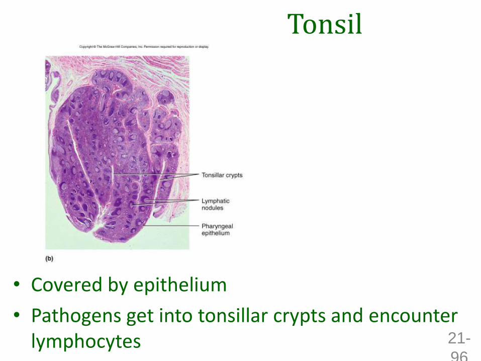
Tonsil
• Covered by epithelium
• Pathogens get into tonsillar crypts and encounter lymphocytes


Orders of Lymphatic Vessels
• Lymph capillaries –smallest lymph vessels
– First to receive lymph
• Lymphatic collecting vessels – collect from lymph capillaries
– Lymph nodes are scattered along collection vessels
Lymphatic system
Lymph duct
Lymph trunk
Lymph node
Lymphatic
capillary
Blood
capillaries
Lymphatic
collecting
vessels, with
valves
(a) Structural relationship between a capillary
bed of the blood vascular system and
lymphatic capillaries
Heart
Arterial systemVenous system

Orders of Lymphatic Vessels
• Lymph nodes
– Scattered along collecting vessels
• Lymph trunks
– Collect lymph from collecting vessels
• Lymph ducts
– Empty into veins of the neck
101

Lymphatic Drainage of Mammary and Axillary Regions
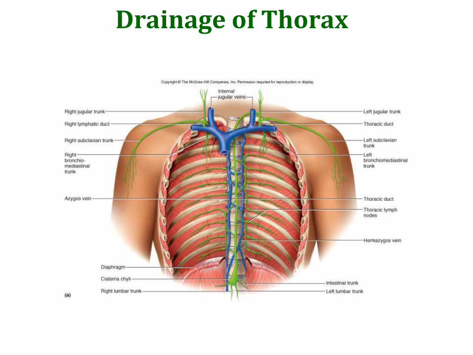
Drainage of Thorax
21-
104
Mechanisms of Lymph Flow
Lymph flows at low pressure and speed
Moved along by rhythmic contractions of lymphatic vessels• stretching of vessels stimulates contraction
Flow aided by skeletal muscle pump
Thoracic pump aids flow from abdominal to thoracic cavity
Valves prevent backward flow
Rapidly flowing blood in subclavian veins, draws lymph into it
Exercise significantly increases lymphatic return

105
Function of the lymphatic system. Theintercellular liquid is soaked through thelymphatic capillaries. It provides thetransfer of lymphocytes, sometimeserythrocytes and even some antigens.

106
The lymph (lympha) is composed aftersoaking of the intercellular liquid in the thecapillaries of the lymphatic system. It is thetransparent liquid that looks like the bloodplasma. The main cells are the lymphocytes.There are about 2-3 liters of lymph. Afterconsuming lots of fatty food the lymph turnsits color into white.
107
The lymph (lympha) is the product offiltration of blood. It is composed of:- Proteins
- Carbohydrates
- Fats
- Salts
- Hormones
- White blood cells
- lymphocytes

108
The lymphatic system (systemalymphoideum) is presented as a big amountof lymphatic capillaries, lymphocapillarnetworks, lymphatic vessels and lymphatictruncks.
109

110

111

112

113
The lymphatic system (systema lymphoideum), asa part of the immune system, consists of:
1. intercellular clefts;
2. lymphatic capillaries;
3. lymphocapillar networks;
4. lymphatic vessels;
5. truncks and ducts.

Distribution and Features of Lymphatic Capillaries
Lymphatic system
Lymph duct
Lymph trunk
Lymph node
Lymphatic
capillary
Blood
capillaries
Lymphatic
collecting
vessels, with
valves
(a) Structural relationship between a capillary
bed of the blood vascular system and
lymphatic capillaries
Heart
Arterial systemVenous system
Filaments anchored
to connective tissue
Fibroblast in loose
connective tissue
Endothelial cell
Flaplike minivalve
(b) Lymphatic capillaries are blind-ended tubes in which
adjacent endothelial cells overlap each other,
forming flaplike minivalves.
Tissue
fluid
Tissue cellBlood
capillaries
Lymphatic
capillaries

115
The lymph is filtered in the lymph nodes
and cleaned from all the wastes.
Macrophages “eat” antigens process them
and transport the immune reaction to the
lymphocytes. The lymphatic system has
the security function.

Lymph Nodes
Cleanse the lymph of pathogens
Human body contains around 500
Superficial lymph nodes located in
• Cervical, axillary, and inguinal regions
Deep nodes are
• Tracheobronchial, aortic, and iliac lymph nodes

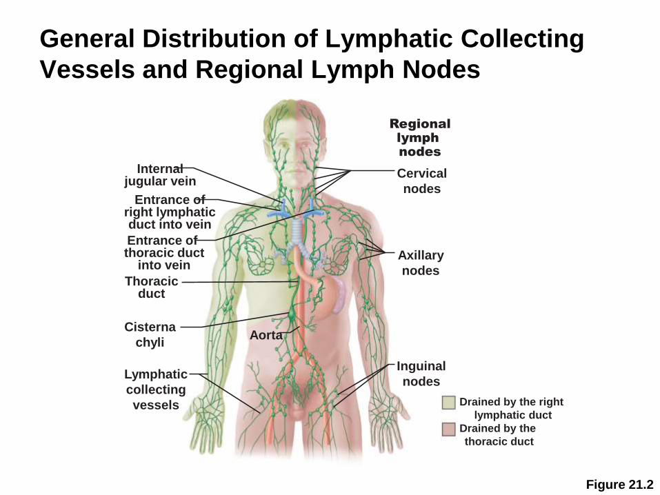
General Distribution of Lymphatic Collecting
Vessels and Regional Lymph Nodes
Figure 21.2
Cervical
nodesEntrance of
right lymphaticduct into vein
Internaljugular vein
Entrance of thoracic duct
into vein
Thoracicduct
Cisterna
chyli
Lymphatic
collecting
vessels
Axillary
nodes
Aorta
Inguinal
nodes
Regional
lymph
nodes
Drained by the right
lymphatic duct
Drained by the
thoracic duct

121

122
The lymphatic capillaries (vasa lymphocapillaria)are the first stage of the system, its roots.
They are located in the all organs and tissues(except of the brain and the spinal cord, epithelialplate of the skin, the internal ear, cartilages, corneaand crystalline lens, bone marrow, placenta andumbilical cord).
21-
123
Lymphatic Capillary

124
125
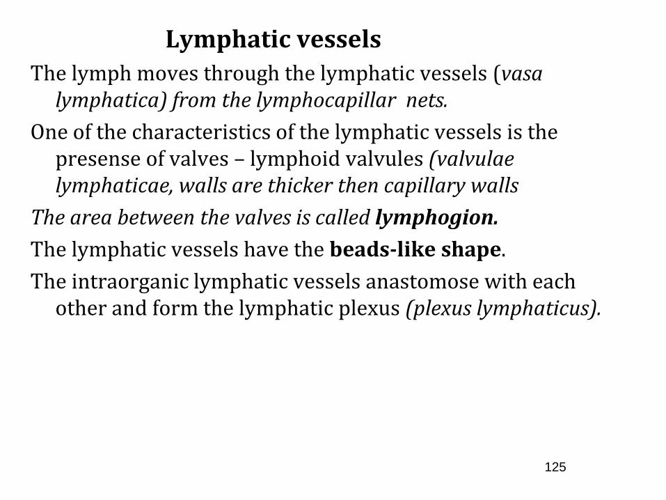
Lymphatic vessels
The lymph moves through the lymphatic vessels (vasa lymphatica) from the lymphocapillar nets.
One of the characteristics of the lymphatic vessels is the presense of valves – lymphoid valvules (valvulae lymphaticae, walls are thicker then capillary walls
The area between the valves is called lymphogion.
The lymphatic vessels have the beads-like shape.
The intraorganic lymphatic vessels anastomose with each other and form the lymphatic plexus (plexus lymphaticus).


127
1622- Gaspare Asselli found the vessels in the mesenterium of the small intestine, that were filled with milk-like liquid. He called them the milk-vessels.

128

129
The lymphatic vessels
Intraorganic lymphatic vessels anastomose with each other and form the lymphatic plexus (plexus lymphaticus).
Superficial lymphatic vessels (vasa lymphatica superficialia), that collect lymph from the skin and superficial fascia.
Profound lymphatic vessels (vasa lymphatica profunda)collect lymph from the lymphocapillar nets of the articular capsules, ligaments, muscles and deep fascia and internal organs.
In the moveable parts lymphatic vessels branch out and form the collateral ways.

130

131
132
Lymphatic vessels are interrupted on their ways to the vein system. Because of this they are divided into afferent lymphatic vessels (vasa lymphatica afferentia) and efferent lymphatic vessels (vasa lymphatica efferentia).
These vessels go to the next lymphatic nodes that are located on the way of the lymph movement.


134

135
The lymphatic nodes (nodi lymphаtici; nodilymphoidei; lymphonodi) are the secondary lymphoidorgans that are located on the lymph ways.
The lymph nodes- are the stations that control thelymph “character” and act like biological filters, wherethe antigens are neutralized.
136
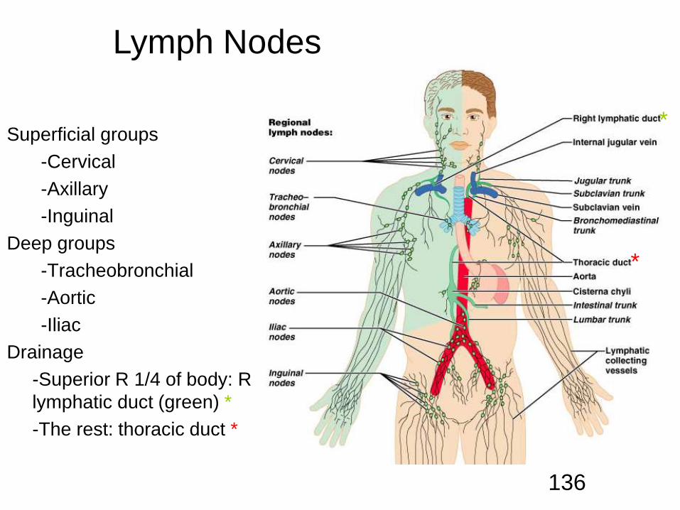
Superficial groups
-Cervical
-Axillary
-Inguinal
Deep groups
-Tracheobronchial
-Aortic
-Iliac
Drainage
-Superior R 1/4 of body: R
lymphatic duct (green) *
-The rest: thoracic duct *
Lymph Nodes
*
*

Microscopic Anatomy of a Lymph Node
Fibrous capsule—surrounds lymph nodes
Trabeculae—connective tissue strands
Lymph vessels
• Afferent lymphatic vessels
• Efferent lymphatic vessels

Microscopic Anatomy of a Lymph Node
Figure 21.3a
Afferent
lymphatic
vessels
Efferent lymphatic
vessels
CapsuleTrabeculae
Hilum
Cortex
Lymphoid follicle
Germinal center
Subcapsular sinus
Medulla
Medullary cord
Medullary sinus
(a) Longitudinal view of the internal structure
of a lymph node and associated lymphatics


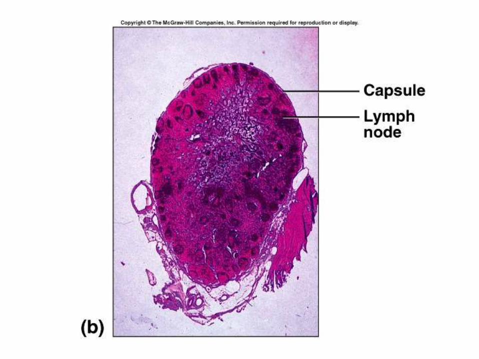
Microscopic Anatomy of a Lymph Node
Figure 21.3b
Follicles
Trabecula
Subcapsular
sinus
Capsule
Medullary
cords
Medullary
sinuses
(b) Photomicrograph of part of a lymph node (14X)
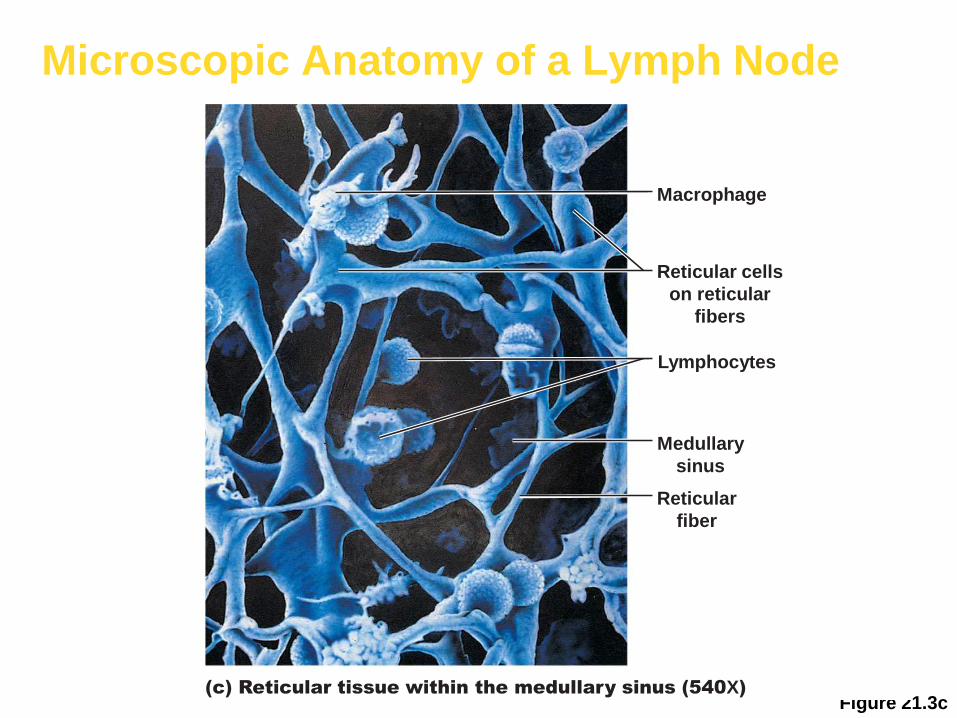
Microscopic Anatomy of a Lymph Node
Figure 21.3c(c) Reticular tissue within the medullary sinus (540X)
Macrophage
Medullary
sinus
Reticular
fiber
Lymphocytes
Reticular cells
on reticular
fibers

142

143
Lymphatic
Trunks
(all are paired except
the intestinal trunk)
Lumbar
Intestinal• Receives fatty
lymph (chyle) absorbed through lacteals in fingerlike villi of intestines
Broncho-mediastinal
Subclavian
Jugular

144
Lymph ducts(variable)
Thoracic duct: everyone has
20% also have a right lymphatic duct
*
20%

145

146

147
148

149
- Lymph nodes have different forms: globular,ovoid, bean-like and others.
- Pink-grey color
- Sizes range between 0,5 mm to 50 mm and more;
- Groups of lymph nodes that are located over thefascia are called superficial lymphatic nodes (nodilymphatici superficiales), and the nodes that are underthe fascia are called the profound lymph nodes (nodilymphatici profundi).

150

151
Considering the location of the lymph nodes andthe lymph movement ways there are regionallymphatic nodes (nodi lymphatici regionales), thatcollect lymph from the different regions of the body.

152

153
Regional lymph nodes, that collect lymph from the
organs of the skeletal system or the body walls
are called somatic lymph nodes (nodi lymphatici
somatici). The name comes from the greek word
soma – body. These nodes are called parietal
lymphatic nodes (nodi lymphatici parietalеs), from
the latin word paries – стінка.
154

155

156

157
158
The nodes that are regional only for the internal
organs are called the visceral lymphatic nodes (nodi
lymphatici viscerales). Nodes that collect lymph as
from the internal organs, as from the muscles, joints
and skin are called the mixed lymph nodes (nodi
lymphatici mixti).

159

160

161

162

163

164

165

166

167

Lymph Trunks
Lymphatic collecting vessels converge
Five major lymph trunks
• Lumbar trunks
• Receives lymph from lower limbs
• Intestinal trunk
• Receives chyle from digestive organs
• Bronchomediastinal trunks
• Collects lymph from thoracic viscera

Lymph Trunks
Five major lymph trunks (continued)
• Subclavian trunks
• Receive lymph from upper limbs and thoracic wall
• Jugular trunks
• Drain lymph from the head and neck
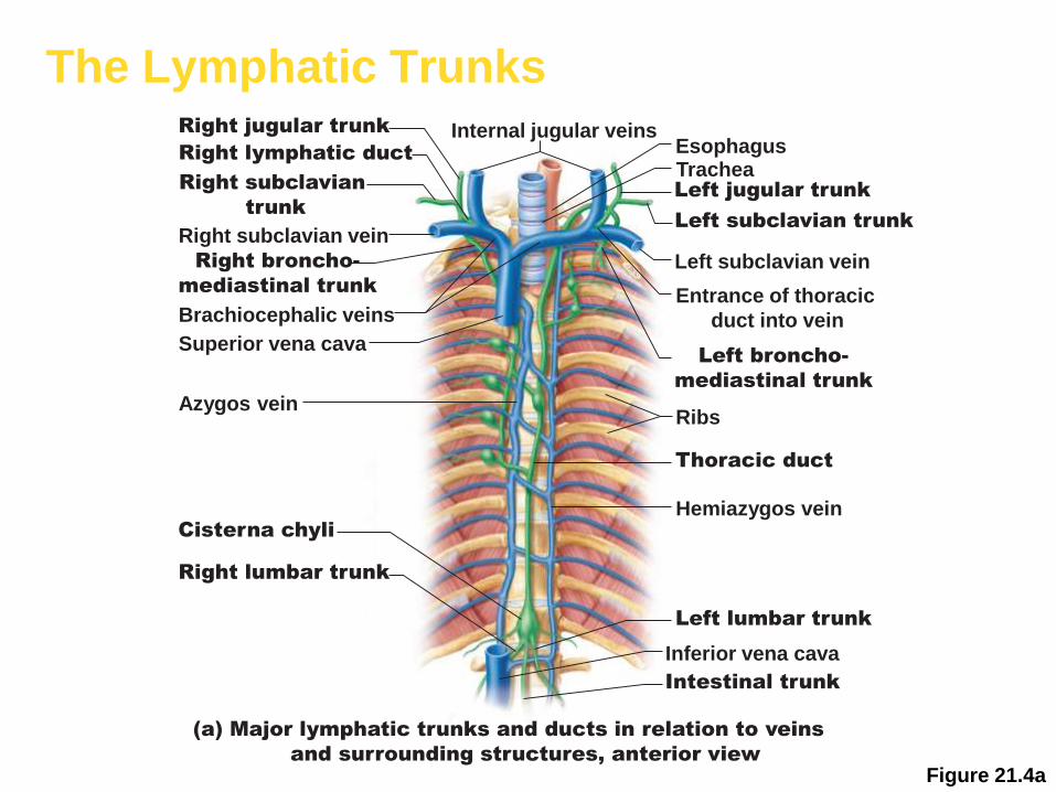
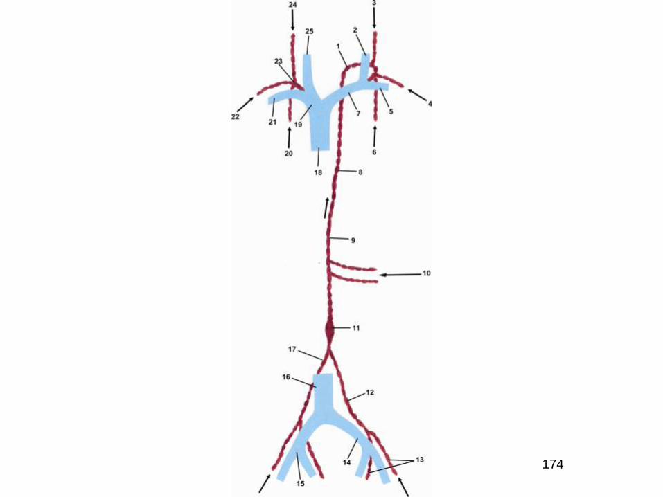
The Lymphatic Trunks
Figure 21.4a
Left jugular trunk
Internal jugular veins
Left subclavian trunk
Left subclavian vein
EsophagusTrachea
Ribs
Left lumbar trunk
Left broncho-
mediastinal trunk
Entrance of thoracic
duct into vein
Thoracic duct
Hemiazygos vein
Intestinal trunk
Inferior vena cava
Right jugular trunk
Right lymphatic duct
Right subclavian
trunk
Right subclavian vein
Right broncho-
mediastinal trunk
Brachiocephalic veins
Superior vena cava
Azygos vein
Cisterna chyli
Right lumbar trunk
(a) Major lymphatic trunks and ducts in relation to veins
and surrounding structures, anterior view

The Lymphatic Trunks
Figure 21.4b
(b) Thoracic duct (colored green)
along the posterior thoracic wall
Azygos vein
on vertebral
bodies
Thoracic duct Aorta

Lymph Ducts
Cisterna chyli
• Located at the union of lumbar and intestinal trunks
Thoracic duct
• Ascends along vertebral bodies
• Empties into venous circulation
• Junction of left internal jugular and left subclavian veins
• Drains three quarters of the body

Right Lymphatic Duct
Empties into right internal jugular and subclavian veins
Internal jugular veinsRight jugular trunk
Right lymphatic duct
Right subclavian
trunk
Right subclavian vein
Right broncho-
mediastinal trunk
Brachiocephalic veins
Superior vena cava
Azygos vein
Cisterna chyli
Right lumbar trunk
174

175

176
177
178
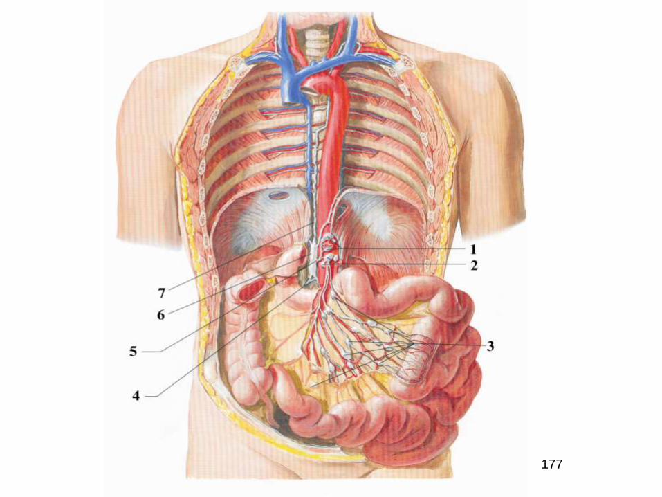
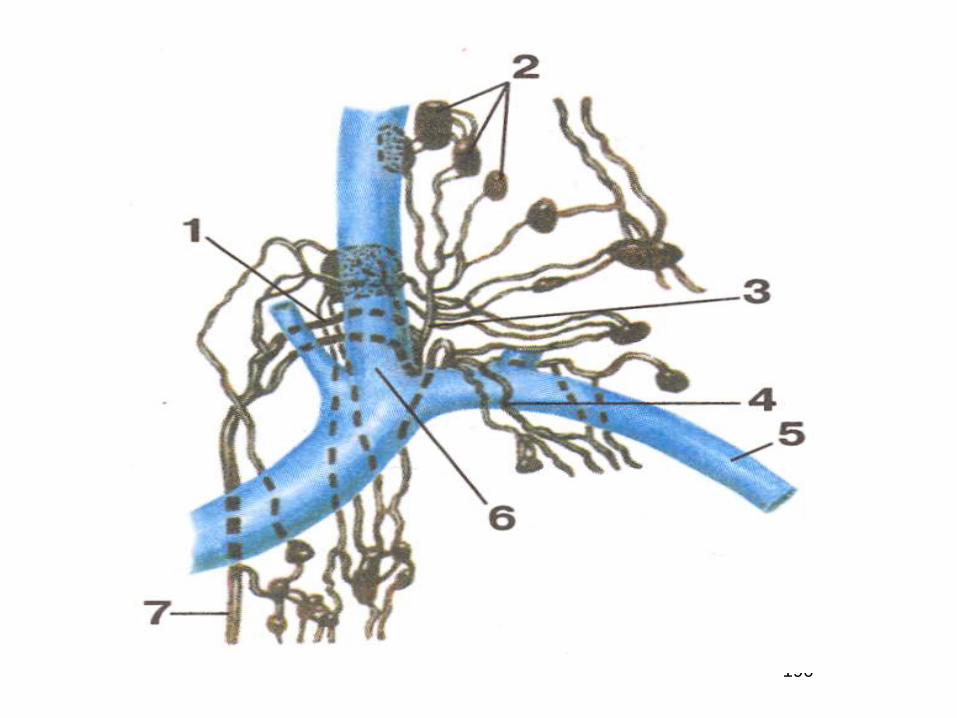
Lymphatic ducts
Lymphatic ducts (ductus lymphatici) are formed because of
combining of lymphatic trunks. There are 2 lymphatic
ducts- right lymphatic duct and pectoral duct.
Right lymphatic duct (ductus lymphaticus dexter) is a
temporal vessel, 10-15 mm.

179

180
181
182

183

184

185

186

187

188

189
190

191

192

193

194

195

196

197

198
Lymphatic System Definitions
• Pathogens—Organisms that cause
disease
• Lymphatic System—Cells, tissues, and
organs that play a central role in the
body’s defenses against pathogens
• Lymphatic system consists of vessels
(lymphatics) filled with lymph connected
to lymphatic organs
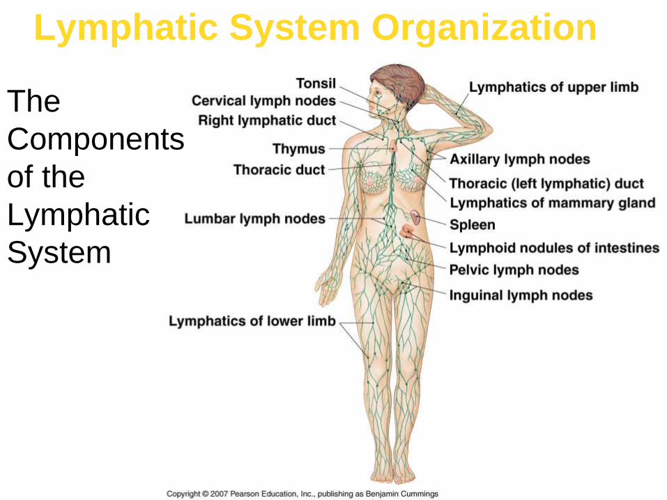
Lymphatic System Organization
The
Components
of the
Lymphatic
System

Functions of the Lymphatic System
• Produce, maintain, distribute lymphocytes
• Lymphocytes attack invading organisms,
abnormal cells, foreign proteins
• Maintain blood volume
• Help eliminate local variations in interstitial
fluid concentration
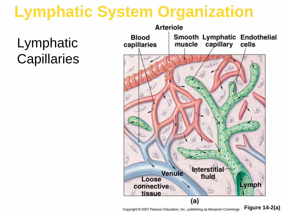
Lymphatic System Organization
Lymphatic
Capillaries
Figure 14-2(a)

Lymphatic System Organization
Figure 14-2(b)
Lymphatic Capillaries

Lymphatic System Organization
Figure 14-3
The Lymphatic Ducts and the Venous System
Lymphocyte Life Cycle
• Continuously migrate between
lymphoid tissues and the blood
• Production and development
(called lymphopoiesis) involves:
• Bone marrow
• Thymus
• Peripheral lymphoid tissues

Lymphatic System Organization
The Tonsils
Figure 14-5

21-
207
Lymph Node
Fig. 21.12 a and b
21-
208
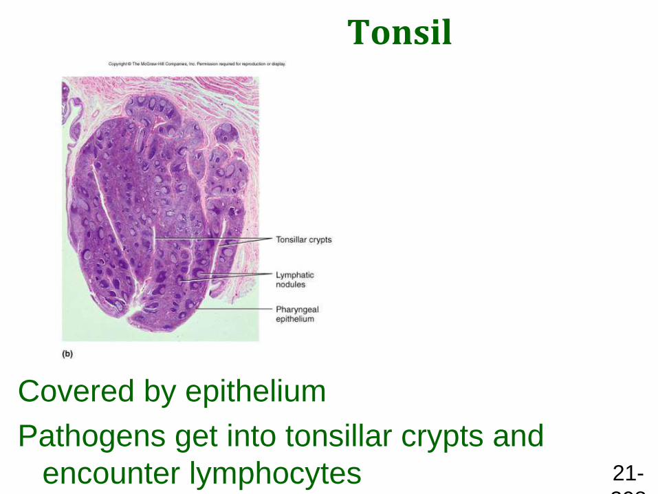
Tonsil
Covered by epithelium
Pathogens get into tonsillar crypts and
encounter lymphocytes


Lymphoid Organs
• Important lymphoid organs
include:
• Lymph nodes
• Thymus
• Spleen
• Located in areas that are
vulnerable to pathogens
Lymph Nodes
• Encapsulated masses of
lymphoid tissue containing
lymphocytes
• Monitor and filter lymph
• Remove antigens
• Initiate immune response

Lymphatic System Organization
Figure 14-6
The Structure
of a Lymph
Node

The Thymus
• Lies behind sternum
• T cells divide and mature there
• Shrinks after puberty
• Produces thymosins
• Hormones that regulate T cell
development

Lymphatic System Organization
The Thymus
Figure 14-7

Lymphatic System Organization
The Thymus
Figure 14-7(a)

Lymphatic System Organization
The Thymus
Figure 14-7(b)

Lymphatic System Organization
The Thymus
Figure 14-7(c)

The Spleen
• White pulp
• Resembles lymphoid nodules
• Removes antigens
• Initiates immune response
• Red pulp
• Contains red blood cells
• Recycles damaged or out-dated RBCs
• Stores iron from recycled RBCs

Lymphatic System Organization
The Spleen
Figure 14-8

Lymphatic System Organization
The Spleen
Figure 14-8(a)
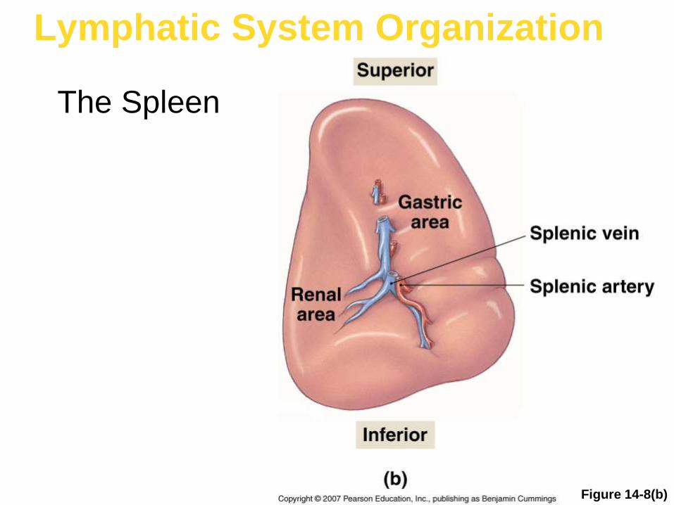
Lymphatic System Organization
Figure 14-8(b)
The Spleen

Overview of the Immune Response
• Purpose is to inactivate or destroy:
• Pathogens
• Abnormal cells
• Foreign molecules
• Based on activation of lymphocytes
by specific antigens by antigen
recognition

Immune Disorders
• Autoimmune disorders• Mistaken attack on body’s own tissues
• Immunodeficiency disease• Disease (e.g., AIDS) or a congenital
block of immunity
• Allergies• Inappropriate or excessive response to
allergens
• Age-related loss of effectiveness

Lymphoid Organs
• Primary lymphoid organs
– Bone marrow
– Thymus
• Secondary lymphoid organs
– Lymph nodes, spleen, tonsils
– Aggregated lymphoid nodules
– Appendix

Lymphoid Organs
• Designed to gather and destroy infectious microorganisms and to store lymphocytes
Figure 21.8
Tonsils (in pharyngeal
region)
Thymus (in thorax; most
active during youth)
Spleen (curves around
left side of stomach)
Aggregated lymphoid
nodule (in intestine)
Appendix

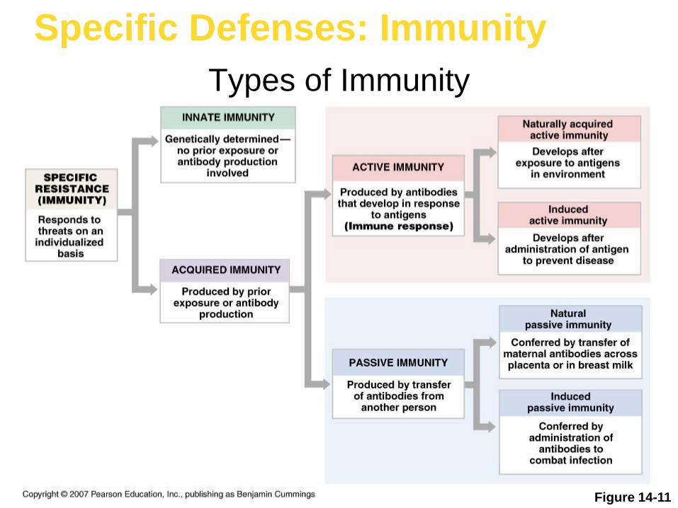
Specific Defenses: Immunity
Types of Immunity
Figure 14-11


