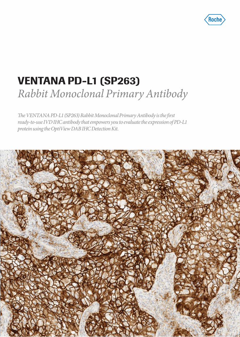
VENTANA PD-L1 (SP263) Rabbit Monoclonal Primary Antibody e VENTANA PD-L1 (SP263) Rabbit Monoclonal Primary Antibody is the first ready-to-use IVD IHC antibody that empowers you to evaluate the expression of PD-L1 protein using the OptiView DAB IHC Detection Kit.
The VENTANA PD-L1 (SP263) Rabbit Monoclonal Primary Antibody is the first ready-to-use IVD IHC antibody that empowers you to evaluate the expression of PD-L1 protein using the OptiView DAB IHC Detection Kit.
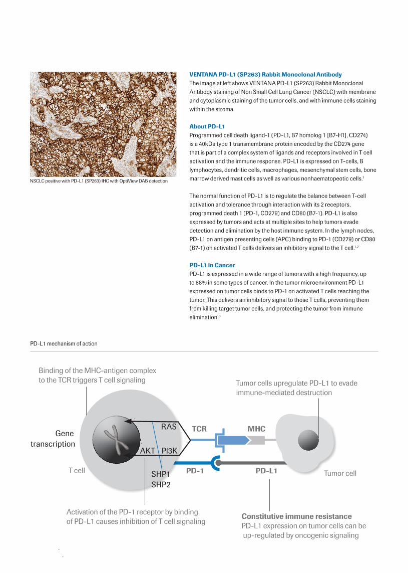
VENTANA PD-L1 (SP263) Rabbit Monoclonal Antibody The image at left shows VENTANA PD-L1 (SP263) Rabbit Monoclonal Antibody staining of Non Small Cell Lung Cancer (NSCLC) with membrane and cytoplasmic staining of the tumor cells, and with immune cells staining within the stroma.
About PD-L1Programmed cell death ligand-1 (PD-L1, B7 homolog 1 [B7-H1], CD274) is a 40kDa type 1 transmembrane protein encoded by the CD274 gene that is part of a complex system of ligands and receptors involved in T cell activation and the immune response. PD-L1 is expressed on T-cells, B lymphocytes, dendritic cells, macrophages, mesenchymal stem cells, bone marrow derived mast cells as well as various nonhaematopeotic cells.1
The normal function of PD-L1 is to regulate the balance between T-cell activation and tolerance through interaction with its 2 receptors, programmed death 1 (PD-1, CD279) and CD80 (B7-1). PD-L1 is also expressed by tumors and acts at multiple sites to help tumors evade detection and elimination by the host immune system. In the lymph nodes, PD-L1 on antigen presenting cells (APC) binding to PD-1 (CD279) or CD80 (B7-1) on activated T cells delivers an inhibitory signal to the T cell.1,2
PD-L1 in CancerPD-L1 is expressed in a wide range of tumors with a high frequency, up to 88% in some types of cancer. In the tumor microenvironment PD-L1 expressed on tumor cells binds to PD-1 on activated T cells reaching the tumor. This delivers an inhibitory signal to those T cells, preventing them from killing target tumor cells, and protecting the tumor from immune elimination.3
NSCLC positive with PD-L1 (SP263) IHC with OptiView DAB detection
PD-L1 mechanism of action
MHCTCR
PD-1 PD-L1
Tumor cells upregulate PD-L1 to evade immune-mediated destruction
Tumor cell
Constitutive immune resistancePD-L1 expression on tumor cells can be up-regulated by oncogenic signaling
Binding of the MHC-antigen complex to the TCR triggers T cell signaling
Activation of the PD-1 receptor by binding of PD-L1 causes inhibition of T cell signaling
RAS
AKT Pl3K
SHP1SHP2
T cell
Gene transcription
References
1. Keir ME, Butte MJ, Freeman GJ, Sharpe AH. PD-1 and its ligands in tolerance and immunity. Annual Rev Immunol 2008;26:677-704.
2. Park JJ, Omiya R, Matsumura Y, Sakoda Y, Kuramasu A, Augustine MM, et al. B7-H1/CD80 interaction is required for the induction and maintenance of peripheral T-cell tolerance. Blood. 2010;116(8):1291-8.
3. Zou W, Chen L. Inhibitory B7-family molecules in the tumour microenvironment. Nat Rev Immunol. 2008;8(6):467-77.
4. GLOBOCAN 2012: Estimated cancer Incidence, Mortality, Prevalence and Disabilityadjusted life years (DALYs) Worldwide in 2012. Available from URL: http://globocan.iarc.fr/Pages/fact_sheets_cancer.aspx. Accessed 23 March 2015.
5. Pisters KM, Le Chevalier T. Adjuvant chemotherapy in completely resected non-small-cell lung cancer [published erratum appears in J Clin Oncol 2008 May 1;26(13):2238]. J Clin Oncol 2005 May 10;23(14):3270-8.
6. D’Addario G, Früh M, Reck M, Baumann P, Klepetko W, Felip E, et al; ESMO Guidelines Working Group. Metastatic non-small-cell lung cancer: ESMO Clinical Practice Guidelines for diagnosis, treatment and follow-up. Ann Oncol 2010 May;21(Suppl 5):v116-9
.
Automation: optimized for use on all VENTANA Benchmark IHC/ISH staining instrumentsDetection: optimized with OptiView DAB IHC Detection Kit (760-700 [06396500001])
Non-Small Cell Lung CancerLung cancer has been the most common cancer in the world for several decades, and by 2012, there were an estimated 1.8 million new cases, representing 12.9% of all new cancers. It was also the most common cause of death from cancer estimated to be responsible for nearly 1 in 5 (1.59 million deaths, 19.4% of the total).4 Non-small cell lung cancer (NSCLC) represents approximately 80% to 85% of all lung cancers. Unfortunately, at the time of diagnosis, approximately 70% of patients with NSCLC already have advanced or metastatic disease not amenable to surgical resection. Furthermore, a significant percentage of patients with early stage NSCLC who have undergone surgery subsequently develop distant recurrence and die as a result of their lung cancer.5 Despite advances in the diagnosis, imaging, staging and treatment of NSCLC, the estimated overall 5-year survival for patients in Europe continues to be low (11%).6
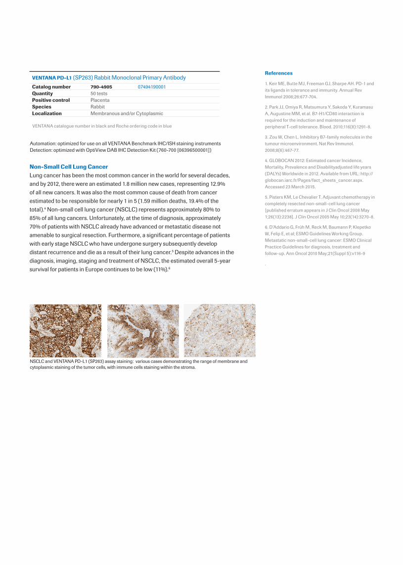
NSCLC and VENTANA PD-L1 (SP263) assay staining: various cases demonstrating the range of membrane and cytoplasmic staining of the tumor cells, with immune cells staining within the stroma.