160
VENTILATION WORKSHOP Dr Despina Demopoulos Paediatric Intensivist

VENTILATION WORKSHOP
Dr Despina Demopoulos Paediatric Intensivist

OUTLINE
■ Introduction
■ Different modes
■ Tricks of the Trade
■ Sick Kids
■ Can we get more out of conventional ventilation?
■ Newer modes- NAVA, PAV
■ Practical session- RCA

INTRODUCTION
■ Respiratory disordersà main cause of respiratory failure
■ Average PICU has about 30% (range 20%–64%) of its patients mechanically ventilated for a mean of 5–6 days
■ Most commonly à conventional ventilation

GOALS OF VENTILATION
■ Provide adequate oxygenation ■ Provide adequate ventilation
■ Optimize lung volumes ■ Optimize circulation
■ Minimize damage to the lungs (and the body) ■ Have a comfortable patient

MODES OF VENTILATION
■ Assist Control (AC) Pressure/Volume
■ Continuous Positive Airway Pressure (CPAP) & Pressure Support
■ Pressure Regulated Volume Control (PRVC)
■ Controlled Mechanical Ventilation (CMV)
■ Intermittent Mandatory Ventilation (IMV)
■ Synchronized Intermittent Mandatory Ventilation (SIMV)


VOLUME VENTILATION
■ Preset – Volume – PEEP – Rate – I-time – FiO2
■ Ventilator Determines – Pressure required
■ Advantages – Guaranteed minute ventilation – More comfortable for patient
■ Draw-backs – Large ETT leak – Not optimal for poorly
compliant lungs


PRESSURE VENTILATION
■ Preset – PIP – PEEP – Rate – I-time – FiO2
■ Vent determines – Tidal volume given
■ Advantages – Provides more support at lower
PIP for poorly compliant lungs
■ Draw back – Minute ventilation not
guaranteed
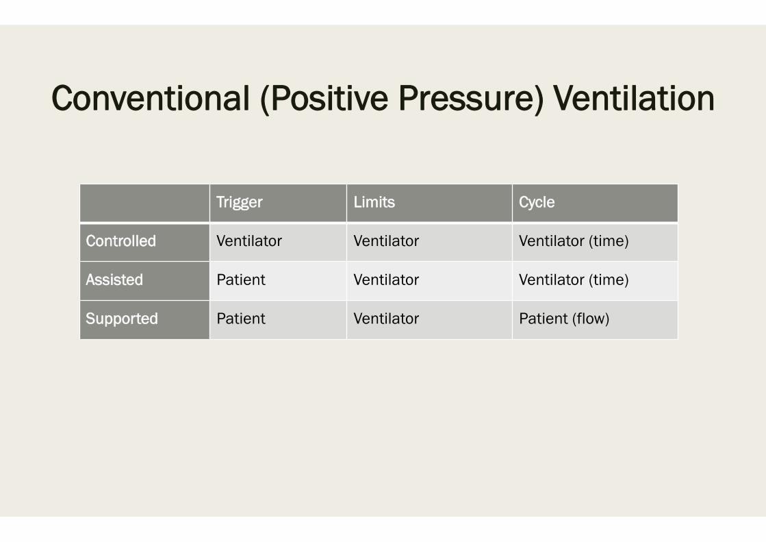
Conventional (Positive Pressure) Ventilation
Trigger Limits Cycle
Controlled Ventilator Ventilator Ventilator (time)
Assisted Patient Ventilator Ventilator (time)
Supported Patient Ventilator Patient (flow)

MODES OF VENTILATION ■ Control Modes:
– every breath is fully supported by ventilator – classic control modes, patients unable to breathe except at the controlled set
rate – in newer control modes, machines may act in assist-control, with a minimum
set rate and all triggered breaths above that rate also fully supported. ■ IMV Modes: intermittent mandatory ventilation modes - breaths “above” set rate not
supported ■ SIMV: vent synchronizes IMV “breath” with patient’s effort

MODES OF VENTILATION ■ Synchronized Intermittent Mandatory Ventilation
– Mandatory number of positive pressure breaths per minute, each synchronized to patient effort.
– Ventilator detects initiation of spontaneous breath and does not deliver machine breath during a spontaneous breath.
– Between mechanical breaths may breathe an indefinite number of times from reservoir
– Spontaneous breaths produce no response from the ventilator

MODES OF VENTILATION
■ Assist/Control Mode Ventilation – Combined mode of ventilation – Ventilator delivers positive pressure breath of predetermined TV in response to
each inspiratory effort (assisted ventilation) – If pt fails to initiate breath within a specific time period, ventilator automatically
delivers a mechanical breath to maintain minimum or “backup” respiratory rate (controlled ventilation)
– To trigger assisted breath must lower airway pressure by preset amount- the trigger sensitivity.
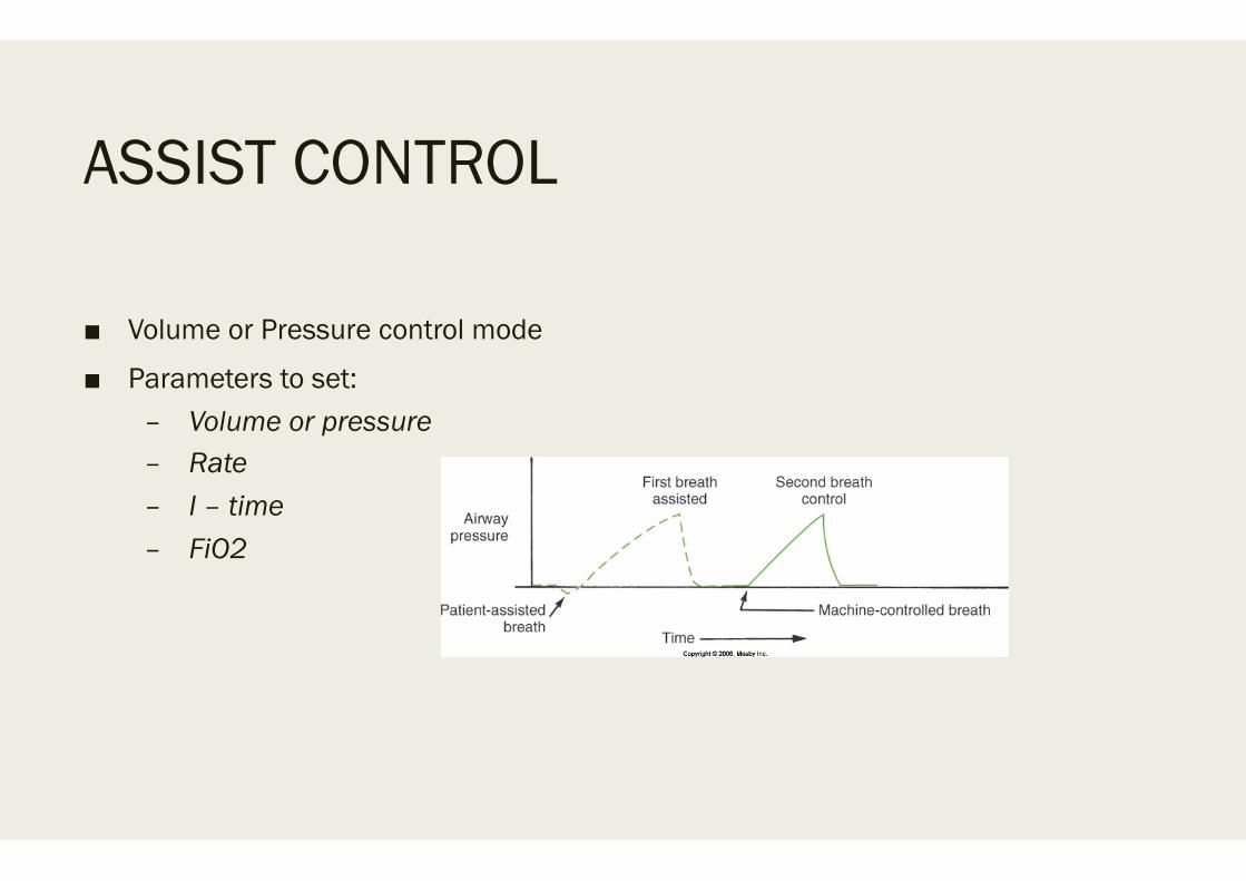
ASSIST CONTROL
■ Volume or Pressure control mode
■ Parameters to set: – Volume or pressure – Rate – I – time – FiO2

ASSIST CONTROL
■ Machine breaths: – Delivers the set volume or pressure
■ Patient’s spontaneous breath: – Ventilator delivers full set volume or pressure & I-time
■ Mode of ventilation provides the most support


MODES OF VENTILATION
■ Pressure-Support Ventilation – When inspiratory flow rate falls below preset threshold, flow of gas terminates. – Patient controls respiratory rate and inspiratory time and flow. – TV + minute ventilation partly determined by patient + partly by ventilator

MODES OF VENTILATION
■ Pressure Control Ventilation (PCV) – Minimises static airway pressures in ARDS patients. – Involves setting target airway pressure on ventilator which then delivers rapid
flow to that set pressure with a square pressure wave form
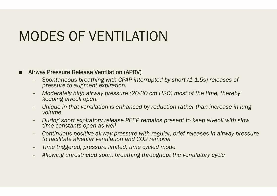
MODES OF VENTILATION
■ Airway Pressure Release Ventilation (APRV) – Spontaneous breathing with CPAP interrupted by short (1-1.5s) releases of
pressure to augment expiration. – Moderately high airway pressure (20-30 cm H2O) most of the time, thereby
keeping alveoli open. – Unique in that ventilation is enhanced by reduction rather than increase in lung
volume. – During short expiratory release PEEP remains present to keep alveoli with slow
time constants open as well – Continuous positive airway pressure with regular, brief releases in airway pressure
to facilitate alveolar ventilation and CO2 removal – Time triggered, pressure limited, time cycled mode – Allowing unrestricted spon. breathing throughout the ventilatory cycle

MODES OF VENTILATION
■ Advantages: Preservation of spontaneous breathing -may improve comfort + decrease sedation need
■ CPAP useful in keeping alveoli open
■ A short expiratory time which favours ventilation of fast compartments
■ Reduced barotrauma risk
■ Relatively low airway pressures - ↓ volutrauma, improve pulmonary circulation + O2 delivery

MODES OF VENTILATION ■ Pressure regulated volume control (PRVC)
– Good alternative to PCV if rapidly changing compliance – Remains a pressure regulated approach but pressure varies to maintain given
tidal volume. – Tidal volume, PEEP, rate, inspiratory time is set – Advantage of having a guaranteed tidal volume with a flow pattern that
doesn’t harm the lungs – Agitated patients may hyperventilate

TRICKS of the TRADE

TRICK #1
■ Know which kids are “sick” and need ventilation

“SICK” KIDS
■ Hypoxia
■ Hypercarbia
■ Airway protection
■ (Decrease demand in cases of poor cardiac output)

“SICK KIDS”
■ SIGNS OF DETERIORATION – Increasing recession – Increasing respiratory rate – Increasing pulse rate – Fatigue – Altered mental status – Cyanosis

TRICK #2
■ Know your ventilator
■ Terminology
■ Different modes
■ Ventilator settings

VENTILATORS-terminology
– TIME
– I - Time: amount of time spent in inspiration – E - Time: amount of time spent in expiration
– Volume
– Amount of tidal volume that a patient receives – Pressure
– Measure of impedance to gas flow rate – Flow
– Measure of rate at which gas is delivered

VENTILATORS-terminology
■ PEEP = positive end expiratory pressure ■ Pressure maintained in the airways at the end of exhalation ■ Keeps Alveoli from collapsing
■ PIP = peak inspiratory pressure ■ Point of maximal airway pressure
■ Delta P = the difference between PIP – PEEP
■ MAP = mean airway pressure

OXYGEN ■ Alveolar gas equation
■ PAO2 = FiO2 (PATM - PH2o) - PaCO2
■ Nasal Prongs 24%-40 %
■ Face mask 28%-80 %
■ NOT “Double oxygen”

CPAP/ NIV ■ 2 main effects ■ Increase pressure in posterior pharynx => increase ΔP across conducting airways =>
improves airflow ■ Increases PEEP, thus FRC > Closing capacity ■ Nasal CPAP increasingly NB in neonates – reduces need for ventilation in pre-term
infants ■ Also useful in small infants
■ After infancy, before childhood – difficult to achieve ■ > 6-8years face mask

HFNC ■ Key Features of Airvo Humidifier:
■ Humidifier with integrated flow generator Oxygen delivery without a blender Variety of interfaces Easy to set up and use Validated high-level disinfection process

HFOV
■ Adjustable Parameters – Mean Airway Pressure: usually set 2-4 higher than MAP on conventional
ventilator – Amplitude: monitor chest rise – Hertz: number of cycles per second – FiO2 – I-time: usually set at 33%
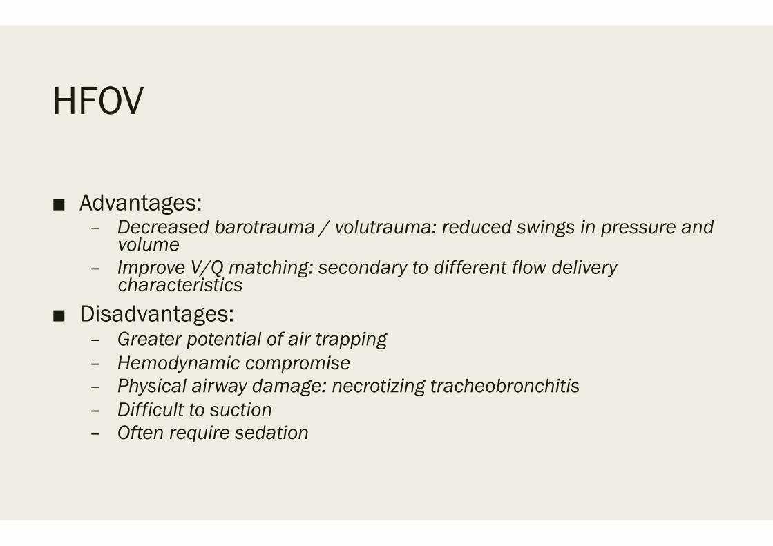
HFOV
■ Advantages: – Decreased barotrauma / volutrauma: reduced swings in pressure and
volume – Improve V/Q matching: secondary to different flow delivery
characteristics ■ Disadvantages:
– Greater potential of air trapping – Hemodynamic compromise – Physical airway damage: necrotizing tracheobronchitis – Difficult to suction – Often require sedation
SETTING THE VENTILATOR
Ventilator Settings- FiO2 ■ Dangerous drug
■ Lowest setting to keep Sats >88-92 %
■ “Closed loop” Auto-weaning
■ Trigger α“Patient comfort”
■ Flow vs Pressure
■ Beware “Auto-triggering‟
■ Beware increased work of breathing
■ New – Neurally Adjusted Ventilatory Assist
Ventilator Settings- PEEP
■ Role in Paediatrics? ■ Improve FRC > Closing capacity ■ Normal healthy lung has PEEP +/- 3-5 cmH2O ■ Intubation removes natural PEEP augmentation – bypassing post pharynx ■ Generally 8-10 cmH2O ■ Higher for recruitment ■ Prior contra-indications
– Isolated head injury – Asthma
Ventilator settings- Driving Pressure
■ PIP vs ΔP
■ Generates mean airway pressure (MAP)– oxygenation
■ Generates TV – Alveolar ventilation
■ Should not exceed 30 cm H2O
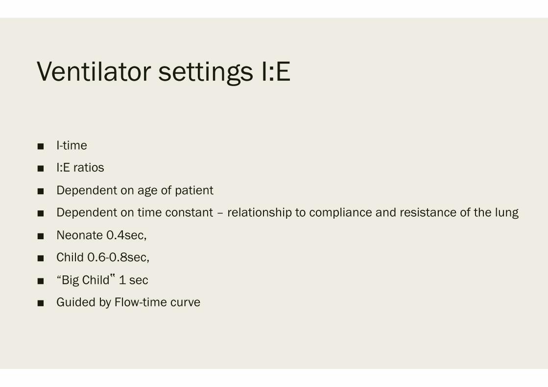
Ventilator settings I:E
■ I-time
■ I:E ratios
■ Dependent on age of patient
■ Dependent on time constant – relationship to compliance and resistance of the lung
■ Neonate 0.4sec,
■ Child 0.6-0.8sec,
■ “Big Child‟ 1 sec
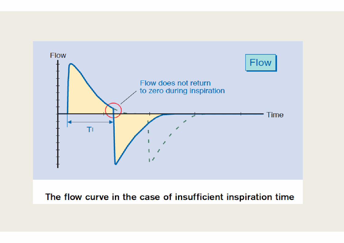
■ Guided by Flow-time curve


Too short Te will not allow to deliver max. possible Vt at given P à will induce PEEPi à increases the risk for
hemodynamic instability
The patient respiratory mechanics dictate the maximal respiratory frequency
Too short Ti will reduce delivered Vt à unneccessary high PIP will be applied à unnecessary high intrathoracic pressures

Ventilator Settings- Rate
■ In Assist control – back-up rate
■ Set to allow spontaneous breaths
■ Air drawn to lower regions
■ Avoid high rates to “blow off CO2”
■ Can cause drop in TV; dead space ventilation
■ Be guided by flow-time curve

TRICK #3
■ Know how to manage different diseases
■ Know the pathophysiology
■ “Lung protective strategies”

HYPOXIA
■ Hypoventilation: decreased alveolar ventilation, i.e. CNS depression
■ Diffusion impairment: abnormality at pulmonary capillary bed
■ Shunt: blood flow without gas exchange – Intra-pulmonary – Intra-cardiac
■ Ventilation-perfusion mismatch: Both dead space and shunt abnormalities

TREATING HYPOXIA
■ Increase FiO2: >60% toxic to lung parenchyma
■ Increase mean airway pressure – PEEP : not too much, not too little – PIP – I-time

HYPERCARBIA
■ Decreased minute ventilation – Respiratory rate – Tidal volume
■ Treatment: – Increase respiratory rate: assure I-time not too short as rate increased – Increase tidal volume – Allow permissive hypercarbia

BRONCHIOLITIS
■ Apnoeas (relatively normal lungs) – Minimise VILI with low Tv
■ Air trapping – Manage like asthma
■ ARDS – Manage like ARDS (including HFOV, iNO, ECMO)
■ HFNC

PULMONARY DISEASE- OBSTRUCTIVE
Airway obstruction causing increase resistance to airflow: e.g. asthma
■ Optimize expiratory time by minimizing minute ventilation
■ Bag slowly after intubation
■ Don’t increase ventilator rate for increased CO2

ASTHMA
■ Pressure controlled ventilation (keep PIP <30) or volume controlled ventilation (Tv 5-8 ml/kg) may be used
■ A long expiratory time (with a optimum inspiratory time) with an I/E ratio of >1:2 and a slow rate allow emptying of the lungs and avoid ‘air trapping’ and progressive hyperinflation
■ Manual decompression of chest à may help to deflate overinflated lungs and improves ventilation.

ASTHMA
■ Sedation à important to avoid the complications of air-leak
■ Preferred drugs for these patients are ketamine (has a bronchodilator effect) & fentanyl
■ ? Suction and physiotherapy à clear mucus plugging and prevent atelectasis
■ Specific treatments for asthmaà nebulised and intravenous salbutamol, systemic steroids, magnesium sulphate & intravenous aminophylline

ARDS
■ Major physiological derangements: – 1) a major defect in oxygenation; – 2) a poor efficiency of the lungs at eliminating CO2 – 3) a major reduction in lung volumes and complianceà severe restrictive lung
disease.


ARDS Management
■ Mechanisms implicated in VILI – Oxygen toxicity from use of high FiO2 – Over distension of alveoli leading to volutrauma and barotrauma – Repetitive opening and closing of alveoli causing shear stress and triggering
further inflammation (atelectrauma) – High respiratory rate

LUNG PROTECTIVE STRATEGIES
– High PEEP – Pressure limiting PIP: <30 cmH2O – Low tidal volume: 4-6 ml/kg – FiO2 <60% – Permissive hypercarbia – Permissive hypoxia

ARDS Management- PEEP
■ PEEP improves oxygenation by providing movement of fluid from the alveolar to interstitial space, recruitment of small airways and collapsed alveoli and an increase in functional residual capacity.
■ PEEP is adjusted between 8 cm H2O and 20 cm H2O; PEEP is progressively increased by 2-3 cm H2O increments to maintain saturation between 90 and 95% with FiO2 < 0.5.
■ The child should be monitored for any evidence of cardiovascular compromise and hyperinflation

ARDS- HFOV
■ The advantages of HFOV are – use of low VT and avoidance of barotrauma – maintenance of near normal PaCO2 with improved minute ventilation.

ARDS- NITRIC OXIDE (NO) ■ Causes pulmonary vasodilation and decrease in pulmonary hypertension. Maximal
improvement in oxygenation is usually achieved with <10 ppm in most patients.
■ 10 min àHours
■ Paediatric studies suggest that iNO improves short-term oxygenation in children with ARDS but little change is seen in long-term oxygenation indices

ARDS- SURFACTANT THERAPY
■ Metanalysis (Crit Care 2007) & systemic review of surfactant use in critically ill children with acute respiratory failure à significant reduction in mortality, as well as a significant reduction of ventilator days and less need for rescue therapy (nitric oxide, High frequency ventilation and ECMO) in these patients
■ Dose: 2ml/kg (50mg/kg/dose)

• Acutehypoxaemicrespiratoryfailurewithnoimprovementwithin48hrsofstartingventilationusingalungprotectivestrategy.(Wilson,2005)• WithOI>7(Wilson2005,Wilson1999)• WithPF<150(Luchetti1998,2002)• DiffusebilateralinfiltratesonCXR(Wilson1999,
2005,Moller2003)
• Aspirationofhydrocarbons.(HorozOO2010,Mastropietro2011)
• Bronchiolitis
• Ventilated>24hrswithoutimprovement(Luchetti1998)
• PF<160(Luchetti2002,1998)• CXRshowingairtrapping(Luchetti2002)

ARDS- CORTICOSTEROIDS ■ Prospective, randomized controlled trialà prolonged administration of
methylprednisolone in adult patients with unresolving ARDS (ARDS>7 days) à improvement in lung injury and MODS scores and reduced mortality.
■ Larger more recent RCTsà have shown improved ventilatory parameters but increased late mortality in the group given steroids
■ Hyperglycaemia S/E
PRONE POSITIONING
■ Changes in regional lung perfusion, regional pleural pressures & recruitment of dorsal lung à improve oxygenation during prone positioning
■ Few risks & costs involved

OTHER DISEASES

PULMONARY DISEASE- RESTRICTIVE
■ Compromised lung volume: – Intrinsic lung disease – External compression of lung
■ Recruit alveoli, optimize V/Q matching
■ Lung protective strategies

PNEUMONIA/LUNG COLLAPSE
■ Minimise oxygen toxicity (FiO2 <0.60)
■ Minimise atelectrauma (adequate PEEP)
■ Minimise volutrauma (low Tv 4-6 ml/kg)
■ Permissive hypercapnia
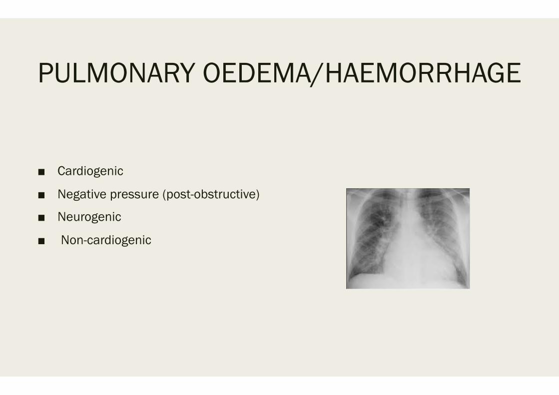
PULMONARY OEDEMA/HAEMORRHAGE
■ Cardiogenic
■ Negative pressure (post-obstructive)
■ Neurogenic
■ Non-cardiogenic

PULMONARY OEDEMA/HAEMORRHAGE
■ Conventional ventilation – Minimise oxygen toxicity (FiO2 <0.60) – Minimise atelectrauma (adequate PEEP) – Minimise volutrauma (low Tv 4-6 ml/kg) – Permissive hypercapnia
■ High frequency ventilation – Constant MAP – Recruitment of lung
TRICK #4
■ Be safe
■ Safety bundle

VENTILATOR-ASSOCIATED PNEUMONIA (VAP)BUNDLE:
– DVT prophylaxis – GI prophylaxis – Head of bed (HOB) elevated to 30-45° – Daily Sedation Vacation – Daily Spontaneous Breathing Trial – Additional- Oral care

TRICK #5
■ Daily assessment trial of readiness to extubate
■ Adjuncts to extubation eg decr airway oedema in UAO (steroids >6 hrs)
■ Sedation holiday
■ Conservative fluid regime
Daily sedation vacation/ Spontaneous Breathing Trials ■ Implement a protocol to lighten sedation daily at an appropriate time to assess for
neurological readiness to extubate. – Include precautions to prevent self-extubation such as increased monitoring
and vigilance during the trial.
■ Include a sedation vacation strategy in your overall plan to wean the patient from the ventilator
– if you have a weaning protocol, add "sedation vacation" to that strategy.

WEANING

CRITERIA FOR EXTUBATION READINESS TEST FAILURE
Pediatr Crit Care Med 2009 Vol. 10, No. 1


Pain Assessment
• Pain Scale • Observation • Non- pharmacologic intervention
If Pain present, choose
analgesic
• Paracetamol, NSAIDs, Tilidine drops
• Morphine • Fentanyl • Remifentanil • Ketamine
Choose sedation strategy
• Daily Interruption of Sedation(DIS)
• Sedation Scale • No sedation
Choose sedative
• Midazolam • Propofol (rapid wake-up) • Dexmedetomidine (↓sedation, rapid wake-up)
Adapted: Upadhyay, S.P., et al. A Practical Guide to Sedation and Analgesia in Paediatric Intensive Care Unit (ICU). (2017) J Anesth Surg 4(1): 1- 6.

TRICK #6
■ Know how to read “problems” on your vent

Air Leak

Autocycling

Secretions

Intrinsic PEEP
■ If the respiratory rate is set high or the expiratory time is not long enough there is a risk for auto PEEP.
■ The patient does not have enough time to exhale and it is evident on the flow curve that flow will not return to zero before the next breath starts.
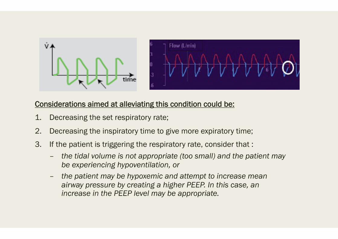
Considerations aimed at alleviating this condition could be:
1. Decreasing the set respiratory rate;
2. Decreasing the inspiratory time to give more expiratory time;
3. If the patient is triggering the respiratory rate, consider that : – the tidal volume is not appropriate (too small) and the patient may
be experiencing hypoventilation, or – the patient may be hypoxemic and attempt to increase mean
airway pressure by creating a higher PEEP. In this case, an increase in the PEEP level may be appropriate.

■ If an imaginary line is drawn to connect the origin of the loop with the PIP, it can estimate the dynamic compliance of the lung.
■ Compliance is mathematically determined by
■ Δ volume/ Δ pressure
■ Is graphically displayed on the LOOP screen.
Pressure–volume loop

Pressure–volume loop
■ A loop indicating good compliance will be described as upright (compliance axis>45)
■ A loop indicating poor compliance is described as flat, or lying on its side.

Pressure–volume loop
■ Inadequate hysteresis, producing a narrow loop, may be indicative of inadequate flow

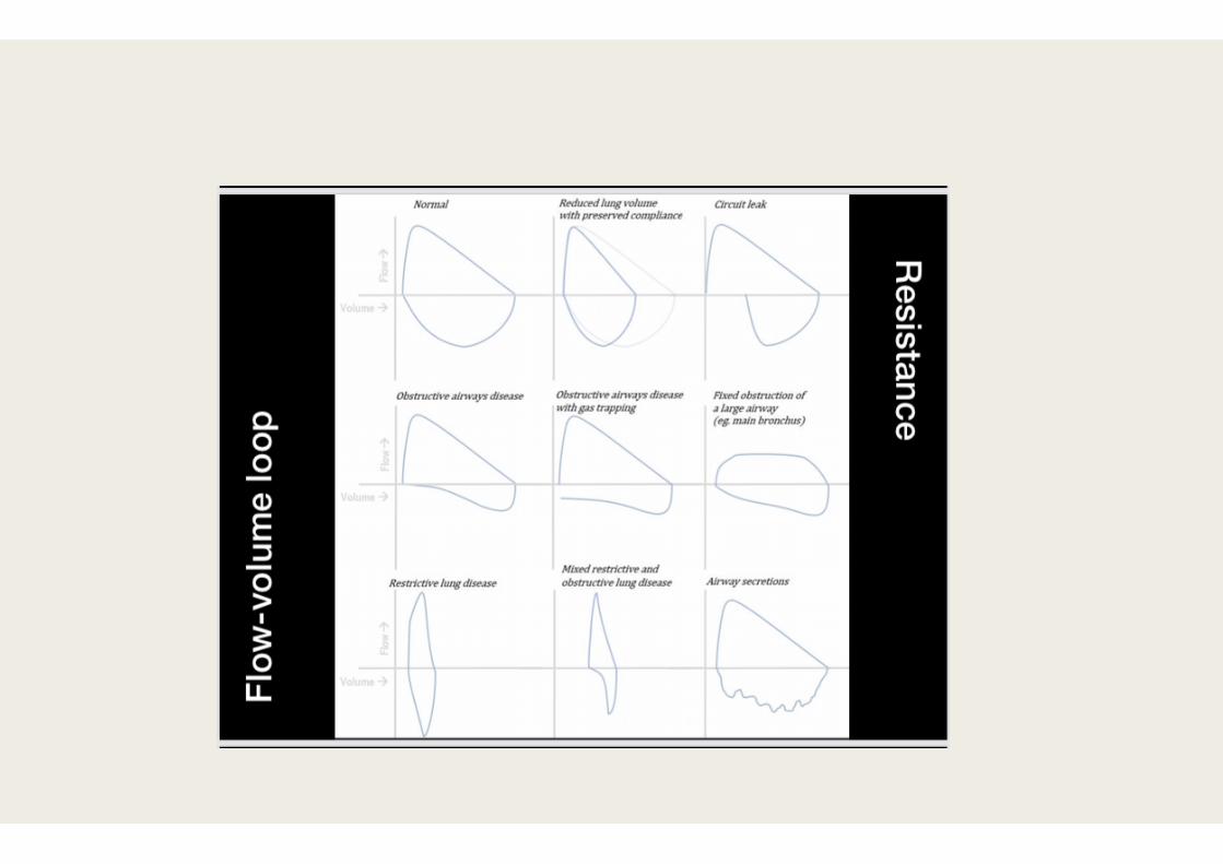
Flow–volume loop
■ The effect of altering resistance by use of a bronchodilator.
■ After treatment, resistance improves, and there is a demonstrable difference in the appearance of the loop.
FLOW SYNCHRONY
■ Defined as the ideal matching of inspiratory flow of a ventilator breath to the patient's inspiratory demand during assisted or supported ventilation
■ Asynchrony: Inadequate inspiratory flow at any point during inspiration causing an increased or irregular patient effort.
– –leads to increased WOB – –“fighting” the ventilator

FLOW ASYNCHRONY

TRIGGER INSENSITIVITY

INSPIRATORY SYNCHRONY
Optimal inspiratory patient - ventilator synchrony is a function of: – inspiratory flow pattern – adequate inspiratory flow – appropriate trigger sensitivity – ETT effects – appropriate lung inflation

PATIENT- VENTILATOR INTERACTIONS
Expiratory synchrony
■ end-expiratory lung volume
■ premature termination of exhalation & intrinsic PEEP
■ expiratory resistance

• Hering-Breuer reflexes • Respiratory Muscle Weakness • Respiratory system mechanics • Pathology • Leaks
Patient
• Ventilator algorithms and control • Trigger signal • Cycling off • Rate and character of inspiratory flow • Intrinsic PEEP • Leaks
Ventilator
• Mode and Settings • Level of support • Level of sedation
Decision making
Patient-ventilator asynchrony

INSPIRATORY RISE TIME
■ Inspiratory rise time is the time taken to reach peak inspiratory flow or pressure at the start of each breath, expressed either as a percentage of the respiratory cycle time or in seconds.

INSPIRATORY RISE TIME
■ The flow and pressure rise time can be adapted in accordance with the patient.
■ The Inspiratory rise time has to be set to a comfortable value for the patient and can be evaluated by the shape of the flow and pressure curves

CONCLUSION
■ Children have natural propensity to have lung collapse
■ Ventilation aims to restore oxygenation, lung volumes, decrease work of breathing
■ Set Ventilator to cause least harm, most benefit and comfort to the patient

CONCLUSION ■ Rare for a child to “fight” ventilation
– Hypoxia – Blocked tube – Inadequate settings
■ DO NOT SEDATE/PARALYSE WITHOUT CAUSE
■ Sedation Protocols – keep patient comfortable, not agitated – allow spontaneous breathing

CONCLUSION ■ Tricks of the Trade:
– Recognise sick kids and goals of ventilation – Know your ventilator & settings – Learn how to manage different diseases ■ Lung protective strategies
– Be safe – Daily assessment trial of readiness to extubate – Know how to read problems on the vent

Can we get more?
■ Individualise therapy
■ Test lung recruitabiity
■ New modes – NAVA – PAV – Noisy ventilation
ARDS
■ Must individualise therapy
■ Application of the same concepts may be beneficial in certain forms of ARDS or at certain stages, and may be risky or harmful in others.
■ This is the case for spontaneous breathing activity, non invasive ventilation (NIV) and high PEEP
ARDS
■ Measurement of oesophageal pressure (Poes) (surrogate for pleural pressure) àdetermine the lung mechanics, separate the effect of the chest wall
■ Lung recruitabilityà helps to individualise the settings for mechanical ventilation & choose the PEEP needed to keep the lung sufficiently open to minimise the risks of repeated opening and closing of alveoli

ARDS
■ Using lower tidal volumes (≤6 versus ≥10 mL·kg−1 PBW) in patients without ARDS was associated with better clinical outcomes, including development of ARDS, mortality and duration of mechanical ventilation
Sinha P, Sanders RD, Soni N, et al. Acute respiratory distress syndrome: the prognostic value of ventilatory ratio – a simple bedside tool to monitor ventilatory efficiency. Am J Respir Crit Care Med 2013; 187: 1150–1153.
NMBAs in ARDS- Passive ventilation
■ Should be considered for early and short-term use in patients
■ Gainnier et al. NMBA group had a sustained improvement in oxygenation and a lower Pplat after 48 h of randomisation
■ Forel et al. demonstrated the same result associated with a decreased concentration of proinflammatory cytokines (interleukin (IL)-1β, IL-6 and IL-8) in both bronchoalveolar lavage fluid and serum

NMBAs in ARDS- Passive ventilation
■ The ACURASYS study (same group) enrolled 340 patients with moderate-to-severe ARDS to receive cisatracurium or placebo for 48 h
■ The early administration of cisatracurium reduced adjusted 90-day mortality and barotrauma, and also increased ventilator free days without increasing muscle weakness

NMBAs in ARDS
■ Mortality benefit of cisatracurium was limited to patients with a PaO2/FiO2 ratio <120.
■ Concern about the development of ICU-acquired weakness
■ Not associated with ICU-acquired weakness when used for a short period
NMBAs in ARDS
■ How do NMBAs improve gas exchange and outcomes? – minimising VILI by minimising the transpulmonary pressure changes during
assisted breathing and reducing patient–ventilator asynchrony, – decreasing oxygen consumption of respiratory muscles, and – reducing pulmonary and systemic inflammation.

Spontaneous breathing
■ Complete inactivity of the diaphragmà disuse atrophy and muscle weaknessà ventilator-induced diaphragmatic dysfunction (VIDD), 18–24 h of mechanical ventilation
■ Contributes to weaning problems and poorer prognosis
■ In experimental studies, allowance of spontaneous breathing using either assist-control ventilation or pressure support ventilation can reduce VIDD
Jaber S, Sebbane M, Verzilli D, et al. Adaptive support and pressure support ventilation behavior in response to increased ventilatory demand. Anesthesiology 2009; 110: 620–62
Sassoon CS, Zhu E, Caiozzo VJ. Assist-control mechanical ventilation attenuates ventilator-induced diaphragmatic dysfunction. Am J Respir Crit Care Med 2004; 170: 626–632.
Spontaneous breathing in ARDS
■ Experimental lung injury models of ARDS demonstrated that preserving spontaneous breathing was associated with:
– 1) reduced markers of lung inflammation and epithelial cell damage; – 2) improved tidal ventilation, gas exchange and oxygen delivery; and – 3) increased systemic blood flow.

Spontaneous breathing in ARDS
■ Neumann et al. and Putensen et al. demonstrated that partial ventilatory support with airway pressure release ventilation (APRV)
– promoted alveolar recruitment in juxta-diaphragmatic areas – improved ventilation/perfusion matching and gas exchange – increased oxygen delivery in comparison with controlled mechanical ventilation

Spontaneous breathing in ARDS
■ BUT in severe lung injury àhigh transpulmonary pressure, worsened oxygenation and lung damage, and could also cause local injury by internal redistribution of volume

■ An ongoing large multicentre randomised controlled study (BiRDS) will examine the efficacy and safety of early spontaneous breathing with APRV mode using normal inspiratory to expiratory ratios in comparison with controlled mechanical ventilation
Early Spontaneous Breathing in Acute Respiratory Distress Syndrome (BiRDS) study; ClinicalTrials.gov identifier: NCT01862016)

PEEP ■ PEEP is able to keep the recruited lung areas reopened by the ventilator, and thus
improve gas exchange in patients with ARDS and reduce the risk of repeated opening and closure
■ A wide variability in the amount of recruitable lung exists among patients (0% and 50% of potentially recruitable lung)
■ High PEEP may be able to keep the lung open only if the lung is recruitable

PEEP
■ Increase in end-expiratory lung volume (EELV)
■ In a highly recruitable patient, a substantial part of the increase in EELV can be due to reopening of previously collapsed lung tissue, referred to as recruitment
■ In a poorly recruitable patient, most of the increase in EELV is generated by inflation of previously open lung tissue potentially leading to overdistension (volutrauma)à failing to recruit the collapsed tissue

Testing alveolar recruitability
■ Different techniques have been proposed: – multiple pressure–volume curves, – measurement of lung volume, – use of Poes and transpulmonary pressure, – use of lung ultrasound – use of a physiological test based on oxygenation
■ In research studies, alveolar recruitability has been assessed using computed tomography (CT)


Multiple pressure–volume curves technique ■ Plotting several pressure–volume curves obtained at different PEEP levels on the
same volume axis, measuring or estimating the volume above functional residual capacity (FRC), i.e. the relaxation volume at zero end-expiratory pressure (ZEEP), at each PEEP level
■ Elastic pressure–volume curves can be obtained using low flow inflation

Multiple pressure–volume curves technique
■ When reducing PEEP to ZEEP during a prolonged expiration, the lung volume expired is the volume above FRC at that PEEP level
■ Numerous studies have demonstrated good reproducibility of pressure–volume curves for assessment of alveolar recruitability.
■ Too complexà remains limited to research areas

Poes monitoring
■ Measuring Poes to estimate pleural pressure à estimating transpulmonary pressure at end-inspiration and expiration from the difference between Pplat or PEEP and oesophageal pressures
■ Proposed method to titrate PEEP and adjust pressures: transpulmonary pressure=Paw–Poes
■ EPVent (Esophageal Pressure directed Ventilation) studyà usefulness of Poes in guiding PEEP therapy in ARDS

Poes monitoring
■ Poes is often elevated in patients with ARDS (reduced chest wall compliance, oedema or abdominal distension) àtranspulmonary pressure can be negative at end-expiration
■ May indicate closed or compressed airways or atelectatic lung
■ PEEP can be increased until transpulmonary pressure becomes positive at end-expiration to keep the airways open
Transpulmonary pressure
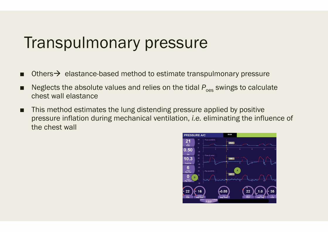
■ Othersà elastance-based method to estimate transpulmonary pressure
■ Neglects the absolute values and relies on the tidal Poes swings to calculate chest wall elastance
■ This method estimates the lung distending pressure applied by positive pressure inflation during mechanical ventilation, i.e. eliminating the influence of the chest wall

Transpulmonary pressure ■ Since any positive pressure applied at the airway opening acts on two elastic
structures connected in series (the lung and the chest wall), Paw is distributed between chest wall and lung elastance
■ This method for partitioning lung and chest wall elastance has been used to
guide a transpulmonary “open lung” approach in a cohort of patients with severe ARDS related to influenza A (H1N1)
■ Helps to decide in severely hypoxaemic patients requiring high Paw pressuresà
increase pressures on the ventilator or ECMO
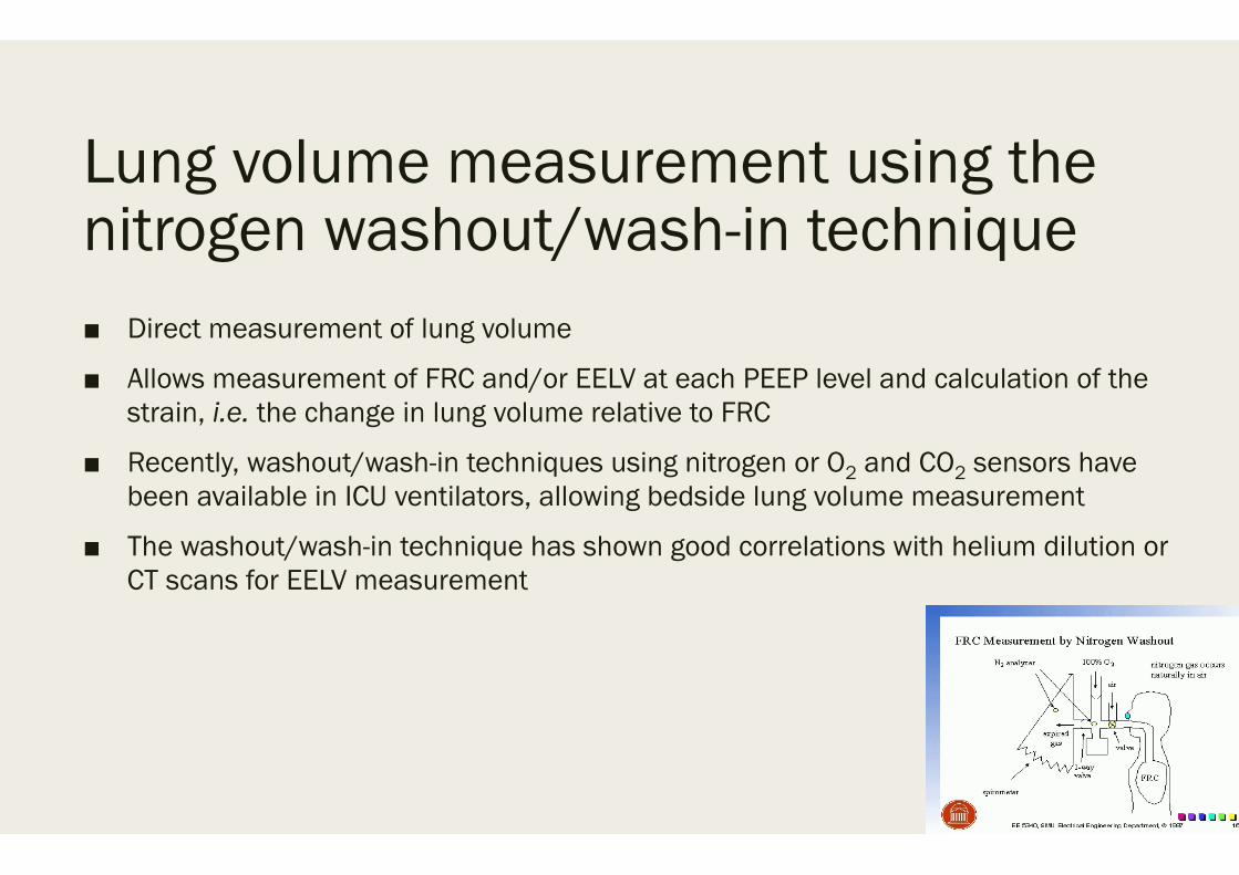
Lung volume measurement using the nitrogen washout/wash-in technique ■ Direct measurement of lung volume
■ Allows measurement of FRC and/or EELV at each PEEP level and calculation of the strain, i.e. the change in lung volume relative to FRC
■ Recently, washout/wash-in techniques using nitrogen or O2 and CO2 sensors have been available in ICU ventilators, allowing bedside lung volume measurement
■ The washout/wash-in technique has shown good correlations with helium dilution or CT scans for EELV measurement

Lung ultrasound ■ Evaluating the response to PEEP could be done by assessing the lung reaeration
with lung ultrasound
■ Bouhemad et al. have used a specific score based on the repeated examination of six lung regions in each lung, before and after increasing PEEP
■ The transthoracic lung ultrasound technique is a method equivalent to the pressure–volume curve method for quantitative assessment of PEEP-induced lung recruitment

Reducing the burden of ventilation by extracorporeal CO2 removal ■ Extracorporeal CO2 removal (ECCO2R) uses a veno–venous (or arterio–venous)
extracorporeal device at low blood flow rates (300–1000 mL·min−1)
■ The major difference with veno–venous extracorporeal membrane oxygenation (ECMO) is that much lower blood flows are needed to remove CO2, compared with 3–5 L·min−1 with ECMO
Reducing the burden of ventilation by extracorporeal CO2 removal
■ The advantage of the low flow is that relatively small vascular cannulas can be used for this amount of blood flow
■ Ultraprotective lung ventilation in many patients with ARDS
■ In addition, the decrease in tidal volume, down to 4 mL·kg−1 PBW, could facilitate an increase in PEEP.


NEWER VENTILATOR FEATURES
■ AUTOFLOW Uses a combination of VC and PC to provide a set VT with a decelerating flow. ■ ATC (AUTOMATIC TUBE COMPENSATION) Measures resistance continuously in the
ventilator circuit and adjusts pressure to maintain flow. Theoretically superior to PS. ■ VOLUME SUPPORT Used for a spontaneously breathing patient. Adjusts level of pressure
support to achieve a set tidal volume. Essentially should be self-weaning. ■ VAPS (VOLUME ASSURED PRESSURE SUPPORT) Similar to VS

NEW MODES
(1) Modes that adapt to the instantaneous inspiratory effort of the patient, such as
proportional assist ventilation (PAV) and neurally adjusted ventilatory assist (NAVA);
(2) Automated modes that can be adapted to the patient demands, such as
adaptive support ventilation (ASV) (3) Modes that introduce biological variability in the ventilatory pattern, such as
variable pressure support ventilation (V-PSV) or “noisy ventilation”


Proportional assist ventilation (PAV)
■ Synchronized assist ventilation mode in which the ventilator provides pressure assistance proportional to the instantaneous effort of the patient
■ Pressure Control ventilation with Assist/Control for spontaneous breaths
■ The rate is a back up rate only

PAV
■ Ventilator detects the inspiratory effort of the patient by precisely measuring the flow and volume leaving the ventilator toward the patient
■ Both parameters are conditioned by the inspiratory decrease in alveolar pressure which the patient generates through muscle contraction
■ The flow and volume are amplified by respective adjustable gain controls, and the sum of both constitutes the control signal that generates the pressure response of the ventilator
■ Rapid delivery of flow in response to this control signal

Functioning of proportional assist ventilation with load-adjustable gain factors (PAV+) ■ A simplified and improved form has recently been introduced, called proportional
assist ventilation with load-adjustable gain factors, or PAV+
■ Offers two essential improvements: (1) the noninvasive and semi-continuous measurement of respiratory mechanics,
allowing automatic closed-loop adjustment of the assist level. This measurement is made by introducing brief pauses (300ms) at the end of inspiration every 8–15 respirations to estimate resistance and elastance
(2) the automatic adjustment of a single level of flow and volume assistance that becomes a constant fraction of the measured values of resistance and elastance

PAV+
■ Simply need to adjust the percentage by which the ventilator must assist patient effort
■ Accordingly, an assist level of 70% means that the ventilator will contribute 70% to the total pressure reached, leaving the remaining 30% to the patient


PAV and PAV+: clinical characteristics
■ Marantz et al. characterized the physiological response to PAV among patients dependent upon mechanical ventilation
■ During PAV, in the absence of limitations imposed by respiratory mechanics, the RCS of the patient determines the tidal volume (Vt) and the frequency in response to variable assist levels
■ Patients tend to lower Vt and to increase the frequency in order to maintain the chosen minute volume
■ Reduction of the inspiratory pressures

PAV & PAV+
■ Compared to PS ventilationà PAV has shown similar muscle discharge and better hypercapnia compensation
■ In response to an increase in elastic loading of 30%, Kondili et al.recorded greater efficiency in compensation (lesser increase of the work of breathing) with PAV+ than with PSV

PAV & PAV+
■ Xirouchaki et al. compared the effectiveness of PSV versus PAV+ in maintaining critical patients dependent upon mechanical ventilation in assisted ventilation
■ They found PAV+ to significantly increase the probability of remaining with spontaneous ventilation, in addition to considerably reducing patient–ventilator asynchrony
■ Bosma et al. showed PAV to afford superior sleep quality, with fewer disruptions, in comparison with PSV

PAV & PAV+
■ The PAV system depends on pneumatic triggering à same limitations for inspiratory cycling in patients with dynamic hyper insufflation and intrinsic positive-end expiratory pressure (PEEP) as the traditional modes
■ Although expiratory cycling, based on flow, accompanies the cessation of inspiratory effort, expiratory asynchronies have been described particularly with high assist levels
Neurally adjusted ventilatory assist ■ Neurally adjusted ventilatory assist (NAVA)
■ As control signal for both assist and for inspiratory and expiratory cycling of the ventilator, this mode uses the electrical activity of the diaphragm (EAdi)
■ Recorded via transesophageal electromyography using a modified nasogastric tubeà EAdi catheter
■ Similar in size and function to a conventional nasogastric tube but equipped with several microelectrodes at the distal tip for recording EAdi

Neurally adjusted ventilatory assist
■ Correct positioning of the catheter is carried out using the transesophageal electrocardiographic signal recorded through the same electrodes as a guide
■ The operator can check correct positioning (at the esophageal hiatus) on the ventilator screen, based on a simple algorithm

NAVA ■ EAdi is a signal that directly measures the efferents from the RCS, integrating the
sum of time and space of the neural respiratory impulse that results in diaphragmatic activation
■ Inspiratory cycling is determined by the detection of the elevation of EAdi over the expiratory level, with a sensitivity threshold determined by the operator
■ Expiratory cycling occurs when EAdi decreases to 70% of the maximum inspiratory value.
■ Allows adjustment of the duration of the mechanical inspiratory and expiratory times to the neural inspiratory and expiratory times of the patient determined by the RCS


NAVA
■ Eliminates the limitations of pneumatic triggering, since it is not affected by leakages or the presence of dynamic hyperinsufflation
■ Ventilatory mode which theoretically offers the greatest level of patient–ventilator synchrony

NAVA
■ Significant improvement in patient–ventilator synchrony
■ Less over-assistance tendency
■ Greater variability of the respiratory pattern in comparison with PSV in different groups of patients
■ Ineffective effort, i.e., inspiratory effort of the patient that is not accompanied by mechanical assist, virtually disappears with NAVA

NAVA
■ In contrast to PSV, increments in assist level have been shown to exert less effect upon the inspiratory and expiratory cycling times ensuring better synchrony over a broad assist range
■ Patroniti et al. have published a detailed description of the ventilatory pattern during NAVA
■ In patients with respiratory failure, the authors compared the response to increasing NAVA levels with increasing PSV levels

N. Patroniti,G. Bellani,E. Saccavino,A. Zanella,G. Grasselli,S. Isgrò Respiratory pattern during neurally adjusted ventilatory assist in acute respiratory failure patients Intensive Care Med, 38 (2012), pp. 230-239

NAVA
■ Patients tend to select a protective tidal volume (6ml/kg) with moderate assist levels and a generally higher respiratory frequency
■ Can facilitate assisted ventilation also in patients with seriously impaired respiratory function
■ Reduced asynchrony in patients subjected to extracorporeal oxygenation support and with severely impaired lung distensibility versus PSV
■ Achieved better auto-regulation of PCO2 during weaning from ECMO
■ Maintaining protective ventilatory parameters with low Vt values

Summary: “We found NAVA to be a safe and feasible primary ventilation mode for use with children. It outscored standard ventilation in some aspects, as it was able to enhance oxygenation even at lower airway pressures and led to reduced use of sedatives during longer periods of treatment.”

Summary: “NAVA is safe and suitable in infants recovering from severe ARDS. It may be valuable in the weaning phase of severe pediatric ARDS, and the present data are useful to build adequately powered randomized trials.”

LIMITATIONS OF NAVA
■ If feeding tube is contra-indicated – Cannot use NAVA or Edi monitoring
■ If no respiratory drive – Cannot use NAVA, but monitoring of Edi possible
■ If respiratory drive is uncontrollable, i.e. no response from patient on parameter changes
– Use NAVA with caution ■ At very high NAVA levels, breathing pattern can become irregular ■ The performance of NAVA can be affected by signal disturbances (e.g. ECG leak
through)

Automated modes adaptable to patient demands ■ Automatically adjust the pressure or minute-volume levels administered to the
patient, adapting to the needs of the latter over time
■ Adaptive support ventilation (ASV) performs cycle-to-cycle adjustments of tidal volume (through changes in pressure) and respiratory frequency, adapting them to changes in respiratory mechanics.
■ Mixed mode àcan function as a controlled or assisted mode according to the contribution of the patient

ASV vs SIMV
■ Tassaux et al.
■ In comparison with synchronized intermittent ventilation (SIMV-PSV), ASV improved synchrony, reducing the muscle load for a similar delivered minute-volume

D. Tassaux,E. Dalmas,P. Gratadour,P. Jolliet Patient–ventilator interactions during partial ventilatory support: a preliminary study comparing the effects of adaptive support ventilation with synchronized intermittent mandatory ventilation plus inspiratory pressure support Crit Care Med, 30 (2002), pp. 801-807

Variable pressure support ventilation (noisy ventilation) ■ V-PSV introduces random variability in the levels of pressure support ventilation,
resulting in a ventilatory pattern that is variable but independent of the demands of the patient and his or her inspiratory effort

V-PSV
■ Based on the recurrent application of a set of 600 pressure values generated on a random basis
■ Values follow a normal distribution, with a mean and standard deviation adjusted to achieve the desired level of variability
■ Mean pressure value is adjusted to obtain a Vt of 6ml/kg, and the pressure limits are determined by the adjusted upper pressure limit and the expiratory pressure level (PEEP or CPAP)
■ Clinician can adjust the level of variability between 0 and 100%, and the system maintains a stable mean pressure

V-PSV
■ Mechanisms underlying the improvement in respiratory mechanics are not fully clearà alveolar recruitment effect postulated, together with possible stimulation of the production and release of surfactant
■ Not enough clinical studies
■ In patients we will have to determine whether this level of variability is also optimum, and whether extrinsic variability offers advantages with respect to the intrinsic variability of the patient (such as that introduced in PAV or NAVA), as well as explore the effects upon patient–ventilator synchrony

NEW MODES
■ New assisted ventilation modesà adapt to the changing patient needs
■ Allow the patient a total control of the ventilatory process, causing the ventilator to act as an accessory muscle in synchrony with patient inspiratory effort
■ New modes that incorporate increasingly complex closed-loop or knowledge-based control systems are paving the way toward gradual automatization of the mechanical ventilation process
F. Suarez-Sipmann Med Intensiva 2014;38:249-60 - Vol. 38 Num.4 DOI: 10.1016/j.medine.2014.04.001

CONCLUSION
■ Ventilation aims to restore oxygenation, lung volumes, decrease work of breathing
■ Set Ventilator to cause least harm, most benefit and comfort to the patient
■ Can get more out of conventional ventilation
■ Measurement of lung recruitability helps individualise the settings
■ New modes à NAVA, PAV, ASV, PSV+
■ Adapt to the changing needs of the patient

Thank you


