28
MECHANICAL MECHANICAL VENTILATION VENTILATION Soumya Ranjan Parida Basic B.Sc. Nursing 4 th year Sum Nursing College
| Date post: | 06-Aug-2015 |
| Category: |
Healthcare |
| Upload: | soumya-ranjan-parida |
| View: | 47 times |
| Download: | 0 times |

MECHANICAL MECHANICAL VENTILATIONVENTILATION
Soumya Ranjan ParidaBasic B.Sc. Nursing 4th year
Sum Nursing College

Indication for ventilation
• Absolute indication:
1. PaO2 below 50 mm Hg at FiO2 above 0.8 in infant more than 32 weeks, and FiO2 above 0.6 in infant < 32 weeks
2. PaCo2 above 60mmHg with persistent acidemia i.e. pH <7.2 in infant >32 weeks
3. PaCo2 above 50 mmHg with persistent acidemia i.e. pH 7.25 in infant <32 weeks
4. Prolonged apnea
5. General anesthesia

Indication for ventilation
• Relative indication
1. Frequent apneas not responding to drug therapy
2. Early treatment in view of deteriorating bood gases
3. Relieving work of breathing in a child of respiratory difficulty
4. Initiating exogenous surfactant therapy in RDS
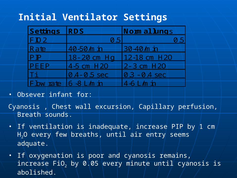
Initial Ventilator SettingsSettings RDS Normal lungsFIO2 0.5 0.5Rate 40-50/min 30-40/minPIP 18- 20 cm Hg 12-18 cm H2OPEEP 4-5 cm H2O 2- 3 cm H2OTi 0.4- 0.5 sec 0.3 - 0.4 secFlow rate 6 -8 L/min 4-6 L/min
• Obsever infant for:
Cyanosis , Chest wall excursion, Capillary perfusion, Breath sounds.
• If ventilation is inadequate, increase PIP by 1 cm H2O every few breaths, until air entry seems adquate.
• If oxygenation is poor and cyanosis remains, increase FiO2 by 0.05 every minute until cyanosis is abolished.
• Draw ABG
• Adjust ventilation as indicated.

Effects of changes in ventilator settings on blood gases:
A] Oxygenation:
1)Fi O2 :The goal is to maintain adequate tissue
oxygen delivery.
• Acceptable upper limits for PaO2 is 100 &
lower limit is 50 mmHg.
• This brings a hemoglobin saturation of 89% - 95%.
• Increasing FiO2 is the most simplest and direct way of increasing oxygenation.
• Increasing FiO2 minimizes barotrauma but there is a chance of oxygen toxicity at levels above 60%

Effects of changes in ventilator settings on blood gases:A] Oxygenation:
2) Mean airway pressure: It is the average area under the curve of the pressure waveform.
• MAP is increased by increases in PEEP, PIP, Inspiratory time (Ti), rate and flow rate.
• PIP: It increases the driving pressure for gas flow into ventilated lung units.
• Ti: This increases the time for gas to distribute the ill perfused areas.
• PEEP: It splints small airways open, ↓airway resistance, ↓ the time constant for inspiration and allows more gas to enter the lung unit for any given PIP orTi
• Thus if an increase in PaO2 is needed then it can be achieved by 1st ↑ PEEP then PIP and then Ti.

Effects of changes in ventilator settings on blood gases:
B] Ventilation:
1)CO2: its elimination depends on minute ventilation.
Minute ventilation= tidal vol * RR
Thus PaCO2 decreases with increase in tidal vol or RR.
↑ in Tidal Vol -- ↑ PIP , ↓ PEEP
For Very immature infants or infants with airleak a PaCO2 of 50- 60mmHg may be tolerated.

Effects of changes in ventilator settings on blood gases:
Bld gas abnormality Corrective measures CommentsFiO2 Rate PIP PEEP Ti
Hypercapnea(PaCO2 >50 mm Hg) --- ↑ ↑ -- -- increase flow rate,Te and reduce dead spaceHypocapnea(PaCO2 <35 mm Hg) -- ↓ ↓ -- -- reduce flow rate and increase dead spaceHyperoxia(PaO2 >100 mm Hg ↓ -- ↓ ↓ ↓ reduce FiO2Hypoxia(PaO2 <50 mm Hg ↑ -- ↑ ↑ ↑ if chest expansions good then it is better to increse
FiO2

Settings in common disease states
RESPIRATORY DISTRESS SYNDROME (RDS):
•It is caused by surfactant deficiency which results in decreased compliance of the lungs.
•There is diffuse alveolar collapse with V/Q mismatch and increased work of breathing.•Mild RDS infants who do not require ventilation can be put
on CPAP early in the course to prevent further atelectasis.•CPAP is given through either nasal prongs or
nasopharyngeal tube.
•CPAP is started at 5 to 6 cm H2O and can be increased to
7 to 8 cm H2O. It is then titrated by observation of
retractions, RR and oxygen saturation.

Settings in common disease states
RESPIRATORY DISTRESS SYNDROME (RDS): cont…
•Mechanical ventilation is used when surfactant has to be administered and when even on high FiO2 , CPAP saturation is not maintained.•A continuous flow, pressure-limited, time cycled ventilator is mainly used.•Initial settings are PIP of 20 – 25 cm H2O, PEEP at 46 cm H2O, flow rate 7 to 12 l/min, rate of 20- 40 breaths/min and Ti of 0.3 – 0.4sec.•Weaning is done when the pt becomes stable and first FiO2 and PIP are weaned, alternating with RR.

Settings in common disease states
RESPIRATORY DISTRESS SYNDROME (RDS): cont…
•Alternatively HFV is used when MAP required exceeds 10 – 11 cm H2O in small infants and 12 cm in larger
infants.•HFJ ventilation:- PIP is set 20% lower than conventional ventilators, PEEP at 8 to 10 cm H2O., RR 420 breaths/min, Ti jet valve at 0.02 sec.•HFO ventilation:- the parameters set are MAP of 2 to 5 cm H2O higher than conventional ventilators, Frequency at 10 to 15 Hz , Ti at 33%, flow rates of 8 to 15 L/min and piston amplitude set to provide adequate chest vibration.

Settings in common disease states
MECONIUM ASPIRATION SYNDROME:
• MAS results from aspiration of meconium stained amniotic fluid.
• Aspirated meconium causes acute airway obstructon, marked airway resistance, scattered atelectasis with V/Q mismatching and hyperexpansion due to ball valve effect.
• CPAP can be used in infants who are stable without respiratory failure. It stabilizes collapsed terminal airways and improves atelectasis.
• When assisted ventilation is indicated (PaCO 60 mm Hg, PaO2 <50mmHg ) following ventilators settings are recommended:

Settings in common disease states
MECONIUM ASPIRATION SYNDROME:
Following ventilators settings are recommended
• PIP 30-35cm H2O, PEEP 4-5 cm H2O, Rate 20-25/min
IT 0.4 to 0.5 sec
• Some infants may require rapid rates with short inspiratory times i.e 0.2 sec
• HFJV and HFOV may be used when the conventional ventilator fails in severe disease.

Settings in common disease states
BRONCHOPULMONARY DYSPLASIA: (BPD)
• BPD has a multifactorial etiology but barotrauma appears to be the root cause.
• It results from injury to the alveoli and airways.
• It is marked by shifting focal atelectasis, hyperinflation with V/Q mismatching, chronic and acute increase in airway resistance, and a significant increase in the work of breathing.
• The optimal strategy is to wean infants as soon as possible so as to minimize barotrauma and oxygen toxicity.
• Ventilator settings are kept at minimum to provide adequate gas exchange.

Settings in common disease states
BRONCHOPULMONARY DYSPLASIA: (BPD)
• Hyperventilation should be avoided and PaCO2 maintained at >55mm Hg, with pH >7.25
• Oxygen saturation should be maintaned at 90% to 95% or lower and PaO2 at 60 to 80mm Hg
• High frequency oscillatory ventilators are not frequently used as they do not prevent BPD in high risk infants.
• Weaning is done by decreasing rate by 1 to 2 breaths/min or 1 cm H2O PIP every day when tolerated

Settings in common disease states
AIR LEAK:
• Pneumothorax and Pulmonary Interstitial Emphysema (PIE) are most common air leak syndromes.
• The primary goal is to reduce MAP through PIP, Ti, PEEP as high pressures drive air into the interstitium during the ventilator cycle & increase expiratory time.
• Adequate oxygenation is maintained by increasing FiO2 and rate.

Settings in common disease states
Apnea:
• Apnea needing ventilation may result from apnea of prematurity, during or following anesthesia or
neuromuscular paralysis.
• In apnea of prematurity CPAP at levels 4-6 cm H2O can reduce the number of apneic spells.
• The goal is to provide physiologic ventilation using moderate PEEP (3-4 cm H2O), low gas flow, and normal rates (30- 40 breaths/min) with PIP adjusted to prevent hyperventilation (10- 18cm H20)
Weaning from ventilator
• Process of weaning begins at the time of initiation of ventilation.
• Duration of ventilation varies with the disease process and is associated with the patients ability to take over >60% work of breathing.
• Eg. In a HMD it may take 3 days to 1 week, whereas in a case of MAS it may be sooner.
• Once a infant has remained stable for at least 24 hours weaning can be attempted.

Weaning from ventilator
Markers of improved condition;
• Improving general condition, fever etc
• Decreasing FiO2 requirment
• Improving breath sounds
• Decreasing ET secretions
• Improving chest xray
• Improved electrolyte and fluid status
• Improving hemodynamic status
• Improving neurological status

Weaning from ventilator
• Before initiating of weaning a chest xray should be done to obtain a baseline
• Increase in compliance and FRC typically heralds recovery from pulmonary disease.
• The ventilator mode should be changed from control mode to SIMV mode with pressure support.
• The first setting to be reduced is PIP by 1.0 cm H2O decrements till it is brought down to 25 cm H2O.
• Then PIP and FiO2 ( decreased 0.05 or 5%) are reduced alternately till a relative safe level of 20 PIP and 0.6 FiO2 are reached

Weaning from ventilator
• After this FiO2 and PEEP should be decreased hand in hand i.e. at 0.6 FiO2, PEEP should be 6. PIP should be reduced by 1.0 cm H2O every 15- 20 mins.
• Ventilatory rate is now reduced in small increments of 2 breaths/min till it is brought down to 10breaths/min
• Extubation is indicated when FiO2 is 0.4, PIP 10- 15cm H2O,PEEP 3 cm H2O, Ti 0.3sec and RR 10/min.
• Some infants can be put on CPAP before extubation.

Extubation from ventilator
Extubation can be performed when the foll criteria are met:
• Control of airway reflexes, minimal secretions.
• Good breath sounds
• Minimal oxygen requirnment <0.3 with SpO2 >94.
• Minimal rate 5/min
• Minimal pressure support
• Adequate muscle tone
• Minimal inotropic support
• Normal electrolytes, no fluid imbalance

Extubation from ventilator
Extubation procedure
• Keep NBM 4 ours before extubation
• Suction the ET tube, oral cavity and nostrils.
• Suction the nasogastric tube to deflate the stomach
• Keep oxygen ready
• Nebulization with beta stimulant and or adrenaline should be ready immediate postextubation.
• IV steroids dexamethasone 0.15mg/kg may be used in prolonged intubation. It can be started 24 hrs prior to extubation and to be continued for 48 hrs.

Extubation from ventilator
• Aminophylline can be started as it decreases resistance and increases respiratory drive
• ABG is usually done 20 min after extubation
• Post extubation Xray should be done

Complications of mechanical ventilaion
Airway injury
• Tracheal inflammation
• Tracheobronchomalacia
• Subglottic stenosis
• Granuloma formation
• Palatal grooving
• Nasal septal injury
• Necrotizing tracheobronchitis

Complications of mechanical ventilaion
Endotracheal tube complications:
• Dislodgement
• Accidental extubation
• Airway erosion
Chronic lung injury
• Bronchopulmonary dysplasia
• Acquired lobar emphysema

Complications of mechanical ventilaion
Air leak
• Pulmonary interstitial emphysema
• Pneumothorax
• Pneumomediastinum
• Pneumopericardium
• Pnemoperitoneum
• Hyperinflation

Complications of mechanical ventilaion
Cardiovascular
Decreased cardiac output
Patent ductus arteriosus
Intraventricular hemorrhage
Miscellaneous
Retinopathy of newborn
Apnea
Infection
Feeding intolerance
Developmental delay


