CASE REPORT PEER REVIEWED | OPEN ACCESS www.edoriumjournals.com International Journal of Case Reports and Images (IJCRI) International Journal of Case Reports and Images (IJCRI) is an international, peer reviewed, monthly, open access, online journal, publishing high-quality, articles in all areas of basic medical sciences and clinical specialties. Aim of IJCRI is to encourage the publication of new information by providing a platform for reporting of unique, unusual and rare cases which enhance understanding of disease process, its diagnosis, management and clinico-pathologic correlations. IJCRI publishes Review Articles, Case Series, Case Reports, Case in Images, Clinical Images and Letters to Editor. Website: www.ijcasereportsandimages.com Very delayed coronary stent fracture presenting as unstable angina: A case report Saurabh Mehrotra, Praful Sharma P., Yashpaul Sharma Y.P. ABSTRACT Introduction: Coronary stent fracture represents an under diagnosed clinical event of drug- eluting stents which is often associated with adverse clinical outcomes of in-stent restenosis. Numerous risk factors are associated with stent fracture that include stent overexpansion, creation of hinge points due to stent overlapping, use of longer stents for complex lesions as well as mechanical fatigue causing stent distortion in the right coronary artery and vein grafts. Case Report: A 64-year old male, a cigarette smoker, presented with rest angina. Coronary angiogram showed discrete 99% stenosis in proximal left anterior descending artery and a mid-eccentric 90% lesion in the right coronary artery (RCA). The patient was taken up for angioplasty of both the vessels. A type V fracture was detected after four years of zotarolimus- eluting stent placement in the right coronary artery. Conclusion: Despite the recent advances in drug-eluting stents design, there remains a potential of stent fracture especially when a long drug-eluting stents is implanted in a tortuous vessel and is exposed to torsion forces at the hinge points. (This page in not part of the published article.)
Transcript
CASE REPORT PEER REVIEWED | OPEN ACCESS
www.edoriumjournals.com
International Journal of Case Reports and Images (IJCRI)International Journal of Case Reports and Images (IJCRI) is an international, peer reviewed, monthly, open access, online journal, publishing high-quality, articles in all areas of basic medical sciences and clinical specialties.
Aim of IJCRI is to encourage the publication of new information by providing a platform for reporting of unique, unusual and rare cases which enhance understanding of disease process, its diagnosis, management and clinico-pathologic correlations.
IJCRI publishes Review Articles, Case Series, Case Reports, Case in Images, Clinical Images and Letters to Editor.
Website: www.ijcasereportsandimages.com
Very delayed coronary stent fracture presenting as unstable angina: A case report
Introduction: Coronary stent fracture represents an under diagnosed clinical event of drug-eluting stents which is often associated with adverse clinical outcomes of in-stent restenosis. Numerous risk factors are associated with stent fracture that include stent overexpansion, creation of hinge points due to stent overlapping, use of longer stents for complex lesions as well as mechanical fatigue causing stent distortion in the right coronary artery and vein grafts. Case Report: A 64-year old male, a cigarette smoker, presented with rest angina. Coronary angiogram showed discrete 99% stenosis in proximal left anterior descending artery and a mid-eccentric 90% lesion in the right coronary artery (RCA). The patient was taken up for angioplasty of both the vessels. A type V fracture was detected after four years of zotarolimus-eluting stent placement in the right coronary artery.Conclusion: Despite the recent advances in drug-eluting stents design, there remains a potential of stent fracture especially when a long drug-eluting stents is implanted in a tortuous vessel and is exposed to torsion forces at the hinge points.
(This page in not part of the published article.)
International Journal of Case Reports and Images, Vol. 8 No. 2, February 2017. ISSN – [0976-3198]
Int J Case Rep Images 2017;8(2):147–150. www.ijcasereportsandimages.com
Mehrotra et al. 147
CASE REPORT PEER REVIEWED | OPEN ACCESS
Very delayed coronary stent fracture presenting as unstable angina: A case report
Introduction: Coronary stent fracture represents an under diagnosed clinical event of drug-eluting stents which is often associated with adverse clinical outcomes of in-stent restenosis. Numerous risk factors are associated with stent fracture that include stent overexpansion, creation of hinge points due to stent overlapping, use of longer stents for complex lesions as well as mechanical fatigue causing stent distortion in the right coronary artery and vein grafts. Case Report: A 64-year old male, a cigarette smoker, presented with rest angina. Coronary angiogram showed discrete 99% stenosis in proximal left anterior descending artery and a mid-eccentric 90% lesion in the right coronary artery (RCA). The patient was taken up for angioplasty of both the vessels. A type V fracture was detected after four years of zotarolimus-eluting stent placement in the right coronary artery. Conclusion: Despite
Affiliations: 1MD, DM, Assistant Professor, Department of Cardiology, Post Graduate Institute of Medical Education and Research (PGIMER), Chandigarh, Punjab, India; 2MD, DM, Senior Resident, Department of Cardiology, Post Grad-uate Institute of Medical Education and Research (PGIM-ER), Chandigarh, Punjab, India; 3MD, DM, Head, Cardiol-ogy, Department of Cardiology, Post Graduate Institute of Medical Education and Research (PGIMER), Chandigarh, Punjab, India.Corresponding Author: S. Mehrotra, Assistant Professor, Cardiology, Post Graduate Institute of Medical Education and Research (PGIMER), Sector-12, Chandigarh- 160012, Punjab, India; Email: [email protected]
Received: 24 July 2016Accepted: 21 November 2016Published: 01 February 2017
the recent advances in drug-eluting stents design, there remains a potential of stent fracture especially when a long drug-eluting stents is implanted in a tortuous vessel and is exposed to torsion forces at the hinge points.
Mehrotra S, Sharma PP, Sharma YYP. Very delayed coronary stent fracture presenting as unstable angina: A case report. Int J Case Rep Images 2017;8(2):147–150.
Article ID: Z01201702CR10766SM
*********
doi:10.5348/ijcri-201727-CR-10766
INTRODUCTION
The introduction of drug-eluting stents has marked a new era in the field of interventional cardiology with significant reduction in the incidence of restenosis as well as repeat revascularization [1, 2]. Although drug-eluting stents has become the standard of care for percutaneous coronary intervention, the occurrence of late stent thrombosis has raised concern over their long-term safety. Stent fracture is being increasingly recognized as a potential cause of in-stent restenosis and stent thrombosis with the clinical manifestation of recurrent angina, myocardial infarction and even sudden death [3–6]. We report a rare case of delayed stent fracture after percutaneous coronary intervention with zotarolimus-eluting stent (ZES).
International Journal of Case Reports and Images, Vol. 8 No. 2, February 2017. ISSN – [0976-3198]
Int J Case Rep Images 2017;8(2):147–150. www.ijcasereportsandimages.com
Mehrotra et al. 148
CASE REPORT
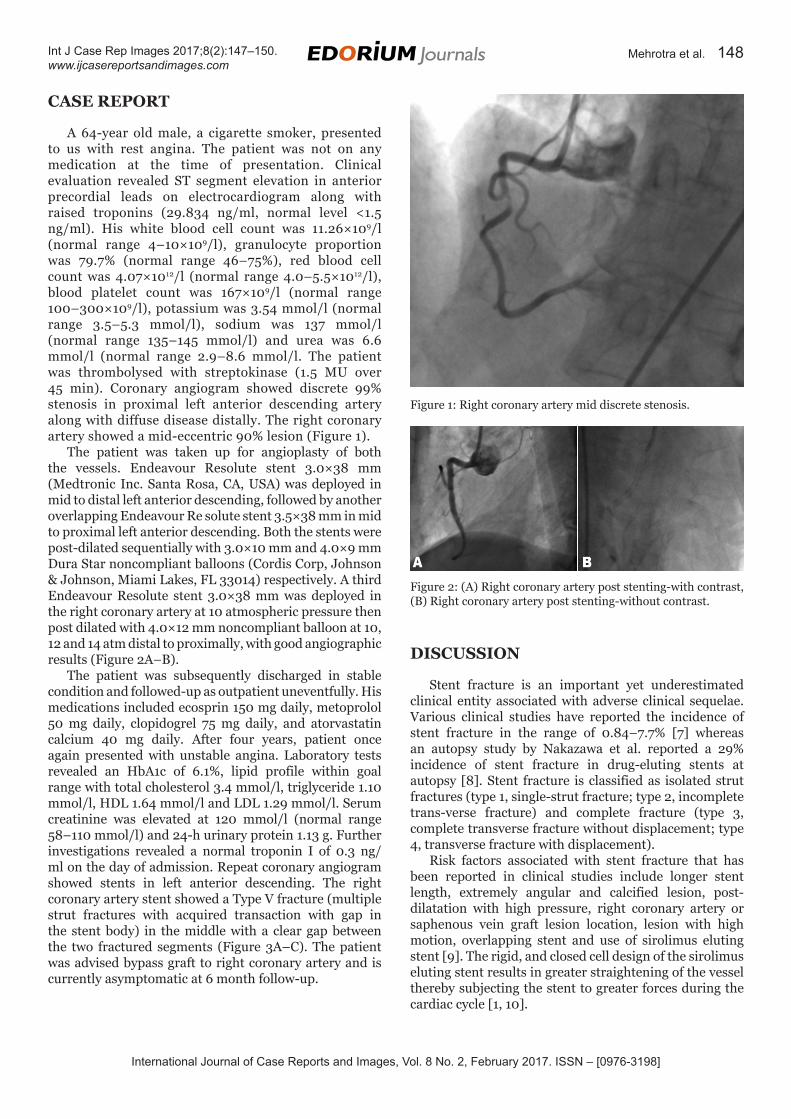
A 64-year old male, a cigarette smoker, presented to us with rest angina. The patient was not on any medication at the time of presentation. Clinical evaluation revealed ST segment elevation in anterior precordial leads on electrocardiogram along with raised troponins (29.834 ng/ml, normal level <1.5 ng/ml). His white blood cell count was 11.26×10⁹/l (normal range 4–10×109/l), granulocyte proportion was 79.7% (normal range 46–75%), red blood cell count was 4.07×10¹²/l (normal range 4.0–5.5×10¹²/l), blood platelet count was 167×109/l (normal range 100–300×109/l), potassium was 3.54 mmol/l (normal range 3.5–5.3 mmol/l), sodium was 137 mmol/l (normal range 135–145 mmol/l) and urea was 6.6 mmol/l (normal range 2.9–8.6 mmol/l. The patient was thrombolysed with streptokinase (1.5 MU over 45 min). Coronary angiogram showed discrete 99% stenosis in proximal left anterior descending artery along with diffuse disease distally. The right coronary artery showed a mid-eccentric 90% lesion (Figure 1).
The patient was taken up for angioplasty of both the vessels. Endeavour Resolute stent 3.0×38 mm (Medtronic Inc. Santa Rosa, CA, USA) was deployed in mid to distal left anterior descending, followed by another overlapping Endeavour Re solute stent 3.5×38 mm in mid to proximal left anterior descending. Both the stents were post-dilated sequentially with 3.0×10 mm and 4.0×9 mm Dura Star noncompliant balloons (Cordis Corp, Johnson & Johnson, Miami Lakes, FL 33014) respectively. A third Endeavour Resolute stent 3.0×38 mm was deployed in the right coronary artery at 10 atmospheric pressure then post dilated with 4.0×12 mm noncompliant balloon at 10, 12 and 14 atm distal to proximally, with good angiographic results (Figure 2A–B).
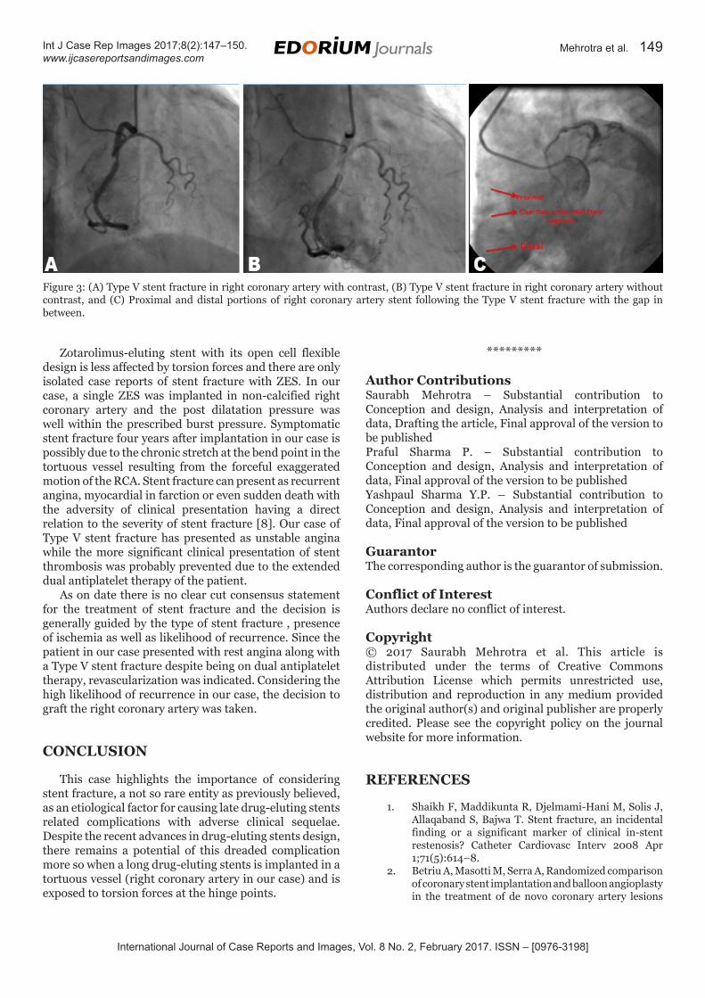
The patient was subsequently discharged in stable condition and followed-up as outpatient uneventfully. His medications included ecosprin 150 mg daily, metoprolol 50 mg daily, clopidogrel 75 mg daily, and atorvastatin calcium 40 mg daily. After four years, patient once again presented with unstable angina. Laboratory tests revealed an HbA1c of 6.1%, lipid profile within goal range with total cholesterol 3.4 mmol/l, triglyceride 1.10 mmol/l, HDL 1.64 mmol/l and LDL 1.29 mmol/l. Serum creatinine was elevated at 120 mmol/l (normal range 58–110 mmol/l) and 24-h urinary protein 1.13 g. Further investigations revealed a normal troponin I of 0.3 ng/ml on the day of admission. Repeat coronary angiogram showed stents in left anterior descending. The right coronary artery stent showed a Type V fracture (multiple strut fractures with acquired transaction with gap in the stent body) in the middle with a clear gap between the two fractured segments (Figure 3A–C). The patient was advised bypass graft to right coronary artery and is currently asymptomatic at 6 month follow-up.
DISCUSSION
Stent fracture is an important yet underestimated clinical entity associated with adverse clinical sequelae. Various clinical studies have reported the incidence of stent fracture in the range of 0.84–7.7% [7] whereas an autopsy study by Nakazawa et al. reported a 29% incidence of stent fracture in drug-eluting stents at autopsy [8]. Stent fracture is classified as isolated strut fractures (type 1, single-strut fracture; type 2, incomplete trans-verse fracture) and complete fracture (type 3, complete transverse fracture without displacement; type 4, transverse fracture with displacement).
Risk factors associated with stent fracture that has been reported in clinical studies include longer stent length, extremely angular and calcified lesion, post-dilatation with high pressure, right coronary artery or saphenous vein graft lesion location, lesion with high motion, overlapping stent and use of sirolimus eluting stent [9]. The rigid, and closed cell design of the sirolimus eluting stent results in greater straightening of the vessel thereby subjecting the stent to greater forces during the cardiac cycle [1, 10].
Figure 1: Right coronary artery mid discrete stenosis.
Figure 2: (A) Right coronary artery post stenting-with contrast, (B) Right coronary artery post stenting-without contrast.
International Journal of Case Reports and Images, Vol. 8 No. 2, February 2017. ISSN – [0976-3198]
Int J Case Rep Images 2017;8(2):147–150. www.ijcasereportsandimages.com
Mehrotra et al. 149
Zotarolimus-eluting stent with its open cell flexible design is less affected by torsion forces and there are only isolated case reports of stent fracture with ZES. In our case, a single ZES was implanted in non-calcified right coronary artery and the post dilatation pressure was well within the prescribed burst pressure. Symptomatic stent fracture four years after implantation in our case is possibly due to the chronic stretch at the bend point in the tortuous vessel resulting from the forceful exaggerated motion of the RCA. Stent fracture can present as recurrent angina, myocardial in farction or even sudden death with the adversity of clinical presentation having a direct relation to the severity of stent fracture [8]. Our case of Type V stent fracture has presented as unstable angina while the more significant clinical presentation of stent thrombosis was probably prevented due to the extended dual antiplatelet therapy of the patient.
As on date there is no clear cut consensus statement for the treatment of stent fracture and the decision is generally guided by the type of stent fracture , presence of ischemia as well as likelihood of recurrence. Since the patient in our case presented with rest angina along with a Type V stent fracture despite being on dual antiplatelet therapy, revascularization was indicated. Considering the high likelihood of recurrence in our case, the decision to graft the right coronary artery was taken.
CONCLUSION
This case highlights the importance of considering stent fracture, a not so rare entity as previously believed, as an etiological factor for causing late drug-eluting stents related complications with adverse clinical sequelae. Despite the recent advances in drug-eluting stents design, there remains a potential of this dreaded complication more so when a long drug-eluting stents is implanted in a tortuous vessel (right coronary artery in our case) and is exposed to torsion forces at the hinge points.
*********
Author ContributionsSaurabh Mehrotra – Substantial contribution to Conception and design, Analysis and interpretation of data, Drafting the article, Final approval of the version to be publishedPraful Sharma P. – Substantial contribution to Conception and design, Analysis and interpretation of data, Final approval of the version to be publishedYashpaul Sharma Y.P. – Substantial contribution to Conception and design, Analysis and interpretation of data, Final approval of the version to be published
GuarantorThe corresponding author is the guarantor of submission.
Conflict of InterestAuthors declare no conflict of interest.
1. Shaikh F, Maddikunta R, Djelmami-Hani M, Solis J, Allaqaband S, Bajwa T. Stent fracture, an incidental finding or a significant marker of clinical in-stent restenosis? Catheter Cardiovasc Interv 2008 Apr 1;71(5):614–8.
2. Betriu A, Masotti M, Serra A, Randomized comparison of coronary stent implantation and balloon angioplasty in the treatment of de novo coronary artery lesions
Figure 3: (A) Type V stent fracture in right coronary artery with contrast, (B) Type V stent fracture in right coronary artery without contrast, and (C) Proximal and distal portions of right coronary artery stent following the Type V stent fracture with the gap in between.
International Journal of Case Reports and Images, Vol. 8 No. 2, February 2017. ISSN – [0976-3198]
Int J Case Rep Images 2017;8(2):147–150. www.ijcasereportsandimages.com
Mehrotra et al. 150
(START): A four-year follow-up. J Am Coll Cardiol 1999 Nov 1;34(5):1498–506.
3. Aoki J, Nakazawa G, Tanabe K, Incidence and clinical impact of coronary stent fracture after sirolimus-eluting stent implantation. Catheter Cardiovasc Interv 2007 Feb 15;69(3):380–6.
4. Lee MS, Jurewitz D, Aragon J, Forrester J, Makkar RR, Kar S. Stent fracture associated with drug-eluting stents: Clinical characteristics and implications. Catheter Cardiovasc Interv 2007 Feb 15;69(3):387–94.
5. Park JS, Shin DG, Kim YJ. Fractured DES with a patent coronary artery: Clinical implications. J Invasive Cardiol 2007 Feb;19(2):E43–5.
6. Shite J, Matsumoto D, Yokoyama M. Sirolimus-eluting stent fracture with thrombus, visualization by optical coherence tomography. Eur Heart J 2006 Jun;27(12):1389.
7. Alexopoulos D, Xanthopoulou I. Coronary stent fracture: How frequent it is? Does it matter? Hellenic J Cardiol 2011 Jan-Feb;52(1):1–5.
8. Nakazawa G, Finn AV, Vorpahl M, et al. Incidence and predictors of drug-eluting stent fracture in human coronary artery a pathologic analysis. J Am Coll Cardiol 2009 Nov 17;54(21):1924–31.
9. Kim HS, Kim YH, Lee SW, et al. Incidence and predictors of drug-eluting stent fractures in long coronary disease. Int J Cardiol 2009 Apr 17;133(3):354–8.
10. Canan T, Lee MS. Drug-eluting stent fracture: Incidence, contributing factors, and clinical implications. Catheter Cardiovasc Interv. 2010 Feb 1;75(2):237–45.
Access full text article onother devices
Access PDF of article onother devices
EDORIUM JOURNALS AN INTRODUCTION
Edorium Journals: On Web
About Edorium JournalsEdorium Journals is a publisher of high-quality, open ac-cess, international scholarly journals covering subjects in basic sciences and clinical specialties and subspecialties.
Edorium Journals www.edoriumjournals.com
Edorium Journals et al.
Edorium Journals: An introduction
Edorium Journals Team
But why should you publish with Edorium Journals?In less than 10 words - we give you what no one does.
Vision of being the bestWe have the vision of making our journals the best and the most authoritative journals in their respective special-ties. We are working towards this goal every day of every week of every month of every year.
Exceptional servicesWe care for you, your work and your time. Our efficient, personalized and courteous services are a testimony to this.
Editorial ReviewAll manuscripts submitted to Edorium Journals undergo pre-processing review, first editorial review, peer review, second editorial review and finally third editorial review.
Early View versionEarly View version of your manuscript will be published in the journal within 72 hours of final acceptance.
Manuscript statusFrom submission to publication of your article you will get regular updates (minimum six times) about status of your manuscripts directly in your email.
Our Commitment
Favored Author programOne email is all it takes to become our favored author. You will not only get fee waivers but also get information and insights about scholarly publishing.
Institutional Membership programJoin our Institutional Memberships program and help scholars from your institute make their research accessi-ble to all and save thousands of dollars in fees make their research accessible to all.
Our presenceWe have some of the best designed publication formats. Our websites are very user friendly and enable you to do your work very easily with no hassle.
Something more...We request you to have a look at our website to know more about us and our services.
We welcome you to interact with us, share with us, join us and of course publish with us.
Browse Journals
CONNECT WITH US
Invitation for article submissionWe sincerely invite you to submit your valuable research for publication to Edorium Journals.
Six weeksYou will get first decision on your manuscript within six weeks (42 days) of submission. If we fail to honor this by even one day, we will publish your manuscript free of charge.*
Four weeksAfter we receive page proofs, your manuscript will be published in the journal within four weeks (31 days). If we fail to honor this by even one day, we will pub-lish your manuscript free of charge and refund you the full article publication charges you paid for your manuscript.*
This page is not a part of the published article. This page is an introduction to Edorium Journals and the publication services.
* Terms and condition apply. Please see Edorium Journals website for more information.