40
VESTIBULAR REHABILITATION By- Dr. Swati Bhattacharya PT MPT-Neurology
| Date post: | 12-Jan-2017 |
| Category: |
Healthcare |
| Upload: | swati-bhattacharya |
| View: | 672 times |
| Download: | 0 times |
VESTIBULAR REHABILITATION
By-Dr. Swati Bhattacharya PT
MPT-Neurology

Anatomy and physiology of inner ear
Bony Labyrinth: Houses the membranous labyrinth. Filled with perilymph fluid (similar to CSF).
Membranous Labyrinth: Consists of the otolith organs and semi-circular canals. Filled with endolymph (similar to intracellular fluid).

Membranous labyrinthOtolith Organs Utricle: Responsible for horizontal translation of
the head and head tiltSaccule: Responsible for vertical translation of
the head.Combined, they sense linear acceleration and
static tilt of the head with respect to the gravitational axis.
Maculae: Sensory receptor for the otolith organs. A gelatinous matrix surrounds the hair cells. Otoconia are embedded on top of the maculae in the otolithic membrane..

Semi-circular Canals (SSC)Three fluid filled loops responsible for
sensory input related to head velocity and angular acceleration.
Enables the VOR to generate eye movements to match head movements, resulting in clear vision during head movement.
Cristae: sensory structure for the SSC that sense angular movement
Cupula: gelatinous mass surrounding the hair cells of the cristae in the SSC.

Hair Cells:Endolymph fluid within the SSC
and otolith organs move the hair cells according to head movement.
The direction of deflection of the hair cells tells the brain how the head is moving.

VESTIBULAR DISORDER
UNILATERAL VESTIBULAR HYPOFUNCTION
BILATERAL VESTIBULAR HYPOFUNCTION

Unilateral vestibular hypofunctionA diagnosis of unilateral
vestibular hypofunction is made when the balance system in your inner ear, the peripheral vestibular system, is not working properly.
There is a vestibular system in both inner ears, so unilateral means that only one system is impaired, while the other is working normally.

Causes of UVHVestibular neuronitisLabyrinthitisWeakness of structures of inner
ear due to ageingToxic reactions to medicationsBlood clots, tumors, brain injuries
impacting inner ear structures,
Symptoms of UVHDizziness or vertigo (described as a
spinning sensation) Poor balance, especially with head
turns Trouble walking, especially outdoors,
in dark rooms, or in crowded places Blurred vision, especially when
turning your head quickly Nausea and vomiting in acute and
severe cases
Exercises for UVHGaze stabilization exercise to help
improve the coordination of head and eye movements
Special balance exercises to incorporate and strengthen your inner ear balance system
Walking exercises to improve balance in challenging environments like walking outdoors, walking on uneven surfaces, walking in dark rooms, and walking in crowded places
Bilateral Vestibular HypofunctionLoss of vestibular function in both
labyrinths leads to characteristic dysfunction in vision and balance.

Causes of bilateral vestibular hypofunctionSevere head injuriesCertain infections, especially meningitis in
children or viralHereditary symptoms, sometimes associated
with migraineMenieres syndrome, which is due to an
increased pressure in the fluid in the inner earInflammatory conditionsAutoimmune diseasesToxic side effect of antibiotics such as
gentamicin
Hopkins treatment The therapy program will
emphasize improving upon what labyrinthine function remains or on developing alternative strategies using other sensory cues such as from the neck, feet or eyes that can substitute for the missing sensations from the labyrinth.
Tai Chi or Chinese exercises may also help patients regain balance

Vertigo
Peripheral Vertigo
Central Vertigo
• Collects the inner ear causes.•These include- BPPV Labyrinthitis Meniere’s disease Vestibular neuronitis
• Collects together central nervous system causes.•These include- migraine stroke Transient ischaemic attack

Peripheral causes of vertigoUnilateral Vestibular
HypofunctionPeripheral vestibular asymmetryLabyrinthitisVestibular neuronitisVestibular infarctVestibular schwannoma/ acoustic
neuroma

Peripheral vertigo history and symptomsSudden onset (illness, trauma or
unknown)Constant dizziness, provoked by
motion especially head and body turns.
Discomfort with watching movement or patterns
Mild-moderate imbalanceHorizontal unidirectional gaze-
evoked nystagmus

Bilateral vestibular hypofunctionNot dizzyOff balanceNo balance in the darkOscillopsia (things bounce
visually)
CAUSES – Chemotherapy, ototoxic antibiotics, autoimmune.

BPPV (Benign paroxysmal positional vertigo)ETIOLOGY: Otoliths in semicircular canal cupula deflects more than usual

BPPV SYMPTOMSDizziness with position changesLying flat, rolling over, sit up,
stand up, bend over, look upStrong spinning for less than one
minuteMay have leftover symptoms for
hours.
BPPV diagnosisBPPV is diagnosed with Dix
Hallpike Maneuver.
https://www.youtube.com/watch?v=P2Jdb8h9MHo
BPPV epley maneuver
Dizziness◦Light headedness
◦Feeling faint
◦Unsteady
◦Dysequilibrium
Definitions
DefinitionsImbalance
◦Stumbling, difficulty walking straight or turning a corner
◦Clumsiness or difficulty with coordination
◦Tendency to fall
DefinitionsVertigo
◦A specific spinning sensation
◦An illusion of motion
◦The feeling that you or your environment is moving
WHAT IS VESTIBULAR REHABILITATION THERAPY?Vestibular rehabilitation therapy (VRT) is
an exercise-based program designed to promote central nervous system compensation for inner ear deficits. VRT can help with a variety of vestibular problems, including benign paroxysmal positional vertigo (BPPV) and the unilateral or bilateral vestibular hypofunction (reduced inner ear function on one or both sides) associated with Ménière’s disease, labyrinthitis, and vestibular neuritis.

What are the Indications for Therapy?
Specific interventions for benign paroxysmal positional vertigo (BPPV)◦ The Epley and Semont maneuvers (see following and BPPV
page)◦ The Brandt-Daroff exercises (also see following section and
BPPV page for details)◦ Log roll exercises (for lateral canal BPPV)
General interventions for vestibular loss◦ Unilateral loss, such as for vestibular neuritis or acoustic
neuroma◦ Bilateral loss, such as for gentamicin
toxicity and related conditions Empirical treatment for common situations where the
diagnosis is unclear◦ Post-traumatic vertigo◦ Multifactorial disequilibrium of the elderly
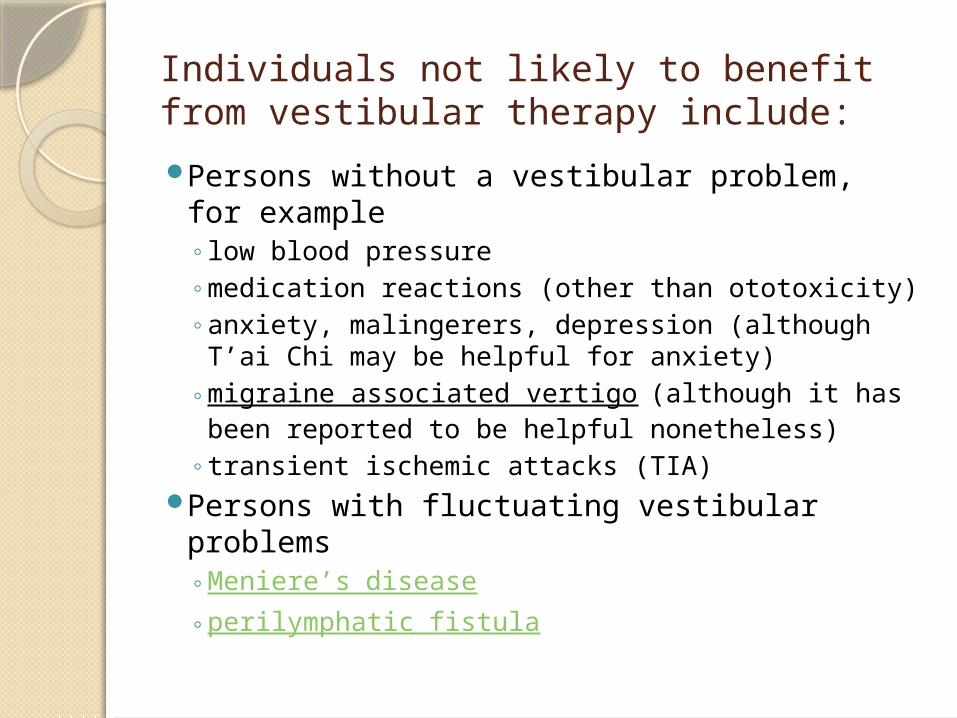
Individuals not likely to benefit from vestibular therapy include:Persons without a vestibular problem, for
example◦low blood pressure◦medication reactions (other than ototoxicity)◦anxiety, malingerers, depression (although T’ai
Chi may be helpful for anxiety)◦migraine associated vertigo (although it has been
reported to be helpful nonetheless)◦transient ischemic attacks (TIA)
Persons with fluctuating vestibular problems◦Meniere’s disease◦perilymphatic fistula

Vestibular compensationVestibular compensation is a process that
allows the brain to regain balance control and minimise dizziness symptoms when there is damage to, or an imbalance between, the right and left vestibular organs (balance organs) in the inner ear. Essentially, the brain copes with the disorientating signals coming from the inner ears by learning to rely more on alternative signals coming from the eyes, ankles, legs and neck to maintain balance.
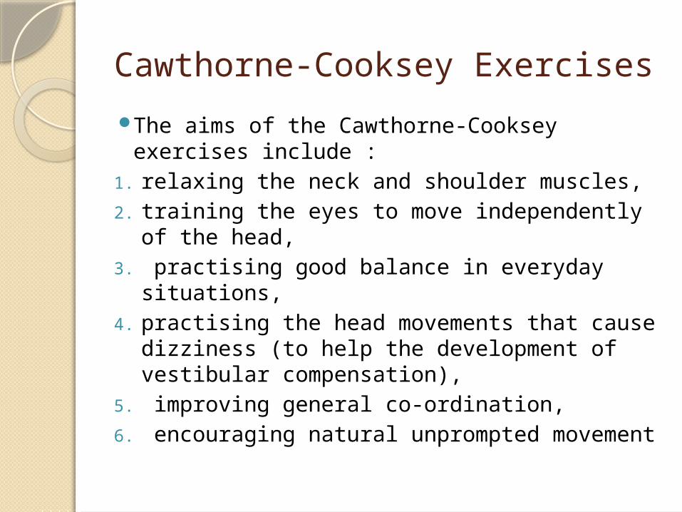
Cawthorne-Cooksey ExercisesThe aims of the Cawthorne-Cooksey exercises
include :1. relaxing the neck and shoulder muscles, 2. training the eyes to move independently of
the head,3. practising good balance in everyday
situations, 4. practising the head movements that cause
dizziness (to help the development of vestibular compensation),
5. improving general co-ordination, 6. encouraging natural unprompted movement
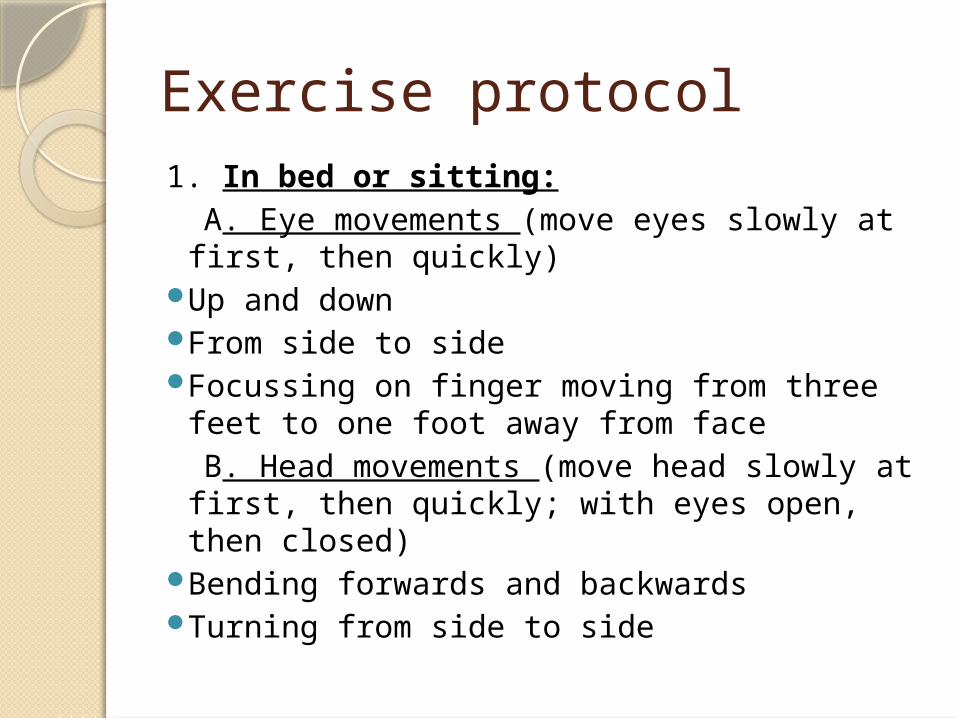
Exercise protocol1. In bed or sitting: A. Eye movements (move eyes slowly at first,
then quickly)Up and downFrom side to sideFocussing on finger moving from three feet to
one foot away from face B. Head movements (move head slowly at
first, then quickly; with eyes open, then closed)
Bending forwards and backwards Turning from side to side

2. Sitting: A. Eye and head movements, as
1 B. Shrug and circle shoulders C. Bend forward and pick up
objects from the ground D. Bend side to side and pick up
objects from the ground

3. Standing: A. Eye, head and shoulder movements, as 1 and
2 B. Change from a sitting to a standing position
with eyes open, then closed (please note this is not advised for the elderly with postural hypertension)
C. Throw a ball from hand to hand above eye level
D. Throw a ball from hand to hand under the knees
E. Change from a sitting to a standing position, turning around in between.

4. Moving about: A. Walk across the room with eyes open B. Walk up and down a slope with eyes
open C. Walk up and down steps with eyes
open D. Throw and catch a ball E. Any game involving stooping,
stretching and aiming (for example, bowls or bowling)

Gaze stabilization exercisesThe aim of gaze stabilization
exercises is to improve vision and the ability to focus on a stationary object while the head is moving.

1. Look straight ahead and focus on a letter (for example, an E) held at eye level in front of you.
2. Move your head from side to side, staying focussed on the target letter. Build up the speed of your head movement. It is crucial that the letter stays in focus. If you get too dizzy, slow down.
3. Try to continue for up to one minute (the brain needs this time in order to adapt). Build up gradually to repeat three to five times a day.

Canalith (or otolith) repositioning procedures (CRP)The aim of Canalith repositioning
procedures (CRP) is to treat people with benign paroxysmal positional vertigo (BPPV) by moving particles or otoliths trapped in the posterior semicircular canals in the inner ear (labyrinth) causing dizziness.
It involves sequential movement of the head into four positions, staying in each position for roughly 30 seconds.


Many cases of Benign Positional Vertigo have their origin in the articular receptors of the cervical spine. Such cases do not respond well to CRP and are better managed by the Brandt-Daroff exercises which activate the cervical-vestibular connections and promote compensation.

Brandt-Daroff exercises 1. Sit on the edge of the bed and turn your head
45 degrees to one side. 2. Quickly lie down on your opposite side (that is,
to the left if you turned your head to the right, and vice versa) so that the back of your head behind your ear touches the bed.
3. Hold this position for about 30 seconds or until the dizziness symptoms stop.
4. Return to the sitting position.
Repeat on the on the other side, alternating until you have completed six repetitions on each side


