Improved processing power & speed Reduction in machine size & footprints Windows-based intuitive software platforms Veterinary Ultrasound & The General Practitioner By: Lorne Caroll DVM Veterinary ultrasound was once considered a diagnostic modality restricted to use in academic institutions and referral centers only. Since its insurrection in the 1970’s, veterinary ultrasound has advanced in a number of ways that now allows for its daily use in general practice. Advances in research and technology, education and competitive pricing have now made ultrasound easier, better and available to all general practitioners. The average pet owner now has expectations that their family veterinarian will have ultrasound diagnostic technology at their disposal should their animal ever require it. Ultrasound is more user friendly and less intimidating and great for use in any general practice technological advances such as: The costs of newer and better veterinary ultrasound machines have become so much more affordable. Features such as, B-mode, M-mode, spectral Doppler with pulse wave and continuous wave and colour that used to be found only in academic and referral facilities are now found on smaller machines with better image quality often at a fraction of the price. The pricing of new ultrasound machines is designed to facilitate its daily use in veterinary practice, augment good veterinary care and generate revenue for the veterinary hospital. Hospitals utilizing ultrasound technology are effectively realizing an economic return similar to or greater than radiology due to ultrasound’s potential broader scope of use. Ultrasound provides valuable diagnostic information with virtually no risk and is non-invasive to the animal. Veterinary staff such as associate veterinarians and registered animal health technologists can receive appropriate training to utilize the benefits of ultrasonography. DECEMBER 2012 Probe frequency ranges & performance 3 and 4 D advanced imaging New modalities-cross beam & speckle reduction
Transcript
Improved processing power & speed
Reduction in machine size & footprints
Windows-based intuitive software platforms
Veterinary Ultrasound & The General Practitioner
By: Lorne Caroll DVM
Veterinary ultrasound was once considered a diagnostic modality restricted to use in academic
institutions and referral centers only. Since its insurrection in the 1970’s, veterinary ultrasound has advanced in a number of ways that now allows for its daily use in general practice. Advances in research and technology, education and competitive pricing have now made ultrasound easier, better and available to all general practitioners.
The average pet owner now has expectations that their family veterinarian will have ultrasound diagnostic technology at their disposal should their animal ever require it. Ultrasound is more user friendly and less intimidating and great for use in any general practice technological advances such as:
The costs of newer and better veterinary ultrasound machines have become so much more affordable. Features such as, B-mode, M-mode, spectral Doppler with pulse wave and continuous wave and colour that used to be found only in academic and referral facilities are now found on smaller machines with better image quality often at a fraction of the price.
The pricing of new ultrasound machines is designed to facilitate its daily use in veterinary practice, augment good veterinary care and generate revenue for the veterinary hospital. Hospitals utilizing ultrasound technology are effectively realizing an economic return similar to or greater than radiology due to ultrasound’s potential broader scope of use. Ultrasound provides valuable diagnostic information with virtually no risk and is non-invasive to the animal.
Veterinary staff such as associate veterinarians and registered animal health technologists can receive appropriate training to utilize the benefits of ultrasonography.
Climbing the Steep Learning Curve Easier and Faster
Veterinarians can now attain high levels of ultrasound training, education and confidence increasing the use of ultrasound in daily practice than ever before. With smaller, improved machines, reference texts, materials and continuing education. The general practitioner can achieve a high level of skill and confidence in a relatively short period of time. Some recommendations to aid veterinarians to become skilled sonographers more easily and quickly are:
1. Purchase the latest and most user friendly ultrasound machine with appropriate features and probe frequency range to perform the studies you are in (ie large or small dog/cat or abdominal versus cardiac)
2. Keep the ultrasound machine accessible to minimize efforts for its use. If the ultrasound machine is tucked away in a corner of the hospital gathering dust…
3. Familiarize yourself with the ultrasound features and navigate through the windows and menus options
4. Have good reference texts and images readily available. Some veterinarians will keep these right on their ultrasound machine
5. Take as many CE courses as possible scil Vet Novations Education Schedule
6. Use the ultrasound daily on all elective procedures such as spays to quickly bring familiarity of normal anatomy, organ location, tissue interfacing and echogenicity
"What is your diagnosis?"
Case Presentation:
10 year male, neutered Cocker Spaniel with coughing and lethargy.
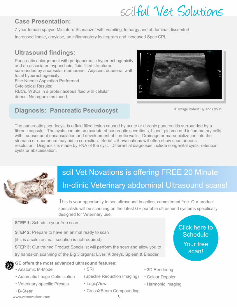
7 year female spayed Miniature Schnauzer with vomiting, lethargy and abdominal discomfort
Increased lipase, amylase, an inflammatory leukogram and increased Spec CPL
Ultrasound findings:
Pancreatic enlargement with peripancreatic hyper echogenicity and an associated hypoechoic, fluid filled structured surrounded by a capsular membrane. Adjacent duodenal wall focal hyperechogenicity.
Fine Needle Aspiration Performed
Cytological Results:
RBCs, WBCs in a proteinaceous fluid with cellular
debris. No organisms found.
Diagnosis: Pancreatic Pseudocyst
The pancreatic pseudocyst is a fluid filled lesion caused by acute or chronic pancreatitis surrounded by a fibrous capsule. The cysts contain an exudate of pancreatic secretions, blood, plasma and inflammatory cells with subsequent encapsulation and development of fibrotic walls. Drainage or marsupialization into the stomach or duodenum may aid in correction. Serial US evaluations will often show spontaneous resolution. Diagnosis is made by FNA of the cyst. Differential diagnoses include congenital cysts, retention cysts or abscessation.
This is your opportunity to see ultrasound in action, commitment free. Our product
specialists will be scanning on the latest GE portable ultrasound systems specifically
designed for Veterinary use.
STEP 1: Schedule your free scan
STEP 2: Prepare to have an animal ready to scan
(if it is a calm animal, sedation is not required)
STEP 3: Our trained Product Specialist will perform the scan and allow you to
try hands-on scanning of the Big 5 organs: Liver, Kidneys, Spleen & Bladder
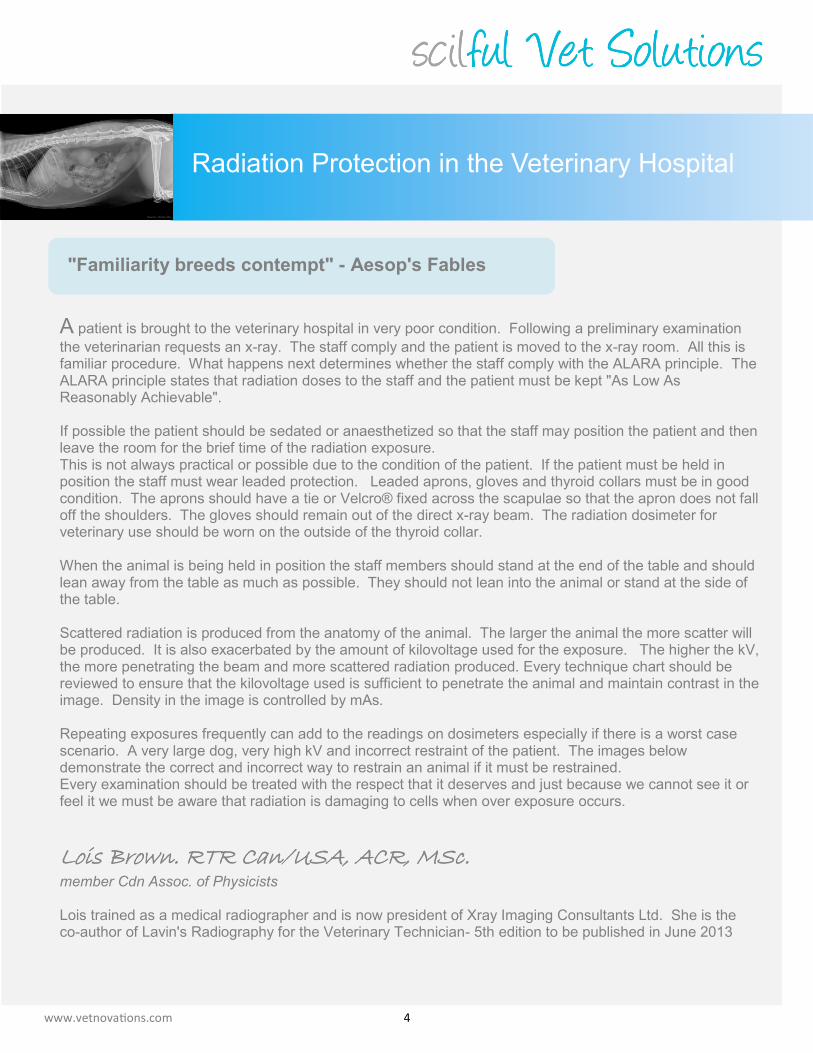
A patient is brought to the veterinary hospital in very poor condition. Following a preliminary examination
the veterinarian requests an x-ray. The staff comply and the patient is moved to the x-ray room. All this is familiar procedure. What happens next determines whether the staff comply with the ALARA principle. The ALARA principle states that radiation doses to the staff and the patient must be kept "As Low As Reasonably Achievable". If possible the patient should be sedated or anaesthetized so that the staff may position the patient and then leave the room for the brief time of the radiation exposure. This is not always practical or possible due to the condition of the patient. If the patient must be held in position the staff must wear leaded protection. Leaded aprons, gloves and thyroid collars must be in good condition. The aprons should have a tie or Velcro® fixed across the scapulae so that the apron does not fall off the shoulders. The gloves should remain out of the direct x-ray beam. The radiation dosimeter for veterinary use should be worn on the outside of the thyroid collar. When the animal is being held in position the staff members should stand at the end of the table and should lean away from the table as much as possible. They should not lean into the animal or stand at the side of the table. Scattered radiation is produced from the anatomy of the animal. The larger the animal the more scatter will be produced. It is also exacerbated by the amount of kilovoltage used for the exposure. The higher the kV, the more penetrating the beam and more scattered radiation produced. Every technique chart should be reviewed to ensure that the kilovoltage used is sufficient to penetrate the animal and maintain contrast in the image. Density in the image is controlled by mAs. Repeating exposures frequently can add to the readings on dosimeters especially if there is a worst case scenario. A very large dog, very high kV and incorrect restraint of the patient. The images below demonstrate the correct and incorrect way to restrain an animal if it must be restrained. Every examination should be treated with the respect that it deserves and just because we cannot see it or feel it we must be aware that radiation is damaging to cells when over exposure occurs.
Lois Brown. RTR Can/USA, ACR, MSc. member Cdn Assoc. of Physicists Lois trained as a medical radiographer and is now president of Xray Imaging Consultants Ltd. She is the co-author of Lavin's Radiography for the Veterinary Technician- 5th edition to be published in June 2013
This year instead of sending Christmas Cards to our customers and partners, scil Vet Novations is making a
donation to Precious Paws Rescue. Precious Paws Rescue is a non-profit rescue organization who is dedicated to saving the lives of homeless pets by rescuing them from pounds and shelters across North America where they are at risk of euthanasia. This will help to reduce paper waste and redirect funds to an organization that makes a positive difference in the world. If you would like to join us in making a donation please visit http://www.preciouspawsrescue.ca/
We wish you and your family a safe
and happy holiday season!
An environmentally friendly way to say
“Happy Holidays”
www.vetnovations.com
Can you spot the eight common technician errors in this photo?
1. Aprons worn incorrectly
(falling off shoulders) 2. No leaded gloves 3. Technician at side of table 4. Technician leaning into table 5. Technician hand on table 6. No collimation 7. Dosimeter on sleeve 8. No leaded eye glasses
Rules your clinic should live by 1. Never lean over the table when the exposure is taken. 2. Always stand at the end of the table, lean away from the table and turn your head to the side when the
exposure is made. 3. Leaded gloves must be worn when restraining an animal. 4. Leaded eyewear should be worn in the x-ray room. 5. The x-ray beam must be collimated to the anatomy within the field of view. If the animal is very large a
piece of leaded rubber should be placed along the table in front of the animal to prevent scattered radia-tion from affecting the image.
6. Dosimeter must be worn outside the apron attached to the thyroid collar. Thank you to the technicians and the beagle who correctly posed incorrectly for this photo!