Grant, I. R., Foddai, A. C. G., Tarrant, J. C., Kunkel, B., Hartmann, F. A., McGuirk, S., Hansen, C., Talaat, A. M.,& Collins, M. T. (2017). Viable Mycobacterium avium subsp. paratuberculosis isolated from calf milk replacer.Journal of Dairy Science, 100(12), 9723-9735. https://doi.org/10.3168/jds.2017-13154
Published in:Journal of Dairy Science
Document Version:Publisher's PDF, also known as Version of record
Queen's University Belfast - Research Portal:Link to publication record in Queen's University Belfast Research Portal
General rightsCopyright for the publications made accessible via the Queen's University Belfast Research Portal is retained by the author(s) and / or othercopyright owners and it is a condition of accessing these publications that users recognise and abide by the legal requirements associatedwith these rights.
Take down policyThe Research Portal is Queen's institutional repository that provides access to Queen's research output. Every effort has been made toensure that content in the Research Portal does not infringe any person's rights, or applicable UK laws. If you discover content in theResearch Portal that you believe breaches copyright or violates any law, please contact [email protected].
When advising farmers on how to control Johne’s disease in an infected herd, one of the main recom-mendations is to avoid feeding waste milk to calves and instead feed calf milk replacer (CMR). This advice is based on the assumption that CMR is free of viable Mycobacterium avium ssp. paratuberculosis (MAP) cells, an assumption that has not previously been chal-lenged. We tested commercial CMR products (n = 83) obtained from dairy farms around the United States by the peptide-mediated magnetic separation (PMS)-phage assay, PMS followed by liquid culture (PMS-culture), and direct IS900 quantitative PCR (qPCR). Conven-tional microbiological analyses for total mesophilic bac-terial counts, coliforms, Salmonella, coagulase-negative staphylococci, streptococci, nonhemolytic Corynebacte-rium spp., and Bacillus spp. were also performed to assess the overall microbiological quality of the CMR. Twenty-six (31.3%) of the 83 CMR samples showed evidence of the presence of MAP. Seventeen (20.5%) tested positive for viable MAP by the PMS-phage as-say, with plaque counts ranging from 6 to 1,212 pfu/50 mL of reconstituted CMR (average 248.5 pfu/50 mL). Twelve (14.5%) CMR samples tested positive for vi-able MAP by PMS-culture; isolates from all 12 of these samples were subsequently confirmed by whole-genome sequencing to be different cattle strains of MAP. Seven (8.4%) CMR samples tested positive for MAP DNA by IS900 qPCR. Four CMR samples tested positive by both PMS-based tests and 5 CMR samples tested positive by IS900 qPCR plus one or other of the PMS-based tests, but only one CMR sample tested positive by all 3 MAP detection tests applied. All conventional microbiology results were within current standards for
whole milk powders. A significant association existed between higher total bacterial counts and presence of viable MAP indicated by either of the PMS-based as-says. This represents the first published report of the isolation of viable MAP from CMR. Our findings raise concerns about the potential ability of MAP to survive manufacture of dried milk-based products.Key words: Mycobacterium avium ssp. paratuberculosis, milk replacer, calf health, Johne’s disease, infectious disease control
INTRODUCTION
Milk replacer has been fed to calves since at least the 1950s, although formulations have changed over the years in terms of percentage of fat and protein. Calf milk replacers (CMR) are generally made with by-products originating from milk processing industries, such as whole milk powder, skim milk powder, casein, whey, and whey protein, although protein sources other than milk by-products such as soy products, dried egg, fish protein concentrates, and single cell protein may also be used (FAO, 2011; Bovine Alliance on Management and Nutrition, 2014). Most dairy calves in the United States are fed milk replacer before weaning for reasons of convenience, biosecurity, and economics (Costello, 2012; Bovine Alliance on Management and Nutrition, 2014). Calves are particularly susceptible to infectious diseases, and some infectious agents such as Mycobac-terium avium ssp. paratuberculosis (MAP), the cause of Johne’s disease (JD), bovine viral diarrhea virus, bovine leukosis virus, Pasteurella multocida, Salmonella sp., and Mycoplasma bovis can be transmitted from cow to calf through feeding unpasteurized milk (Costello, 2012). Feeding CMR, as an alternative to feeding waste unpasteurized milk or farm-pasteurized milk, is a com-mon practice in the United States. The latest statistics from the National Herd Monitoring Scheme indicate that 49.9% of all US dairy operations (of all sizes) fed
Viable Mycobacterium avium ssp. paratuberculosis isolated from calf milk replacerIrene R. Grant,*1 Antonio C. G. Foddai,* James C. Tarrant,† Brenna Kunkel,† Faye A. Hartmann,‡ Sheila McGuirk,§ Chungyi Hansen,† Adel M. Talaat,† and Michael T. Collins†*Institute for Global Food Security, School of Biological Sciences, Queen’s University Belfast, Belfast, Northern Ireland, United Kingdom BT9 7BL†Department of Pathobiological Sciences, and‡Clinical Microbiology Laboratory, University of Wisconsin Veterinary Care, and§Department of Medical Sciences, School of Veterinary Medicine, University of Wisconsin, Madison 53706
Received May 26, 2017.Accepted August 19, 2017.1 Corresponding author: [email protected]
9724 GRANT ET AL.
Journal of Dairy Science Vol. 100 No. 12, 2017
some kind of CMR to pre-weaned heifers during 2014; 16.4% of operations fed nonmedicated CMR and 37.6% fed medicated CMR (USDA, 2016).
As mentioned above, calves may be fed CMR to pre-vent diseases such as JD, caused by MAP, which is shed in the milk and feces of infected cows. Transmission of MAP is considered to be an early event in a calf’s life, and there are recognized risk factors for transmission of MAP to calves within dairy herds (Doré et al., 2012). A focus of JD control programs is calf-related inter-ventions as part of herd management plans. Avoiding feeding waste milk and feeding CMR instead is a key recommendation within JD control programs worldwide (Doré et al., 2012; Garcia and Shalloo, 2015; Pieper et al., 2015). As stated by Cooper and Watson (2013), the assumption has always been that the risk of viable MAP organisms in commercial CMR powders is negli-gible because CMR is invariably pasteurized and often highly processed, but is that really the case? Seemingly, to date, no one has ever challenged that assumption.
Demonstrating the existence of viable MAP in pro-cessed milk or dairy products, such as pasteurized milk, cheeses, yogurt, milk powders, and powdered infant for-mula, has proven difficult because, until recently, cul-ture always necessitated inclusion of a chemical decon-tamination step to inactivate non-MAP contaminants; the latter is known to adversely affect the viability of some or all of the MAP cells present in milk, poten-tially leading to negative culture results (Dundee et al., 2001; Gao et al., 2005). However, detection methods for viable MAP in milk and dairy products have improved considerably over recent years with the advent of im-munomagnetic separation (Grant et al., 1998; O’Brien et al., 2016) and subsequently peptide-mediated mag-netic separation (PMS; Stratmann et al., 2002, 2006; Foddai et al., 2010; O’Brien et al., 2016), which permit selective capture, separation, and concentration of whole MAP cells from a sample before culture or PCR, and novel mycobacteriophage-based methods of MAP detection (Stanley et al., 2007; Foddai et al., 2010; Swift et al., 2013; Botsaris et al., 2016), which require the MAP cells to be viable to obtain a positive result (plaques in a lawn of fast-growing Mycobacterium smeg-matis). In particular, a method combining PMS and a phage amplification assay to detect MAP (PMS-phage assay), developed and optimized by Foddai et al. (2009, 2011), and used in combination with an optimized milk sample preparation protocol (Foddai and Grant, 2015), is proving to be a very sensitive method of detecting viable MAP in cow milk. The optimized PMS-phage assay was recently reported to have a limit of detection 50% of ~1 MAP cell per 50 mL of milk, making it a more sensitive detection method than existing quanti-
tative PCR for MAP and conventional culture methods (Foddai and Grant, 2017).
As time passes and the novel optimized PMS-phage assay is applied to test various milk and dairy products, new information on the presence and numbers of viable MAP in these foods is emerging (Foddai and Grant, 2017). We previously reported the outcome of testing of powdered infant milk formula (PIF) by the PMS-phage assay (Grant et al., 2014). Of 68 PIF samples tested, 30 (44.1%) samples tested positive for viable MAP by the PMS-phage assay, with viable MAP numbers ranging from 4 to 678 pfu/50 mL of reconstituted PIF indi-cated by the plaque counts obtained. Because PIF and CMR are similar milk-based, powdered dairy products, probably with not dissimilar production processes, our viable MAP in PIF findings led us to query whether testing of CMR by the PMS-phage assay might also yield similar results with respect to the presence of vi-able MAP. Preliminary testing of a small number of CMR samples sourced in Wisconsin by the PMS-phage assay (carried out before the CMR testing reported here) found that 1 (12.5%) of 8 CMR samples tested positive for viable MAP. We hypothesized that viable MAP may be more widely prevalent in commercial powdered CMR products, so we decided to carry out a larger study. The objectives of the study were (1) to test commercial CMR products sourced from within the United States using standard culture methods, 2 PMS-based methods (PMS-phage assay and PMS plus liquid culture) and IS900 quantitative PCR (qPCR) to detect the presence of viable MAP and MAP DNA, respectively, and (2) to assess the overall hygienic qual-ity of the CMR samples by performing conventional microbiological analyses, to determine if the presence of any hygiene indicator microorganism might correlate with detection of viable MAP. An optimized method for detecting MAP in powdered dairy products has yet to be published, so during this study multiple methods, including several published and unpublished cultural and qPCR approaches (detailed below), were employed in the 2 CMR testing laboratories to maximize chances of detecting low numbers of viable MAP, if present, in the CMR samples.
MATERIALS AND METHODS
Acquisition of CMR Samples
The CMR samples were acquired in 2 stages. In the first stage, 50 samples were acquired during the sum-mer of 2014 from dairy farms in southern Wisconsin by author Tarrant. Hygiene precautions were taken to avoid on-farm contamination. Samples were col-
lected with a sterile plastic scoop into sterile transport bags, with a separate sterile scoop being used for each sample. If a sealed bag of CMR was available on the farm, that bag was used for sampling; the majority of the 50 Wisconsin-sourced CMR samples were obtained from unopened bags of CMR (James Tarrant, Univer-sity of Wisconsin–Madison, personal communication). Subsequently, during the spring of 2015, an additional 35 CMR samples were acquired from across the United States. For CMR collection outside Wisconsin, sample collection kits, including sterile scoops and bags, were mailed to veterinarians and veterinary students with detailed instructions about hygienic sampling technique and a request to use a sealed CMR bag if possible; all except 2 of the 35 CMR samples obtained in other US states were recorded as being from unopened bags. The 83 CMR samples ultimately tested originated from 15 US states and included 35 different brands of CMR representing 6 unique manufacturers.
CMR Sample Reconstitution
Unless otherwise stated, 6 to 9 g of each CMR sample (as indicated by the manufacturer’s label) was asep-tically weighed into a sterile 50-mL centrifuge tube, reconstituted to 50 mL with pre-warmed (37°C) sterile distilled water, shaken thoroughly for several minutes to ensure resuspension of all powder, and then placed at 4°C overnight to fully rehydrate. Next day, the re-constituted CMR samples were removed from the fridge and allowed to equilibrate to room temperature for at least 1 h before being centrifuged at 3,000 × g for 15 min at room temperature. Each CMR pellet was then resuspended in 1 to 5 mL of PBS (pH 7.4) containing 0.05% Tween 20 (PBS-T), as appropriate depend-ing on the size of pellet obtained, to obtain what was considered to be a suitable consistency for PMS. The volume of PBS-T added was recorded so that a correc-tion factor could be applied to plaque counts obtained when 1 mL of each CMR pellet sample was subjected to the PMS-phage assay. Finally, the reconstituted CMR pellet samples were ultrasonicated (pulse mode 37 kHz for 4 min in ice water) in an Ultrasonic bath (FB-11201, Fisher Scientific Ltd., Loughborough, UK) to break up any MAP clumps in the sample before PMS was performed. Only one sample of each CMR was tested by each of the methods described below, so intra- or inter-laboratory variation for the methods employed was not evaluated during this study.
PMS of MAP from Reconstituted CMR
The PMS was performed on 1 mL of each sample (prepared as indicated above) using 5 µL of biotinyl-
ated-aMp3 peptide- and 5 µL of biotinylated-aMptD peptide-coated MyOne Tosylactivated Dynabeads (Life Technologies, Paisley, Scotland, UK), prepared in-house as previously described (Foddai et al., 2010). A positive (1 mL of a 10−1 dilution of a broth culture of MAP B2) and negative (1 mL of 7H9 broth) control sample was included with each batch of 15 to 20 CMR samples processed. Magnetic separation was carried out using a Dynal BeadRetriever (Life Technologies). The MAP cell capture was carried out for 30 min at room temperature under continuous mixing, followed by 2 washes in 1 mL of PBS-T, and final resuspension of the beads in 1 mL of Middlebrook 7H9 broth contain-ing 10% (vol/vol) OADC supplement (7H9/OADC broth) and NOA antibiotic supplement (Abtek Bio-logicals Ltd., Liverpool, UK; final concentrations per liter: 50,000 IU of nystatin, 2 mg of oxacillin, and 30 mg of aztreonam). This final 1-mL bead sample was split equally between the phage assay and culture, carried out as described below. The positive and negative PMS controls were also processed through the phage assay (PMS-phage assay) and culture (PMS-culture) along with each batch of test samples.
PMS-Phage Assay
The PMS-phage assay and confirmatory plaque-PCR were carried out as follows: after PMS, 500 µL of each bead sample was transferred to a 11-mL flip-top vial (Capitol Vials, Auburn, AL) containing 500 µL of 7H9/OADC/4 mM CaCl2/NOA antibiotics (final CaCl2 con-centration in sample, 2 mM) before being incubated overnight at 37°C. The phage assay was carried out as described by Foddai et al. (2009). Briefly, 100 µL of D29 mycobacteriophage suspension (109 pfu/mL) was added to each bead sample, before incubation for 2 h at 37°C. Then, to inactivate exogenous/nonadsorbed seed phage, 100 µL of freshly prepared 100 mM ferrous ammonium sulfate (Sigma-Aldrich, Poole, Dorset, UK) was added to each sample and allowed to incubate for 10 min at room temperature, with thorough vortexing of the sample after 5 min, before the addition of 5 mL of 7H9/OADC/2 mM CaCl2/NOA broth. Samples were returned to the incubator at 37°C for a further 90 min before being plated with tempered (55°C) Middlebrook 7H9 agar containing 10% OADC (both Difco) and 1 mL Mycobacterium smegmatis mc2 155 (108 cfu/mL). Plaques were counted following overnight incubation of plates at 37°C. Plaque counts obtained were expressed as plaque-forming units per 50 mL of reconstituted CMR; direct plaque counts were multiplied by a fac-tor of 2 to take account of the fact that only half of the bead sample (equivalent of 25 mL of reconstituted CMR) was processed through the phage assay, and by
9726 GRANT ET AL.
Journal of Dairy Science Vol. 100 No. 12, 2017
a factor of 1, 3, or 5 to take account of the differing volumes of PBS-T originally added to resuspend the CMR pellets.
Plaque-IS900 PCR, essentially as described by Swift et al. (2013), was carried out on DNA extracted from plaques to confirm that the DNA present was MAP DNA and not from some other Mycobacterium sp. Up to a maximum of 10 plaques were randomly selected from each PMS-phage assay positive sample plate to be excised for DNA extraction. The center of each plaque was excised using a sterile loop and transferred to an Eppendorf tube. The DNA was extracted from the plaques using the Zymoclean Gel DNA Recovery kit (Cambridge Bioscience Ltd., Cambridge, UK), ac-cording to the manufacturer’s instructions. The DNA was eluted from the Zymoclean columns using 20 µL of elution buffer (supplied with kit). The DNA was stored at −20°C until required for plaque-PCR. A protocol modified from Whittington et al. (1998) was used to target the IS900 insertion element. To 40 µL of master mix containing 1 × DreamTaq Green Buffer, 2.5 mM MgCl2, 200 µM of each dNTP, 1 U Fermentas DreamTaq DNA polymerase (Thermo Fisher Scientific, UK), and 250 ng of P90 5′GAAGGGTGTTCGGGGC-CGTCGCTTAGG′3 and P91 5′GGCGTTGAGGTC-GATCGCCC ACGTGAC′3 primers, 10 µL of plaque DNA was added. The PCR cycling conditions were 94°C for 5 min, 37 cycles of 94°C for 30 s, 62°C for 30 s and 72°C for 1 min, final extension at 72°C for 4 min, and then the sample was cooled to 4°C. The PCR products were visualized by 2% agarose gel electrophoresis. The expected IS900 PCR product size was 400 bp.
PMS Culture at Queen’s University Belfast
The PMS-culture and confirmation of suspected posi-tive cultures was carried out as follows: 500 µL of each bead suspension after PMS was inoculated into screw-capped glass test tubes containing 5 mL of modified Middlebrook 7H9 liquid medium (Pozzato et al., 2011), consisting of (per 900 mL) 4.7 g of Middlebrook 7H9 powder, 1.0 g of casitone, 5 mL of glycerol supplement-ed with 10% vol/vol OADC supplement and PANTA plus antibiotic supplement (all Becton Dickinson) and mycobactin J (2 mg/L; Synbiotics Europe SAS, Lyon, France), but without the addition of 16% egg yolk. The broth cultures were incubated at 37°C and absorbance at optical density at 600 nm (OD600nm) was measured at time 0 and then from 4 wk onward every 2 wk up to 16 wk using a Biowave CO8000 Density meter (Biochrom Ltd., Cambridge, UK). When an increase in OD600nm was observed for any broth culture, Ziehl-Neelsen (ZN) staining was employed to determine if acid-fast bacteria were present, and if so, IS900 PCR (Moss et al., 1992)
was applied to confirm presence of MAP cells. Any pri-mary PMS-culture suspected of containing viable MAP was sub-cultured to Dubos broth medium, prepared as described by Hammer et al. (2002) including the addi-tion of 20% Newborn Calf Serum (Life Technologies), PACT antibiotics (50 IU/mL of polymyxin B, 5 µg/mL of amphotericin B, 25 µg/mL of carbenicillin, and 2.5 µg/mL of trimethoprim, all Sigma) and mycobactin J (2 mg/L, Synbiotics Europe SAS), and onto Herrold’s egg yolk medium (HEYM) containing 2 mg/L of my-cobactin J (prepared in-house). Once again, OD600nm of broth cultures was measured periodically until an increase was observed, at which point IS900 PCR was applied to confirm the presence of MAP DNA. IS900 PCR was also applied to suspect colonies growing on HEYM plates to confirm their identity.
PMS Culture at University of Wisconsin–Madison
Reconstituted CMR was processed by PMS using the KingFisher Duo Prime Purification System (Thermo-Fisher Scientific, Fitchburg, WI) to concentrate MAP and then inoculated into MGIT ParaTB medium with 1.0 mL of MGIT ParaTB supplement (which contains mycobactin J), 500 µL of egg yolk emulsion and 200 µL of MGIT PANTA added, and also onto HEY slants (all culture media and supplements from BD Diagnostic Systems, Sparks, MD). For one set of MGIT ParaTB medium cultures, the CMR was reconstituted as previ-ously described (as per manufacturer label instructions; 6–9 g/mL). For the other cultures, all CMR samples were reconstituted at a standard 5 g/50 mL and al-lowed to rehydrate before testing commenced.
If suspicious colonies were seen on HEY or if the MGIT 960 instrument was signal-positive, a 5-target multiplex PCR was performed to establish if MAP was present (Shin et al., 2010). The nature of the CMR ma-terial inoculated onto solid media tended to resemble bacterial growth, causing many cultures to be unnec-essarily tested. In total 149 cultures (including both PMS cultures and conventional cultures) were tested by multiplex PCR.
Conventional Culture for MAP at University of Wisconsin–Madison
The sample processing method described by Botsaris et al. (2016) in a study of powdered infant formula was adapted and used with 3 different culture media. The CMR was first reconstituted in PBS pH 7.2 at 1.0 g/5 mL, allowed to sit at 20°C for 1 h, centrifuged at 2,500 × g for 15 min at room temperature. The pellet was resuspended in 0.75% hexadecylpyridinium chloride (HPC) and allowed to sit for 4 h at 20°C. After cen-
trifugation at 2,500 × g for 15 min at room temperature the pellet was resuspended in 1.0 mL of sterile water. Three media were inoculated: 200 µL onto HEY slants (BD Diagnostic Systems); 100 µL into MGIT ParaTB medium (BD Diagnostic Systems) supplemented with mycobactin J, egg yolk, and PANTA; and 100 µL onto modified 7H10 agar plates supplemented with myco-bactin-J, ADC, and VAN. Suspicious colonies on solid culture media (HEY or 7H10) and signal-positive MGIT cultures were tested by multiplex PCR, as described above for PMS-cultures, to establish if MAP had been isolated. The nature of the CMR material inoculated onto solid media tended to resemble bacterial growth causing many cultures to be unnecessarily tested.
IS900 qPCR at University of Wisconsin–Madison
Three method variations were used to test CMR samples for presence of MAP by IS900 qPCR.
Method 1. For the first qPCR method, the CMR was reconstituted at 5 g/50 mL in sterile water, then held at 4°C for 18 to 24 h before centrifugation at 2,500 × g for 15 min. The pellet was resuspended in 3.0 mL of sterile PBS containing 0.05% Tween 20. The DNA was extracted from 1.0 mL of this suspension using the Tetracore Extraction System (Tetracore, Rock-ville, MD) using the 1.0-g protocol after agitation in the Benchmark BeadBlaster 24 (Benchmark Scientific, Inc., Edison, NJ). From the 25-µL extraction product, 5 µL was used as template DNA for IS900 qPCR.
Method 2. For the second qPCR method, CMR was first reconstituted in PBS at 1.0 g/5 mL, allowed to sit at 20°C for 1 h, centrifuged at 2,500 × g for 15 min. The pellet was resuspended in 0.75% HPC and allowed to sit for 4 h at 20°C. After centrifugation at 2,500 × g for 15 min the pellet was resuspended in 1.0 mL of sterile water. The DNA was extracted from this suspension using the Tetracore Extraction System (Tetracore) using the 1.0-g protocol after agitation in the Benchmark BeadBlaster 24 (Benchmark Scientific). From the 25-µL extraction product, 5 µL was used as template DNA for IS900 qPCR.
Method 3. For the third qPCR method, the CMR was reconstituted at 1.0 g/9 mL, then held at 20°C for 1 h, then overnight at 4°C for 18 to 24 h. After centrifugation at 2,500 × g for 15 min, the pellet was reconstituted in 1.0 mL of sterile Tris-EDTA buffer pH 8.0. The DNA was extracted from this suspension us-ing the Tetracore Extraction System (Tetracore) using the 1.0-g protocol after agitation in the Benchmark BeadBlaster 24 (Benchmark Scientific). From the 25-µL extraction product, 5 µL was used as template DNA for IS900 qPCR.
Conventional Microbiological Analyses at University of Wisconsin–Madison
All CMR samples were reconstituted as per manu-facturer label instructions: 6 to 9 g/50 mL. The result-ing milk sample was then tested by the same methods used to characterize the microbiological quality of bulk tank milk. Freshly reconstituted CMR was tested at the following 4 final dilutions: 1:50, 1:500, 1:5,000, and 1:50,000. Heat-stressed reconstituted milk re-placer (37°C for 6 h) was tested at the same dilutions with the addition of 2 higher dilutions to anticipate the higher microbial counts: 1:100,000 and 1:500,000. Final dilutions were achieved by inoculating 0.2 mL of each primary dilution onto Trypticase soy agar (BAP) supplemented with 5% sheep blood (Remel Inc., Lenexa, KS) for determination of microorganism growth and total bacterial counts and eosin methylene blue agar (Remel Inc.) to quantify lactose fermenters (coliforms) and noncoliform, gram-negative bacteria. Culture plates were incubated at 36°C in 5% CO2 and examined for growth after 24, 48, and 36 h of incu-bation. Each dilution was also inoculated onto XLT4 agar (Remel Inc.), incubated in ambient air at 35°C and examined for growth of Salmonella after 24, 48, and 36 h of incubation. In addition, enrichment culture for Salmonella was performed to increase sensitivity of Salmonella detection. Undiluted reconstituted CMR (0.2 mL) was inoculated into selenite and Rappaport-Vassiliadis enrichment broths (Remel Inc.). Enrichment broths were incubated for 18 h in ambient air at 35°C and then subcultured onto XLT4 agar (Remel Inc.). Subcultures were incubated in ambient air at 35°C and examined for colonies resembling Salmonella after 24 and 48 h of incubation.
Colony counts were recorded on the last day of in-cubation. All colony types were identified and classi-fied into the following groups: coliform or noncoliform, gram-negative rods (lactose positive or negative on eosin methylene blue, respectively), streptococci (aga-lactiae or non-agalactiae), staphylococci (Staph. aureus or CNS), Corynebacterium spp., Bacillus spp., or other microorganism using standard microbiological proce-dures (Hogan et al., 1999). Other microorganisms or species level identifications were performed by MALDI-TOF MS (Bruker Daltonics, Bremen, Germany).
Genomic Analysis of MAP Isolates
When MAP isolates were successfully cultured at Queen’s University Belfast (QUB), genomic DNA was extracted from cells grown in Dubos broth according to a method supplied by Adel Talaat, University of
9728 GRANT ET AL.
Journal of Dairy Science Vol. 100 No. 12, 2017
Wisconsin–Madison (Hsu et al., 2011). Briefly, this involved heating at 80°C for 20 min to kill the myco-bacterial cells, lysozyme, and proteinase K treatments to weaken the cell wall, and addition of 5 M NaCl and cetrimonium bromide/NaCl to lyse the MAP cells. This was followed by (1) phenol/chloroform/isoamyl alcohol (24:24:1) extraction, (2) chloroform/isoamyl alcohol (24:1) extraction, (3) precipitation of DNA with isopro-panol overnight, (4) washing with 70% ethanol, and (5) resuspension of DNA pellet in 30 µL of sterile molecular grade water. The purity and yield of DNA from each suspect MAP isolate was provisionally checked using a Biophotometer (Eppendorf, Hamburg, Germany) be-fore the DNA samples were sent to the DNA Sequencing Facility, University of Wisconsin Biotechnology Center (Madison, WI). All DNA samples were sequenced using Illumina Miseq platform and were run in paired-end with 250 bp for each read. Raw sequencing files were imported to CLCBio Genomic workbench version 8 for reference assembly using MAP K10 (NC_002944). Single nucleotide polymorphisms, multiple nucleotide variant, and whole genome insertions/deletions were also analyzed using CLCBio software. The criteria for variant calling included that a variant has ≥20 times sequence coverage where this variant is present in ≥50% of the sequence reads. The consensus sequence of each sample was then extracted from CLCBio software and used to build a phylogenetic tree using HarvestTools (Treangen et al., 2014). Finally, to distinguish MAP genotypes (type I vs. II or III), hsp65, gyrA, and gyrB PCR followed by enzyme digestion were performed as detailed previously by Castellanos et al. (2007) and Ghosh et al. (2012).
Statistical Analysis
The microbiological quality of CMR was compared for CMR samples that tested positive for viable MAP by phage assay or culture and those that did not, using the Mann-Whitney test with GraphPad Prism version 5.00 for Windows (GraphPad Software, San Diego, CA); a difference with P-value ≤0.05 was considered significant at the 95% confidence level.
RESULTS
PMS-Phage Assay (QUB)
Thirty-three (39.8%) of the 83 CMR samples tested by the PMS-phage assay yielded plaques, in which the presence of MAP DNA was detected by plaque-PCR in 17 cases (Table 1); thus, the presence of viable MAP was confirmed in 17 (20.5%) of the 83 CMR samples tested. Viable MAP numbers, indicated by plaque num-
bers in confirmed PMS-phage assay positive samples, ranged from 6 to 1,212 pfu/50 mL of reconstituted CMR (mean 248.5 pfu/50 mL).
PMS Culture (QUB) or Conventional MAP Culture (University of Wisconsin–Madison)
At QUB, primary liquid cultures of 29 of the 83 CMR samples in modified Middlebrook 7H9 medium (Poz-zato et al., 2011) without egg yolk demonstrated an increase in OD600nm of 0.1 to 0.7 units within the 16-wk incubation period. When ZN stained, 18 of these cul-tures were observed to have acid-fast cells present, so they were deemed suspect positive cultures. When the 18 suspect positive primary broth cultures were sub-cultured to Dubos medium and HEYM, 12 continued to show evidence of growth (an increase in OD600nm) in liquid culture or appearance of suspect colonies typical of MAP after 4 to 6 wk on HEYM. All 12 suspect cultures tested IS900 PCR so were provisionally identi-fied as MAP. Ultimately, pure Dubos broth cultures of viable MAP were obtained from 12 (14.5%) of the 83 CMR samples originally tested. The DNA was subse-quently extracted from these pure broth cultures and sent to University of Wisconsin–Madison (UW) for whole-genome sequencing.
At UW, CMR samples were cultured after PMS or conventional MAP culture protocols involving HPC decontamination in MGIT ParaTB medium supple-mented with egg yolk. None of the UW cultures were positive for viable MAP.
IS900 qPCR for MAP (UW)
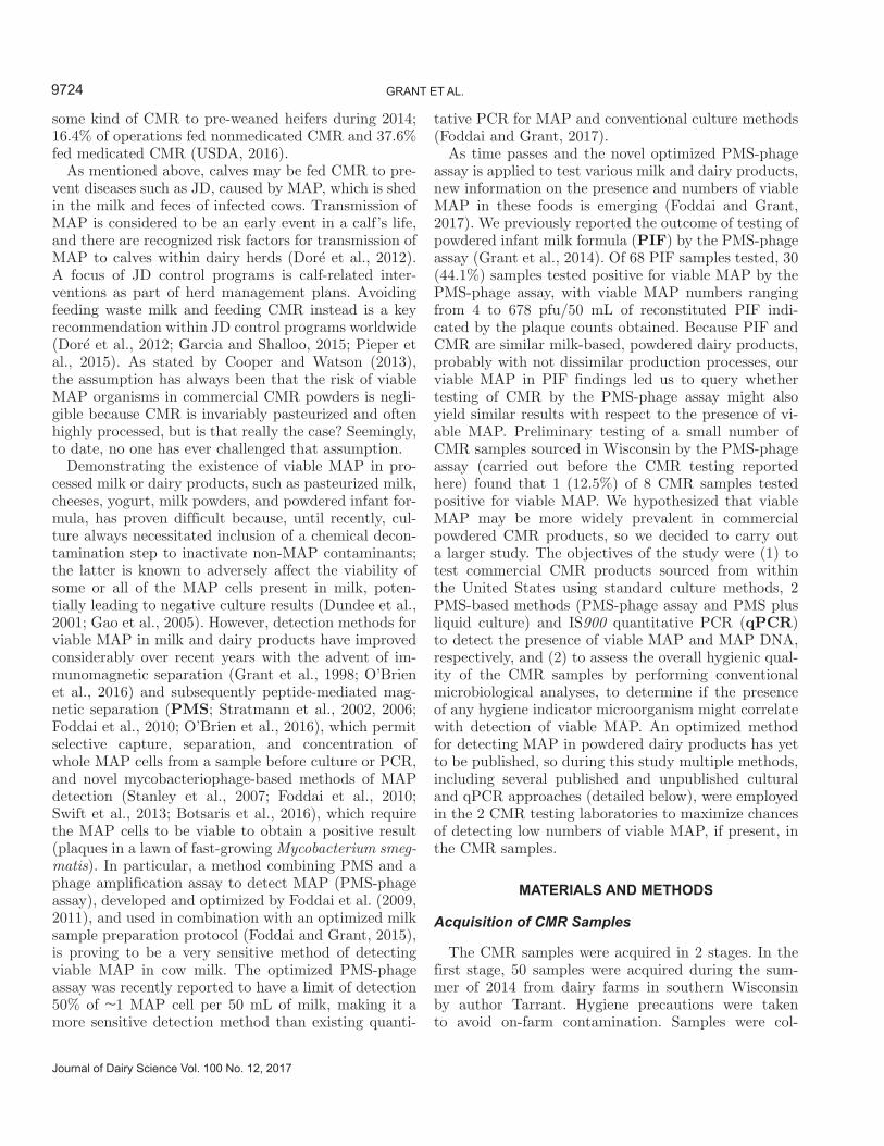
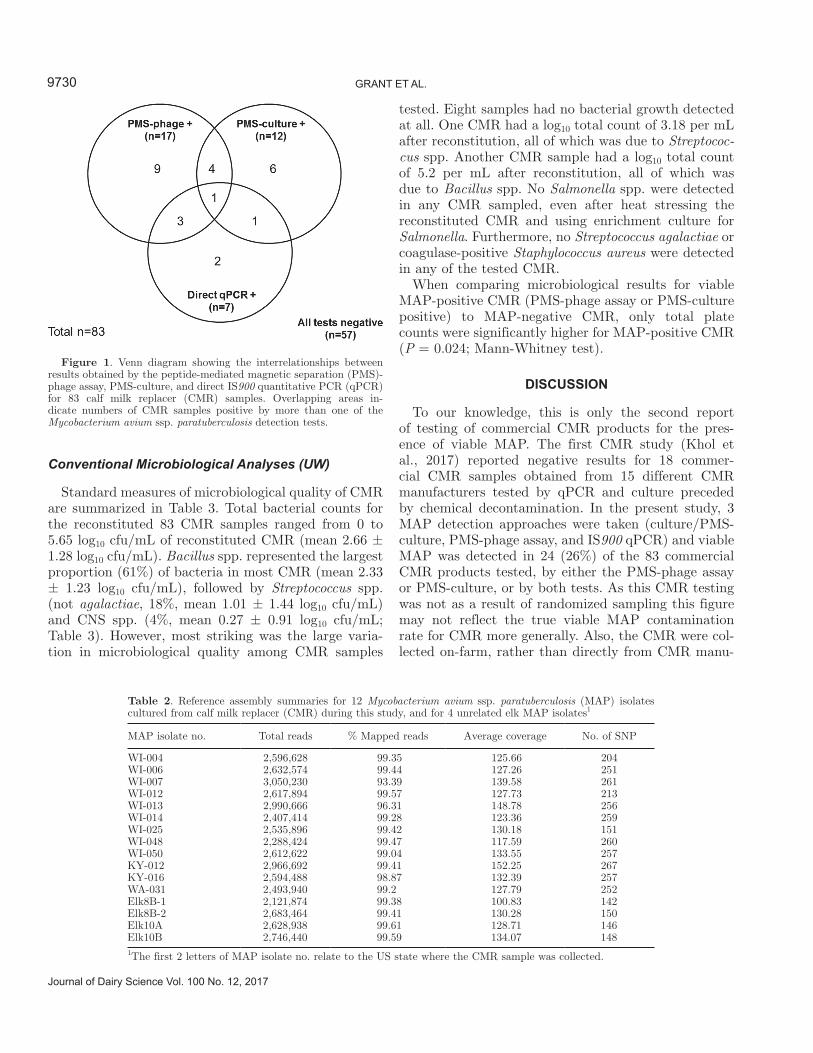
Seven (8.4%) of the 83 CMR samples were IS900 qPCR-positive with Ct values of 28.3 to 35.6, which translates to roughly 10 to 100 cfu of viable and non-viable MAP/g of CMR. Five of the 7 qPCR-positive samples (71%) were also positive by a test for viable MAP (PMS-phage assay or PMS-culture) at QUB (Fig-ure 1).
Inter-Relationships Between Test Results
Table 1 provides details of the PMS-phage assay, PMS-culture, and qPCR results for 26 (31.3%) of the 83 CMR samples, which yielded a positive result by any of the 3 tests applied. Twenty-four (92.3%) of these CMR samples tested positive for the presence of viable MAP by either PMS-phage assay or PMS-culture, or both tests. The MAP isolates were successfully cultured from 12 CMR samples overall. Only one CMR sample (KY-12), originating from a dairy farm in Kentucky, tested positive for MAP by all 3 tests applied (Table 1).
Limited and Whole-Genome Sequencing of Suspect MAP Isolates from CMR
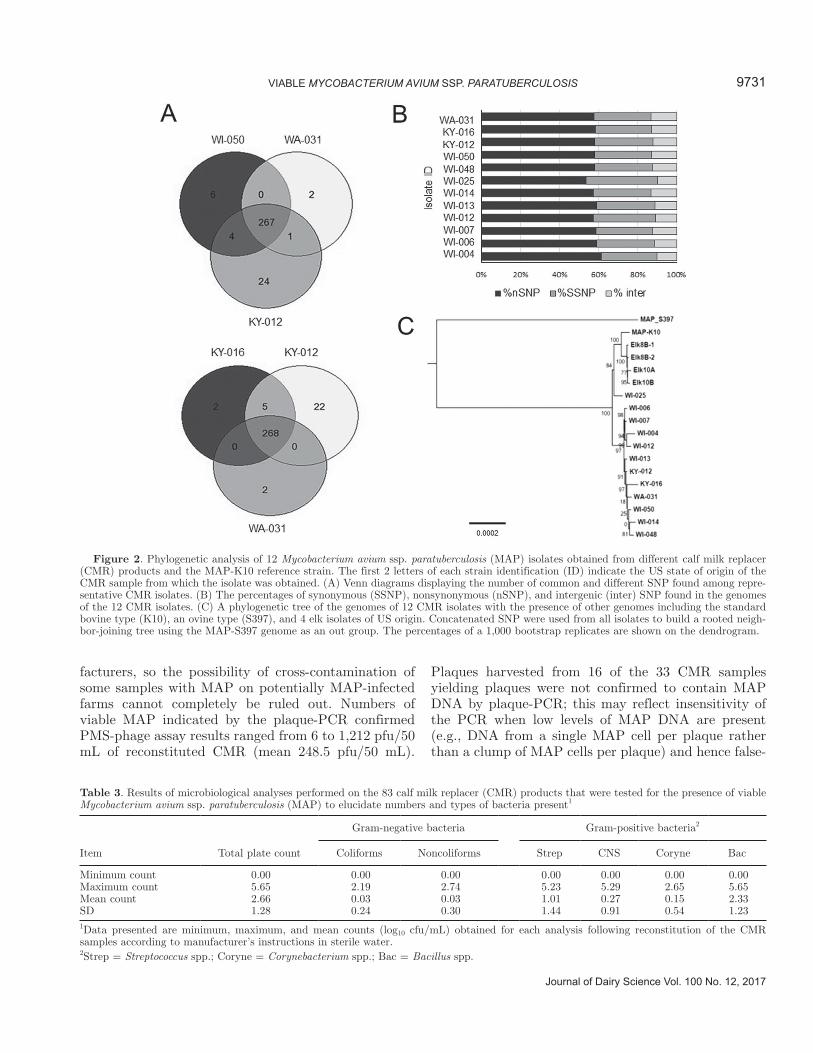
Limited genotyping using hsp65, gyrA, and gyrB in-dicated that the 12 suspected MAP isolates are from cattle source (type II) and do not belong to types I or III. These isolates were from 3 different US states, but the majority of isolates (n = 10/12) were from Wisconsin. We also performed whole-genome sequence analysis to further assay genomic diversity among iso-lates and begin to track the prevalence of MAP col-lected from CMR. The whole-genome sequence analysis confirmed that the 12 CMR isolates were MAP strains, as expected from the limited genotyping. In fact, more than 99% of the sequencing reads were mapped to ref-erence cattle strain, MAP-K10 (Table 2). Identification of SNP among isolates and the standard laboratory strain (MAP-K10) revealed large numbers of SNP that ranged between 151 and 267 SNP per genome, most
of which were shared among isolates from Wisconsin source CMR (Figure 2A). Interestingly, the percent-ages of nonsynonymous SNP were much higher than synonymous SNP, and only a few SNP were present in the intergenic regions (Figure 2B). As expected, a phylogenetic tree of SNP predicted from each genome (Figure 2C) showed a clear distinction from the MAP_S397 strain originally isolated from a sheep (Bannan-tine et al., 2012), but was closely related to the K10 isolate from a cow (Wynne et al., 2010). In addition, genomes from all CMR isolates clustered separately from genomes recently isolated from elk circulating in California (David Press, Wildlife Ecologist, Point Reyes National Seashore, Inverness, CA, personal communi-cations). The sheer number of SNP and their cluster pattern, different from traditional bovine strains used in most Johne’s testing laboratories, suggests a com-mon source for these MAP isolates from CMR collected in 4 different states.
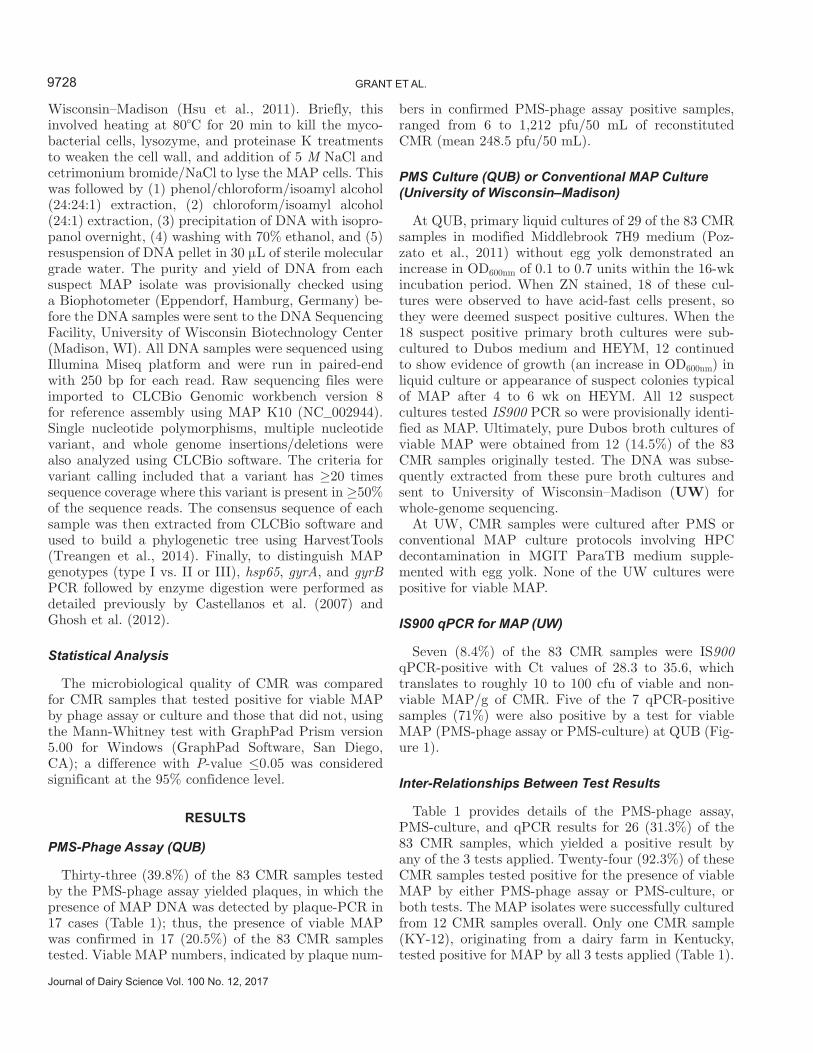
Table 1. Detailed peptide-mediated magnetic separation (PMS)-phage assay, PMS-culture, and direct IS900 quantitative PCR (qPCR) results, and US state of origin information, for the 26 calf milk replacer (CMR) samples that tested positive by one or more of the Mycobacterium avium ssp. paratuberculosis (MAP) detection tests
1Only CMR sample known to have been collected from an already opened bag of CMR.2All 12 isolates from culture-positive CMR samples were subjected to whole-genome sequencing with the results shown in Figure 2.3The average number of pfu/50 mL of reconstituted CMR for PMS-culture and qPCR positive samples were calculated based on the number of pfu indicated by the corresponding PMS-phage assay result for those samples; a negative PMS-phage assay result equated to 0 pfu/50 mL of reconstituted CMR.
9730 GRANT ET AL.
Journal of Dairy Science Vol. 100 No. 12, 2017
Conventional Microbiological Analyses (UW)
Standard measures of microbiological quality of CMR are summarized in Table 3. Total bacterial counts for the reconstituted 83 CMR samples ranged from 0 to 5.65 log10 cfu/mL of reconstituted CMR (mean 2.66 ± 1.28 log10 cfu/mL). Bacillus spp. represented the largest proportion (61%) of bacteria in most CMR (mean 2.33 ± 1.23 log10 cfu/mL), followed by Streptococcus spp. (not agalactiae, 18%, mean 1.01 ± 1.44 log10 cfu/mL) and CNS spp. (4%, mean 0.27 ± 0.91 log10 cfu/mL; Table 3). However, most striking was the large varia-tion in microbiological quality among CMR samples
tested. Eight samples had no bacterial growth detected at all. One CMR had a log10 total count of 3.18 per mL after reconstitution, all of which was due to Streptococ-cus spp. Another CMR sample had a log10 total count of 5.2 per mL after reconstitution, all of which was due to Bacillus spp. No Salmonella spp. were detected in any CMR sampled, even after heat stressing the reconstituted CMR and using enrichment culture for Salmonella. Furthermore, no Streptococcus agalactiae or coagulase-positive Staphylococcus aureus were detected in any of the tested CMR.
When comparing microbiological results for viable MAP-positive CMR (PMS-phage assay or PMS-culture positive) to MAP-negative CMR, only total plate counts were significantly higher for MAP-positive CMR (P = 0.024; Mann-Whitney test).
DISCUSSION
To our knowledge, this is only the second report of testing of commercial CMR products for the pres-ence of viable MAP. The first CMR study (Khol et al., 2017) reported negative results for 18 commer-cial CMR samples obtained from 15 different CMR manufacturers tested by qPCR and culture preceded by chemical decontamination. In the present study, 3 MAP detection approaches were taken (culture/PMS-culture, PMS-phage assay, and IS900 qPCR) and viable MAP was detected in 24 (26%) of the 83 commercial CMR products tested, by either the PMS-phage assay or PMS-culture, or by both tests. As this CMR testing was not as a result of randomized sampling this figure may not reflect the true viable MAP contamination rate for CMR more generally. Also, the CMR were col-lected on-farm, rather than directly from CMR manu-
Figure 1. Venn diagram showing the interrelationships between results obtained by the peptide-mediated magnetic separation (PMS)-phage assay, PMS-culture, and direct IS900 quantitative PCR (qPCR) for 83 calf milk replacer (CMR) samples. Overlapping areas in-dicate numbers of CMR samples positive by more than one of the Mycobacterium avium ssp. paratuberculosis detection tests.
Table 2. Reference assembly summaries for 12 Mycobacterium avium ssp. paratuberculosis (MAP) isolates cultured from calf milk replacer (CMR) during this study, and for 4 unrelated elk MAP isolates1
MAP isolate no. Total reads % Mapped reads Average coverage No. of SNP
WI-004 2,596,628 99.35 125.66 204WI-006 2,632,574 99.44 127.26 251WI-007 3,050,230 93.39 139.58 261WI-012 2,617,894 99.57 127.73 213WI-013 2,990,666 96.31 148.78 256WI-014 2,407,414 99.28 123.36 259WI-025 2,535,896 99.42 130.18 151WI-048 2,288,424 99.47 117.59 260WI-050 2,612,622 99.04 133.55 257KY-012 2,966,692 99.41 152.25 267KY-016 2,594,488 98.87 132.39 257WA-031 2,493,940 99.2 127.79 252Elk8B-1 2,121,874 99.38 100.83 142Elk8B-2 2,683,464 99.41 130.28 150Elk10A 2,628,938 99.61 128.71 146Elk10B 2,746,440 99.59 134.07 1481The first 2 letters of MAP isolate no. relate to the US state where the CMR sample was collected.
facturers, so the possibility of cross-contamination of some samples with MAP on potentially MAP-infected farms cannot completely be ruled out. Numbers of viable MAP indicated by the plaque-PCR confirmed PMS-phage assay results ranged from 6 to 1,212 pfu/50 mL of reconstituted CMR (mean 248.5 pfu/50 mL).
Plaques harvested from 16 of the 33 CMR samples yielding plaques were not confirmed to contain MAP DNA by plaque-PCR; this may reflect insensitivity of the PCR when low levels of MAP DNA are present (e.g., DNA from a single MAP cell per plaque rather than a clump of MAP cells per plaque) and hence false-
Figure 2. Phylogenetic analysis of 12 Mycobacterium avium ssp. paratuberculosis (MAP) isolates obtained from different calf milk replacer (CMR) products and the MAP-K10 reference strain. The first 2 letters of each strain identification (ID) indicate the US state of origin of the CMR sample from which the isolate was obtained. (A) Venn diagrams displaying the number of common and different SNP found among repre-sentative CMR isolates. (B) The percentages of synonymous (SSNP), nonsynonymous (nSNP), and intergenic (inter) SNP found in the genomes of the 12 CMR isolates. (C) A phylogenetic tree of the genomes of 12 CMR isolates with the presence of other genomes including the standard bovine type (K10), an ovine type (S397), and 4 elk isolates of US origin. Concatenated SNP were used from all isolates to build a rooted neigh-bor-joining tree using the MAP-S397 genome as an out group. The percentages of a 1,000 bootstrap replicates are shown on the dendrogram.
Table 3. Results of microbiological analyses performed on the 83 calf milk replacer (CMR) products that were tested for the presence of viable Mycobacterium avium ssp. paratuberculosis (MAP) to elucidate numbers and types of bacteria present1
Item Total plate count
Gram-negative bacteria
Gram-positive bacteria2
Coliforms Noncoliforms Strep CNS Coryne Bac
Minimum count 0.00 0.00 0.00 0.00 0.00 0.00 0.00Maximum count 5.65 2.19 2.74 5.23 5.29 2.65 5.65Mean count 2.66 0.03 0.03 1.01 0.27 0.15 2.33SD 1.28 0.24 0.30 1.44 0.91 0.54 1.231Data presented are minimum, maximum, and mean counts (log10 cfu/mL) obtained for each analysis following reconstitution of the CMR samples according to manufacturer’s instructions in sterile water.2Strep = Streptococcus spp.; Coryne = Corynebacterium spp.; Bac = Bacillus spp.
9732 GRANT ET AL.
Journal of Dairy Science Vol. 100 No. 12, 2017
negative plaque-PCR results may have been obtained. However, the more likely explanation is that the viru-cide step did not achieve complete inactivation of the D29 phages added to the PMS sample at the start of the phage assay, so some plaques observed were due to residual D29 phages and not mycobacterial cells burst-ing to release progeny D29 phages. This latter scenario is the main reason why the confirmatory plaque-PCR step must be included. The MAP DNA was directly detected in just 7 CMR samples by IS900 qPCR, 5 of which also tested PMS-culture or PMS-phage as-say positive. It is unknown whether the mean number of viable MAP indicated by the PMS-phage assay for the MAP culture-positive CMR samples (248.5 pfu/50 mL of reconstituted CMR), would represent sufficient inoculum to infect a calf, but the cumulative MAP dose from birth to weaning would not be insignificant, given the volume of CMR consumed by a calf over this period.
Suspected viable MAP isolates were cultured from 12 different CMR samples overall by liquid culture, with no chemical decontamination applied before culture. From the outset of the study, we were very cognizant that the suggestion may be made that laboratory cross-contam-ination could have contributed to any MAP positive cultures obtained, so the following practical measures were taken to avoid cross-contamination with viable MAP within the laboratory at QUB: (1) CMR samples were weighed out and/or reconstituted in a laboratory where MAP had never been worked with, and not in the CL2 laboratory where viable MAP may have been present; (2) sterile, single-use disposable plasticware was routinely used for all manipulations and transfers in the CL2 laboratory; (3) brand new screw-cap glass culture tubes were used for the CMR PMS-cultures and Dubos sub-cultures, and not recycled glassware that may have been previously used to culture MAP; and (4) CMR cultures were not opened until there was evi-dence of an increase in OD600nm, at which point a ZN stain was carried out to check for presence of acid-fast cells. If acid-fast positive, the culture was aseptically sub-cultured into Dubos broth, which, likewise, was not opened until an increase in OD600nm was observed. All 12 of the suspect MAP isolates were subsequently confirmed to be MAP by limited and whole-genome sequencing; all were found to be of the cattle type and all different from one another. The isolation of multiple different MAP strain types provides reassurance that the isolates obtained are not laboratory contaminants, but potentially have a common source that affected herds in 4 different states out of the 15 where farms were visited and CMR collected. The findings of this study suggest that the common source could be a MAP-contaminated CMR ingredient originating from MAP-infected dairy cattle.
Culture of CMR was carried out using several differ-ent published methods. Conventional culture methods normally used for bovine fecal samples at UW or the method used to detect MAP in powdered infant formula by Botsaris et al. (2016), in the hands of very experi-enced technicians in UW, failed to recover any viable MAP; these methods involved chemical decontamina-tion before culture, which was similar to the approach taken by Khol et al. (2017) in their recent CMR study. Only a milk sample preparation and culture approach, optimized over recent years by researchers at QUB, was successful in isolating viable MAP from 12 CMR samples during this study. This approach included (1) allowing time (overnight at 4°C following reconstitu-tion of CMR) for complete rehydration of MAP cells present in CMR before testing commenced, (2) PMS to selectively capture MAP cells from CMR rather than exposing potentially injured MAP cells to a chemical decontamination treatment, (3) primary culture for up to 16 weeks in a modified Middlebrook 7H9 liquid me-dium (first described by Pozzato et al., 2011), which had casitone added but no egg yolk, to permit resuscitation of sub-lethally injured MAP cells, and (4) sub-culture of any primary cultures once evidence of MAP growth was observed into richer Dubos liquid medium (without egg yolk) and Herrold’s egg yolk medium to stimulate more copious growth of MAP. It is difficult to explain the discrepant culture results at UW and QUB; we can only speculate on possible reasons. There were several distinct differences in terms of CMR sample prepara-tion and culture media employed. First, at QUB, once the CMR pellet was resuspended, the samples were subjected to an ultrasonication treatment to break up clumps of MAP cells. This may have had the effect of releasing viable or sub-lethally injured MAP cells from among predominantly dead cells (particularly after heat treatment) in clumps, giving them (greater) access to nutrients during primary culture in Pozzato medium. Second, no egg yolk was added to either the primary (Pozzato modified Middlebrook medium) or secondary (Dubos broth) culture media at QUB, whereas UW adds egg yolk routinely to liquid MGIT medium. The purpose of adding egg yolk to culture media used for MAP is still unclear, but neutralization of the chemi-cal decontaminant HPC seems to be a common reason (Whittington 2010). Whittington et al. (2013) subse-quently suggested that egg yolk provides major carbon and energy sources as well as the surfactant lecithin. The question arises, are sub-lethally injured MAP cells likely to benefit from, or be adversely affected by, such a rich source of nutrients when trying to repair heat or dessication damage? Third, the primary culture medium adopted by QUB contained added casitone (0.1%, 1 g/L) and a higher than normal amount of
glycerol (0.5%, as per the Pozzato et al. (2011) recipe, rather than 0.2%, which is the amount indicated by the manufacturer (Difco) of Middlebrook 7H9 broth). The additional ingredient, casitone, is a pancreatic en-zymatic digest of casein (milk protein) that contains a particularly high content of AA and peptides of varying sizes, making it a nutritious hydrolysate (BD Biosci-ences, 2017). Glycerol, or alternatively Tween 80, both of which are surfactants, can be added to Middlebrook 7H9 broth to reduce clumping of mycobacterial cells during culture, although, as stated above, 0.2% is the recommended glycerol concentration for this purpose. Glycerol would also represent a carbon source; a very old publication (Sattler and Youmans, 1948) relating to the effects of glycerol on tubercle bacilli (M. tuber-culosis) reported that glucose and glycerol increased their total amount of growth but not their initial rate of multiplication. In light of the MAP isolation success at QUB during this study, we would like to suggest that the optimal culture approach for isolation of MAP from powdered milk products could be liquid culture in the modified 7H9 medium of Pozzato et al. (2011), with PANTA but without egg yolk, following PMS to capture MAP cells from reconstituted CMR that has had time to completely rehydrate before testing com-mences. Chemical decontamination with HPC should definitely be avoided because of its known deleterious effects on the viability of MAP cells (Dundee et al., 2001, Gao et al., 2005), and therefore, the distinct possibility that false-negative results will be obtained when testing dairy products containing low numbers of MAP. The latter, plus the fact that solid Herrold’s egg yolk medium rather than a liquid culture medium was used by Khol et al. (2017), could explain their negative culture findings for CMR. Liquid culture is likely to be more conducive to recovery of stressed MAP cells than culture on solid agar medium simply because the bacterial cells are able to access water and nutrients more readily.
When CMR samples were being sourced for this study, every effort was made to hygienically collect CMR on the farms visited and to collect CMR samples from unopened bags. The vast majority of the 26 CMR sam-ples that tested MAP positive (Table 1) were sampled from unopened bags; just one of the positive samples (OH-013) is known to be from an already opened bag of CMR. However, the possibility of contamination of CMR samples by manure on the farm during collection cannot be totally ruled out because of the difficulties in maintaining hygienic practices in a dirty sampling envi-ronment, despite the best efforts of sampling personnel involved. Generally, all of the conventional measures of microbiological quality applied to the CMR samples were within normal limits for whole milk powders as set
down in USDA-Agricultural Marketing Service Stan-dards for milk and dairy products. These standards stipulate total bacterial counts <500,000 cfu/g and coliform counts ≤10 cfu/g for dry whole milk powders (USDA-AMS, 2001). The only statistically significant association between the conventional microbiological analyses and viable MAP presence in CMR was in relation to total plate counts, which were found to be significantly higher for viable MAP-positive CMR than for MAP-negative CMR (P = 0.024; Mann-Whitney test). In our opinion, this association does not neces-sarily indicate greater contamination of the CMR with MAP on-farm, but rather could reflect the possibility that the raw milk used to produce CMR ingredients contained higher numbers of MAP that have been able to survive heat treatments applied during milk powder production.
CONCLUSIONS
Viable MAP were detected in 24 (28.9%) of 83 CMR samples collected at US dairy farms; 12 were positive by PMS-culture, 17 by PMS-phage assay, and 5 samples were positive by both of these tests. A further 2 CMR samples tested positive for MAP DNA by IS900 qPCR. The presence of viable MAP in CMR was significantly associated with higher total bacterial counts, but with none of the other microbiological parameters. This is the first report of viable MAP in CMR, but not the first report of viable MAP in powdered milk products. The source of the viable MAP detected cannot be veri-fied, whether pre- or postprocessing contamination. It is unknown if the quantity of MAP detected in CMR would be sufficient to cause infection of a calf. However, the prospect that MAP has survived the manufacture of dried milk and whey-based products, which are destined for consumption by food animals could have far-reaching potential consequences; further testing of CMR collected directly at manufacturing sites using the PMS and liquid culture approach described above is warranted to verify our findings. The broader food safety implications of detecting viable MAP in this type of dried dairy product are not insignificant given that powdered infant formulae is consumed by young babies with immature immune systems.
ACKNOWLEDGMENTS
Funding for this study was obtained from the Wis-consin Agriculture Experiment Station (Madison; award number MSN169060), the American Association of Bovine Practitioners (Ashland, OH), and the Wal-ter and Martha Renk Endowed Laboratory for Food Safety (Madison, WI). James Tarrant’s involvement in
9734 GRANT ET AL.
Journal of Dairy Science Vol. 100 No. 12, 2017
the study was funded by the Merial-National Institutes of Health Summer Scholars Program (Duluth, GA). Whole-genome sequencing of the suspect MAP isolates obtained from CMR was partially supported by grant NIFA 2013-67015 from the USDA (Washington, DC) to Adel Talaat.
REFERENCES
Bannantine, J. P., C. W. Wu, C. Y. Hsu, S. G. Zhou, D. C. Schwartz, D. O. Bayles, M. L. Paustian, D. P. Alt, S. Sreevatsan, V. Kapur, and A. M. Talaat. 2012. Genome sequencing of ovine isolates of Mycobacterium avium subspecies paratuberculosis offers insights into host association. BMC Genomics 13:89. https:// doi .org/ 10 .1186/ 1471 -2164 -13 -89.
BD Biosciences. 2017. Casitone. Accessed May 8, 2017. https:// www .bdbiosciences .com/ us/ cell -culture/ media -supplements/ media -supplements/ ao -animal -origin/ bovine/ casitone/ p/ 225930.
Botsaris, G., B. M. C. Swift, I. Slana, M. Liapi, M. Christodoulou, M. Hatzitofi, V. Christodoulou, and C. E. D. Rees. 2016. Detection of viable Mycobacterium avium subspecies paratuberculosis in pow-dered infant formula by phage-PCR and confirmed by culture. Int. J. Food Microbiol. 216:91–94.
Bovine Alliance on Management and Nutrition (BAMN). 2014. A Guide to Calf Milk Replacers: Types, Use and Quality, Revised 2014. Accessed Mar. 21, 2017. https:// www .aphis .usda .gov/ animal _health/ nahms/ dairy/ downloads/ bamn/ BAMN14 _GuideMilkRepl .pdf.
Castellanos, E., A. Aranaz, B. Romero, L. de Juan, J. Alvarez, J. Bezos, S. Rodríguez, K. Stevenson, A. Mateos, and L. Domínguez. 2007. Polymorphisms in gyrA and gyrB genes among Mycobacte-rium avium ssp. paratuberculosis type I, II, and III isolates. J. Clin. Microbiol. 45:3439–3442. https:// doi .org/ 10 .1128/ JCM .01411 -07.
Cooper, R., and I. Watson. 2013. A guide to feeding and assessment of calf milk replacer. Livestock 18:217–222.
Doré, E., J. Paré, G. Côté, S. Buczinski, O. Labrecque, J. P. Roy, and G. Fecteau. 2012. Risk factors associated with transmission of Mycobacterium avium ssp. paratuberculosis to calves within dairy herd: A systematic review. J. Vet. Intern. Med. 26:32–45.
Dundee, L., I. R. Grant, H. J. Ball, and M. T. Rowe. 2001. Compara-tive evaluation of four protocols for the isolation of Mycobacte-rium avium ssp. paratuberculosis from milk. Lett. Appl. Microbiol. 33:173–177.
Foddai, A., C. T. Elliot, and I. R. Grant. 2010. Maximizing capture efficiency and specificity of magnetic separation for Mycobacte-rium avium ssp. paratuberculosis cells. Appl. Environ. Microbiol. 76:7550–7558.
Foddai, A., C. T. Elliott, and I. R. Grant. 2009. Optimization of a phage amplification assay to permit accurate enumeration of vi-able Mycobacterium avium ssp. paratuberculosis cells. Appl. Envi-ron. Microbiol. 75:3896–3902.
Foddai, A., S. Strain, R. H. Whitlock, C. T. Elliot, and I. R. Grant. 2011. Application of a peptide-mediated magnetic separation-phage assay for the detection of viable Mycobacterium avium ssp. paratuberculosis in bovine bulk-tank milk and feces samples. J. Clin. Microbiol. 49:2017–2019.
Foddai, A. C. G., and I. R. Grant. 2015. An optimised milk testing protocol to ensure accurate enumeration of viable Mycobacterium avium ssp. paratuberculosis by the PMS-phage assay. Int. Dairy J. 51:16–23.
Foddai, A. C. G., and I. R. Grant. 2017. Sensitive and specific detec-tion of viable Mycobacterium avium ssp. paratuberculosis in raw milk by the peptide-mediated magnetic separation (PMS)-phage assay. J. Appl. Microbiol. 122:1357–1367. https:// doi .org/ 10 .1111/ jam .13425.
Food and Agriculture Organisation (FAO). 2011. Rearing young rumi-nants on milk replacers and starter feeds. FAO Animal Production and Health Manual No. 13. FAO, Rome, Italy. Accessed Mar. 21, 2017. http:// www .fao .org/ docrep/ 014/ i2439e/ i2439e00 .pdf.
Gao, A., J. Odumeru, M. Raymond, and L. Mutharia. 2005. Develop-ment of improved method for isolation of Mycobacterium avium ssp. paratuberculosis from bulk tank milk: Effect of age of milk, centrifugation, and decontamination. Can. J. Vet. Res. 69:81–87.
Garcia, A. B., and L. Shalloo. 2015. The economic impact and control of paratuberculosis in cattle. J. Dairy Sci. 98:5019–5039.
Ghosh, P., C. Hsu, E. J. Alyamani, M. M. Shehata, M. A. Al-Dubaib, A. Al-Naeem, M. Hashad, O. M. Mahmoud, K. B. J. Alharbi, K. Al-Busadah, A. M. A. Swailem, and A. M. Talaat. 2012. Genome-wide analysis of the emerging infection with Mycobacterium avium ssp. paratuberculosis in the Arabian Camels (Camelus dromedar-ies). PLoS One 7:e31947. https:// doi .org/ 10 .1371/ journal .pone .0031947.
Grant, I. R., H. J. Ball, and M. T. Rowe. 1998. Isolation of Mycobac-terium paratuberculosis from milk by immunomagnetic separation. Appl. Environ. Microbiol. 64:3153–3158.
Grant, I. R., A. Foddai, B. Kunkel, and M. T. Collins. 2014. Detec-tion of viable Mycobacterium avium ssp. paratuberculosis (MAP) in infant formula. Page 306 in Proceedings of the 12th International Colloquium on Paratuberculosis. (Abstr.)
Hammer, P., C. Kiesner, H.-G. Walte, K. Knappstein, and P. Teufel. 2002. Heat resistance of Mycobacterium avium ssp. paratuberculo-sis in raw milk tested in a pilot plant pasteurizer. Kieler Milch-wirtschaftliche Forschungsberichte 54:275–303.
Hogan, H. S., R. N. Gonzalez, R. J. Harmon, S. C. Nickerson, S. P. Oli-ver, J. W. Pankey, and K. L. Smith. 1999. Laboratory Handbook on Bovine Mastitis, Second Edition. National Mastitis Council, Verona, WI.
Hsu, C.-Y., C.-W. Wu, and A. M. Talaat. 2011. Genome-wide sequence variation among Mycobacterium avium subspecies paratuberculosis isolates: A better understanding of Johne’s disease transmission dynamics. Front. Microbiol. 2:236. https:// doi .org/ 10 .3389/ fmicb .2011 .00236.
Khol, J. L., A. L. Braun, I. Slana, P. Kralik, and T. Wittek. 2017. Test-ing of milk replacers for Mycobacterium avium ssp. paratuberculosis by PCR and bacterial culture as a possible source for Johne’s disease (Paratuberculosis) in calves. Prev. Vet. Med. 144:53–56.
Moss, M. T., J. D. Sanderson, M. L. V. Tizard, J. Hermon-Taylor, F. A. K. El-Zaatari, D. C. Markesich, and D. Graham. 1992. Poly-merase chain reaction detection of Mycobacterium paratuberculosis and Mycobacterium avium ssp. silvaticum in long term cultures from Crohn’s disease and control tissues. Gut 33:1209–1213.
O’Brien, L. M., L. D. Stewart, S. A. J. Strain, and I. R. Grant. 2016. Novel monoclonal antibody and peptide binders for Mycobacte-rium avium ssp. paratuberculosis and their application for mag-netic separation. PLoS One 11:e0147870. https:// doi .org/ 10 .1371/ journal .pone .0147870.
Pieper, L., U. S. Sorge, T. J. DeVries, A. Godkin, K. Lissemore, and D. F. Kelton. 2015. Evaluation of the Johne’s disease risk assess-ment and management plan on dairy farms in Ontario, Canada. J. Dairy Sci. 98:6792–6800.
Pozzato, N., J. Gwozdz, M. Gastaldelli, K. Capello, C. Dal Ben, and E. Stefani. 2011. Evaluation of a rapid and inexpensive liquid cul-ture system for the detection of Mycobacterium avium ssp. para-tuberculosis in bovine faeces. J. Microbiol. Methods 84:413–417.
Sattler, T. H., and G. P. Youmans. 1948. The effect of “Tween 80”, bovine albumin, glycerol, and glucose on the growth of Mycobacte-rium tuberculosis var. hominis (H37Rv). J. Bacteriol. 56:235–243.
Shin, S., B. S. Lee, W.-J. Koh, E. J. B. Manning, K. Anklam, S. Sreevatsan, R. S. Lambrecht, and M. T. Collins. 2010. Efficient differentiation of Mycobacterium avium complex species and sub-species by use of five-target multiplex PCR. J. Clin. Microbiol. 48:4057–4062.
Stanley, E. C., R. J. Mole, R. J. Smith, S. M. Glenn, M. R. Barer, M. McGowan, and C. E. D. Rees. 2007. Development of a new, com-bined rapid method using phage and PCR for detection and iden-
tification of viable Mycobacterium paratuberculosis bacteria within 48 hours. Appl. Environ. Microbiol. 73:1851–1857.
Stratmann, J., K. Dohmann, J. Heinzmann, and G. F. Gerlach. 2006. Peptide aMptD-mediated capture PCR for detection of Mycobac-terium avium ssp. paratuberculosis in bulk milk samples. Appl. Environ. Microbiol. 72:5150–5158.
Stratmann, J., B. Strommenger, K. Stevenson, and G. F. Gerlach. 2002. Development of a peptide-mediated capture PCR for detec-tion of Mycobacterium avium ssp. paratuberculosis in milk. J. Clin. Microbiol. 40:4244–4250.
Swift, B. M. C., E. J. Denton, A. Sophie, S. A. Mahendran, J. N. Huxley, and C. E. D. Rees. 2013. Development of a rapid phage-based method for the detection of viable Mycobacterium avium ssp. paratuberculosis in blood within 48 h. J. Microbiol. Methods 94:175–179.
Treangen, T. J., B. D. Ondov, S. Koren, and A. M. Phillippy. 2014. The Harvest suite for rapid core-genome alignment and visualiza-tion of thousands of intraspecific microbial genomes. Genome Biol. 15:524.
USDA. 2016. Dairy 2014: Dairy Cattle Management Practices in the United States, 2014. Accessed Mar. 21, 2017. https:// www .aphis .usda .gov/ animal _health/ nahms/ dairy/ downloads/ dairy14/ Dairy14 _dr _PartI .pdf.
USDA-AMS (Agricultural Marketing Service Dairy Program). 2001. United States Standards for Grades of Dry Whole Milk, effective
Whittington, R. 2010. Cultivation of Mycobacterium avium ssp. para-tuberculosis. Chapter 22, Pages 244–266 in Paratuberculosis: Or-ganisms, Disease, Control. M. A. Behr and D. M. Collins, ed. CAB International, Wallingford, Oxfordshire, UK.
Whittington, R. J., I. Marsh, M. J. Turner, S. McAllister, E. Choy, G. J. Eamens, D. J. Marshall, and S. Ottaway. 1998. Rapid detection of Mycobacterium paratuberculosis in clinical samples from rumi-nants and in spiked environmental samples by modified BACTEC 12B radiometric culture and direct confirmation by IS 900 PCR. J. Clin. Microbiol. 36:701–707.
Whittington, R. J., A. M. Whittington, A. Waldron, D. J. Begg, K. de Silva, A. C. Purdie, and K. M. Plain. 2013. Development and validation of a liquid medium (M7H9C) for routine culture of My-cobacterium avium ssp. paratuberculosis to replace modified Bactec 12B medium. J. Clin. Microbiol. 51:3993–4000.
Wynne, J. W., T. Seemann, D. M. Bulach, S. A. Coutts, A. M. Ta-laat, and W. P. Michalski. 2010. Resequencing the Mycobacterium avium ssp. paratuberculosis K10 genome: Improved annotation and revised genome sequence. J. Bacteriol. 192:6319–6320.


![Presence of Mycobacterium avium Subspecies ... · 1. Introduction Mycobacterium avium subspecies paratuberculosis (Map) is a very slow growing member of the Mycobacteriumaviumcomplex[1,2].](https://static.documents.pub/doc/80x56/600962ca54e6680b3669b7b3/presence-of-mycobacterium-avium-subspecies-1-introduction-mycobacterium-avium.jpg)


