1 THE SKELETON The adult human skeleton has a total of 213 bones, excluding the sesamoid bones. The appendicular skeleton has 126 bones, axial skeleton 74 bones, and auditory ossicles six bones. Each bone constantly undergoes modeling during life to help it adapt to changing biomechanical forces, as well as remodeling to remove old, microdamaged bone and replace it with new, mechanically stronger bone to help preserve bone strength. Do NOT memorize the number of bones, these numbers vary and I just wanted to give you an idea of the numbers of bones in the human skeleton. The four general categories of bones are long bones, short bones, flat bones, and irregular bones. Long bones include the clavicles, humeri, radii, ulnae, metacarpals, femurs, tibiae, fibulae, metatarsals, and phalanges. Short bones include the carpal and tarsal bones, patellae, and sesamoid bones. Flat bones include the skull bones, mandible, scapulae, sternum, and ribs. Irregular bones include the vertebrae, sacrum, coccyx, and hyoid bone. Flat bones form by membranous bone formation (intramembranous ossification), whereas long bones are formed by a combination of endochondral and membranous bone formation.
Transcript
1
THE SKELETON
The adult human skeleton has a total of 213 bones, excluding the sesamoid bones. The appendicular skeleton has 126 bones, axial skeleton 74 bones, and auditory ossicles six bones. Each bone constantly undergoes modeling during life to help it adapt to changing biomechanical forces, as well as remodeling to remove old, microdamaged bone and replace it with new, mechanically stronger bone to help preserve bone strength. Do NOT memorize the number of bones, these numbers vary and I just wanted to give you an idea of the numbers of bones in the human skeleton.
The four general categories of bones are long bones, short bones, flat bones, and irregular bones. Long bones include the clavicles, humeri, radii, ulnae, metacarpals, femurs, tibiae, fibulae, metatarsals, and phalanges. Short bones include the carpal and tarsal bones, patellae, and sesamoid bones. Flat bones include the skull bones, mandible, scapulae, sternum, and ribs. Irregular bones include the vertebrae, sacrum, coccyx, and hyoid bone. Flat bones form by membranous bone formation (intramembranous ossification), whereas long bones are formed by a combination of endochondral and membranous bone formation.
The skeleton serves a variety of functions. The bones of the skeleton provide structural support for the rest of the body, permit movement and locomotion by providing levers for the muscles, protect vital internal organs and structures, provide maintenance of mineral homeostasis calcium storage), and provide the environment for hematopoiesis (the production of all blood cells) within the marrow spaces.
BONE TISSUE
Bone tissue is a specialized form of connective tissue and is the main element of the skeletal tissues. It is composed of cells and an extracellular matrix (all the material outside and in between these bone cells) in which fibers are embedded. Bone tissue is unlike other connective tissues in that the extracellular matrix becomes calcified.
2
FUNCTIONS OF BONE TISSUE
1. SupportThe skeleton is the framework of the body, it supports the softer tissues and provides points of attachment for most skeletal muscles.
2. Protection The skeleton provides mechanical protection for many of the body's internal organs, reducing risk of injury to them. For example, cranial bones protect the brain, vertebrae protect the spinal cord, and the ribcage protects the heart and lungs.
3. Assisting in MovementSkeletal muscles are attached to bones, therefore when the associated muscles contract they cause bones to move. Remember that muscles attach to bone via tendons. Ligaments connect bone to bone.
4. Storage of MineralsBone tissues store several minerals, including calcium and phosphorus. When required, bone releases minerals into the blood - facilitating the balance of minerals in the body.
5. Production of Blood CellsThis process takes place in the red bone marrow inside some larger bones. All of the blood cells (red blood cells, the five types of white blood cells and platelets) are formed in the bone marrow. This process is called ‘hematopoiesis’ or also called ‘hemopoiesis’.
6. Storage of Chemical EnergyWith increasing age some bone marrow changes from 'red bone marrow' to 'yellow bone marrow'.Yellow bone marrow consists mainly of adipose cells, and a few blood cells. It is an important chemical energy reserve. Adipose cells (fat cells) store triglycerides and these triglycerides can be used for the production of energy (ATP).
Bone cells develop as a result of ossification of cells of cartilaginous hyaline tissue or connective tissue proper. Bone tissue is metabolically active. It is a reserve of ions (mainly calcium ions) that, when needed, can be used by the body.
The skeleton is built of bone tissue. Bone provides the internal support of the body and provides sites of attachment of tendons and muscles, essential for locomotion.
Bone provides protection for the vital organs of the body: the skull protects the brain; the ribs protect the heart and lungs.
The hematopoietic bone marrow is protected by the surrounding bony tissue.
The main store of calcium and phosphate is in bone. Bone has several metabolic functions especially in calcium homeostasis.
Bone is a hard, but brittle, tissue and is relatively light per unit volume. Bone is a dynamic tissue, which throughout life bone tissue is continually being formed and resorbed. This remodeling and reorganization of bone tissue is the result of many factors including:
o mechanical stimuli
3
o metabolic causes (lack of dietary calcium, illness, aging)
o endocrine changes
o effects of drugs.
STRUCTURE OF BONE
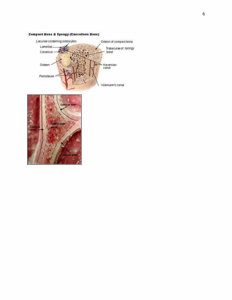
The long bones are composed of a hollow shaft, or diaphysis; flared, cone-shaped metaphyses below the growth plates; and rounded epiphyses above the growth plates. The diaphysis is composed primarily of dense cortical bone (compact bone), whereas the metaphysis and epiphysis are composed of spongy (also called trabecular or cancellous) bone surrounded by a relatively thin shell of dense cortical bone (compact bone).
The adult human skeleton is composed of 80% cortical bone and 20% spongy (trabecular) bone overall. Different bones and skeletal sites within bones have different ratios of cortical (compact) to trabecular (spongy) bone. The vertebra is composed of cortical to trabecular bone in a ratio of 25:75. This ratio is 50:50 in the femoral head and 95:5 in the radial diaphysis. Do not memorize these ratios, they are included just to illustrate a point.
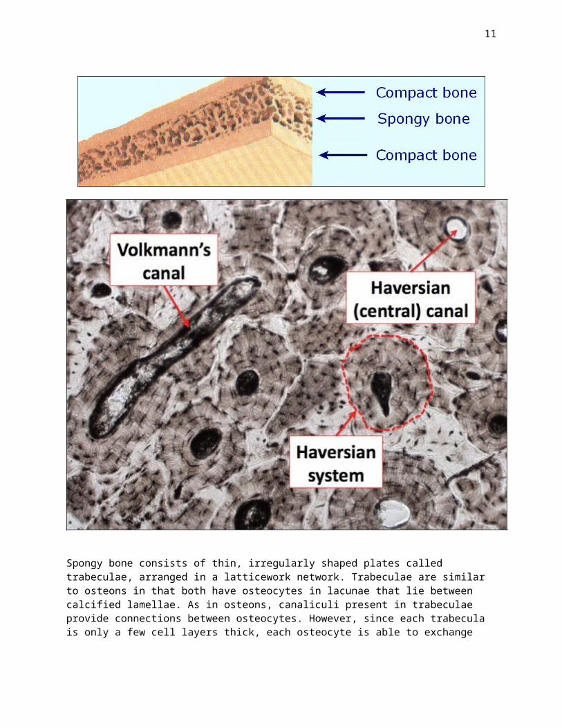
Compact bone is dense and solid and surrounds the marrow space, whereas spongy (trabecular or cancellous) bone is composed of a honeycomb-like network of trabecular plates and rods interspersed in the bone marrow compartment. Both compact and spongy bone are composed of Haversian systems or osteons.
Compact bone has an outer periosteal surface and inner endosteal surface. Periosteal surface activity is important for appositional growth and fracture repair. Bone formation typically exceeds bone resorption on the periosteal surface, so bones normally increase in diameter with aging. Bone resorption typically exceeds bone formation on the endosteal surface, so the medullary cavity normally expands with aging.
The periosteum is a fibrous connective tissue sheath that surrounds the outer cortical surface of bone. The periosteum is tightly attached to the outer cortical surface of bone by thick collagenous fibers, called Sharpeys’ fibers, which extend into underlying bone tissue. The endosteum is a membranous structure covering the inner surface of cortical bone, the medullary cavity.
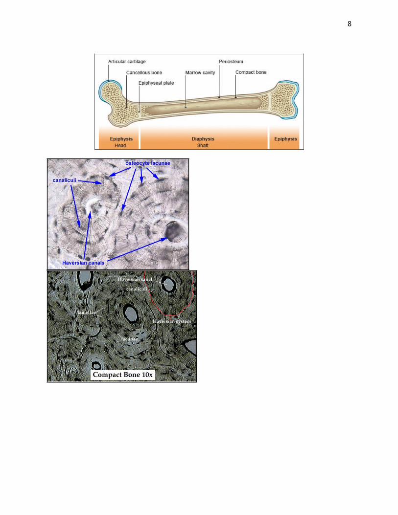
Long Bone:-The Diaphysis: the shaft of a long bone, between the epiphyses, consisting of a tube of compact
bone enclosing the medullary cavity.-The Epiphysis: the end of a long bone that is originally separated from the main bone by a layer
of cartilage but later becomes united to the main bone through ossification. It is initially separated from the shaft (diaphysis) by a section of cartilage that eventually ossifies so that the two portions fuse together.
-The Metaphysis: the zone of growth between the epiphysis and diaphysis during development of a bone. In growing bone each metaphysis includes the epiphyseal plate, a layer of hyaline cartilage that allows the diaphysis of the bone to grow in length. When bone growth in length stops, the cartilage in the epiphyseal plate is replaced by bone and the resulting bony structure is known as the epiphyseal line.
-The Articular Cartilage: a type of hyaline connective tissue that covers the articulating surfaces of bones within synovial joints.
-The Periosteum: the thick fibrous membrane covering the entire surface of a bone except its articular cartilage and serving as an attachment for muscles and tendons. It consists of an outer layer of
4
collagenous tissue containing a few fat cells and an inner layer of fine elastic fibers. The periosteum is permeated with the nerves and blood vessels that innervate and nourish underlying bone. The membrane is thick and markedly vascular over young bones but thinner and less vascular in later life. The periosteum is attached to the underlying bone by perforating (Sharpey’s) fibers, thick bundles of collagen that extend from the periosteum into the bone matrix.
-The Medullary Cavity: it is the central cavity of bone shafts where red bone marrow and/or yellow bone marrow (adipose tissue) is stored; hence, the medullary cavity is also known as the marrow cavity. Located in the main shaft of a long bone (diaphysis) it is lined with a thin, vascular membrane (endosteum).
-The Endosteum: the thin layer of cells (membrane) lining the medullary cavity of a bone.
5
6
There are two main types of bone tissue, compact bone and spongy (or called trabecular or cancellous) bone. Individual bones in the body can be formed from both of these types of bone tissue.
Compact bone is the hard material that makes up the shaft of long bones and the outside surfaces of other bones. Compact bone consists of cylindrical units called osteons (or called Haversian systems). Each osteon contains concentric lamellae (layers) of hard, calcified matrix with osteocytes (bone cells) lodged in lacunae (spaces) between the lamellae. Smaller canals, or canaliculi, radiate outward from a central canal (Haversian canal), which contains blood vessels and nerve fibers. Osteocytes within an osteon are connected to each other and to the central canal by fine cellular extensions. Through these cellular extensions, nutrients and waste are exchanged between the osteocytes and the blood vessels. Perforating canals provide channels that allow the blood vessels that run through the central canals to connect to the blood vessels in the periosteum that surrounds the bone.
8
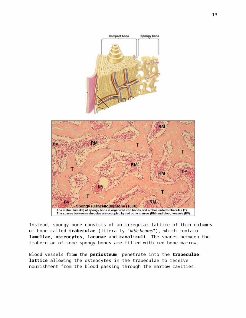
Spongy bone consists of thin, irregularly shaped plates called trabeculae, arranged in a latticework network. Trabeculae are similar to osteons in that both have osteocytes in lacunae that lie between calcified lamellae. As in osteons, canaliculi present in trabeculae provide connections between osteocytes. However, since each trabecula is only a few cell layers thick, each osteocyte is able to exchange nutrients with nearby blood vessels. Thus, no central canal is necessary.
Spongy bone (trabecular bone, cancellous bone) Compact bone (cortical bone)
Spongy bone
Spongy bone is composed of a lattice or network of branching bone spicules or trabeculae. The spaces between the bone spicules contain bone marrow.
Spongy Bone does not include osteons (Haversian systems) (the basic unit/s of Compact Bone).
9
Instead, spongy bone consists of an irregular lattice of thin columns of bone called trabeculae (literally "little beams"), which contain lamellae, osteocytes, lacunae and canaliculi. The spaces between the trabeculae of some spongy bones are filled with red bone marrow.
Blood vessels from the periosteum, penetrate into the trabeculae lattice allowing the osteocytes in the trabeculae to receive nourishment from the blood passing through the marrow cavities.
10
11
Compact bone
Compact bone appears as a mass of bony tissue lacking spaces visible to the unaided eye. Compact bone forms the outer layer of all bones and most of the structure of long bone. It contains few spaces and provides protection and support to the bone around which it is the outer-layer, as well as helping to enable the long bones to bear the stress placed on them by the weight of the body and the use to which the limbs are put, e.g. due any heavy physical work.
The basic unit of Compact Bone is an "osteon", which is also known as a "Haversian System". Each Haversian System (unit) has a cylindrical structure that consists of four parts:
1. A central tube called a Haversian Canal, which contains blood vessels and nerves.The Haversian Canal is surrounded by alternate layers of:
2. Lamellae (the word lamellae literally means "little plates") are concentric rings of a strong matrix formed from mineral salts including calcium and phosphates and collagen fibers. The mineral salts result in the hardness of the bone structure, while the collagen fibers contribute its strength.
3. Lacunae are the small spaces between the lamellae in which contain the bone cells (called "osteocytes") are located.
4. The lacunae are linked together by minute channels called canaliculi.The canaliculi provide routes by which nutrients can reach the osteocytes and waste products can leave them.
Anatomical classification of bones
Bones are characterized anatomically as:
long bones (e.g. humerus, femur) flat bones (membrane bones)
irregular bones (such as the vertebrae)
All these bone types, regardless of their anatomical form, are composed of both spongy and compact bone.
Macroscopic structure of long bones
The main shaft of long bones is called the diaphysis. At the extremities of the long bone are the epiphyses (in articulating joints). The region involved in bone elongation between the diaphysis and epiphysis in growing bones is called the metaphysis. The shaft (or diaphysis) is composed of compact (cortical or diaphyseal) bone. The epiphyses are mainly composed of trabeculae of spongy bone. The articulating surface of the epiphyses of synovial joints is covered with articular cartilage.
Bones are covered with a connective tissue called the periosteum (absent from the articular cartilage surfaces). A thinner layer of connective tissue, known as the endosteum, surrounds the bone marrow
spaces and trabeculae of spongy bone. The periosteum and endosteum are a source of new bone-forming cells (osteoprogenitor cells) and are described as possessing osteogenic potential. The periosteum and endosteum are also involved in bone repair after injury. Blood vessels of the periosteum and endosteum are involved in nutrition of the bone.
Macroscopic structure of flat bones
The flat bones or "membrane" bones of the skull are composed in a sandwich-like fashion of an outer layer of compact bone (outer table), a middle layer of spongy bone (diploe), and an inner layer of compact bone (inner table). Periosteum covers the flat bone on the outer side (near the scalp) and on the inner side the periosteum is thicker and continuous with the duramater (outer meningeal layer of the brain).
BONE CELLS
4 different cell types are found in developing bone:
o Osteoprogenitor cellso Osteoblasts
o Osteocytes
o Osteoclasts
Osteoprogenitor cells
13
Bone, like other connective tissue in the embryo, is derived from mesenchyme cells. After birth, flattened, poorly-differentiated, mesenchyme-like cells, are found in the periosteum and endosteum. These cells can divide (mitosis) and differentiate into bone cells (osteogenic potential) and as a result are known as osteoprogenitor cells. I will not ask you to be able to identify any osteoprogenitor cells in bone tissue.
Osteoblasts
The first cells to develop from the osteoprogenitor cells are the osteoblasts. Osteoblasts are involved in the formation of bone and are found on the boundaries of developing and growing bone. The cells are typically oval, with a large eccentric nucleus, and the cytoplasm is fairly basophilic. These cells are very active in synthesizing and secreting the components of the bone matrix and have well-developed rough endoplasmic reticulum (RER), Golgi bodies and granules. Osteoblasts are rich in the enzyme alkaline phosphatase, which plays a major role in the formation of the mineral deposits in the matrix. The collagen fibers are synthesized and secreted by the osteoblasts.
The matrix closest to the osteoblasts is not yet calcified and is known as osteoid or prebone. This osteoid is rich in collagen fibers. Small membrane-bound matrix vesicles (not visible by light microscopy) are budded off processes of the osteoblast cell membrane and secreted to the matrix. These contain the minerals (calcium and phosphorus) and play an important role in the calcification process of the matrix.
Osteocytes
Osteocytes are mature bone cells that develop from osteoblasts and are located in lacunae within the bony matrix. Osteocytes have cytoplasmic processes located in canaliculi, which penetrate the bony matrix. Cytoplasmic processes from one osteocyte make contact with the processes from neighboring osteocytes and can communicate via gap junctions. Because the bony matrix is calcified there is no possibility of diffusion except via the network of canaliculi.
Osteoclasts
Osteoclasts are the only cells that are known to be capable of resorbing bone. Bone marrow monocyte-macrophage precursor cells are thought to give rise to most osteoclasts. Osteoclasts are the largest of the bone cells and are multinuclear (with up to 50 nuclei). Osteoclasts are involved in bone resorption and can be found on the eroding surfaces of bone, often in cavities known as Howship's lacunae. The osteocytic cell membrane closest to the bone undergoing resorption has multiple invaginations and is known as the "ruffled border". These cells are metabolically very active, possess large numbers of mitochondria (resulting in the acidophilia of regular staining) and have well-developed Golgi bodies. Osteocytes secrete the enzyme acid phosphatase, which is involved in the breakdown of the bony matrix (collagen fibers; proteoglycans; glycoproteins and minerals-calcium and phosphorus).
Bone Growth, Modeling, and Remodeling
Bone undergoes longitudinal and radial growth, modeling, and remodeling during life. Longitudinal and radial growth during growth and development occurs during childhood and adolescence. Longitudinal growth occurs at the growth plates, where cartilage proliferates in the epiphyseal and metaphyseal areas of long bones, before subsequently undergoing mineralization to form primary new bone.
Modeling is the process by which bones change their overall shape in response to physiologic influences or mechanical forces, leading to gradual adjustment of the skeleton to the forces that it encounters. Bones
14
may widen or change axis by removal or addition of bone to the appropriate surfaces by independent action of osteoblasts and osteoclasts in response to biomechanical forces. Bones normally widen with aging in response to periosteal apposition of new bone and endosteal resorption of old bone. During bone modeling, bone formation and resorption are not tightly coupled. Bone modeling is less frequent than remodeling in adults.
Bone remodeling is the process by which bone is renewed to maintain bone strength and mineral homeostasis. Remodeling involves continuous removal of discrete packets of old bone, replacement of these packets with newly synthesized proteinaceous matrix (collagen fibers; proteoglycans and glycoproteins), and subsequent mineralization of the matrix to form new bone. The remodeling process resorbs old bone and forms new bone to prevent accumulation of bone microdamage. Remodeling begins before birth and continues until death. The bone remodeling unit is composed of a tightly coupled group of osteoclasts and osteoblasts that sequentially carry out resorption of old bone and formation of new bone.
Bone formation takes approximately 4 to 6 months to complete. Osteoblasts synthesize new collagenous organic matrix and regulate mineralization of matrix by releasing small, membrane-bound matrix vesicles that concentrate calcium and phosphate. Osteoblasts surrounded by and buried within matrix become osteocytes with an extensive canalicular network connecting them to bone surface lining cells, osteoblasts, and other osteocytes, maintained by gap junctions between the cytoplasmic processes extending from the osteocytes. The osteocyte network within bone serves as a functional syncytium (an interconnected series of cells that then acts as an interconnected tissue). At the completion of bone formation, approximately 50 to 70% of osteoblasts undergo apoptosis (programmed cell death), with the balance becoming osteocytes.
The end result of each bone remodeling cycle is production of a new Haversian System or osteon. The remodeling process is essentially the same in compact and spongy (trabecular) bone. Bone balance is the difference between the old bone resorbed and new bone formed. Periosteal bone balance is mildly positive, whereas endosteal and trabecular bone balances are mildly negative, leading to cortical and
15
trabecular thinning with aging. These relative changes occur with endosteal resorption outstripping periosteal formation.
The main recognized functions of bone remodeling include preservation of bone mechanical strength by replacing older, microdamaged bone with newer, healthier bone and calcium and phosphate homeostasis. The relatively low adult cortical bone turnover rate of 2 to 3%/year is adequate to maintain biomechanical strength of bone. The rate of trabecular bone turnover is higher, more than required for maintenance of mechanical strength, indicating that trabecular bone turnover is more important for mineral metabolism (calcium and phosphorus storage and release).
OSTEOGENESIS
Woven bone (Immature bone, Primary bone)
Osteogenesis is the name given to the development of bone tissue. The first bone to develop is a form of spongy bone known as woven bone (immature bone or primary bone). This is a primitive form of bone tissue that can be identified by the lack of order of the lacunae (of osteocytes) and the thick, irregular "woven" network of collagen fibers in the matrix. Woven bone is found temporarily in the developing embryo, before undergoing rearrangement (remodeling) resulting in the development of lamellar bone.
Woven bone is not usually found in people aged over 14 except for some specific locations including the vicinity of sutures of flat bones of the skull, in tooth sockets, and some tendon insertions. Woven bone also develops temporarily in cases of bone fracture and repair.
Lamellar bone (Mature bone, Secondary bone)
Most bone tissue is lamellar bone in which the tissue is well organized and regular. The lacunae (of osteocytes) are regularly arranged as are the collagen fibers of the matrix. The term lamella ("leaf") refers to the layer of matrix between two rows of lacunae. The lamellar arrangements are best illustrated in the cortical (compact) bone of the diaphysis of long bones.
Mature compact bone is composed of three lamellar arrangements :
Osteons (Haversian Systems) Circumferential Systems
Interstitial Systems
Osteons (Haversian Systems)
Osteons (or Haversian Systems) are cylindrical structures of compact bone, which in transverse section are seen to be formed of 4-20 regular concentric lamella surrounding a central vascular channel (Haversian canal). The diameter of each osteon typically ranges from 20-110 m. The collagen fibers in each lamella are regularly arranged and display anisotropy (birefringence) when examined by polarizing microscopy. The direction of the collagen fibers alternates from lamella to lamella, so that at any one time the anisotropy is visible only in every alternate lamella. At the periphery of each osteon, and separating it from adjacent osteons or interstitial systems, is a cement line. The cement lines do not calcify, have relatively little collagen, but are rich in glycoproteins and stain differently from the matrix of lamella.
16
Each osteon (Haversian system) comprises a single functional unit. Each Haversian canal contains a blood vessel and nerve involved in the common nutrition of the osteon, consequently osteons represent the main functional unit of compact bone. The blood vessels of the Haversian canals are supplied with blood from vessels from the periosteum. These blood vessels penetrate the osteons in a transverse direction and are known as Volkmann’s canals. Volkmann's canals can be identified as they do not have concentric lamella surrounding them.
Circumferential Systems
Immediately below the periosteum, at the periphery of compact bone of the diaphysis, the lamellae surround the bone in a continuous manner. These are known as the outer circumferential lamellae. A similar system of continuous lamellae adjacent to the endosteum is also found and is known as the inner circumferential lamellae. Bundles of collagen fibers, known as Sharpey’s fibers or perforating fibers, anchor the periosteum to the outer circumferential lamellae, especially in sites of tendon insertions.
Interstitial Systems
Remodeling of bone is a continuous process involving resorption of osteons and the rebuilding of new osteons. Interstitial systems of compact bone represent the remnants of osteons after remodeling. They are present between regular osteons and can be identified as irregular lamellar structures that lack a central Haversian canal.
Remodelling
The resorption of osteons involves osteoclasts from the Haversian canals eroding parts of lamella leading to the formation of resorption cavities. These may connect with resorption cavities from adjacent osteons. When sufficient resorption has occurred, osteoblasts appear in the resorption cavity and start building a new generation of osteons. When the new osteon is completed, the remnants of the previous osteon result in an interstitial system. This process of remodeling continues throughout life.
Trabecular bone
17
The spicules or trabeculae of spongy bone are also formed of lamellae, however, these are not arranged into systems as in compact bone. The trabeculae of spongy bone are not penetrated by blood vessels, but receive their nutrition via diffusion from the endosteum lining the bone marrow spaces.
Osteogenesis
There are two different types of bone formation (osteogenesis):
In both cases the first bone tissue to be formed is primary (woven or immature) bone, which is temporary only, prior to its replacement by secondary (lamellar or mature) bone. Intramembranous ossification involves the direct formation of bone within primitive connective tissue, whereas with endochondral ossification there is a cartilage model prior to the development of the bone.
Intramembranous ossification
Intramembranous ossification occurs during the embryonic development of many flat bones of the skull ("membrane bones") and jaw. During the initial stages of the process there is a proliferation and aggregation of mesenchyme cells, and simultaneously in the area one finds the development of many small blood vessels. The long processes of the mesenchyme cells are in contact with those of neighboring mesenchyme cells. The mesenchyme cells begin to synthesize and secrete fine collagen fibrils and an amorphous gel-like substance into the intercellular spaces. This is followed by the differentiation of the mesenchyme cells into osteoblasts (identified by their basophilia and eccentric nuclei). The osteoblasts synthesize and secrete the components of the osteoid (prebone) which, at a later stage, becomes calcified resulting in the development of bone spicules or trabeculae.
The process of intramembranous ossification is well seen in histological preparations of the embryonic calvaria. The newly formed bone matrix of developing trabeculae is stained acidophilic (pink) after regular staining. A layer of osteoblasts is present on the surface of the developing trabeculae, whereas osteocytes occupy lacunae in the bone matrix. Even at this early stage osteoclasts are present on the surface of the trabeculae and are active in bone resorption. Primitive blood vessels are seen in the connective tissue located between the trabeculae. At a later stage the connective tissue surrounding the developing flat bone forms the periosteum.
18
19
Endochondral ossification
Endochondral ossification is best illustrated in the developing long bones.
The first stages involve the development of a hyaline cartilage model with surrounding perichondrium. A layer of woven bone (the periosteal collar) develops around the central shaft of the cartilage as a result of intramembranous ossification.
Primary (diaphyseal) center of ossification.
The chondrocytes in the developing central shaft (primary center of ossification) hypertrophy (enlarge with swollen cytoplasm) and their lacunae also become enlarged. The intercellular matrix becomes calcified. As a result, there is no diffusion via the matrix and the chondrocytes degenerate and die, leaving a network of calcified cartilage. At the same time, blood vessels and mesenchyme-like cells from the periosteum penetrate this region of the diaphysis. Osteoblasts differentiate from the mesenchymal cells and begin forming primary bone tissue on the calcified cartilage framework.
A bone marrow cavity forms in the developing diaphysis as a result of osteoclastic activity eroding the primary spongy bone trabeculae. The bone cavity enlarges accompanied by further vascularization. The further elongation of long bones occurs in the growth plates of the metaphysis.
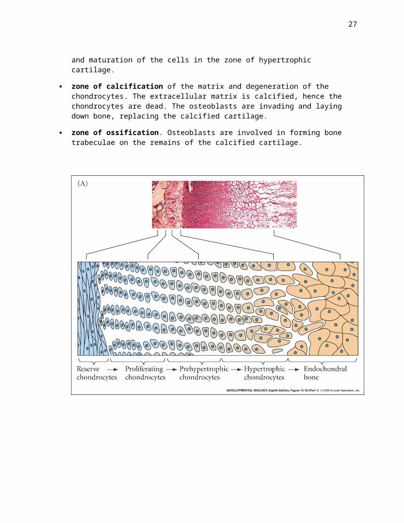
Examination of the growth plates reveals an orderly columnar arrangement of chondrocytes involved in the process of endochondral ossification.
Several zones can be identified according to the arrangement and appearance of the chondrocytes:
20
resting zone (small flattened lacunae). This layer is nearest the epiphysis and consists of small, scattered chondrocytes. These cells do not function in bone growth.
zone of proliferation (site of mitoses, and larger elliptical lacunae). They are arranged like stacks of coins.
zone of hypertrophy (greatly enlarged and rounded chrondrocytes in enlarged lacunae). The lengthening of the diaphysis is the result of cell division in the zone of proliferating cartilage and maturation of the cells in the zone of hypertrophic cartilage.
zone of calcification of the matrix and degeneration of the chondrocytes. The extracellular matrix is calcified, hence the chondrocytes are dead. The osteoblasts are invading and laying down bone, replacing the calcified cartilage.
zone of ossification. Osteoblasts are involved in forming bone trabeculae on the remains of the calcified cartilage.
21
During bone elongation there is a continuous addition of new cartilage cells and subsequent endochondral ossification, accompanied by the enlargement of the diaphyseal bone marrow cavity and erosion of the primary spongy bone. At about age 18 in females and 21 in males, the epiphyseal plates close. All cartilage is replaced with bone. The epiphyseal plate is replaced with the epiphyseal line.
Secondary (epiphyseal) center of ossification
At a later stage of development blood vessels penetrate the epiphyses accompanied by hypertrophy of the more central cartilage cells and calcification of the matrix and degeneration of the chondrocytes. Osteoblasts start building trabecular bone on the skeleton of the calcified cartilage. The trabecula are radially arranged.
Closure of the epiphyses. At ages 14-17, the bone cavities of the diaphysis and epiphyses unite, with the loss of the growth plates. This closure of the epiphyses prevents the further elongation of the long bones.
22
Long bones have two sources of bone trabeculae: the trabeculae formed by endochondral ossification at the growth plates and the trabeculae of the diaphysis formed by intramembranous ossification. During developmental stages the trabeculae formed by endochondral ossification can be recognized by the more basophilic staining of their calcified cartilage (lacking in trabeculae formed from the diaphyseal collar).
Long bones grow in width by the addition of bone tissue by osteoblastic activity in the region of the periosteum, whereas in parallel there is erosion of bone tissue by osteoclastic activity from the inner regions of the bone. As a consequence the bone marrow cavity is enlarged.
Bone is continuously being remodelled throughout life. The bone mass is constantly changing and with aging there is a net loss of bone and the quality of bone becomes impaired. Osteoporosis is common in the elderly.
23
Bone Matrix
Bone is composed of 50 to 70% mineral, 20 to 40% organic matrix, 5 to 10% water, and <3% lipids. The mineral content of bone is mostly hydroxyapatite [Ca10(PO4)6(OH)2], with small amounts of carbonate,
24
magnesium, and acid phosphate, with missing hydroxyl groups that are normally present. Compared with geologic hydroxyapatite crystals, bone hydroxyapatite crystals are very small, measuring only approximately 200 Å in their largest dimension. These small, poorly crystalline, carbonate-substituted crystals are more soluble than geologic hydroxyapatite crystals, thereby allowing them to support mineral metabolism (movement of calcium and phosphorus in and out of hydroxyapatite crystals). Bone mineral provides mechanical rigidity and load-bearing strength to bone, whereas the organic matrix provides elasticity and flexibility.
Structure of bone tissue – similar to cartilaginous tissue. Tissue consists of ground substance, collagen fibers, and bone cells (osteocytes), surrounded by osseous lacunas. Ground substance contains large amount of mineral salts (calcium and phosphorus). Tissue reconstruction after injuries is similar to cartilaginous tissue – osteoclastic cells dissolve the bone and osteogenic cells fill cavity.
Chemical composition of bone tissue – organic compounds, mineral compounds and water. With age the amount of mineral compounds grows bigger and the amount of organic compounds grows smaller. Bones become more brittle and heal with difficulty. Bone tissue, unlike cartilaginous, is vascularized and innervated.
Curvature of the spine:
In the context of human biology (or human anatomy & physiology) the spine is another word for the vertebral column (assuming that the word "spine" is used on its own - as opposed to in reference to specific features on bones, e.g. the spine of scapula).
Curvature of the Spine in AdultsThe shape of a normal adult human spine is shown in the diagram below. This labels the 4 curves of the vertebral column:
Cervical curve - formed by 7 cervical vertebrae Thoracic curve - formed by 12 thoracic vertebrae Lumbar curve - formed by 5 lumbar vertebrae Sacral curve - formed by 5 sacral vertebrae
This differs from the shape of the spine of a human fetus - which consists of one single curve formed by all 4 of the cervical, thoracic, lumbar and sacral regions that eventually form the 4 curves that form that adult spine.
25
Babies are born with a straight backbone (vertebral column) that begins to become S-shaped when they begin to sit up and eventually walk.END OF REQUIRED TEXTBOOK READING. The following pages have information that we will not be tested on, but nonetheless is interesting.
We as humans are vertebrates. What exactly is a vertebrate?
1.Animals having a bony or cartilaginous skeleton with a segmented spinal column and a large brain enclosed in a skull or cranium.
2.Having a backbone or spinal column; "fishes and amphibians and reptiles and birds and mammals are vertebrate animals".
VERTEBRATE BASICS
Vertebrates are the most advanced organisms on Earth. The traits that make all of the animals in this section special are their spinal cords, vertebrae, and notochords. It's all about having a series of nerves along your back (dorsal side). If you are an organism, you can't just have the nerves sitting there. You need to give those nerves support and protection. That need brings us to the backbones and a rod of cartilage called the notochord.
NOT SO MANY SPECIESFifty thousand species might seem like a lot. Compared to the invertebrates, there are not that many species of vertebrates. You might be asking why. One reason is that vertebrates are usually larger than invertebrates. They need more space. Another reason is that, even though they are more advanced, there are many limitations on the environments that are available to them.
Think about it this way. If you are smart mammal, would you rather live near the ocean or in the frozen tundra of the arctic? Many land animals can make that decision and move to more desirable areas for living. Those nicer areas can only support so many species of animals.
THEY'VE GOT THE BRAINSVertebrates are smart. Some of them are very smart. We mean you. Most vertebrates have very advanced nervous systems. While a goldfish might not compare to your intelligence, when you compare a goldfish to a sea anemone, a goldfish is like Einstein. Octopi are probably the smartest invertebrates and may equal or be smarter than some vertebrates. Octopi are the exception in the invertebrate category.
TREPHINATION IN HISTORY Primitive cranial trephining, the surgical opening of the skull performed with primitive tools and techniques, is one of the most fascinating surgical practices in human history. It probably started in the Neolithic at least 7000 years ago. Remarkably, it is performed yet today in parts of Africa, South America, and Melanesia. The word trepanation is derived from the Greek. It means auger or borer. The word trephination more specifically means an opening made by a circular saw of any type.
Skull trepanation in early times was independently practiced in many areas of the world, with the highest concentration of activity in Peru and Bolivia. Evidence for it also is found in Europe, Asia, New Zealand, some Pacific Islands, and North America.
The operation consisted of removing a piece of the skull (frontal, parietal, or occipital bones) from a living patient to expose the dura mater. The dura mater is the tough fibrous membrane forming the outer envelope of the brain. If it is not breached, a patient in the pre-anaesthesia, pre-antisepsis era had a fair to good chance of surviving without brain infection.
Although the operation was performed on men, women, and children, it was most often performed on adult males. Overall, patients that underwent the operation had an impressive recovery rate. As many as two thirds of the skulls examined reveal various degrees of healing--which is the evidence for survival. Considering the danger of severe bleeding, shock, brain edema, and infection, the achievements of such postoperative results suggest considerable skill and experience.
WHY WAS IT DONE?
The motives for Neolithic trephining have been the subject of speculation since the first specimens were discovered in the nineteenth century. Generally, it is surmised that, on the living, it was performed for the escape or entrance of spirits. This, of course, is conjectural. It may have been done for therapeutic reasons, such as for headaches, fractures, infections, insanity, or for convulsions. It might have been done for religious reasons. Frequently, there is evidence of skull fracture, suggesting that the procedure was done to relieve intra cranial pressure.
The following essential aspects of trephining must be accounted for in any explanation of the practice:
a. The practice was astonishingly widespread. b. It was practiced in the presence, and absence of head trauma. c. Only a small percentage of discovered skulls are trephined. d. The practice was performed on the living and on the dead. e. Men, women, and children were operated upon. f. Some skulls show multiple operations. g. In some skulls, the trephining was incomplete, as if the procedure was abandoned mid-operation.
- Antlers are made entirely of bone, horns are made of densely packed hair fibers surrounding a small core of bone.
- A giraffe also has 7 cervical vertebrae.- A sea horse and turtle have both an endoskeleton and an exoskeleton.- If a bone is buried, over the years minerals seep into the bones and replace the soft
bone tissue, thus the bone turns to rock or a fossil.- Soaking any bone, say a chicken bone, in vinegar for several days will cause it to become
soft and flexible. The acidic vinegar eats away at the bone minerals, leaving only the flexible collagen.
- A good forensic anthropologist should be able to determine just from studying bones the gender, height, weight, age, race, possibly their occupation and some types of diseases they might have had.


![[PPT]PowerPoint Presentation - Forsiden - Norges … · Web viewCompact LNG Heat Exchangers Conclusions Spiral Wound LNG heat Exchanger LNG in general The project Conclusions Map](https://static.documents.pub/doc/80x56/5b4aab247f8b9a5c278c58b2/pptpowerpoint-presentation-forsiden-norges-web-viewcompact-lng-heat-exchangers.jpg)


