Page 1
ORIGINAL ARTICLE
Virtual setup: application in orthodontic practice
Virtuelles Setup: Anwendung in der kieferorthopadischen Praxis
Leonardo T. Camardella1• Eduardo Kant C. Rothier1
• Oswaldo V. Vilella1•
Edwin M. Ongkosuwito2• Karel Hero Breuning2
Received: 8 September 2015 / Accepted: 21 March 2016
� Springer-Verlag Berlin Heidelberg 2016
Abstract
Background A plaster dental model is a patient’s tradi-
tional three-dimensional (3D) record. If the dental crowns
from a plaster model are separated and positioned in wax,
this setup of the crowns can be used to simulate orthodontic
treatment. The traditional way to make this dental setup
requires significant time by the orthodontist and in the
orthodontic lab. New developments in dentistry and
orthodontics include the possibility of virtual setups.
Aim In this article, the differences between conventional
setups with plaster models and virtual setups are discussed.
Methods A clinical patient is described for whom two
different setups were made and compared by model
superimposition with Geomagic Qualify software.
Results According to the literature and the results from this
study, virtual setups and conventional setups with plaster
models are equally accurate.
Conclusion Virtual setups present several advantages, e.g.,
digital storage, digital models cannot be damaged, the same
model can undergo several treatment simulations, and
communication between dental and surgical professionals
and between dental professionals and patients is facilitated.
Despite these advantages, considerable time and training
are needed for dental professionals to master and adopt the
general use of digital models and virtual setups in dentistry.
Keywords Orthodontics � Dental model � Diagnosis �Treatment outcome
Zusammenfassung
Hintergrund Ein Gipsmodell ist die traditionelle dreidi-
mensionale Akte des Patienten. Die vom Gipsmodell
getrennten und in Wachs eingebrachten Zahnkronen kon-
nen dazu dienen, die kieferorthopadische Behandlung zu
simulieren. Das traditionelle Verfahren fur dieses Setup
bedarf eines erheblichen Zeitaufwandes sowohl fur den
Kieferorthopaden als auch fur den Zahntechniker. Zu den
neuen Entwicklungen in der Zahnheilkunde und in der
Kieferorthopadie zahlt die Moglichkeit virtueller Setups.
Ziel Diskutiert wird der Unterschied zwischen konventio-
nellen Setups mit Gipsmodellen und virtuellen Setups.
Methoden Fur einen klinischen Patienten wurden 2 ver-
schiedene Setups erstellt, die anhand der Uberlagerung der
Modelle unter Verwendung der Software Geomagic Qua-
lify miteinander verglichen wurden.
Ergebnisse Der Literatur und den Ergebnissen der Studie
zufolge ist die Genauigkeit virtueller Setups und konven-
tioneller Setups mit Gipsmodellen gleich.
Schlussfolgerung Virtuelle Setups bieten eine Reihe von
Vorteilen, u. a. lassen sie sich digital archivieren, digitale
Modelle konnen nicht beschadigt werden, und dasselbe
Modell kann fur mehrere Behandlungssimulationen ein-
gesetzt werden. Ferner erleichtern sie die Kommunikation
zwischen Kieferorthopaden und Kieferchirurgen sowie
zwischen Kieferorthopaden und Patienten. Trotz dieser
Vorteile ist ein erhebliches Maß an Zeit- und Fortbil-
dungsaufwand notwendig, um den Einsatz von digitalen
Dr. Leonardo Tavares Camardella.
& Leonardo T. Camardella
[email protected]
1 Department of Orthodontics, Dental School, Federal
Fluminense University, Mario Santos Braga Street, 30, 28Floor, Room 214, Niteroi, RJ 24020-140, Brazil
2 Department of Orthodontics and Craniofacal Biology and
Cleft Palate, Craniofacial Centre, Radboud University
Medical Centre, Nijmegen, The Netherlands
123
J Orofac Orthop
DOI 10.1007/s00056-016-0048-y
Page 2
Modellen und virtuellen Setups in der Zahnheilkunde zu
erlernen und anzuwenden.
Schlusseworter Kieferorthopadie � Zahnmodell �Diagnose � Behandlungsergebnis
Introduction
Diagnosis and treatment planning are essential steps for
successful orthodontic treatment. Capturing the face (if
possible in 3D), including the patient’s dentition in pho-
tographs, radiographs, and dental models, is fundamental.
Dental models provide a great deal of information on the
mesiodistal dimensions of teeth, arch length discrepancies,
dental asymmetries, and arch relationships in three dimen-
sions. A dental model can also be used to produce a 3D
simulation of a treatment plan called a dental ‘‘setup’’ [2].
Through these simulations, potential therapeutic objectives
such as the need for tooth extractions or interproximal
stripping can be evaluated. A setup is thus a valuable
diagnostic tool that can be used to confirm, modify or reject
a suggested treatment plan and can be particularly valuable
in complex cases. An alternative to the traditional setup
(using a plaster model) is ‘‘the virtual setup,’’ which was
introduced in the last decade. In this article, the advantages
and disadvantages associated with the use of the conven-
tional setup and virtual setup are discussed and these two
setup methods for a clinical case are compared by model
superimposition technique with Geomagic Qualify software
(3D Systems�, Rock Hill, SC, USA).
Setup in orthodontics
Harold Kesling introduced the setup in orthodontics to
manufacture a dental positioner for finishing orthodontic
treatment. After the orthodontic bands were removed, the
remaining spaces could be closed with the positioner.
Shortly thereafter, Kesling realized the importance of this
setup for orthodontic diagnosis and treatment planning [14].
The original technique to make a setup, using separated
plaster crowns of the dentition fixed in dental wax, has
been improved over time. One of these improvements was
to position of the lower incisor in the setup according to the
cephalometric planning (Fig. 1) [2]. After correcting the
position of the lower incisors, the next step traditionally
was to manufacture a setup maintaining the vertical
dimension of the patients dentition by keeping the posterior
teeth such as third and second molars, or placing wax or
resin stops in the model’s posterior region.
A major disadvantage of plaster models and setups in
plaster is that superimposition is not possible. It is thus
difficult to compare two plaster models made at different
times [8]. To analyze tooth movement, dental models need to
be superimposed on a stable structure [6, 31]. Nowadays,
digital dental models can be made using a model scanner, a
scanned impression, or an intraoral scanner
[17, 23, 27, 28, 30, 32]. If digital models are available, they
can be superimposed using specialized software. For upper
model superimposition, the third rugae is suggested as a
stable reference landmark [31]. However, the use of a specific
palate volume when superimposing digital models seems to
be more accurate [6]. Attempts to use stable bone structures
on the mandible to superimpose cone beam computed
tomography and digital models have been tested [22].
Versatility of a virtual setup
The use of digital models in orthodontics has several
advantages. They eliminate the need for storage space
[17, 21, 23, 25, 27, 28, 30, 32] as these models can be
stored on hard drives, memory sticks, CDs and DVDs. If
digital models are available, they can be used to obtain
information for diagnosis and treatment planning [10, 29];
they facilitate the transfer of dental models
[9, 12, 17, 25, 27, 28, 32] and can be used to make custom
appliances based on a virtual setup [11, 20]. These models
also allow visualization of orthodontic treatment plans
[5, 7, 13, 15, 18].
The actual construction of a virtual setup takes less time
than making a conventional setup in plaster. To make a
virtual setup, no actual cutting of the plaster or positioning
of the dental crowns in wax is needed. Setup accuracy can
be improved when digital models are used, because any
loss of tooth structure during the cutting process of the
plaster is avoided during the digital dental crown separa-
tion procedure. The virtual teeth are cut from the model
using virtual segmentation techniques, according to the
software used. In Ortho Analyzer software (3Shape�,
Copenhagen, Denmark) this segmentation process starts
with marking mesial and distal points on each tooth. Then,
the software draws a segmentation line along the gingival
margin. This process is executed semi-automatically, but
the suggested segmentation lines still need to be manually
corrected. After that, the software separates the dental
crowns from the virtual gingiva and defines the inter-
proximal contacts. The time consuming process of lami-
nating and polishing the dental wax needed to make
conventional setups, is not needed for virtual setups.
Plaster model duplication (as used for the traditional fab-
rication of a setup) is also not required [12].
In the conventional setup, the dental arch form is plan-
ned using a brass wire or pre-established wire shape dia-
grams available from different companies. In virtual
L. T. Camardella et al.
123
Page 3
setups, the arch form can be easily adjusted for each
individual patient using software tools that can create an
individual digital arch form. As an alternative, the
orthodontist can select reference points on the digital dental
arch and selects a digital template arch to choose the best
arch form for the patient, for instance by using the WALA
ridge.
References such as dental midlines, the position of
upper and lower molars and the buccal surface of the most
protruded lower incisor are needed for plaster setups [2].
For virtual setups, the original occlusal and vertical plane
serves as a reference. The orthodontist can quantify and
visualize the applied tooth movement in all directions
during the actual virtual setup process and, when required,
applied tooth movement can be easily reversed. The effect
of gradual dental arch expansion, reduction of interdental
tooth material (‘‘stripping’’), or the decision to extract teeth
can be evaluated in a virtual setup for any patient.
Although dental changes on a plaster setup can be com-
pared with the original plaster model, in a virtual setup the
differences between the original position of the dentition
and the planned teeth movement can be visualized after
superimposition of the digital models in different colors.
With digital models it is even possible to make a simula-
tion video demonstrating the planned movements of the
teeth. This virtual setup facilitates efficient communication
between the orthodontist, patients and dental professionals.
If a proposed treatment plan is not accepted, an alternative
plan can be made within minutes.
It is important to mention that tooth movements on
computers are unlimited. Tooth alignment and levelling
can be planned on the computer screen but this result may
not be realistic for that specific patient. Obviously, tooth
movement has its biological limitations. Therefore, too
much expansion or compression of the dental arches as
planned in virtual setups may result in unstable results and
periodontal recessions [5]. In a setup of the custom
orthodontic appliance system ‘‘Insignia’’ (Ormco�,
Orange, CA, USA), the outline of the alveolar mandible
bone at a distance of approximately 4 mm below the
Fig. 1 The setup manufacturing process with plaster models. a Initial
plaster model, b documenting the most protruded lower incisor
position, c splitting the crowns and finishing, d positioning the teeth
in wax and checking the lower incisor position, e finished conven-
tional setup
Abb. 1 Herstellungsprozess beim Setup mit Gipsmodellen. a Initiales
Gipsmodell, b Dokumentation der starksten Protrusion im unteren
Schneidezahnbereich, c Spleißen der Kronen und Finishing/Finieren,
d Einbringen der Zahne in Wachs und Lagekontrolle der unteren
Schneidezahne, e fertiges konventionelles Setup
Virtual setup: application in orthodontic practice
123
Page 4
gingival margin (the so-called ‘‘Mantrough’’) can be
drawn; it reveals the limitations for moving the mandibular
dentition in the virtual setup and during treatment [4]. After
adapting the lower dentition to the mandibular alveolar
bone’s dimensions, the upper dentition can be adapted to
the setup of the lower arch. In some software programs
such as the Ortho Analyzer, the occlusion of the dentition
in the setup can be simulated and visualized in a virtual
articulator. Obviously, all setups should be based on bio-
logical principles, and their utility depends on the clini-
cian’s experience; so although a trained dental technician
can make an initial virtual setup, the orthodontist should
check each setup and make the corrections needed.
Virtual setup applications in orthodontictreatment
There is ample evidence that digital models are as accurate
and reliable as plaster models [17, 21, 23, 25, 27,
28, 30, 32]. With the introduction of digital models, virtual
setups and arch wire bending robots, new individual (cus-
tom) orthodontic appliances have been developed. The
virtual setup of a specific case can be used to gradually
move the dentition into the planned position. A series of
3D printed dental models can be used to fabricate a series
of aligners which move the teeth gradually into the planned
position [19]. Digitally designed attachments can be bon-
ded on the teeth to improve the efficiency of specific tooth
movement with aligners.
A digital model can also be used to virtually position
images of a scanned series of standard brackets. This vir-
tual bracket positioning can be done on the virtual dentition
before treatment or on a virtual treatment setup. This
planned bracket position should be transferred to the den-
tition before treatment and printed dental models can be
used to construct indirect bracket bonding trays [26].
Recent software programs can even be used to design a
virtual indirect bracket transfer bonding tray which can be
printed with 3D printers, without the need to actually print
the dental models. A virtual setup can also be used to
design individualized (‘‘custom’’) brackets and custom
wires for buccal and lingual fixed appliance therapy. These
virtual custom brackets can be printed in wax and casted in
a gold alloy using digital technology [11, 20]. A set of
individual wires can be bent by a wire-bending robot to
complete an individual tooth movement system such as
Incognito.
Of course, the dental roots are not visible in a setup
made from a plaster model or from an intraoral scan of the
dentition. Root parallelism or bone dehiscence of the
alveolar ridge cannot be evaluated on these models. If both
cone beam computed tomography (CBCT) radiographs and
digital models are available, these 3D images can be
Tab. 1 Summary of characteristics of conventional and virtual setups
Tab. 1 Zusammenfassung der charakteristischen Merkmale konventioneller und virtueller Setups
Conventional setup Virtual setup
More time-consuming Less time-consuming
Difficult to duplicate Easy to duplicate
Dental arch form planned using a brass wire or
diagrams
Dental arch form planned digitally
Need for dental and facial references Digital references and quantification of the movements of all teeth
Physical comparison with initial dental model Comparison with initial model via digital superimposition
Verbal communication requiring the presence of dental
professionals and patient
Efficient digital communication between the orthodontist, patient and dental
professionals
Potential of tooth fracture during separation Effective digital segmentation of the teeth
Enables only one setup from each model Enables different treatment plans on the same model
Conventional orthodontic analysis Analysis facilitated by software programs
Need for storage space Digital storage and a copy in the cloud
Deteriorates over time Easy digital back-up maintaining the same quality
Difficulty of sharing diagnostic information with other
professionals
Easy transfer and sharing of dental models and setups via the internet
Used only for treatment planning Also used to design and make custom appliances (aligners, fixed appliances) and
evaluation of treatment progress and result
Difficult to reproduce the same setup Possibility to reproduce the same setup according to the pre-determined records of
movements
CBCTs cannot be combined with plaster models CBCTs can be combined the digital models to make a virtual head
CBCT cone beam computed tomography
L. T. Camardella et al.
123
Page 5
accurately superimposed [15, 18, 24]. Some current CBCT
machines such as the Planmeca ProMax 3D (Planmeca
Inc., Roselle, IL, USA) can make 2D and 3D radiographs
and a 3D facial scan. The software available can be used
for combined 3D information (a ‘‘virtual head’’). Evalua-
tion of the available alveolar bone and the effect of planned
tooth movement on the soft tissues is now possible
[15, 18, 24]. Some companies, such as SureSmile
(Orametrix�, Richardson, TX, USA) use intraoral scans as
well as CBCT images for treatment planning and evalua-
tion. A major advantage of a virtual treatment plan using
the 3D documentation of the head in a 1:1 ratio is that the
orthodontist can evaluate and plan the dentition’s correc-
tion and if needed, the jaws including correction of the
dental roots. Progress intraoral scans and CBCTs allow a
progress setup to be made, which can be used for the
fabrication of finishing wires or finishing aligners. Studies
have shown that the use of computer-bent custom wires as
used in the SureSmile system can reduce orthodontic
treatment time and improve treatment outcomes [1]. But
according to Larson et al. [16] the effectiveness of
orthodontic treatment using SureSmile technology to
achieve predicted tooth position varies according to the
tooth types and movements needed. Table 1 illustrates the
advantages and disadvantages of the conventional and
virtual setups.
Clinical case
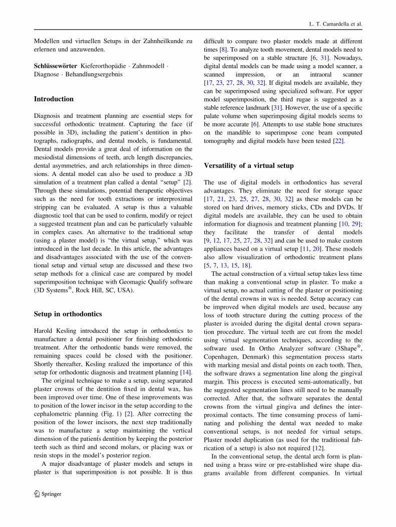
A female patient aged 17 years and 11 months presented
for consultation in the Orthodontic Clinic at the Federal
Fluminense University (Niteroi, Brazil). Her main com-
plaint was her lip prominence. After the anamnesis and
clinical examination, regular orthodontic documentation
was planned. The diagnosis for this patient was a Class I
malocclusion with an anterior open bite, mild anterior
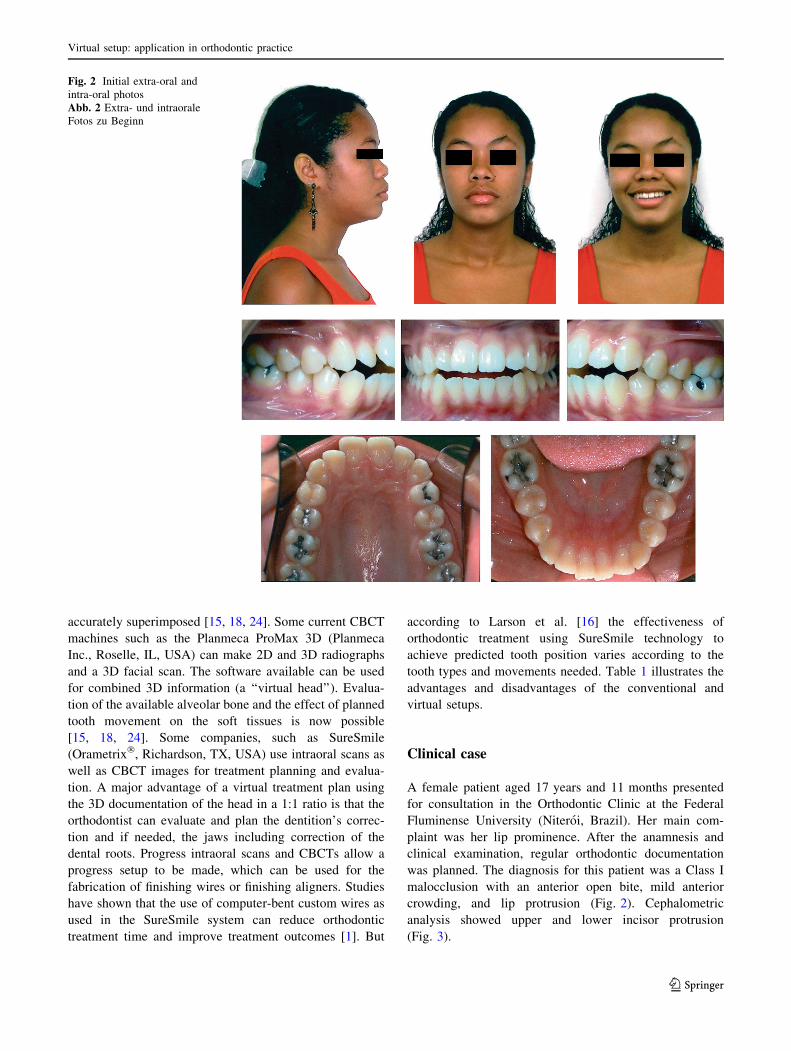
crowding, and lip protrusion (Fig. 2). Cephalometric
analysis showed upper and lower incisor protrusion
(Fig. 3).
Fig. 2 Initial extra-oral and
intra-oral photos
Abb. 2 Extra- und intraorale
Fotos zu Beginn
Virtual setup: application in orthodontic practice
123
Page 6
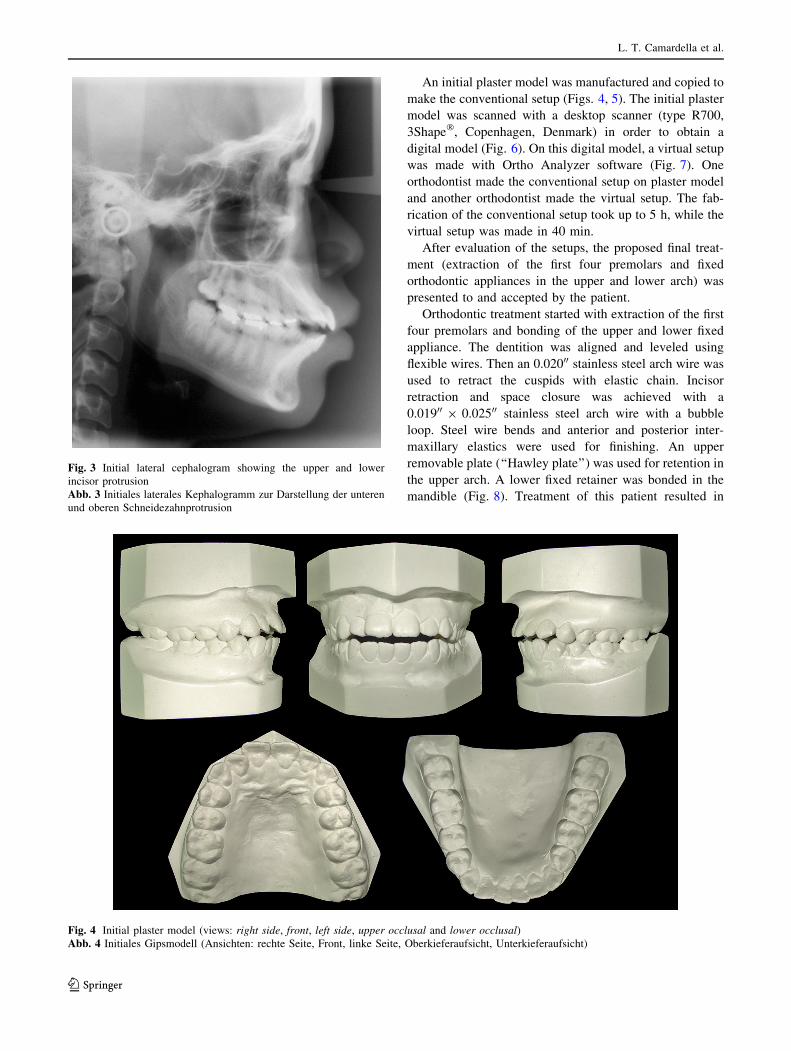
An initial plaster model was manufactured and copied to
make the conventional setup (Figs. 4, 5). The initial plaster
model was scanned with a desktop scanner (type R700,
3Shape�, Copenhagen, Denmark) in order to obtain a
digital model (Fig. 6). On this digital model, a virtual setup
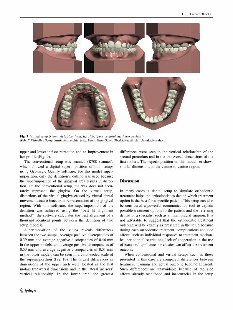
was made with Ortho Analyzer software (Fig. 7). One
orthodontist made the conventional setup on plaster model
and another orthodontist made the virtual setup. The fab-
rication of the conventional setup took up to 5 h, while the
virtual setup was made in 40 min.
After evaluation of the setups, the proposed final treat-
ment (extraction of the first four premolars and fixed
orthodontic appliances in the upper and lower arch) was
presented to and accepted by the patient.
Orthodontic treatment started with extraction of the first
four premolars and bonding of the upper and lower fixed
appliance. The dentition was aligned and leveled using
flexible wires. Then an 0.02000 stainless steel arch wire was
used to retract the cuspids with elastic chain. Incisor
retraction and space closure was achieved with a
0.01900 9 0.02500 stainless steel arch wire with a bubble
loop. Steel wire bends and anterior and posterior inter-
maxillary elastics were used for finishing. An upper
removable plate (‘‘Hawley plate’’) was used for retention in
the upper arch. A lower fixed retainer was bonded in the
mandible (Fig. 8). Treatment of this patient resulted in
Fig. 3 Initial lateral cephalogram showing the upper and lower
incisor protrusion
Abb. 3 Initiales laterales Kephalogramm zur Darstellung der unteren
und oberen Schneidezahnprotrusion
Fig. 4 Initial plaster model (views: right side, front, left side, upper occlusal and lower occlusal)
Abb. 4 Initiales Gipsmodell (Ansichten: rechte Seite, Front, linke Seite, Oberkieferaufsicht, Unterkieferaufsicht)
L. T. Camardella et al.
123
Page 7
Fig. 5 Conventional plaster setup (views: right side, front, left side, upper occlusal and lower occlusal)
Abb. 5 Konventionelles Gips-Setup (Ansichten: rechte Seite, Front, linke Seite, Oberkieferaufsicht, Unterkieferaufsicht)
Fig. 6 Initial digital model (views: right side, front, left side, upper occlusal and lower occlusal)
Abb. 6 Initiales digitales Modell (Ansichten: rechte Seite, Front, linke Seite, Oberkieferaufsicht, Unterkieferaufsicht)
Virtual setup: application in orthodontic practice
123
Page 8
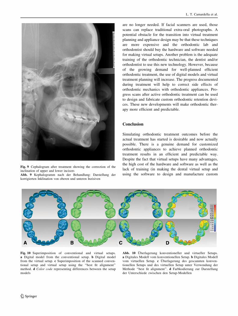
upper and lower incisor retraction and an improvement in
her profile (Fig. 9).
The conventional setup was scanned (R700 scanner),
which allowed a digital superimposition of both setups
using Geomagic Qualify software. For this model super-
imposition, only the dentition’s outline was used because
the superimposition of the gingival area results in distor-
tion. On the conventional setup, the wax does not accu-
rately represent the gingiva. On the virtual setup,
distortions of the virtual gingiva caused by virtual dental
movements cause inaccurate representation of the gingival
region. With this software, the superimposition of the
dentition was achieved using the ‘‘best fit alignment
method’’ (the software calculates the best alignment of a
thousand identical points between the dentition of two
setup models).
Superimposition of the setups reveals differences
between the two setups. Average positive discrepancies of
0.39 mm and average negative discrepancies of 0.46 mm
in the upper models, and average positive discrepancies of
0.53 mm and average negative discrepancies of 0.51 mm
in the lower models can be seen in a color-coded scale of
the superimposition (Fig. 10). The largest differences in
dimensions of the upper arch were located in the first
molars transversal dimensions and in the lateral incisors’
vertical relationship. In the lower arch, the greatest
differences were seen in the vertical relationship of the
second premolars and in the transversal dimensions of the
first molars. The superimposition on this model set shows
similar dimensions in the canine-to-canine region.
Discussion
In many cases, a dental setup to simulate orthodontic
treatment helps the orthodontist to decide which treatment
option is the best for a specific patient. This setup can also
be considered a powerful communication tool to explain
possible treatment options to the patient and the referring
dentist or a specialist such as a maxillofacial surgeon. It is
not advisable to suggest that the orthodontic treatment
outcome will be exactly as presented in the setup because
during each orthodontic treatment, complications and side
effects such as individual responses to treatment mechan-
ics, periodontal restrictions, lack of cooperation in the use
of extra oral appliances or elastics can affect the treatment
outcome.
When conventional and virtual setups such as those
presented in this case are compared, differences between
treatment planning and actual outcome become apparent.
Such differences are unavoidable because of the side
effects already mentioned and inaccuracies in the setup
Fig. 7 Virtual setup (views: right side, front, left side, upper occlusal and lower occlusal)
Abb. 7 Virtuelles Setup (Ansichten: rechte Seite, Front, linke Seite, Oberkieferaufsicht, Unterkieferaufsicht)
L. T. Camardella et al.
123
Page 9
manufacturing process [7, 13]. Nevertheless, virtual setups
are at least as effective and accurate as conventional setups
and are an effective tool for diagnosis and orthodontic
treatment planning, appliance fabrication, and treatment
assessments [3].
According to the literature, significant differences in
the Original American Board of Orthodontics Objective
Grading System (ABO-OGS) scores between two virtual
setups of the same original models made by a single
clinician were reported [7]. Virtual setups of the same
original model made by different clinicians differed also
[7]. Such differences also occur if conventional setups
with plaster models are made, because dental setups
depend on a practitioner’s subjective decisions. In this
article, two different orthodontists made the conven-
tional and virtual setups. The differences between these
setups, especially those in the transversal posterior
relationship, are influenced by how the planned arch
form had been selected (brass wire for the conventional
setup versus a virtual arch form for the virtual setup).
However, the advantage of a virtual setup is that a
report with a script of all movements performed during
the setup fabrication can be generated; thus, an identical
setup can be made.
For each setup, references to the original dental position
are needed. In the conventional setup, the second molar
position was maintained to preserve the vertical occlusion
dimension. In the virtual setup, all posterior teeth were
moved but the original occlusal and transversal plane were
used as a reference. Tooth movement limitations (con-
strained movement) can be selected in the planning soft-
ware to prevent very large and clinically impossible tooth
movements.
As progress in digital imaging techniques and tools to
plan medical treatments accelerates, the use of virtual
setups in orthodontics before and during treatment will
become the ‘‘main stream’’ in orthodontics. If intra-oral
color scanners are used, traditional intra-oral photographs
Fig. 8 Extra-oral and intra-oral
photos after treatment
Abb. 8 Extra- und intraorale
Fotos nach Behandlung
Virtual setup: application in orthodontic practice
123
Page 10
are no longer needed. If facial scanners are used, those
scans can replace traditional extra-oral photographs. A
potential obstacle for the transition into virtual treatment
planning and appliance design may be that these techniques
are more expensive and the orthodontic lab and
orthodontist should buy the hardware and software needed
for making virtual setups. Another problem is the adequate
training of the orthodontic technician, the dentist and/or
orthodontist to use this new technology. However, because
of the growing demand for well-planned efficient
orthodontic treatment, the use of digital models and virtual
treatment planning will increase. The progress documented
during treatment will help to correct side effects of
orthodontic mechanics with orthodontic appliances. Pro-
gress scans after active orthodontic treatment can be used
to design and fabricate custom orthodontic retention devi-
ces. These new developments will make orthodontic ther-
apy more efficient and predictable.
Conclusion
Simulating orthodontic treatment outcomes before the
actual treatment has started is desirable and now actually
possible. There is a genuine demand for customized
orthodontic appliances to achieve planned orthodontic
treatment results in an efficient and predictable way.
Despite the fact that virtual setups have many advantages,
the high cost of the hardware and software as well as the
lack of training (in making the dental virtual setup and
using the software to design and manufacture custom
Fig. 9 Cephalogram after treatment showing the correction of the
inclination of upper and lower incisors
Abb. 9 Kephalogramm nach der Behandlung: Darstellung der
korrigierten Inklination von oberen und unteren Inzisiven
Fig. 10 Superimposition of conventional and virtual setups.
a Digital model from the conventional setup. b Digital model
from the virtual setup. c Superimposition of the scanned conven-
tional setup and virtual setup using the ‘‘best fit alignment’’
method. d Color code representing differences between the setup
models
Abb. 10 Uberlagerung konventioneller und virtueller Setups.
a Digitales Modell vom konventionellen Setup. b Digitales Modell
vom virtuellen Setup. c Uberlagerung des gescannten konven-
tionellen Setups und des virtuellen Setup unter Verwendung der
Methode ‘‘best fit alignment’’. d Farbkodierung zur Darstellung
der Unterschiede zwischen den Setup-Modellen
L. T. Camardella et al.
123
Page 11
orthodontic appliances) currently limit the use of this
technology in orthodontics.
Compliance with ethical guidelines
Conflict of interest L. T. Camardella, E. K. C. Rothier, O. V. Vilella,
E. M. Ongkosuwito, and K. H. Breuning declare that they have no
competing interests.
All procedures performed in studies involving human participants
were in accordance with the ethical standards of the institutional and/
or national research committee and with the 1964 Helsinki declaration
and its later amendments or comparable ethical standards. Informed
consent was obtained from all individual participants included in the
study. Additional informed consent was obtained from all individual
participants from whom identifying information is included in this
article.
References
1. Alford TJ, Roberts WE, Hartsfield JK et al (2011) Clinical out-
comes for patients finished with the SureSmile method compared
with conventional fixed orthodontic therapy. Angle Orthod
81:383–388
2. Araujo TM, Fonseca LM, Caldus LD et al (2012) Preparation and
evaluation of orthodontic setup. Dental Press J Orthod
17:146–165
3. Barreto MS, Faber J, Vogel CJ et al (2016) Reliability of digital
orthodontic setups. Angle Orthod 86:255–259
4. Breuning KH (2011) Efficient tooth movement with new tech-
nologies for customized treatment. J Clin Orthod 45:257–262
(quiz 87)5. Chen S, Xu TM (2013) Treatment of a severe transverse dental
arch discrepancy assisted by 3-dimensional planning. Am J
Orthod Dentofac Orthop 143:105–115
6. Choi DS, Jeong YM, Jang I et al (2010) Accuracy and reliability
of palatal superimposition of three-dimensional digital models.
Angle Orthod 80:497–503
7. Fabels LN, Nijkamp PG (2014) Interexaminer and intraexaminer
reliabilities of 3-dimensional orthodontic virtual setups. Am J
Orthod Dentofac Orthop 146:806–811
8. Flugge TV, Schlager S, Nelson K et al (2013) Precision of
intraoral digital dental impressions with iTero and extraoral
digitization with the iTero and a model scanner. Am J Orthod
Dentofac Orthop 144:471–478
9. Goonewardene RW, Goonewardene MS, Razza JM et al (2008)
Accuracy and validity of space analysis and irregularity index
measurements using digital models. Aust Orthod J 24:83–90
10. Gracco A, Buranello M, Cozzani M et al (2007) Digital and
plaster models: a comparison of measurements and times. Prog
Orthod 8:252–259
11. Grauer D, Proffit WR (2011) Accuracy in tooth positioning with a
fully customized lingual orthodontic appliance. Am J Orthod
Dentofac Orthop 140:433–443
12. Horton HM, Miller JR, Gaillard PR et al (2010) Technique
comparison for efficient orthodontic tooth measurements using
digital models. Angle Orthod 80:254–261
13. Im J, Cha JY, Lee KJ et al (2014) Comparison of virtual and
manual tooth setups with digital and plaster models in extraction
cases. Am J Orthod Dentofac Orthop 145:434–442
14. Kesling H (1956) The diagnostic setup with consideration of the
third dimension. Am J Orthod 42:740–748
15. Kihara T, Tanimoto K, Michida M et al (2012) Construction of
orthodontic setup models on a computer. Am J Orthod Dentofac
Orthop 141:806–813
16. Larson BE, Vaubel CJ, Grunheid T (2013) Effectiveness of
computer-assisted orthodontic treatment technology to achieve
predicted outcomes. Angle Orthod 83:557–562
17. Leifert MF, Leifert MM, Efstratiadis SS et al (2009) Comparison
of space analysis evaluations with digital models and plaster
dental casts. Am J Orthod Dentofacial Orthop 136:16e1–16e4
(discussion)18. Macchi A, Carrafiello G, Cacciafesta V et al (2006) Three-di-
mensional digital modeling and setup. Am J Orthod Dentofac
Orthop 129:605–610
19. Miller RJ, Derakhshan M (2004) Three-dimensional technology
improves the range of orthodontic treatment with esthetic and
removable aligners. World J Orthod 5:242–249
20. Mujagic M, Fauquet C, Galletti C et al (2005) Digital design and
manufacturing of the Lingualcare bracket system. J Clin Orthod
39:375–382 (quiz 0)21. Mullen SR, Martin CA, Ngan P et al (2007) Accuracy of space
analysis with emodels and plaster models. Am J Orthod Dentofac
Orthop 132:346–352
22. Park TJ, Lee SH, Lee KS (2012) A method for mandibular dental
arch superimposition using 3D cone beam CT and orthodontic 3D
digital model. Korean J Orthod 42:169–181
23. Quimby ML, Vig KW, Rashid RG et al (2004) The accuracy and
reliability of measurements made on computer-based digital
models. Angle Orthod 74:298–303
24. Rangel FA, Maal TJ, Bronkhorst EM et al (2013) Accuracy and
reliability of a novel method for fusion of digital dental casts and
Cone Beam Computed Tomography scans. PLoS ONE 8:e59130
25. Rheude B, Sadowsky PL, Ferriera A et al (2005) An evaluation of
the use of digital study models in orthodontic diagnosis and
treatment planning. Angle Orthod 75:300–304
26. Sachdeva RC (2001) SureSmile technology in a patient–centered
orthodontic practice. J Clin Orthod 35:245–253
27. Sousa MV, Vasconcelos EC, Janson G et al (2012) Accuracy and
reproducibility of 3-dimensional digital model measurements.
Am J Orthod Dentofac Orthop 142:269–273
28. Stevens DR, Flores-Mir C, Nebbe B et al (2006) Validity, reli-
ability, and reproducibility of plaster vs digital study models:
comparison of peer assessment rating and Bolton analysis and
their constituent measurements. Am J Orthod Dentofac Orthop
129:794–803
29. Tomassetti JJ, Taloumis LJ, Denny JM et al (2001) A comparison
of 3 computerized Bolton tooth-size analyses with a commonly
used method. Angle Orthod 71:351–357
30. Torassian G, Kau CH, English JD et al (2010) Digital models vs
plaster models using alginate and alginate substitute materials.
Angle Orthod 80:474–481
31. van der Linden FP (1978) Changes in the position of posterior
teeth in relation to ruga points. Am J Orthod 74:142–161
32. Wiranto MG, Engelbrecht WP, Nolthenius HET et al (2013)
Validity, reliability, and reproducibility of linear measurements
on digital models obtained from intraoral and cone-beam com-
puted tomography scans of alginate impressions. Am J Orthod
Dentofac Orthop 143:140–147
Virtual setup: application in orthodontic practice
123