Construction and Properties of a Recombinant Herpes SimplexVirus 1 Lacking Both S-Component Origins of DNA Synthesis
KAZUHIKO IGARASHI, RANDALL FAWL, RICHARD J. ROLLER, AND BERNARD ROIZMAN*The Marjorie B. Kovler Viral Oncology Laboratories, The University of Chicago, Chicago, Illinois 60637
Received 11 September 1992/Accepted 23 December 1992
The herpes simplex virus 1 (HSV-1) genome contains three origins of DNA synthesis (Ori) utilized by viralDNA synthesis proteins. One sequence (OriL) maps in the L component, whereas two sequences (Oris) map inthe S component. We report the construction of a recombinant virus, R7711, from which both Oris sequenceshave been deleted, and show that the Oris sequences are not essential for the replication of HSV-1 in culturedcells. In addition to the deletions of Oris in R7711, the a47 gene and the 5' untranscribed and transcribednoncoding regions of the Us11 gene were deleted, one of the a4 promoter-regulatory regions was replaced withthe simian virus 40 promoter, and the at22 promoter was substituted with the av27 promoter. The total amountof viral DNA synthesized in Vero cells infected with the Oris-negative (Oris-) virus was approximately thatseen in cells infected with the Oris-positive virus. However, cells infected with the Oris- virus accumulatedviral DNA more slowly than those infected with the wild-type virus during the first few hours after the onsetof DNA synthesis. In single-step growth experiments, the yield of Orins progeny virus was reduced at mostfourfold. Although a single Oris (R. Longnecker and B. Roizman, J. Virol. 58:583-591, 1986) and the singleOriL (M. Polvino-Bodnar, P. K. Orberg, and P. A. Schaffer, J. Virol. 61:3528-3535, 1987) have been shownto be dispensable, this is the first indication that both copies of Oris are dispensable and that one copy of anOri sequence may suffice for the replication of HSV-1.
The herpes simplex virus 1 (HSV-1) genome consists oftwo covalently linked components, L and S, that are in-verted relative to each other such that DNA extracted fromvirions consists of four populations differing solely in therelative orientations of the components (7, 11). Each com-ponent consists of a unique sequence (UL or Us) flanked byinverted repeats (31, 38). In addition, each componentcontains either one (OriL; L component) or two (Oris; Scomponent) sequences that confer upon DNA fragments thecapacity to be amplified by proteins specified by the virus(15, 21, 33, 34, 35, 37, 41). The OriL sequence mapsapproximately in the middle of the unique sequence of the Lcomponent (UL), whereas the two Oris sequences mapwithin the repeats flanking the unique sequence (Us) of the Scomponent. Both OriL and Oris are located between diver-gently transcribed genes; OriL is located between the UL29and UL30 genes, whereas Oris maps between the a4 geneand either the a22 or the a47 gene. The two Oris sequencesare identical but inverted relative to each other. Moreover,they share homologous sequences with the OriL sequence(15, 41). The evidence that both Oris sequences and the OriLsequence can function as a DNA replication origin utilizedby HSV DNA replication proteins has been obtained in atransient plasmid replication assay (15, 35, 37, 41). Also, aprotein specified by the UL9 open reading frame has beenshown to bind to specific sites within each origin (9, 23, 40),and the UL9 gene is essential for the replication of HSV-1(4). Nevertheless, mysteries regarding the functions of theorigins of DNA synthesis during viral replication abound.A key problem stems from the nature of the replication of
HSV DNA. This DNA circularizes within a very short timeafter infection in the absence of de novo protein synthesis(24). In principle, circular DNA could replicate by a thetamodel, which depends heavily on each initiation event in the
* Corresponding author.
origins of DNA synthesis, or by a rolling-circle model, inwhich the dependence on the origins is minimal and occursat most only once at the initiation of DNA synthesis.Attempts to uncover forms consistent with theta replicationhave not been successful. The evidence is consistent withthe conclusion that both defective genomes, amplified intrans by viral enzymes furnished by the expression ofnondefective genomes, and nondefective genomes them-selves accumulate as head-to-tail concatemers, in accor-dance with a rolling-circle model of DNA replication (3, 13,14, 37). If this model is correct, it is not clear why the HSVgenome contains and presumably conserves three origins ofDNA replication.
In previous studies, workers in our laboratory and subse-quently others (16, 25) reported that at least one origin,either one Oris or OriL, can be deleted without affecting theability of the virus to replicate in cells in culture. Further-more, OriL is not required for the establishment and reacti-vation of the virus in trigeminal neurons harboring latentvirus in mice inoculated by the corneal route (25). Attemptsto delete both Oris sequences from the HSV-2 genome wereunsuccessful, and it has been suggested that at least one Orissequence is essential for viral DNA replication (32). In thispaper, we report the properties of a genetically engineeredvirus from which both Oris sequence have been deleted. Thephenotypic properties of the recombinant cannot be differ-entiated from those of the wild-type parent with respect toDNA synthesis in cells in culture.
MATERIALS AND METHODS
Cells and viruses. The properties and propagation ofHSV-1 strain F [HSV-1(F)] and the thymidine kinase (tk)deletion mutant HSV-1(F)A305 were previously described(8, 26). Vero cells (American Type Culture Collection),rabbit skin cells (obtained from J. McClaren), and 143TK-
2123
Vol. 67, No. 4
2124 IGARASHI ET AL.
cells (obtained from Carlo Croce) were propagated as de-scribed elsewhere (2).
Plasmids. Standard procedures were used in all construc-tions described in this report (29). Plasmid pRB124 carriesthe BamHI X fragment of HSV-1(F) in pBR322 (16). pRB179carries the BamHI T fragment of HSV-1(F) in pGEM-3Z (1).The sequence arrangement of plasmids carrying the a4 gene
fused to the promoter-enhancer sequence of simian virus 40(SV40) is shown in Fig. 1. Plasmid pRB4325, in which theHSV-1 tk gene fused to the ao27 promoter-regulatory se-
quences is inserted into the a22 promoter-regulatory region,was constructed in several steps. In the first step, a Hinflfragment of pRB257 containing nucleotides -968 to +53 ofthe a27 gene was cloned into the NruI site located down-stream of the ao47 promoter in pRB421 to generate plasmidpRB4012. The orientation of the ct27 promoter inserted inpRB4012 was the same as that of the a47 promoter. In thesecond step, plasmid pRB4012 was partially digested withTthlllI, filled in with the Klenow fragment of Eschenchiacoli DNA polymerase, and ligated with a 1.5-kb BglII-NcoIfragment encoding the tk gene, which had been excised frompRB3351 and filled in with the Klenow fragment. Theresulting plasmid, pRB4276, carries the tk gene cloned in theTthlllI site of the xt27 promoter-regulatory domain, in a
direction such that the tk gene is transcribed from the a47promoter. In the final step, pRB138, which carries theBamHI N fragment of HSV-1(F) DNA in the BamHI site ofpBR322, was digested with RsrII, and the resulting largerfragment, which contains the at22 coding sequences, was
ligated with the 2.7-kb RsrII fragment of pRB4276 to gener-
ate pRB4325. The direction of ligation was such that the tkgene was under the control of the ao22 promoter, whereas thea22 gene was under the control of the ot27 promoter. Forgeneration of pRB4397, in which the functional tk gene andOris sequences are deleted, pRB4325 was digested withBssHII, and the larger fragment was recircularized with T4
DNA ligase. Two DNA fragments with deletions across Orisin the R7711 virus (see below) were cloned from the viralDNA as BamHI fragments into pGEM-3Zf(+) (Promega,Madison, Wis.). pRB4488 carries the 1.3-kb BamHI frag-ment extending from the at4 5' transcribed nontranslatedleader region to the BamHI site present in the at27 promoter-
regulatory sequence. pRB4489 carries the 2.7-kb BamHIfragment, which includes the SV40 promoter-ot4 fusion junc-tion.
Viral DNA. Vero cells in 850-cm2 roller bottles were
infected with virus at a multiplicity of infection of 0.01 PFUper cell and incubated for 3 to 4 days at 34°C. For harvest,the cultures were washed once with phosphate-bufferedsaline (PBS) (140 mM NaCl, 3 mM KCl, 10 mM Na2HPO4,
1.5 mM KH2PO4), scraped into PBS, and recovered by
centrifugation at 3,000 rpm in a Beckman JS-13 rotor for 5min at 4°C. The cell pellet was resuspended in 150 mMNaCl-1.5 mM MgCl2-0.1% Nonidet P-40-10 mM Tris-HCl
(pH 7.4), the suspension was vortexed briefly, and thennuclei were removed by centrifugation as described above.
The supernatant fluid was removed to a fresh tube, adjusted
to 0.2% sodium dodecyl sulfate-5 mM EDTA-50 mM 2-mer-
captoethanol, and extracted twice by gentle shaking with 1:1
phenol-chloroform. Nucleic acids were precipitated with 2
volumes of ethanol and recovered by centrifugation at 10,000
rpm in a Sorval SS-34 rotor for 15 min at 4°C. The nucleic
acid pellet was suspended in 1 ml of TE (1 mM EDTA, 10
mM Tris-HCl [pH 8.0]) and treated with RNase A (20 ,ug/ml)for 15 min at 37°C. Viral DNA was separated from oligori-
bonucleotides and other small contaminants by sedimenta-
tion through a gradient of 5 to 20% potassium acetate in 10mM Tris-HCI (pH 8.0)-5 mM EDTA at 27,000 rpm in aBeckman SW41 rotor for 17 h at 4°C. The supernatant fluidwas discarded, and the viral DNA pellet was resuspended inTE. Excess salt was removed by precipitation of the viralDNA with ethanol. The precipitate was resuspended in TEand stored at 4°C.Recombinant viruses. Procedures for the construction of
recombinant HSVs by insertion and deletion of the tkselectable marker were previously described (27, 28). Thestructure and purity of recombinant viruses were evaluatedby hybridization of diagnostic probes with restriction en-zyme-digested DNAs from the recombinant viruses. Sam-ples of DNA were digested with restriction enzymes, elec-trophoretically separated on 0.8% agarose gels, andtransferred to ZetaProbe membranes (Bio-Rad Laboratories,Richmond, Calif.). Blots were probed with 32P-labeled DNAby use of protocols recommended by the manufacturer.Double-stranded DNAs to be used as probes were labeledwith [a-32P]deoxynucleotide triphosphates by use of a nicktranslation kit (DuPont, Wilmington, Del.). A syntheticDNA oligonucleotide probe was labeled at its 5' terminuswith [-y- P]ATP and T4 polynucleotide kinase.
Transient replication assay. Test plasmids (0.5 ,ug) weretransfected into Vero cells grown in 25-cm2 flasks by thecalcium precipitation method (29) with 4.5 ,ug of salmonsperm DNA as a carrier. Four hours later, the cells wereincubated with 15% glycerol in PBS for 2 min, washed withPBS without glycerol, and reincubated at 37°C. At 19 hposttransfection, the cells were exposed to 10 PFU ofHSV-1(F) per cell for 1 h. At 32 h posttransfection, total cellDNA was extracted from the cells as described elsewhere(5). Portions of the resulting DNA samples equivalent to 106cells were digested with 40 U of BamHI and 100 U of DpnIovernight. The digests were separated on 0.8% agarose gels,transferred to ZetaProbe membranes, and hybridized withnick-translated pGEM-3Zf(+) DNA.Time course of viral DNA synthesis. Vero cells in 25-cm2
culture flasks were infected with 5 PFU of test virus per cell.At various times after infection, total cell DNA was isolatedas described elsewhere (5), and one-fifth of each sample wasdigested with BamHI. Digests were electrophoretically sep-arated on agarose gels, transferred to ZetaProbe mem-branes, and hybridized successively with excess 32P-labeledpRB179 and pRB124 DNAs carrying BamHI T and BamHIX fragments of HSV-1(F) DNA, respectively. The amount ofradioactive DNA hybridizing to each band was quantifiedwith a Betascope 603 blot analyzer (Betagen).
RESULTS
Experimental design. To determine whether HSV-1 re-quired at least one Oris sequence, we systematically deletedboth Oris sequences. The strategy used in these experimentswas as described earlier (27, 28) and was based on atechnique in which the tk gene selectable marker, sand-wiched between flanking sequences homologous to the tar-get sequence, was first recombined at a site at or near thetarget sequence and then deleted along with the targetsequence in a second recombinational event. The proce-dures were straightforward and relied on the observationthat although double recombinational events through flank-ing sequences are relatively infrequent, procedures for theselection of either tk-positive or tk-negative virus progenyresulting from such events are available (27, 28).The initial strategy was to insert one copy of the tk gene
J. VIROL.
VOL. 67, 1993
A Plasmids~~~~~L U1
------_-------------------------- -' _
Orisr
rsI 'v47~~~~IOrsw22_-A4 0 _ i 4 a47 ( a40
kI ( 2H NE Ba S Ba Na E S Ba Bst N S
HSV-1 Oris- VIRUS 2125
B. VirusesI ULusU
TK =: [
TK - o [
,, -......--, -I kS Ba Bst P/N E Ba S Ba
Ps
Nd P
Onsa47 *4 0
a4 Us51 _- Isv40 Us1o( _I
II A Ii 3H/S Ba NaE S Ba Bst E (E)
d a bc
a L- - d _ __- --'a d 'c '
OnS
Psv4Oa4IS a S A
S Ba Na E (E)
Bst/Nd
OnsU51O0_l F-
Psv40a411I A 7r7k
S Ba/PH/S Ba NOE EBaSBst/Nd
Us5° ( H_Psv4Oa4
IS Ba/ P H/S
- -Bst/Nd
- a4
II
Ba Na
- a4
Ba Na
4
Ba Sp P H/S Ba
i
i2
36
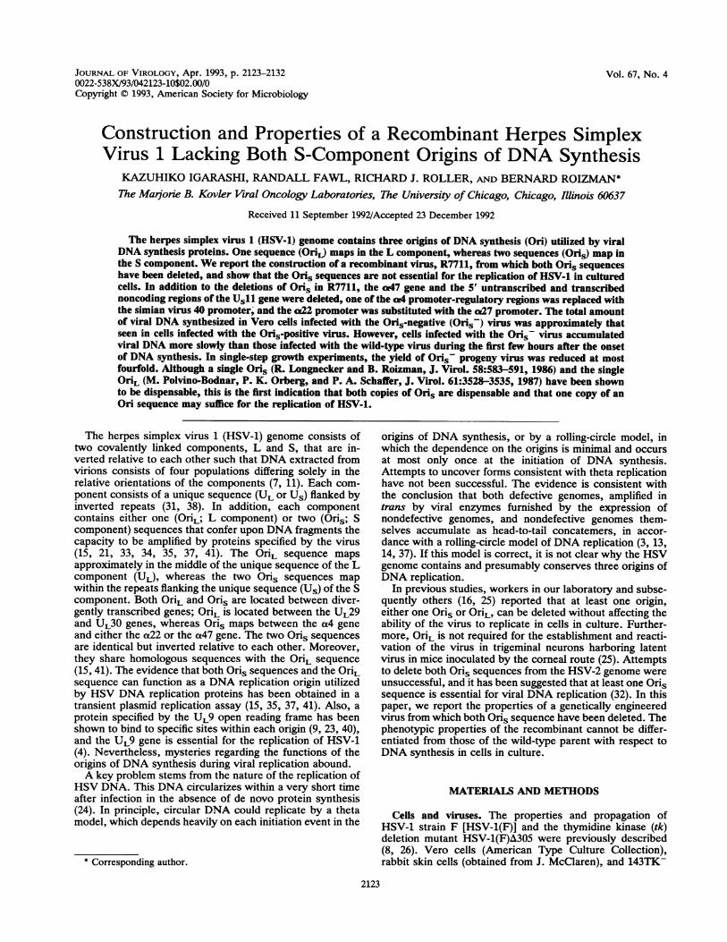
FIG. 1. Schematic construction of recombinant virus R3914. (A) Plasmid construction. Line 1, prototype sequence arrangement of theHSV-1 genome showing unique sequences of the L and S components (thin lines) flanked by inverted repeat sequences (open rectangles). Line2, expanded representation of the left and right ends of the S component, showing the organization of sequences contained within plasmidspRB3094 and pRB421, respectively. Transcribed sequences are indicated by arrows, protein coding sequences are indicated by filled rectangles,and Oris sequences are indicated by open rectangles. Line 3, right, pRB421 was altered by end filling of the unique EcoRI site (shown inparentheses) and by insertion of an EcoRI linker into the NruI site to create plasmid pRB441. To simplify the presentation of the construction,in this diagram of pRB441, we designated the sequences between the SalI and BstEII sites with the letter a, those between the BstEII site andthe EcoRI insert with the letter b, and those between the EcoRI insert and the end-filled EcoRI site with the letter c. Line 3, left, the sequencesbetween the Sall and HindIII sites of pRB3094 were replaced with the HindIII-NdeI fragment of pSV2CAT (10), which contains the SV40enhancer-promoter sequences from PvuII to HindIII (hatched rectangle) and the pBR322 sequences from NdeI to PvuII (open rectangle). Theresulting plasmid, pRB3906, had the a4 leader coding sequences under the control of the SV40 promoter (Psv4O); this chimeric construct couldbe excised as an NdeI-EcoRI fragment, designated by the letter d. Line 4, the b sequences of pRB441 were replaced with the d sequences ofpRB3906, so that the segments of pRB441 designated a and c would provide flanking sequences for the Psv4O-a4 construct (in the order a-d-c)within the resulting plasmid, pRB3915. (B) Virus construction. Line 1, DNA sequence arrangement of HSV-1 recombinant R3630 (19) containingdeletions in the natural position of the tk gene and in the S component. Line 2, expanded representation of the S component (right end) ofrecombinant virus R3630, in which the a4-tk chimeric gene has replaced the coding sequences between the BstEII and NruI sites of the a47 gene.Transcribed sequences, protein coding sequences, and Oris sequences are indicated as in panel A; a4 promoter-regulatory sequences are
depicted by a stippled rectangle; and the tk gene 5' transcribed noncoding and coding sequences are indicated by a cross-hatched rectangle. Line3, diagram showing the intended replacement of the a4-tk chimeric gene in R3630 with the chimeric Psv4O-a4 construct in plasmid pRB3915 byrecombination within the regions of Us10 and Oris. Line 4, schematic representation of the S component (right end) of R3914, with a deletionof Oris resulting from recombination within the regions of Us10 and a4. Line 5, expansion of the mutagenized region in R3914, which iscontained within a 2.7-kb novelBamHI fragment. Line 6, three restriction fragments, which together span the mutagenized region in R3914, wereused as probes in Fig. 2. Probe 1, sequences between the left BamHI site and the nearest SphI site in the HSV-1 BamHI Z fragment; probe 2,SV40 promoter-enhancer sequences between the PvuII and HindIII sites excised from pSV2CAT; probe 3, entire HSV-1 BamHI Y fragment.Restriction enzymes: Ba, BamHI; Bst, BstEII; E, EcoRI; H, HindIII; N, NruI; Na, Narl; Nd, NdeI; P, PvuII; S, Sall; Sp, SphI. Note that atthe junction indicated as H/S, the HindIII site but not the Sall site was regenerated.
adjacent to each of the two Oris sequences within theinverted repeats of the S component, i.e., between Oris andthe a22 gene in one repeat and between Oris and the a47gene in the other. However, because such a recombinantcould not be selected in a single step, we resorted tosequential mutagenesis of the Oris sequences.
Construction of HSV-1 lacking one copy of Oris. Duringconstruction of a virus in which a chimeric cx4 gene consist-
ing of the coding domain of the gene fused to the SV40promoter was recombined into the viral genome, a recombi-
nant virus designated R3914 and lacking one copy of the Orissequence was isolated. The parent virus used for this con-
_Wa4
2Na
3
4
Iss
2126 IGARASHI ET AL.
.-I
Ncr
l- 11
o' F.--4.
C
cn -
gr cc =
T)ifC) r-- Nlce N
C
_4_-
-
* b
-- 4
40 -S
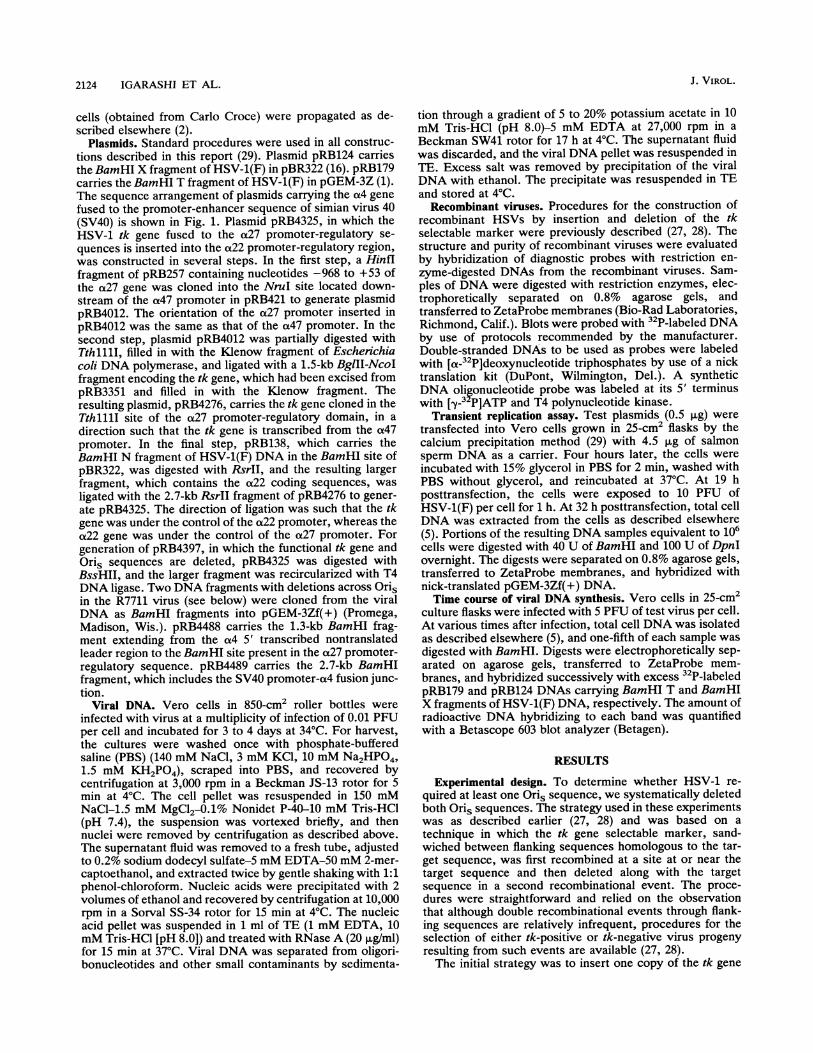
1 2 3 4 1 2 3 4 5 6 1 2 3 4 1 2 3FIG. 2. Autoradiographic images of electrophoretically separated digests of wild-type and recombinant viruses probed for structure. (A)
BamHI digests of R7710 (lanes 1 and 3) and R3914 (lanes 2 and 4) DNAs probed first with 32P-labeled pRB138 DNA (lanes 1 and 2), whichcontains the HSV-1(F) BamHI N DNA fragment in pBR322, stripped, and reprobed with 32P-labeled pRB4540 DNA (lanes 3 and 4), whichcontains the BglII-NruI DNA fragment of the HSV-1 tk gene (20). Bands hybridizing to the HSV-1 sequences are indicated with dots. Theband marked with the arrowhead is the 2.7-kb fragment containing the SV40 promoter-a4 chimeric gene and pBR322 sequences hybridizingwith the probes (Fig. 1B, line 4). (B) BamHI digests of R3914 (lanes 1 and 4), R7710 (lanes 2 and 5), and R7711 (lanes 3 and 6) DNAs probedfirst with the 50-mer oligodeoxynucleotide probe shown in Fig. 3, line 2 (lanes 4 to 6), and then reprobed with the 1.6-kb BamHI-SacI DNAfragment of pRB138, which contains sequences between the left BamHI site and the Sacl site of the a22 gene, including Oris (lanes 1 to 3).(C) BamHI digests of HSV-1(F) (lane 1), R3914 (lane 2), R7710 (lane 3), and R7711 (lane 4) DNAs probed with the 32-mer oligodeoxynu-cleotide probe shown in Fig. 3, line 3. (D) BamHI-BssHII digests of R3914 (lane 1), R7710 (lane 2), and R7711 (lane 3) DNAs probed withthe same 1.6-kb BamHI-SacI DNA fragment of pRB138 as that used in panel B. The positions of marker DNAs are indicated to the left ofeach blot; their sizes are 8.5, 7.2, 6.4, 5.7, 4.8, 4.3, 3.7, 2.3, and 1.9 kb. In panel D, the positions of 1.4-, 1.3-, and 0.7-kb bands are also shown.
struction, R3630 (19), carried an a(4 promoter-tk chimericgene in place of the a47 gene (Fig. 1B, line 2). DNA ofplasmid pRB3915 (Fig. 1A, line 4) was cotransfected withR3630 DNA to isolate a tk-negative virus. The possible viralDNA structures within the mutagenized region resultingfrom the double recombinational event in which the tk genewas deleted are depicted in lines 3 and 4 of Fig. 1B. If theright-hand crossover relative to the SV40 promoter occurredwithin the region near Oris, the progeny virus would acquirethe structure depicted in line 3, in which a total of threecopies of the ot4 gene would be present in the viral genomeand one copy of the a4 gene would be fused to the SV40promoter. If the right-hand crossover relative to the SV40promoter occurred within the a4 gene domain, the progenyvirus would acquire the structure depicted in line 4, in whichone of two copies of the a4 gene would be fused to the SV40promoter and one of the Oris sequences would be deleted.On the basis of tests defined by two criteria (see below), weconcluded that isolate R3914 has the structure depicted inFig. 1B, line 4.The first criterion was the presence or absence of a 3.5-kb
BamHI fragment that contained Oris and the 3' portion ofthe a4 gene (Fig. 1B, line 3). As shown in Fig. 2C, lane 2, thediagnostic 3.5-kb fragment was not detected by hybridiza-
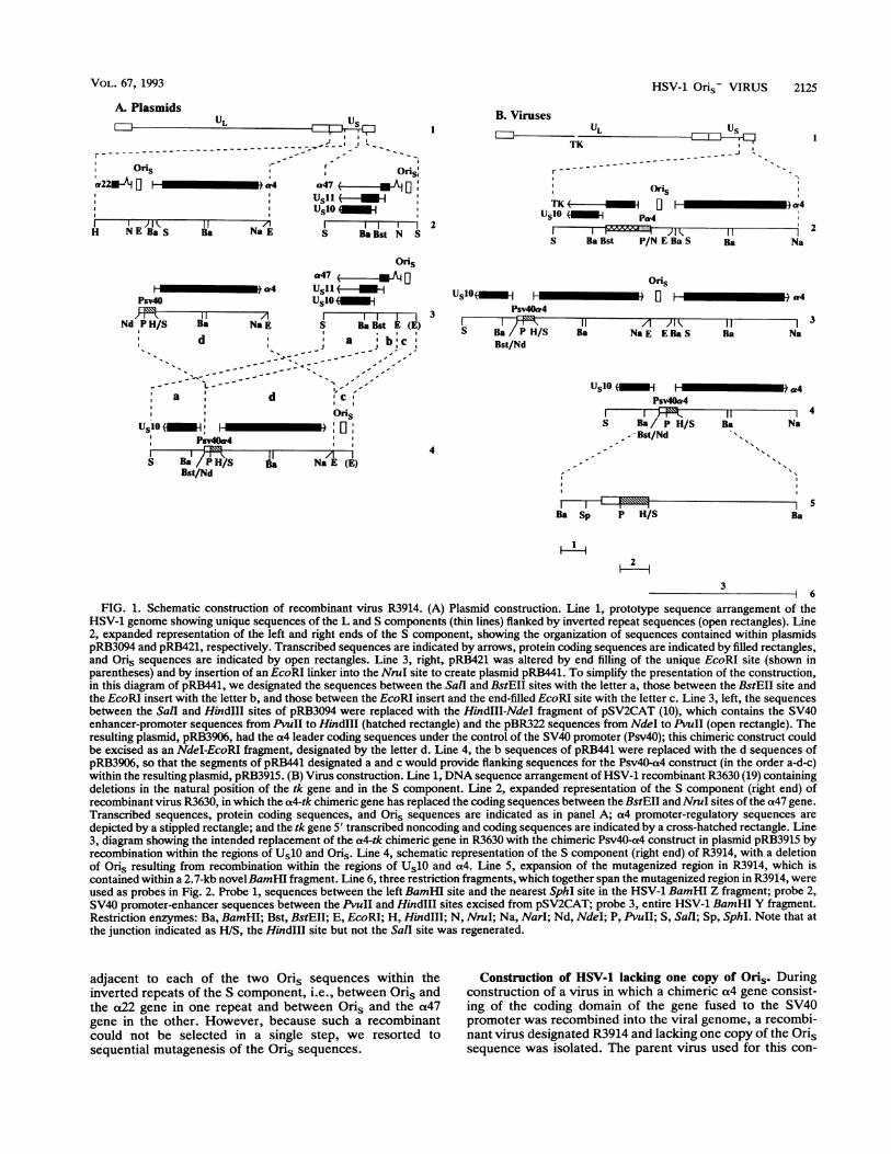
tion of the BamHI digest of R3914 DNA with a labeled DNAprobe specific for Oris and shown in Fig. 3.The second criterion was the location of an SV40 promot-
er-a4 chimeric gene in the DNA fragments generated by Salldigestion. If the recombination yielded the construct shown
FIG. 3. Diagram of the structure of Oris and sequences of theoligodeoxynucleotides that were used as probes for Oris sequences.Line 1, schematic structure of Oris. Rectangles labeled I to IIIrepresent UL9 protein binding sites (I and II) or a presumed bindingsite (III) (9, 22, 39). A/T represents an AlT-rich sequence. Lines 2and 3 show the synthetic oligodeoxynucleotides that served asprobes for Oris sequences in viral DNAs in the studies shown in Fig.2. The 50-mer and 32-mer sequences show 80% (40 of 50) and 97%(31 of 32) identity to OriL, respectively.
J. VIROL.
I
qp
HSV-1 Oris- VIRUS 2127
BamHI BamHl BamHl/HindIllW. WA_.f
_l% 01,M r=;X - X,, X,v
BamHl BamHl/Hindill
P" II.
_4f-
-, o.-r-
II0L;E'S , uS
~u :
1 2 3
SV404 5 6
SV407 8 9 10 11 12BamHI/SphI
13 14 15 16 17 18BamHI Y
of Bamlit Z
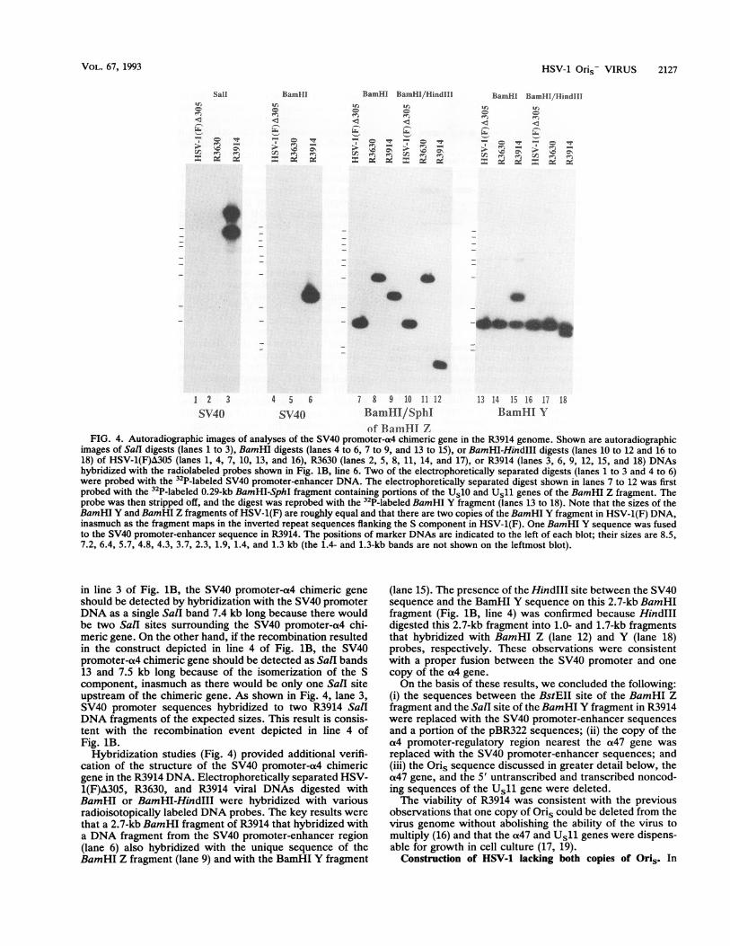
FIG. 4. Autoradiographic images of analyses of the SV40 promoter-a4 chimeric gene in the R3914 genome. Shown are autoradiographicimages of Sall digests (lanes 1 to 3), BamHI digests (lanes 4 to 6, 7 to 9, and 13 to 15), or BamHI-HindIII digests (lanes 10 to 12 and 16 to18) of HSV-1(F)A305 (lanes 1, 4, 7, 10, 13, and 16), R3630 (lanes 2, 5, 8, 11, 14, and 17), or R3914 (lanes 3, 6, 9, 12, 15, and 18) DNAshybridized with the radiolabeled probes shown in Fig. 1B, line 6. Two of the electrophoretically separated digests (lanes 1 to 3 and 4 to 6)were probed with the 32P-labeled SV40 promoter-enhancer DNA. The electrophoretically separated digest shown in lanes 7 to 12 was firstprobed with the 32P-labeled 0.29-kb BamHI-SphI fragment containing portions of the Us10 and Us11 genes of the BamHI Z fragment. Theprobe was then stripped off, and the digest was reprobed with the 32P-labeled BamHI Y fragment (lanes 13 to 18). Note that the sizes of theBamHI Y and BamHI Z fragments of HSV-1(F) are roughly equal and that there are two copies of the BamHI Y fragment in HSV-1(F) DNA,inasmuch as the fragment maps in the inverted repeat sequences flanking the S component in HSV-1(F). One BamHI Y sequence was fusedto the SV40 promoter-enhancer sequence in R3914. The positions of marker DNAs are indicated to the left of each blot; their sizes are 8.5,7.2, 6.4, 5.7, 4.8, 4.3, 3.7, 2.3, 1.9, 1.4, and 1.3 kb (the 1.4- and 1.3-kb bands are not shown on the leftmost blot).
in line 3 of Fig. 1B, the SV40 promoter-a4 chimeric geneshould be detected by hybridization with the SV40 promoterDNA as a single SalI band 7.4 kb long because there wouldbe two SalI sites surrounding the SV40 promoter-a4 chi-meric gene. On the other hand, if the recombination resultedin the construct depicted in line 4 of Fig. 1B, the SV40promoter-a4 chimeric gene should be detected as Sall bands13 and 7.5 kb long because of the isomerization of the Scomponent, inasmuch as there would be only one Sall siteupstream of the chimeric gene. As shown in Fig. 4, lane 3,SV40 promoter sequences hybridized to two R3914 SalIDNA fragments of the expected sizes. This result is consis-tent with the recombination event depicted in line 4 ofFig. 1B.
Hybridization studies (Fig. 4) provided additional verifi-cation of the structure of the SV40 promoter-a4 chimericgene in the R3914 DNA. Electrophoretically separated HSV-1(F)A305, R3630, and R3914 viral DNAs digested withBamHI or BamHI-HindIII were hybridized with variousradioisotopically labeled DNA probes. The key results werethat a 2.7-kb BamHI fragment of R3914 that hybridized witha DNA fragment from the SV40 promoter-enhancer region(lane 6) also hybridized with the unique sequence of theBamHI Z fragment (lane 9) and with the BamHI Y fragment
(lane 15). The presence of the HindIII site between the SV40sequence and the BamHI Y sequence on this 2.7-kb BamHIfragment (Fig. 1B, line 4) was confirmed because HindIIIdigested this 2.7-kb fragment into 1.0- and 1.7-kb fragmentsthat hybridized with BamHI Z (lane 12) and Y (lane 18)probes, respectively. These observations were consistentwith a proper fusion between the SV40 promoter and onecopy of the a4 gene.On the basis of these results, we concluded the following:
(i) the sequences between the BstEII site of the BamHI Zfragment and the SalI site of the BamHI Y fragment in R3914were replaced with the SV40 promoter-enhancer sequencesand a portion of the pBR322 sequences; (ii) the copy of thea4 promoter-regulatory region nearest the a47 gene wasreplaced with the SV40 promoter-enhancer sequences; and(iii) the Oris sequence discussed in greater detail below, thea47 gene, and the 5' untranscribed and transcribed noncod-ing sequences of the Usll gene were deleted.The viability of R3914 was consistent with the previous
observations that one copy of Oris could be deleted from thevirus genome without abolishing the ability of the virus tomultiply (16) and that the aL47 and Us11 genes were dispens-able for growth in cell culture (17, 19).
Construction of HSV-1 lacking both copies of Ori5. In
Sall
c T
xS -
- E
VOL. 67, 1993
40 a
2128 IGARASHI ET AL.
Jl UL us,_____ ______---------------@, _
Oris _- OIO°fia4 V - a22 a0-4u7a4
Ba S Ba Bss N Ba Ba BaBst N Bss Ba S Ba
Bam Hi N Bam HI Z
USIO a a4
Ba S Ba Bss N Ba Ba Psv4O Ba
A'Oris TK
a4 _1W- 0 HOW' _ a22a b a
Ba SBaBss Baa Ba BaTK Pa27
a4 in-4 I-*.
b a
Ba SBa Bss BaPa27
L 11.J
Ba
FIG. 5. Summary of genome structures of recombinant viruses.Line 1, schematic representation of the prototype HSV-1 genomearrangement. Line 2, sequence organization of the BamHI N and Zfragments at the left and right ends of the unique short component ofthe HSV-1 genome, respectively. Transcribed sequences are indi-cated by arrows, protein coding sequences are indicated by filledrectangles, and Oris sequences are indicated by open rectangles.The positions of the BamHI N and Z fragments of HSV-1 are
indicated by the double-headed arrows. Line 3, correspondingsequence organization in recombinant R3914. The sequence locatedbetween the BstEII and SaII sites of HSV-1(F)A305, shown in line 2,which includes most of the BamHI Z fragment, was replaced withthe SV40 enhancer-promoter fragment (rectangle b) and NdeI-PvuIIfragment of pBR322 (rectangle a). The unique 2.7-kb BamHI frag-ment spanning this mutation (line A) was cloned from the progeny
virus R7711, giving rise to pRB4489. Line 4, same as line 2, exceptthat the structure of virus R7710 is shown. Rectangle b, tk gene
coding sequence; rectangle a, a27 promoter-regulatory sequence.Line 5, same as line 3, except that the structure of recombinantR7711 is shown. The BamHI DNA fragment that contained thejunction of the deletion (line B) was cloned into pGEM-3Zf(+) toyield pRB4488. Restriction enzymes: Ba, BamHI; Bss, BssHII; Bst,BstEII; N, NruI; S, SalIl.
wild-type HSV-1, Oris is located in the promoter-regulatorysequences of both theat22 and the a47 genes (Fig. 5, line 2).In making insertions and deletions around the remainingcopy of Oris in R3914, we wanted to ensure continuedexpression of the aL22 gene, since this gene is important forgrowth in primary human and rodent cells (30) and since theproperties of a double deletion ofao47 and a22 could not bepredicted. To accomplish this, we created two plasmids: (i)pRB4325 (Fig. 5, line 4), in which both the tk selectablemarker and the promoter-regulatory sequences of the(x27gene were inserted near Oris in thea22 gene in the BamHIN fragment such that the tk gene was under the control of theat22 promoter, whereas the a22 gene was under the controlof the aL27 promoter, and (ii) pRB4397 (Fig. 5, line 5), inwhich Oris, theaL22 promoter-regulatory sequences, and thetk selectable marker were deleted. The deletion in pRB4397did not remove any TAATGARAT motifs, important fora4gene expression, from the region upstream of the a4 gene
promoter. The tk insertion plasmid pRB4325 was cotrans-fected with R3914 viral DNA, and the tk-positive virusR7710 was isolated from the transfection progeny. Subse-
quently, pRB4397 was cotransfected with R7710 viral DNA,and the Oris-negative tk-negative virus R7711 was isolated.For confirmation of both the purity and the presence of the
appropriate insertions and deletions in R7710 and R7711,their DNAs and that of R3419 were digested with restrictionenzymes, electrophoretically separated, blotted, and probedwith radiolabeled DNAs containing either sequences fromthe BamHI N fragment or a portion of the tk gene (Fig. 2).The BamHI N fragment of HSV-1(F) and R3914 is -4.9 kb
in length. Insertion of the tk gene and a27 promoter se-quences into the BamHI N fragment expanded the fragmentand resulted in a new BamHI cleavage site (Fig. 5, line 4),dividing the BamHI N fragment into two fragments. ForR7710, these fragments were expected to be 4.4 and 3.0 kb insize. Digestion of R3914 and R7710 DNAs with BamHIreleased fragments that were of the expected sizes and thathybridized to probe sequences derived from BamHI N (Fig.2A and B, lanes 1 and 2). Furthermore, as expected, the3.0-kb fragment from R7710 also hybridized to a probecontaining tk gene sequences (Fig. 2A, lane 3). In R7711, thesmaller fragment hybridizing to the BamHI N probe wasreduced to 1.3 kb in size (Fig. 2B, lane 3), consistent with thedeletion of the Oris, tk, and a22 sequences.For ensuring that the region containing Oris had been
deleted in R7711, electrophoretically separated BamHI andBssHII digests of R3914, R7710, and R7711 DNAs wereprobed with sequences from the BamHI N fragment (Fig.2D). As expected, the 1.7-kb BssHII fragment of R7710,which contained the Oris sequence, a22 promoter-regula-tory sequences, and part of the tk gene, was absent fromR7711.Absence of Oris sequences and function in R7711. The
minimum sequence required for the Oris replication functionis quite small (6, 36); the smallest HSV-1 DNA fragmentshown to possess replication origin function is 67 bp inlength (6). Such a small sequence, if retained in R7711 bysome rearrangement, might not be detected by the relativelylow resolution analyses described above. For addressing thisissue, the same DNA blot as that shown in lanes 1 to 3 in Fig.2B was probed with an oligodeoxynucleotide probe contain-ing Oris sequence (Fig. 2B, lanes 4 to 6). The sequence of the50-mer oligodeoxynucleotide probe (Fig. 3, line 2) is con-tained within the minimal HSV-1 DNA segment required foramplification by viral factors, and mutations in this sequenceabolish the ability of the DNA to act as an origin (18, 40).The BamHI N fragment (4.9 kb) of R3914 DNA (Fig. 2B,lane 4) and the 3.0-kb fragment derived from the chimericBamHI N fragment of R7710 DNA (Fig. 2B, lane 5) hybrid-ized with this probe, consistent with the localization of Orison these fragments. The absence of other hybridizing bandsis consistent with the absence of Oris at the other end of Us.In contrast, R7711 DNA failed to hybridize with this probe(Fig. 2B, lane 6), suggesting a complete loss of Oris se-quences from this virus.An additional hybridization experiment was carried out
with a shorter oligodeoxynucleotide probe to detect simul-taneously both Oris and OriL sequences (Fig. 2C). The32-mer oligodeoxynucleotide (Fig. 3, line 3) is shorter inlength and shows greater similarity to Ori, than that shownin Fig. 3, line 2, and used in the hybridization experimentsdescribed above. In BamHI digests of HSV-1(F) DNA, thisprobe detected the Oris sequences as two stronger bands(BamHI N and Z fragments) and the OriL sequence as aweaker band (BamHI V fragment). Both R3914 and R7710yielded only one stronger band in addition to the weakerband, indicating that these viruses possessed only one copy
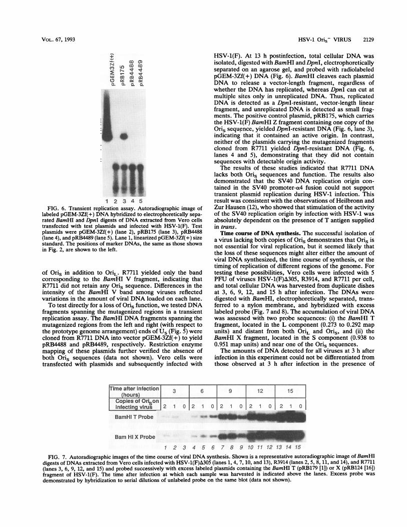
labeled pGEM-3Zf(+) DNA hybridized to electrophoretically sepa-rated BamHI and DpnI digests of DNA extracted from Vero cellstransfected with test plasmids and infected with HSV-1(F). Testplasmids were pGEM-3Zf(+) (lane 2), pRB175 (lane 3), pRB4488(lane 4), and pRB4489 (lane 5). Lane 1, linearized pGEM-3Zf(+) sizestandard. The positions of marker DNAs, the same as those shownin Fig. 2, are shown to the left.
of Oris in addition to OriL. R7711 yielded only the bandcorresponding to the BamHI V fragment, indicating thatR7711 did not retain any Oris sequence. Differences in theintensity of the BamHI V band among viruses reflectedvariations in the amount of viral DNA loaded on each lane.To test directly for a loss of Oris function, we tested DNA
fragments spanning the mutagenized regions in a transientreplication assay. The BamHI DNA fragments spanning themutagenized regions from the left and right (with respect tothe prototype genome arrangement) ends of Us (Fig. 5) werecloned from R7711 DNA into vector pGEM-3Zf(+) to yieldpRB4488 and pRB4489, respectively. Restriction enzymemapping of these plasmids further verified the absence ofboth Oris sequences (data not shown). Vero cells were
transfected with plasmids and subsequently infected with
HSV-1(F). At 13 h postinfection, total cellular DNA wasisolated, digested with BamHI and DpnI, electrophoreticallyseparated on an agarose gel, and probed with radiolabeledpGEM-3Zf(+) DNA (Fig. 6). BamHI cleaves each plasmidDNA to release a vector-length fragment, regardless ofwhether the DNA has replicated, whereas DpnI can cut atmultiple sites only in unreplicated DNA. Thus, replicatedDNA is detected as a DpnI-resistant, vector-length linearfragment, and unreplicated DNA is detected as small frag-ments. The positive control plasmid, pRB175, which carriesthe HSV-1(F) BamHI Z fragment containing one copy of theOris sequence, yielded DpnI-resistant DNA (Fig. 6, lane 3),indicating that it contained an active origin. In contrast,neither of the plasmids carrying the mutagenized fragmentscloned from R7711 yielded DpnI-resistant DNA (Fig. 6,lanes 4 and 5), demonstrating that they did not containsequences with detectable origin activity.The results of these studies indicated that R7711 DNA
lacks both Oris sequences and function. The results alsodemonstrated that the SV40 DNA replication origin con-tained in the SV40 promoter-a4 fusion could not supporttransient plasmid replication during HSV-1 infection. Thisresult was consistent with the observations of Heilbronn andZur Hausen (12), who showed that stimulation of the activityof the SV40 replication origin by infection with HSV-1 wasabsolutely dependent on the presence of T antigen suppliedin trans.Time course of DNA synthesis. The successful isolation of
a virus lacking both copies of Oris demonstrates that Oris isnot essential for viral replication, but it seemed likely thatthe loss of these sequences might alter either the amount ofviral DNA synthesized, the time course of synthesis, or thetiming of replication of different regions of the genome. Fortesting these possibilities, Vero cells were infected with 5PFU of viruses HSV-1(F)A305, R3914, and R7711 per cell,and total cellular DNA was harvested from duplicate dishesat 3, 6, 9, 12, and 15 h after infection. The DNAs weredigested with BamHI, electrophoretically separated, trans-ferred to a nylon membrane, and hybridized with excesslabeled probe (Fig. 7 and 8). The accumulation of viral DNAwas assessed with two probe sequences: (i) the BamHI Tfragment, located in the L component (0.273 to 0.292 mapunits) and distant from both OriL and Ons, and (ii) theBamHI X fragment, located in the S component (0.938 to0.951 map units) and near one of the Ons sequences.The amounts of DNA detected for all viruses at 3 h after
infection in this experiment could not be differentiated fromthose observed at 3 h after infection in the presence of
ime after infection 3 6 19 12 15Copiso O
Copies of Orisoninfecting virus 2
BamHI T Probe
1 0 2 1 0 2
Bam Hi X Probe
1 2 3 4 5 6 7 8 9 10 11 12 13 14 15
FIG. 7. Autoradiographic images of the time course of viral DNA synthesis. Shown is a representative autoradiographic image of BamHI
digests of DNAs extracted from Vero cells infected with HSV-1(F)A305 (lanes 1, 4, 7, 10, and 13), R3914 (lanes 2, 5, 8, 11, and 14), and R7711(lanes 3, 6, 9, 12, and 15) and probed successively with excess labeled plasmids containing the BamHI T (pRB179 [1]) or X (pRB124 [16])fragment of HSV-1(F). The time after infection at which each sample was harvested is indicated above the lanes. Excess probe wasdemonstrated by hybridization to serial dilutions of unlabeled probe on the same blot (data not shown).
0 2 2 1 0dbu&bl
VOL. 67, 1993
1 01
2130 IGARASHI ET AL.
2 4001 1/200 /2 ~~~~~~~~~~~~~0)
200- 100
0 -- 00 3 6 9 12 15 0 3 6 9 12 15
Time after infection (hours)
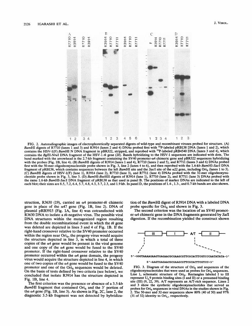
FIG. 8. Graphic representation of the time course of viral DNA synthesis. (A) The amount of labeled BamHI T fragment hybridized (incounts per minute) to HSV-1(F)A&305, R3914, and R7711 DNAs recovered from infected Vero cells as a function of time postinfection (inhours) is shown. Each time point is the mean of two independent infections. (B) As in panel A, except that the BamHI X fragment was usedas the hybridization probe.
cycloheximide (data not shown). Therefore, the amounts ofDNA detected at 3 h indicated the levels of unreplicatedinput DNA. Amplification of DNA could be detected in allinfected-cell samples harvested at 6 h postinfection, suggest-ing that the deletion of one or both copies of Oris did notcause a delay in the onset of viral DNA synthesis. Althoughthe amounts of viral DNA made by R7711 were reduced at 9h, all three viruses showed similar time courses of DNAaccumulation and produced nearly equal amounts of prog-eny DNA. These results indicate that the viruses lackingboth Oris sequences are not significantly or demonstrablyimpaired with respect to their capacity to make viral DNA.
Effect of the deletion of both Oris sequences on virusmultiplication. Vero cells grown in 25-cm2 dishes wereinfected at either a low (0.01 PFU per cell) or a high (5 PFUper cell) multiplicity of infection with HSV-1(F)A305, R3914,or R7711. At 24 h after infection, cells were lysed by a singlecycle of freezing and thawing and sonicated, and the titer ofinfectious virus in each lysate was determined by a plaqueassay with Vero cells (Table 1). Both R3914 and R7711showed small reductions in virus yield at 24 h. These
TABLE 1. Virus yields from Vero cells at 24 h postinfection
Virus yield, PFU/ml of culture lysate, at aVirus multiplicity of infection ofa:
5 PFU/celi 0.01 PFU/cell
HSV-1(F)A305 4.7 x 10⁷ (1) 9.3 x 10⁶ (1)R3914 3.3 x 10⁷ (0.70) 5.4 x 10⁶ (0.58)R7711 2.5 x 10⁷ (0.53) 2.7 x 10⁶ (0.29)
a Mean of duplicate infections. Titration was done with Vero cells. Theyields of the recombinant viruses relative to those of the parent virus,HSV-1(F)A305, are shown in parentheses.
reductions were more apparent at a low multiplicity ofinfection but were at most fourfold for R7711. Both mutantviruses also multiplied efficiently in 143TK-, BHK, andHEp-2 cells (data not shown).
DISCUSSIONWe report that we have deleted both Oris sequences from
HSV-1 recombinant virus R7711. Evidence supporting thisconclusion is based on both hybridization analyses of theviral DNA with specific probes and functional assays show-ing that the DNA sequences that in the wild-type viruscontain the Oris sequences are not amplified in trans byHSV-1 proteins expressed in the transfected cells by super-infecting virus.The Oris-negative virus R7711 yielded approximately
fourfold less infectious progeny than the parent virus fromwhich it was derived in single-step growth experiments at alow multiplicity of infection. Cells infected with the viruslacking both Oris sequences showed a demonstrable in-crease in DNA content at 6 h postinfection and, although theamounts of viral DNA were clearly and reproducibly re-duced at 9 h postinfection, appeared to accumulate DNA atleast as rapidly as cells infected with the wild-type virusthereafter. The basis of the reduced accumulation at 9 hpostinfection requires further investigation, but we speculatethat the R7711 virus is delayed in the transition from anearly, slow phase of replication to a late, rapid phase. Thetotal amounts of viral DNA accumulated in cells infectedwith wild-type and mutant viruses by 15 h after infectionwere approximately similar, suggesting that the decreasedvirus yield of R7711 after single-step replication did notresult directly from a defect in DNA synthesis. Indeed, it ispossible that the small difference between R7711 and HSV-1(F)A305 in virus yield is unrelated to the Oris deletion,
J. VIROL.
HSV-1 Oris- VIRUS 2131
since our manipulations affected the structure of at least fourviral genes in addition to Oris.We conclude on the basis of our data as well as on the
basis of the reports published previously (16, 25) the follow-ing. (i) Both Oris sequences are dispensable for viral repli-cation in cells in culture. (ii) The evidence indicates that if anorigin of viral DNA synthesis is required for the amplifica-tion of a wild-type genome, 0riL is sufficient for this pur-pose. (iii) Inasmuch as the OriL sequence was shown to bedispensable under conditions in which both Oris sequenceswere intact (25) and in this study we showed that both Orissequences are dispensable, we may conclude that none ofthe three origins is uniquely required for viral replication incell culture. Rather, if an origin ofDNA synthesis is requiredat all, any one of the three origins will most likely suffice.
In view of the evidence that the absence of two origins ofDNA synthesis does not grossly affect the quantity of viralDNA synthesized, the reason for the evolution and conser-vation of three origins of DNA synthesis remains obscure.Three broad hypotheses can address this point: (i) Oris andOriL mediate different types of replication initiation (i.e.,mediate the assembly of different types of replication com-plexes, perhaps leading to different mechanisms of replica-tion); (ii) the two origin types are used differently in differentcells or at specific stages of the viral life cycle (e.g., lyticversus latent infection); and (iii) these origins are indeedfunctionally redundant, but the presence of multiple originsin the virus genome increases the probability and/or the rateof successful initiation of DNA replication or decreases thetime required to replicate genome DNA. Interestingly, it canbe argued that the placement of each of the three origins inthe HSV-1 genome could conceivably provide for theirconservation independently of selective pressure derivedfrom their function in DNA replication. OriL maps betweenthe genes encoding the single-stranded DNA binding proteinICP8 and the viral DNA polymerase. Although the viabilityof the Ori.-negative virus (25) demonstrates that the OriLsequence is not essential for the expression of these genes,the OriL sequence may contribute to proper temporal andquantitative expression. The Oris sequences are present inreiterated sequences, and their stability could conceivablybe maintained by a sequence conversion mechanism, even inthe absence of selective pressure.
ACKNOWLEDGMENTS
This study was aided by Public Health Service grants from theNational Cancer Institute (CA47451) and the National Institute ofAllergy and Infectious Diseases (AI124009) and by an unrestrictedgrant from the Bristol-Myers Squibb Program in Infectious Dis-eases.
REFERENCES1. Baines, J. D. 1992. Unpublished data.2. Baines, J. D., and B. Roizman. 1991. The open reading frames
UL3, UL4, UL10, and UL16 are dispensable for the growth ofherpes simplex virus 1 in cell culture. J. Virol. 65:938-944.
3. Becker, Y., Y. Asher, E. Weinberg-Zahlering, S. Rabkin, A.Friedmann, and E. Kessler. 1978. Defective herpes simplexvirus DNA: circular and circular-linear molecules resemblingrolling circles. J. Gen. Virol. 40:319-335.
4. Carmichael, E. P., M. J. Kosovsky, and S. K. Weller. 1988.Isolation and characterization of herpes simplex virus type 1host range mutants defective in viral DNA synthesis. J. Virol.62:91-99.
5. Challberg, M. D. 1986. A method for identifying the viral genesrequired for herpesvirus DNA replication. Proc. Natl. Acad.Sci. USA 83:9094-9098.
6. Deb, S., and M. Doelberg. 1988. A 67-base-pair segment fromthe Oris region of herpes simplex virus type 1 encodes originfunction. J. Virol. 62:2279-2286.
7. Delius, H., and J. B. Clements. 1976. A partial denaturation mapof herpes simplex virus type 1 DNA: evidence for inversions ofthe unique DNA regions. J. Gen. Virol. 33:125-133.
8. Ejercito, P. M., E. D. Kieff, and B. Roizman. 1968. Character-ization of herpes simplex virus strains differing in their effectson social behavior of infected cells. J. Gen. Virol. 2:357-364.
9. Elias, P., C. M. Gustafsson, and 0. Hammarsten. 1990. Theorigin binding protein of herpes simplex virus 1 binds coopera-tively to the viral origin of replication Oris. J. Biol. Chem.265:17167-17173.
10. Gorman, C. M., L. A. Moffat, and B. H. Howard. 1982.Recombinant genomes which express chloramphenicol acetyl-transferase in mammalian cells. Mol. Cell. Biol. 2:1044-1051.
11. Hayward, G. S., R. J. Jacob, S. C. Wadsworth, and B. Roizman.1975. Anatomy of herpes simplex virus DNA: evidence for fourpopulations of molecules that differ in the relative orientationsof their long and short segments. Proc. Natl. Acad. Sci. USA72:4243-4247.
12. Heilbronn, R., and H. ZurHausen. 1989. A subset of herpessimplex virus replication genes induces DNA amplificationwithin the host cell genome. J. Virol. 63:3683-3692.
13. Jacob, R. J., and B. Roizman. 1979. Anatomy of herpes simplexvirus DNA. XII. Accumulation of head-to-tail concatamers innuclei of infected cells and their role in the generation of fourisomeric arrangements of viral DNA. J. Virol. 29:448-457.
14. Jongeneel, C. V., and S. L. Bachenheimer. 1981. Structure ofreplicating herpes simplex virus DNA. J. Virol. 39:656-660.
15. Lockshon, D., and D. A. Galloway. 1986. Cloning and charac-terization of OriL2, a large palindromic DNA replication originof herpes simplex virus type 2. J. Virol. 58:513-521.
16. Longnecker, R., and B. Roizman. 1986. Generation of an invert-ing herpes simplex virus 1 mutant lacking the L-S junction asequences, an origin of DNA synthesis, and several genesincluding those specifying glycoprotein E and the a47 gene. J.Virol. 58:583-591.
17. Longnecker, R., and B. Roizman. 1987. Clustering of genesdispensable for growth in culture in the S component of theHSV-1 genome. Science 236:573-576.
18. Martin, D. W., S. P. Deb, J. S. Klauer, and S. Deb. 1991.Analysis of the herpes simplex virus type 1 Oris sequence:mapping of functional domains. J. Virol. 65:4359-4369.
19. Mavromara-Nazos, P., M. Ackermann, and B. Roizman. 1986.Construction and properties of a viable herpes simplex virus 1recombinant lacking coding sequences of the a47 gene. J. Virol.60:807-812.
20. Michael, N. 1992. Unpublished data.21. Mocarski, E. S., and B. Roizman. 1981. Site specific inversion
sequence of herpes simplex virus genome: domain and struc-tural features. Proc. Natl. Acad. Sci. USA 78:7047-7051.
22. Mocarski, E. S., and B. Roizman. 1982. Herpesvirus-dependentamplification and inversion of a cell-associated viral thymidinekinase gene flanked by viral a sequence and linked to an originof viral DNA replication. Proc. Natl. Acad. Sci. USA 79:5626-5630.
23. Olivo, P. D., N. J. Nelson, and M. D. Challberg. 1988. Herpessimplex virus DNA replication: the UL9 gene encodes an originbinding protein. Proc. Natl. Acad. Sci. USA 85:5414-5418.
24. Poffenberger, K. L., and B. Roizman. 1985. A noninvertinggenome of a viable herpes simplex virus 1: presence of head-to-tail linkages in packaged genomes and requirements for circu-larization after infection. J. Virol. 53:587-595.
25. Polvino-Bodnar, M., P. K. Orberg, and P. A. Schaffer. 1987.Herpes simplex virus type 1 oriL is not required for virusreplication or for the establishment and reactivation of latentinfection in mice. J. Virol. 61:3528-3535.
26. Post, L. E., S. Mackem, and B. Roizman. 1981. Regulation of agenes of herpes simplex virus: expression of chimeric genesproduced by fusion of thymidine kinase with a gene promoters.Cell 25:227-232.
27. Post, L. E., and B. Roizman. 1981. A generalized technique for
VOL. 67, 1993
2132 IGARASHI ET AL.
deletion of specific genes in large genomes: a gene 22 of herpessimplex virus 1 is not essential for growth. Cell 25:227-232.
28. Roizman, B., and F. J. Jenkins. 1985. Genetic engineering ofnovel genomes of large DNA viruses. Science 229:1208-1214.
29. Sambrook, J., E. F. Fritsch, and T. Maniatis. 1989. Molecularcloning: a laboratory manual. Cold Spring Harbor Laboratory,Cold Spring Harbor, N.Y.
30. Sears, A. E., I. W. Halliburton, B. Meignier, S. Silver, and B.Roizman. 1985. Herpes simplex virus 1 mutant deleted in thea22 gene: growth and gene expression in permissive and restric-tive cells and establishment of latency in mice. J. Virol. 55:338-346.
31. Sheldrick, P., and N. Berthelot. 1975. Inverted repetitions in thechromosome of herpes simplex virus. Cold Spring HarborSymp. Quant. Biol. 39:667-678.
32. Smith, C. A., M. E. Marchetti, P. Edmonson, and P. A. Schaffer.1989. Herpes simplex virus type 2 mutants with deletions in theintergenic region between ICP4 and ICP22/47: identification ofnonessential cis-acting elements in the context of the viralgenome. J. Virol. 63:2036-2047.
33. Spaete, R. R., and N. Frenkel. 1982. The herpes simplex virusamplicon: a new eucaryotic defective-virus cloning-amplifyingvector. Cell 30:295-304.
34. Spaete, R. R., and N. Frenkel. 1985. The herpes simplex virusamplicon: analysis of cis-acting replication functions. Proc.
Natl. Acad. Sci. USA 82:694-698.35. Stow, N. D. 1982. Localization of an origin of DNA replication
within the TRs/IRs repeated region of the herpes simplex virustype 1 genome. EMBO J. 1:863-867.
36. Stow, N. D., and E. C. McMonagle. 1983. Characterization ofthe TRs/IRs origin of DNA replication of herpes simplex virustype 1. Virology 130:427-438.
37. Vlazny, D. A., and N. Frenkel. 1981. Replication of herpessimplex virus DNA: location of replication recognition signalswithin defective virus genomes. Proc. Natl. Acad. Sci. USA78:742-746.
38. Wadsworth, S., R. J. Jacob, and B. Roizman. 1975. Anatomy ofherpes simplex virus DNA. II. Size, composition, and arrange-ment of inverted terminal repetitions. J. Virol. 15:1487-1497.
39. Weir, H. M., J. M. Calder, and N. D. Stow. 1989. Binding of theherpes simplex virus type 1 UL9 gene product to an origin ofviral DNA replication. Nucleic Acids Res. 17:1409-1425.
40. Weir, H. M., and N. D. Stow. 1990. Two binding sites for theherpes simplex virus type 1 UL9 protein are required for efficientactivity of the Oris replication origin. J. Gen. Virol. 71:1379-1385.
41. Weller, S. K., A. Spadoro, J. E. Schaffer, A. W. Murray, A. M.Maxam, and P. A. Schaffer. 1985. Cloning, sequencing, andfunctional analysis of OriL, a herpes simplex virus type 1 originof DNA synthesis. Mol. Cell. Biol. 5:930-942.