Visual evoked potentials elicited by chromatic motion onset
D.J. McKeefry *Vision Science Research Group, School of Biomedical Sciences, Uni�ersity of Ulster, Coleraine, Northern Ireland BT52 1SA, UK
Received 21 December 1999; received in revised form 3 October 2000
Abstract
Visually Evoked Potentials (VEPs) were recorded in response to the onset of chromatic and luminance motion gratings of 1 cpdand luminance 40 cd m−2 subtending a 7° field. At slow speeds (�2 cycles s−1) the motion onset response exhibits a clearamplitude minimum at isoluminance. Over the Michelson contrast range tested (0.05–0.75) the chromatic response at 2 cycles s−1
The discovery that colour and motion are processedby separate cortical areas in the macaque monkey brainprovided the initial inspiration for the theory of func-tional specialisation and segregation within the visualsystem (Zeki, 1978). Subsequent psychophysical andanatomical studies (Ramachandran & Gregory, 1978;Livingstone & Hubel, 1987) appeared to substantiatethe idea that colour and motion are processed byseparate pathways within the visual system. In apparentsupport of this view are reports that motion perceptionis somehow impaired or impoverished at isoluminance(e.g. Ramachandran & Gregory; Cavanagh, Tyler, &Favreau, 1984; Troscianko & Fahle, 1988; Mullen &Boulton, 1992). But it would be erroneous to considerthe motion system as being simply ‘colour blind’ asnumerous studies have shown that colour can giveunambiguous cues about motion (Derrington & Hen-ning, 1993; Cropper & Derrington, 1996; Willis &Anderson, 1998). What is less clear is the extent of
independence or interaction between the chromatic andluminance inputs to the motion system. Some studieshave suggested independence (Krauskopf & Farrell,1990; Metha, Vingrys, & Badcock, 1994; Cropper, Mul-len, & Badcock, 1996), with recent reports raising thepossibility that chromatic motion is processed solely bya ‘third-order’ motion mechanism (Lu, Lesmes, & Sper-ling, 1999). Other studies demonstrate that there arestrong interactions between colour and motion path-ways (Cavanagh & Favreau, 1985; Derrington & Bad-cock, 1985; Kooi & DeValois, 1992; Chichilnisky,Heeger, & Wandell, 1993; Papathomas, Gorea, &Julesz, 1993; Ffytche, Skidmore, & Zeki, 1995; Edwards& Badcock, 1996). A recent model (Gegenfurtner &Hawken, 1996), may provide a limited degree of recon-ciliation between some of these conflicting ideas andproposes the existence of two motion processing path-ways that differ mainly in their temporal properties.One mechanism operates at low temporal rates and hasdifferent channels for luminance and chromatic motion.The other operates at higher temporal rates possessinga single motion channel with both chromatic and lumi-nance inputs. The fast and slow mechanisms are bothsensitive to colour, but they respond in different ways.
* Present address: Dept. Optometry, University of Bradford, Brad-ford BD7 1DP, UK. Tel.: +44-1274-236240; fax: +44-1274-235570.
D.J. McKeefry / Vision Research 41 (2001) 2005–20252006
The latter encodes colour veridically, but not velocity;the former velocity veridically, but not colour (howeversee Gorea, Papathomas, & Kovacs, 1993; Metha &Mullen, 1997; for alternative views).
Visually Evoked Potentials (VEPs) are a measure ofcortical activity in response to a visual stimulus, whichwhen suitably chosen, they can selectively reflect theoperation of specific neural processes (Kulikowski,Robson, & McKeefry, 1996; Kulikowski, McKeefry, &Robson, 1997). Like reaction times, VEPs offer theadvantage of allowing the examination ofsuprathreshold visual performance. Many studies havereported the existence of VEPs that reflect motionrelated processing in the visual system (Clarke, 1972,1973, 1974; Tyler & Kaitz, 1977; Muller & Gopfert,1988; Muller, Gopfert, Schlykowa, & Anke, 1990;Gopfert, Muller, & Simon, 1990; Kuba & Kubova,1992; Bach & Ullrich, 1994; Snowden, Ullrich, & Bach,1995; Kubova, Kuba, Spekreijse, & Blakemore, 1995;Bach & Ullrich, 1997; Odom, DeSmedt, Van Malderen,& Spileers, 1999). The consensus appears to be that theresponse is motion specific if the VEP fulfils the follow-ing criteria: (i) possesses high contrast sensitivity; (ii)exhibits a saturating contrast response characteristic;(iii) is susceptible to motion adaptation. These criteriaare met by the N200 component of the motion onsetVEP (Kuba & Kubova, 1992; Bach & Ullrich, 1994,1997; Kubova et al., 1995) and by steady-state VEPselicited by directional changes in motion (Snowden etal., 1995).
The criteria for motion-specificity have been derivedfrom VEPs generated by luminance motion stimuli.Fewer studies in comparison have examined VEPs elic-ited by chromatically defined motion (Morrone, Fioren-tini, & Burr, 1996). This study will attempt to addressthe basic question of whether motion specific VEPs canbe produced by an isoluminant chromatic motion stim-ulus. Motion specificity will be tested in terms of theadherence of the chromatic motion VEP to criteria (i)and (ii) listed above [adherence to criterion (iii) will bedealt with in a subsequent study].
In addition to the examination of the motion specifi-city of the chromatic motion VEP, I will also testwhether motion VEPs show any sign of the segregation,suggested by the Gegenfurtner & Hawken (1996) modelof motion processing. The rationale is that if separatechannels do exist for luminance and colour at slowspeeds, presumably with separate neural substrates,then this would be revealed by different response prop-erties of the potentials generated by the two types ofmotion. Furthermore, if, as the model also proposes,the mechanisms signalling slow and fast chromatic in-formation are different, one would expect the propertiesof the fast and slow chromatic motion VEPs to exhibitsigns of this segregation.
2. Methods
2.1. VEP recording
VEPs were recorded using silver–silver chloride elec-trodes. An active electrode was placed at Oz and refer-enced to linked ear electrodes with a ground electrodeplaced on the forehead. The VEPs were averaged usinga CED 1401 ‘micro’ and accompanying Signal software(version 1.72). Amplifier (CED 1902) bandwidth was0.5–30 Hz and signals were sampled at a rate of 250 Hzover 1.496 s.
Simultaneous electro-oculogram recordings per-formed on one experienced VEP subject and two naivesubjects, demonstrated that motion VEPs were notcontaminated by eye movement artefacts when subjectswere instructed to maintain fixation on a centrallyplaced cross.
2.2. Subjects
A total of 15 undergraduate and postgraduate stu-dents aged between 22 and 35 years were used assubjects during the course of these series of VEP andpsychophysical experiments (though not all of themtook part in every experiment). All subjects had 6/6 (orbetter) unaided vision or corrected acuity and wereclassified as colour normal according to theFarnsworth–Munsell 100 Hue test.
2.3. Stimuli
Vertically oriented sinusoidal gratings of 1 cpd weregenerated on an Eizo T562-T colour monitor with aframe rate of 120 Hz, under the control of a VSG2/3graphics card (version 5, Cambridge Research Systems).The stimulus subtended a circular field of 7° with aconstant mean luminance of 40 cd m−2 and was sur-rounded by a neutral background (CIE 1931 chromatic-ity co-ordinates x=0.310, y=0.316) of the sameluminance.
The luminance contrast content of the stimulus couldbe systematically varied by manipulation of the relativemean luminance of the red and green phosphors, ex-pressed as the G/(G+R) ratio. G/(G+R) ratios=1and 0 produce green-dark green and red-dark red lumi-nance modulated (achromatic) grating stimuli, respec-tively. At G/(G+R)=0.5 the stimulus takes on theappearance of a bichromatic red–green grating (chro-maticity co-ordinates Rx=0.366, Ry=0.248 and Gx=0.390, Gy=0.517). Calibrations and measurementswere performed using a PR650 Spectrascan SpectraCol-orimeter. Theoretically, a G/(G+R) ratio=0.5 shouldconstitute a purely isoluminant (chromatic) stimulus.However, the isoluminant ratio can vary between sub-jects and chromatic aberration can introduce luminance
D.J. McKeefry / Vision Research 41 (2001) 2005–2025 2007
contrast modulation in an erstwhile isoluminance pat-tern (Charman, 1991). Therefore, prior to the start ofeach recording session, subjects set their own individualisoluminant points using a minimum motion method(Anstis & Cavanagh, 1983) for each of the experimentalconditions employed. The average setting across allsubjects for isoluminance was G/(G+R)=0.45(S.D.=1.38).
The use of a 7° stimulus was necessary in order tominimise the effects of luminance intrusions which havebeen shown to compromise the selectivity and specific-ity of chromatic VEPs in response to isoluminant stim-uli (Kulikowski et al., 1996). These intrusions arisefrom changes in the isoluminant point as a function ofretinal eccentricity and also as a result of chromaticaberrations (both longitudinal and transverse). Empiri-cal evidence suggests that isoluminant red/green grat-ings should contain less than eight cycles in order tominimise such intrusions (Kulikowski et al., 1996).
VEPs were elicited by the motion onset of the verti-cally orientated gratings, the speed of which could bevaried (1–10 cycles s−1). Response averaging was trig-
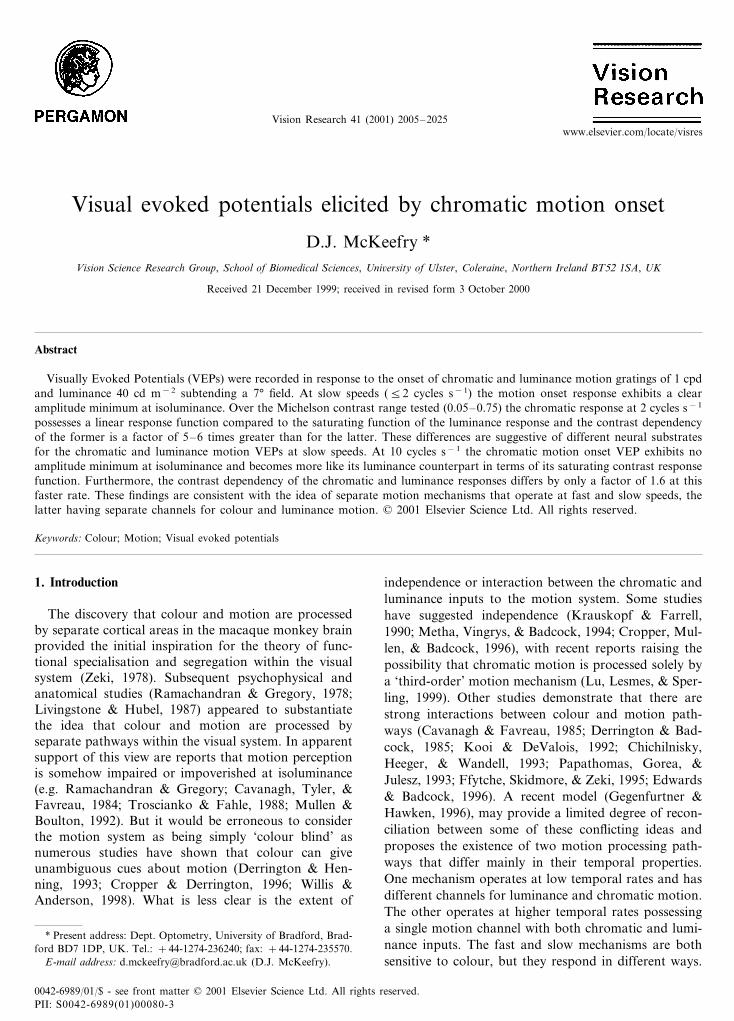
gered by the onset of horizontal motion that lasted 350ms, followed by a stationary grating phase lasting 1170ms (Fig. 1A) giving a duty cycle of approximately 23%.A typical response generated by this stimulus is shownin Fig. 1B, which indicates the major components ofthe motion onset response, and how intra-responseamplitudes and latencies were measured.
2.4. Luminance and chromatic contrast
When comparing chromatic and luminance visualfunction the problem of how to express chromaticmodulation arises (see Lennie & D’Zmura, 1988; Der-rington & Henning, 1993). Achromatic contrast can besimply expressed in terms of the Michelson contrast(Lmax−Lmin/Lmax+Lmin). But for the expression ofchromatic contrast two different approaches have beenadopted. In the majority of instances chromatic con-trast is assigned as being equal to the luminance con-trast of the grating at G/(G+R)=0 or 1. In caseswhere a direct comparison of contrast sensitivity be-tween luminance and chromatic motion was required,chromatic contrast was calculated in terms of L and Mcone modulations. This was done using the luminanceand CIE chromaticity co-ordinates of the red–greenstimulus (x, y, Y) which are then converted to Juddmodified CIE 1931 values (x �, y �, Y �). Calculated X �, Y �and Z � values can be then be used in conjunction withcone fundamentals (e.g. Walraven, 1974; Smith &Pokorny, 1975; Vos, 1978) to obtain a value for coneexcitation by each colour from which modulation canbe calculated. This allows the expression of cone con-trast as a percentage of luminance modulation. For theisoluminant chromatic stimulus, M cone contrast wascalculated as 27% of luminance modulation and L conecontrast as 8%. A mean value of L and M cone contrastwas then employed as a measure of chromatic contrastand used as a scaling factor to adjust chromatic re-sponse data, enabling a more appropriate comparisonto be made with the luminance data.
3. Results
3.1. Motion onset responses as a function of G/(G+R)ratio
The use of gratings of varying G/(G+R) ratio offersthe advantage of allowing a gradual and systematictransformation of the stimulus from achromatic tochromatic. Furthermore, the intermediate G/(G+R)ratios generate stimuli containing a mixture of bothchromatic and luminance contrast. Thus we can ob-serve the effects of such transformations on the motiononset VEP.
Fig. 1. (A). The spatial and temporal configuration of the stimulusused to elicit motion onset VEPs, the leftward pointing arrow indi-cates the direction of motion. Subjects were instructed to fixate on acentrally placed target (not shown). (B). A typical motion onset(luminance) VEP indicating the position, in terms of latency to peakmeasurements, of the main components examined as well as how theamplitudes of the various components were measured.
D.J. McKeefry / Vision Research 41 (2001) 2005–20252008
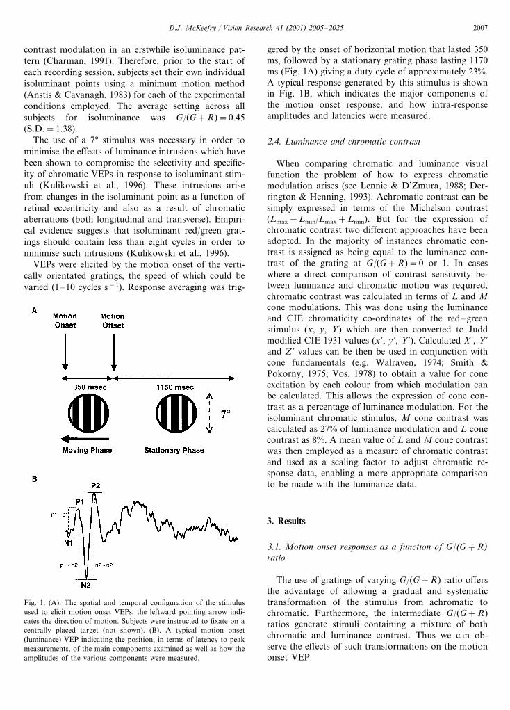
Fig. 2A shows motion onset VEPs elicited by a 2cycles s−1 stimulus as a function of G/(G+R) ratio forsix subjects. As has been described elsewhere (Gallichio& Andreassi, 1982; Gopfert et al., 1990; Kubova et al.,1995; Kuba & Kubova, 1992), the motion onset VEPfor luminance motion (G/(G+R)=0 or 1) exhibits atriphasic positive–negative–positive (P1–N2–P2) com-plex with a prominent negativity occurring at around200 ms (the N200 or N2 component). Fig. 2 (B–D)plots the latency variations of the P1, N2 and P2components as a function of G/(G+R) ratio. It can beseen that as the stimulus tends towards isoluminance(G/(G+R)=0.45), there is an increase in latency forthe P1, N2 and P2 components of the motion onsetVEP, which is highly significant P�0.0001 for all threecomponents (repeated measures ANOVA). In additionto this increase in latency, the motion onset response
also exhibits a reduction in amplitude at isoluminance(Fig. 2 E–G) which is only just significant for then1–p1 (P�0.05) component, but highly significant forthe p1–n2 and n2–p2 (P�0.0001) components. Con-sistent with the findings of Kubova et al. (1995);Spileers, Mangelschots, Maes, and Orban (1996) theearliest n1–p1 component was of small amplitude atthis slow speed.
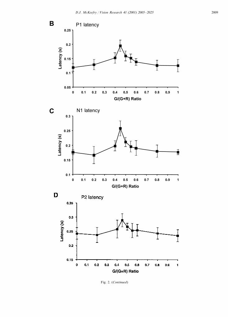
The notion that different mechanisms may subservechromatic motion perception at fast and slow speedsraises the question as to whether the VEP exhibits anysign of this segregation. Fig. 3 shows the responsevariations as a function of G/(G+R) for a 10 cycless−1 stimulus. The most obvious difference between theVEPs generated by the faster and slower motion is thatfor the former a robust motion onset VEP is main-tained at isoluminance. This is indicated in Fig. 3
Fig. 2. (A) Group averaged (n=6) motion onset VEPs elicited as a function of G/(G+R) ratio for a 2 cycles s−1 stimulus. Each trace is theaverage of at least 126 repetitions for each subject. At G/(G+R)=0.45 the stimulus is an isoluminant red/green grating, at values of 0 and 1 thestimulus contains only luminance modulation. The stimulus had a Michelson contrast of 0.25 and mean luminance=40 cd m−2. The responseswere recorded from electrode position Oz referenced to linked ears. (B–D). Latency variation of the P1, N2 and P2 components as a function ofG/(G+R) ratio. (E–G). Amplitude variation of n1–p1, p1–n2 and n2–p2 components as a function of G/(G+R) ratio. The data pointsrepresent the mean across subjects and the bars= �1 S.D.
D.J. McKeefry / Vision Research 41 (2001) 2005–2025 2009
Fig. 2. (Continued)
D.J. McKeefry / Vision Research 41 (2001) 2005–20252010
Fig. 2. (Continued)
D.J. McKeefry / Vision Research 41 (2001) 2005–2025 2011
(E–G) where the amplitude of the motion onset com-ponents are plotted as a function of G/(G+R) ratio atthis faster speed. Unlike for the 2 cycles s−1 data, thereis no statistically significant amplitude minimum atisoluminance for the 10 cycles s−1 stimulus for then1–p1, p1–n2 or n2–p2 components. However, re-sponse latency at this faster rate, as shown in Fig.3(B–D), does increase significantly for all three compo-nents (P1, P�0.005; N2, P�0.001; P2, P�0.05).
3.2. Contrast response functions of chromatic andluminance motion onset VEPs
The dependence of the luminance motion onset VEPupon contrast has been previously examined (Muller &Gopfert, 1988; Snowden et al., 1995; Kubova et al.,1995; Bach & Ullrich, 1997) and has been significant inthe ascription of motion specificity to this response. Inthe next experiment the contrast dependence of thechromatic motion onset VEP was compared to that ofthe luminance response.
The group averaged (n=6) motion onset VEPs forluminance and chromatic stimuli are plotted in Fig. 4A–B, respectively. Fig. 5 plots the amplitude variationof the p1–n2 component as a function of contrast(n2–p2 was found to behave in a similar fashion and isnot shown). Chromatic contrast in this instance isdefined as equivalent to the Michelson contrast of thegrating at G/(G+R)=0 or 1. Consistent with earlierstudies, the luminance motion VEP exhibits a saturat-ing response function that reaches saturation around10% contrast and can be fitted by a Naka–Rushtonequation:
A=Amax
cn
cn+c50n (1)
where: Amax=maximum VEP amplitude, c=contrast,c50=contrast at which VEP amplitude reaches halfmaximum (Bach & Ullrich, 1997).
The behaviour of the chromatic motion onset re-sponse is different. Rather than being described by asaturating function, like its luminance counterpart, the
Fig. 3. (A) Group averaged (n=6) motion onset VEPs elicited as a function of G/(G+R) ratio for a 10 cycles s−1 stimulus. Stimulus andrecording protocols are otherwise as for Fig. 2. (B–D) Latency of the P1, N2 and P2 components plotted as a function of G/(G+R) ratio. (E–G)n1–p1, p1–n2 and n2–p2 amplitude plotted in a similar manner. Note that the vertical scale is different from that in Fig. 2A.
D.J. McKeefry / Vision Research 41 (2001) 2005–20252012
Fig. 3. (Continued)
D.J. McKeefry / Vision Research 41 (2001) 2005–2025 2013
Fig. 3. (Continued)
D.J. McKeefry / Vision Research 41 (2001) 2005–20252014
chromatic motion VEP is well described by a linearfunction (r=0.89; P�0.001) when contrast is plotted ona logarithmic scale. Attempts to fit the achromatic datawith a similar linear function produces a correlationco-efficient that is not significant (r=0.599, P�0.05).
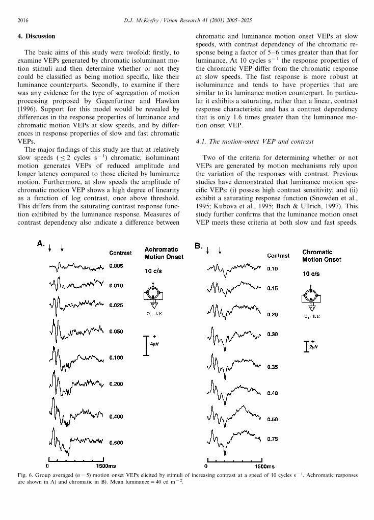
Differences between the response behaviour of the fast(10 cycles s−1) and slower (2 cycles s−1) motion onsetresponses in the first experiment, prompted furtherexamination of the contrast response characteristics atthis faster speed. Fig. 6A–B) shows the luminance andchromatic VEP waveforms and Fig. 7A plots the p1–n2amplitude variation as a function of contrast. Theluminance motion onset VEP, like its counterpart atslower speed, exhibits a saturating contrast responsefunction. The chromatic motion VEP however, unlikethe slower chromatic response, can no longer be ade-quately described by a simple linear function (correlationanalysis indicating a weaker and non-significant associa-tion (r=0.38, P=0.31)). In fact the chromatic responsefunction now appears to be described better by asaturating Naka–Rushton function.
Fig. 7B shows the variation of n1–p1 amplitude as afunction of contrast for the chromatic and luminance
stimuli. This component appears to be more prominentat faster speeds, as has been noted in other studies(Kubova et al., 1995; Spileers et al., 1996), and thebehaviour of the n1–p1 is quite different from the laterp1–n2 component in the luminance motion VEP. Whilstthe later component reaches saturation around contrastlevels of 10%, the earlier n1–p1 amplitude exhibits littleevidence of saturation below 70% contrast. This be-haviour would appear to be consistent with the findingsof Spileers et al. (1996) and the non-saturating nature ofn1–p1 also appears similar to the prominent positivecomponent recorded by Bach and Ullrich (1997) usingan electrode at Oz referenced to a frontal electrode (Fpz).As a result of this less prominent saturation the lumi-nance n1–p1 amplitude data, like the chromatic data,can be fitted with a linear function (r=0.92; P�0.01).
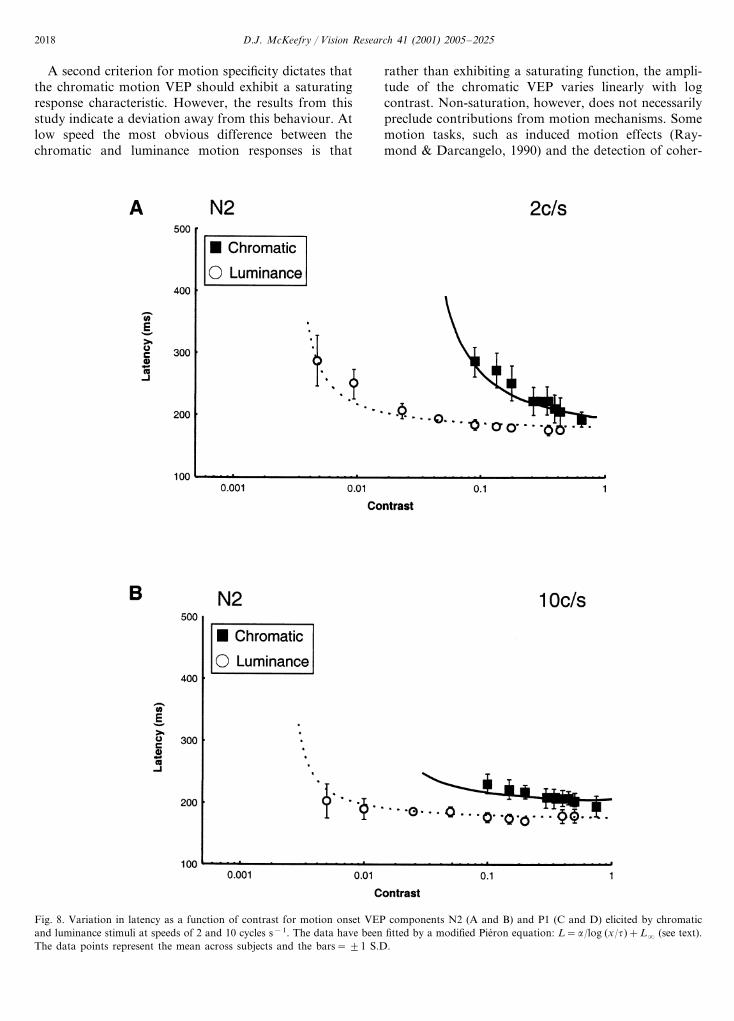
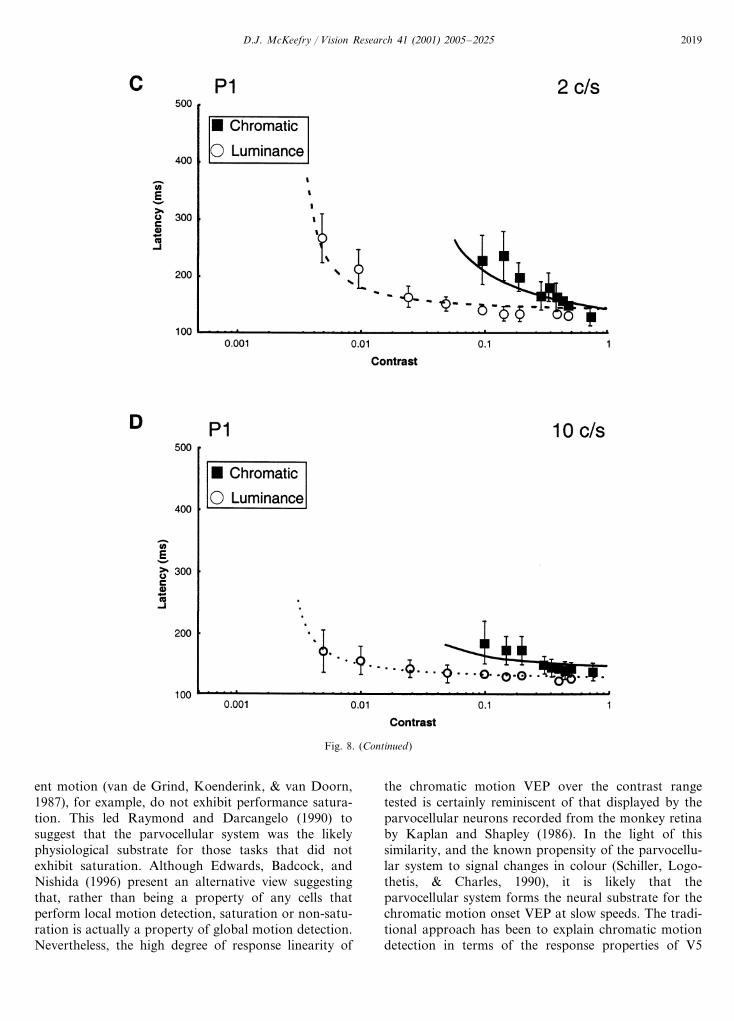
Fig. 8 shows the latency variations as a function ofcontrast for two of the components in the luminance andchromatic motion onset VEP. In order to obtain anestimate of contrast dependency of the componentselicited by luminance and chromatic motion the ap-proach of Burr, Fiorentini, and Morrone (1998), in theirstudy of reaction times (RTs), was adopted. They
Fig. 4. Group averaged (n=6) motion onset VEPs elicited by stimuli of increasing contrast at a speed of 2 cycles s−1. Achromatic responses areshown in A) and chromatic in B). Contrast in this case is defined as Michelson contrast and mean luminance=40 cd m−2. Note that there aredifferences in vertical scale for the achromatic and chromatic reponses.
D.J. McKeefry / Vision Research 41 (2001) 2005–2025 2015
Fig. 5. Amplitude of the p1–n2 motion onset VEP componentplotted as a function of contrast for a 2 cycles s−1 stimulus. Theachromatic data (circles) have been fitted by a Naka–Rushton equa-tion (A=Amax cn/(cn+c50
n )) and the chromatic data (filled squares)by a linear regression line. The data points represent the mean acrosssubjects and the bars= �1 S.D.
luminance motion. However, at 10 cycles s−1 the dif-ferences between luminance and chromatic responsesare less marked, with �=40–50 ms log unit for lumi-nance and �=60–100 ms log unit for chromatic mo-tion.
One of the key characteristics of the (luminance)motion onset VEP is that it exhibits high contrastsensitivity (Muller & Gopfert, 1988; Snowden et al.,1995; Kubova et al., 1995; Bach & Ullrich, 1997).The basic question is whether the chromatic motiononset VEP exhibits similar high contrast sensitivity.The answer is complicated by the fact that it dependsupon how one chooses to define chromatic contrast.When chromatic contrast is defined simply in termsof the Michelson contrast of the constituent gratingswhich are added in antiphase, sensitivity to luminancemotion is higher than for chromatic motion (Ca-vanagh & Anstis, 1991). However, when expressed interms of a more physiologically meaningful metric,i.e. the modulation of cone excitation, it has beenshown that the converse is true (Stromeyer, Cole, &Kronauer, 1987). Subsequent studies using the samemetric have confirmed that chromatic motion sensitiv-ity is higher for low temporal frequencies, but at hightemporal frequencies sensitivity to luminance motionis greater (Derrington & Henning, 1993; Stromeyer,Kronauer, Ryu, Chaparro, & Eskew, 1995). Conemodulation has become a widely applied metric ofchromatic contrast (see for example: Lennie & D’Z-mura, 1988; Chaparro, Stromeyer, Huang, Kronauer,& Eskew, 1993). Therefore in Fig. 10A contrast re-sponse functions for motion onset VEPs show thechromatic response data now plotted in terms ofchromatic contrast rather than Michelson contrast.The effect is that the chromatic data have undergonea simple linear transformation that shifts the functionleftwards along the x-axis, thus equating cone withluminance modulation. The contrast response func-tions in Fig. 10 differ further from earlier figures inthat the luminance data contains only points that oc-cur before response saturation (contrast�0.1). This isto enable the data to be fitted with regression lineswhich when extrapolated to zero amplitude give anestimate of contrast threshold. This technique wouldseem to be justifiable as Fig. 10 indicates that thethreshold estimates obtained by VEP extrapolationagree closely with the psychophysically measured lu-minance and chromatic motion detection thresholds.The results (Fig. 10A) show that at slow speeds thereis no significant difference between the VEP thresholdestimates of contrast sensitivity for chromatic and lu-minance motion. However, when the results for 10cycles s−1 are plotted in the same fashion (Fig. 10B),chromatic contrast sensitivity is shown to be lowerthan for luminance, consistent with psychophysicalfindings (Derrington & Henning, 1993).
employed a modification of Pieron’s equation to de-scribe the effects of contrast on reaction times. Thismodification has the advantage of possessing only oneparameter that determines slope, rather than two inthe more traditional form of the Pieron equation, fur-thermore, it asymptotes to infinity at threshold (Burret al., 1998). Obviously, in this instance the relation-ship to be examined is between VEP response latencyand contrast rather than reaction time and the equa-tion is given as:
L=�
log (x/�)+L� (2)
where: L=VEP latency, �=constant determiningslope of curve (i.e. contrast dependency), x=contrast,�=detection threshold, L�= latency asymptote. Thedata in Fig. 8 have been fitted with this type of equa-tion and it can be seen that, similar to the RT data(Burr et al., 1998), it provides a good description ofboth the luminance and chromatic data at fast andslow presentation speeds for the components shown.
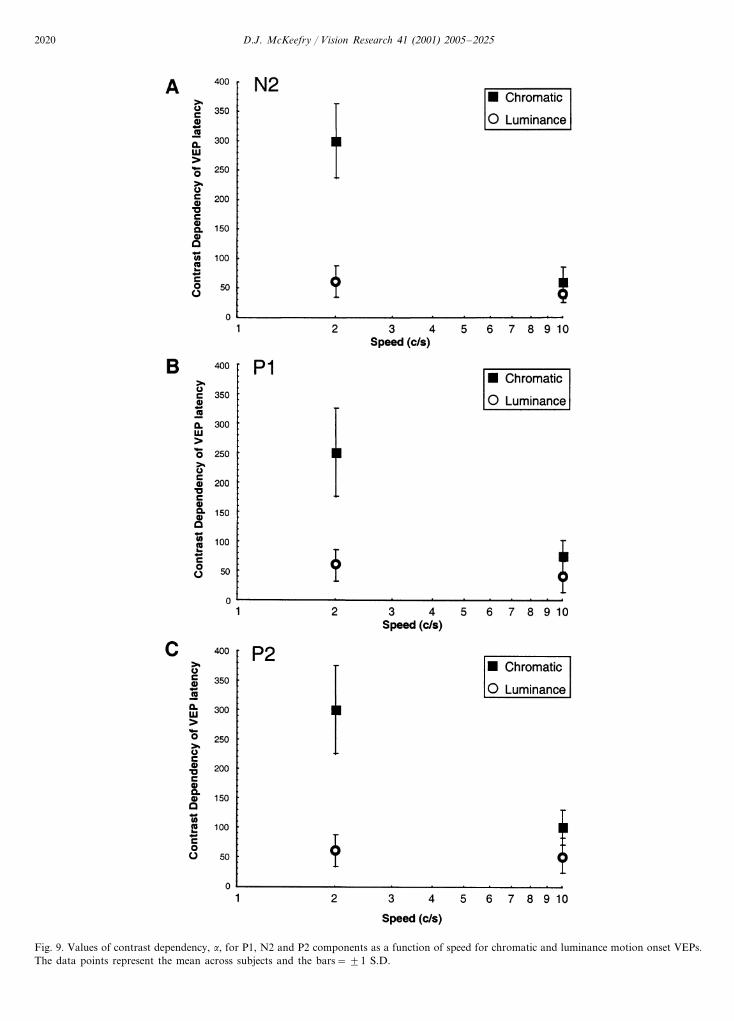
Fig. 9 plots the values for �, the contrast depen-dency, as a function of stimulus speed for the N2, P1and P2 components of the chromatic and luminancemotion onset VEPs. The general trend appears to bethe same across all of these components. At 2 cycless−1 there is considerable difference between the con-trast dependency of the chromatic and luminance re-sponses, �=250–300 ms log unit for colour but withlower values of between 50 and 60 ms log unit for
D.J. McKeefry / Vision Research 41 (2001) 2005–20252016
4. Discussion
The basic aims of this study were twofold: firstly, toexamine VEPs generated by chromatic isoluminant mo-tion stimuli and then determine whether or not theycould be classified as being motion specific, like theirluminance counterparts. Secondly, to examine if therewas any evidence for the type of segregation of motionprocessing proposed by Gegenfurtner and Hawken(1996). Support for this model would be revealed bydifferences in the response properties of luminance andchromatic motion VEPs at slow speeds, and by differ-ences in response properties of slow and fast chromaticVEPs.
The major findings of this study are that at relativelyslow speeds (�2 cycles s−1) chromatic, isoluminantmotion generates VEPs of reduced amplitude andlonger latency compared to those elicited by luminancemotion. Furthermore, at slow speeds the amplitude ofchromatic motion VEP shows a high degree of linearityas a function of log contrast, once above threshold.This differs from the saturating contrast response func-tion exhibited by the luminance response. Measures ofcontrast dependency also indicate a difference between
chromatic and luminance motion onset VEPs at slowspeeds, with contrast dependency of the chromatic re-sponse being a factor of 5–6 times greater than that forluminance. At 10 cycles s−1 the response properties ofthe chromatic VEP differ from the chromatic responseat slow speeds. The fast response is more robust atisoluminance and tends to have properties that aresimilar to its luminance motion counterpart. In particu-lar it exhibits a saturating, rather than a linear, contrastresponse characteristic and has a contrast dependencythat is only 1.6 times greater than the luminance mo-tion onset VEP.
4.1. The motion-onset VEP and contrast
Two of the criteria for determining whether or notVEPs are generated by motion mechanisms rely uponthe variation of the responses with contrast. Previousstudies have demonstrated that luminance motion spe-cific VEPs: (i) possess high contrast sensitivity; and (ii)exhibit a saturating response function (Snowden et al.,1995; Kubova et al., 1995; Bach & Ullrich, 1997). Thisstudy further confirms that the luminance motion onsetVEP meets these criteria at both slow and fast speeds.
Fig. 6. Group averaged (n=5) motion onset VEPs elicited by stimuli of increasing contrast at a speed of 10 cycles s−1. Achromatic responsesare shown in A) and chromatic in B). Mean luminance=40 cd m−2.
D.J. McKeefry / Vision Research 41 (2001) 2005–2025 2017
Fig. 7. (A). Amplitude of the p1–n2 motion onset VEP componentplotted as a function of contrast for a 10 cycles s−1 stimulus. Boththe achromatic (circles) and chromatic (filled squares) data have beenfitted by Naka–Rushton functions. The data points represent themean across subjects and the bars= �1 S.D. (B) Amplitude of then1–p1 motion onset VEP component plotted as a function of con-trast for the same stimulus. In this case the achromatic (circles) andchromatic (filled squares) data have been fitted by linear functions.
system that form the physiological substrate of theluminance motion onset VEP (Kubova et al., 1995).
The main question is whether the chromatic motiononset VEP meets the criteria for motion specificity. Inthe case of contrast sensitivity, whether or not it can bedescribed as higher or lower than achromatic contrastsensitivity, depends crucially upon how chromatic con-trast is defined. In this instance, in order for a meaning-ful comparison to be made with luminance, chromaticcontrast has been expressed in terms of cone modula-tion. Not surprisingly, as a result of this definition, thefindings are consistent with those psychophysical stud-ies that have used a similar metric (Derrington &Henning, 1993), in that chromatic motion sensitivity isas high as luminance sensitivity at low speeds, but islower at faster temporal rates. This presumably is aresult of the low pass nature of chromatic temporalprocessing and the band-pass nature of achromaticprocessing (De Lange, 1958; Regan & Tyler, 1971;Kelly, 1974). Comparisons of absolute sensitivity toluminance and chromatic stimuli are always going to besubject to how one chooses to define chromatic con-trast. But the dichotomy that exists between luminanceand chromatic responses, based upon differences in thetheir respective contrast dependencies and gain func-tions, is unaffected by the metric chosen to quantifychromatic contrast.
It could be argued that the reduced motion-onsetVEP amplitude observed at isoluminance for slowspeeds, is simply the result of reduced chromatic con-trast. The visual system could, in effect, be treating anisoluminant chromatic grating in the same way as a lowcontrast luminance grating, rather than as an intrinsi-cally different kind of stimulus (Troscianko & Fahle,1988). However, other evidence tends to suggest thatchromatic motion processing is not as straightforwardas this. Cavanagh et al. (1984), for example, haveshown that the addition of chromatic contrast to aluminance motion stimulus can reduce its perceivedspeed, as well as its ability to generate a motion after-effect (Cavanagh & Favreau, 1985). Reductions in VEPamplitude have also been reported at isoluminance forS-cone isolating stimuli that have much higher chro-matic contrast (�70%) than the stimuli employed inthis study (McKeefry, 2001). Even if the effects uponthe motion onset VEP at isoluminance for slow speedscould be explained purely in terms of a reduction inchromatic contrast, it fails to explain why there is not asimilar diminution in the response for chromatic mo-tion at fast speeds. One would need to resort to explain-ing these differences in terms of separate motionmechanisms operating at slow and fast speeds, theformer in which chromatic contrast is an importantmeans of coding and the latter in which it is not (seebelow).
Such properties are in keeping with single-unit (Der-rington & Lennie, 1984; Hawken & Parker, 1984; Blas-del & Fitzpatrick, 1984; Kaplan & Shapley, 1986; Sclar,Maunsell, & Lennie, 1990), fMRI (Tootell et al., 1995)and certain psychophysical studies (Nakayama & Sil-verman, 1985; McKee, Silverman, & Nakayama, 1986;Derrington & Goddard, 1989; Cropper, 1994). All theseinvestigations confirm the idea of a motion system thatis dominated by input from the magnocellular system,and as a result, possesses high contrast sensitivity andsaturates with increasing contrast. This accounts for thesuggestion that it is the neurons of the magnocellular
D.J. McKeefry / Vision Research 41 (2001) 2005–20252018
A second criterion for motion specificity dictates thatthe chromatic motion VEP should exhibit a saturatingresponse characteristic. However, the results from thisstudy indicate a deviation away from this behaviour. Atlow speed the most obvious difference between thechromatic and luminance motion responses is that
rather than exhibiting a saturating function, the ampli-tude of the chromatic VEP varies linearly with logcontrast. Non-saturation, however, does not necessarilypreclude contributions from motion mechanisms. Somemotion tasks, such as induced motion effects (Ray-mond & Darcangelo, 1990) and the detection of coher-
Fig. 8. Variation in latency as a function of contrast for motion onset VEP components N2 (A and B) and P1 (C and D) elicited by chromaticand luminance stimuli at speeds of 2 and 10 cycles s−1. The data have been fitted by a modified Pieron equation: L=�/log (x/�)+L� (see text).The data points represent the mean across subjects and the bars= �1 S.D.
D.J. McKeefry / Vision Research 41 (2001) 2005–2025 2019
Fig. 8. (Continued)
ent motion (van de Grind, Koenderink, & van Doorn,1987), for example, do not exhibit performance satura-tion. This led Raymond and Darcangelo (1990) tosuggest that the parvocellular system was the likelyphysiological substrate for those tasks that did notexhibit saturation. Although Edwards, Badcock, andNishida (1996) present an alternative view suggestingthat, rather than being a property of any cells thatperform local motion detection, saturation or non-satu-ration is actually a property of global motion detection.Nevertheless, the high degree of response linearity of
the chromatic motion VEP over the contrast rangetested is certainly reminiscent of that displayed by theparvocellular neurons recorded from the monkey retinaby Kaplan and Shapley (1986). In the light of thissimilarity, and the known propensity of the parvocellu-lar system to signal changes in colour (Schiller, Logo-thetis, & Charles, 1990), it is likely that theparvocellular system forms the neural substrate for thechromatic motion onset VEP at slow speeds. The tradi-tional approach has been to explain chromatic motiondetection in terms of the response properties of V5
D.J. McKeefry / Vision Research 41 (2001) 2005–20252020
Fig. 9. Values of contrast dependency, �, for P1, N2 and P2 components as a function of speed for chromatic and luminance motion onset VEPs.The data points represent the mean across subjects and the bars= �1 S.D.
D.J. McKeefry / Vision Research 41 (2001) 2005–2025 2021
Fig. 10. Contrast response functions of the p1–n2 component for luminance and chromatic motion onset VEPs elicited by a stimulus of speed1 cycles s−1 (A) and 10 cycles s−1 (B). Chromatic contrast in this instance has been calculated in terms of cone modulation (see text). Regressionlines were fitted to the chromatic data (A: r=0.89, P=0.006; B: r=0.94, P=0.016) and to the pre-saturation luminance data points (A: r=0.89,P=0.04; B: r=0.86, P=0.13). These lines were extrapolated to zero amplitude in order to give an estimate thresholds (horizontal lines indicate95% confidence intervals). Psychophysical measures of thresholds for chromatic (filled triangle) and luminance (empty triangle) motion detectionare also shown.
D.J. McKeefry / Vision Research 41 (2001) 2005–20252022
(MT) neurons which have been shown to be responsiveto moving isoluminant stimuli (Saito, Tanaka, Isono,Yasuda, & Mikami, 1989; Gegenfurtner, Kiper,Beusmans, Cardandini, & Zaidi, 1994; Dobkins & Al-bright, 1994). But the parvocellular system must playsome part in signalling chromatic motion (Cropper &Derrington, 1996). Selective lesioning of the magnocel-lular pathway (Merigan, Byrne, & Maunsell, 1991) hasshown that although motion detection thresholds maybe raised, the perception of motion is not otherwiseaffected, implying that the parvocellular system must beable to support some form of motion analysis. Inaddition, Ferrera, Rudolph, and Maunsell (1994) havedemonstrated that neurons in the parvocellular domi-nated infero-temporal visual pathway are capable ofresponding to motion, when it is important in objectidentification.
At high velocity the linear contrast response functionof the chromatic motion VEP is replaced by a saturat-ing function, which is similar to that observed for theluminance response. This change in response behaviourmay be indicative of a shift in the underlying nature ofthe cellular substrate from linear parvocellular to non-linear magnocellular mechanisms. It is well known thatthe magnocellular system is quite capable of respondingto isoluminant stimuli modulated at high temporal rates(Lee, Martin, & Valberg, 1989a,b) but that encoding ofchromatic contrast occurs in an ‘unsigned’ (Dobkins &Albright, 1993) manner. This proposed change in thenature of the cellular substrate of the VEP is consistentwith the idea (see Gegenfurtner & Hawken, 1996) of averidical colour sensitive (i.e. signed) channel that oper-ates at low stimulus speeds and a non-veridical colourinsensitive (i.e. unsigned) channel that operates at highspeeds. Furthermore, it also consistent with the moregeneral hypothesis that the mediation of particularvisual functions, by either parvo- or magno- cellularpathways, is dictated primarily by the spatio-temporalcharacteristics of the stimulus (Merigan, 1990).
4.2. Consistency with reaction times
VEPs, like the study of reaction times (RTs), allowthe assessment of supra-threshold visual performance.Combined RT and VEP studies have been used in thepast to compare chromatic and luminance spatial visionwhich have been shown to exhibit different contrastdependencies (Parry, Kulikowski, Murray, Kranda, &Ott, 1988). The properties of the luminance and chro-matic motion, as revealed in this study by VEP latencyvariation, show similar differences and are highly con-sistent with the RT data of Burr et al. (1998). The mainarea of consistency lies in the fact that there is astronger contrast dependency for VEPs and RTs tochromatic stimuli than for VEPs and RTs to luminancestimuli, especially at slow speeds. In addition, the dif-
ferences between luminance and chromatic VEPs andRTs become less marked under conditions of highcontrast and high speed. This divergence in the re-sponse properties of the chromatic and luminance mo-tion VEPs at low speed and convergence at high, andthe consistency with RTs, further validates the notionthat different mechanisms operate for the perception ofchromatic motion at fast and slow speeds (Gegenfurt-ner & Hawken, 1996).
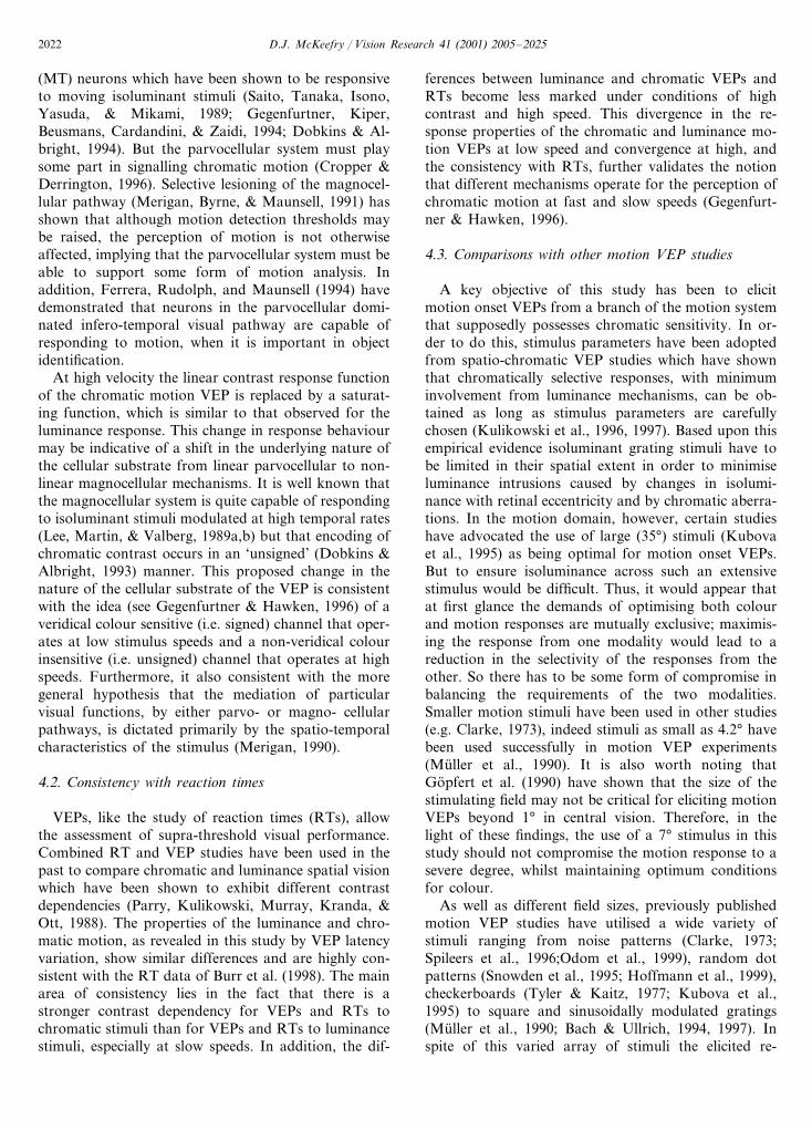
4.3. Comparisons with other motion VEP studies
A key objective of this study has been to elicitmotion onset VEPs from a branch of the motion systemthat supposedly possesses chromatic sensitivity. In or-der to do this, stimulus parameters have been adoptedfrom spatio-chromatic VEP studies which have shownthat chromatically selective responses, with minimuminvolvement from luminance mechanisms, can be ob-tained as long as stimulus parameters are carefullychosen (Kulikowski et al., 1996, 1997). Based upon thisempirical evidence isoluminant grating stimuli have tobe limited in their spatial extent in order to minimiseluminance intrusions caused by changes in isolumi-nance with retinal eccentricity and by chromatic aberra-tions. In the motion domain, however, certain studieshave advocated the use of large (35°) stimuli (Kubovaet al., 1995) as being optimal for motion onset VEPs.But to ensure isoluminance across such an extensivestimulus would be difficult. Thus, it would appear thatat first glance the demands of optimising both colourand motion responses are mutually exclusive; maximis-ing the response from one modality would lead to areduction in the selectivity of the responses from theother. So there has to be some form of compromise inbalancing the requirements of the two modalities.Smaller motion stimuli have been used in other studies(e.g. Clarke, 1973), indeed stimuli as small as 4.2° havebeen used successfully in motion VEP experiments(Muller et al., 1990). It is also worth noting thatGopfert et al. (1990) have shown that the size of thestimulating field may not be critical for eliciting motionVEPs beyond 1° in central vision. Therefore, in thelight of these findings, the use of a 7° stimulus in thisstudy should not compromise the motion response to asevere degree, whilst maintaining optimum conditionsfor colour.
As well as different field sizes, previously publishedmotion VEP studies have utilised a wide variety ofstimuli ranging from noise patterns (Clarke, 1973;Spileers et al., 1996;Odom et al., 1999), random dotpatterns (Snowden et al., 1995; Hoffmann et al., 1999),checkerboards (Tyler & Kaitz, 1977; Kubova et al.,1995) to square and sinusoidally modulated gratings(Muller et al., 1990; Bach & Ullrich, 1994, 1997). Inspite of this varied array of stimuli the elicited re-
D.J. McKeefry / Vision Research 41 (2001) 2005–2025 2023
sponses exhibit a remarkable degree of consistency, witha characteristic P1–N2–P2 triphasic response beingobtained in most cases. However, the relative predomi-nance of the constituent components tend to vary, forexample, as a function of stimulus speed as well aselectrode placement. In the case of the latter, the N2component in particular appears to be most prominentwhen the electrodes are placed over the lateral occipitalcortex, rather than the more centrally placed Oz elec-trode (Kubova et al., 1995; Bach & Ullrich). Thispresumably is the result of the closer proximity of thelateral electrodes to the motion processing area of thehuman brain, V5, which is found nearby (Watson,Myers, Frackowiak, Hajnal, Woods, Mazziotta, Shipp,& Zeki, 1993). Further experimentation will be requiredin order to ascertain whether the N2 component of thechromatic motion onset VEP follows a similar patternto its luminance counterpart. The fact that the process-ing of colour information takes place along the ventraloccipito-temporal cortex in the human cortex (McK-eefry & Zeki, 1997), would lead to the prediction thatthere may be a difference in behaviour between thechromatic and luminance N2 component as a functionof lateral electrode placement. On the other hand, recentexperiments have revealed that human V5 is capable ofprocessing information about moving isoluminantcolour stimuli (Dougherty, Press, & Wandell, 1999;Wandell, Poirson, Newsome, Baseler, Boynton, Huk,Gandhi, & Sharpe, 1999). This would imply that chro-matic and luminance N2 should behave in a similarfashion as a function of lateral placement.
5. Conclusions
This study has established that motion specific VEPscan be elicited by moving isoluminant red/green stimuli.VEPs elicited by luminance and chromatic motion stim-uli at low speeds have different response characteristics.Luminance motion VEPs saturate at low contrasts,whilst chromatic motion VEPs increase in a linearfashion, chromatic responses also have a higher contrastdependency. These differences imply that there are sep-arate channels for the mediation of colour and lumi-nance motion, with the parvocellular and magnocellularsystems, respectively, being the likely physiological sub-strates. At faster speeds the nature of the chromaticmotion VEP changes and becomes more like the lumi-nance response, it exhibits a saturating contrast re-sponse characteristic and its contrast dependency is notvery different from that for luminance. These shifts inresponse properties represent changes in the propertiesof the neural substrate of the VEP and may be indica-tive of a shift away from parvocellular to magnocellularinvolvement, the neurons of which are known to beresponsive to rapidly changing isoluminant stimuli.
The differences in the properties of VEPs elicited byluminance and chromatic motion stimuli at fast andslow rates are consistent with ideas about the motionsystem comprising of two systems: one for fast motionthat is responsive to both colour and luminance, butwhich does not encode chromaticity. The other for slowmotion that has separate luminance and colour inputs.
Acknowledgements
The author gratefully acknowledges Professor J. Ku-likowski, Drs I. Murray and N. Parry for helpfulcomments on previous versions of this paper. This workhas been supported by the Wellcome Trust.
References
Anstis, S., & Cavanagh, P. (1983). A minimum motion technique forjudging isoluminance. In J. D. Mollon, & L. T. Sharpe, ColourVision, Physiology and Psychophysics (pp. 155–166). London:Academic Press.
Bach, M., & Ullrich, D. (1994). Motion adaptation governs the shapeof motion evoked cortical potentials. Vision Research, 34, 1541–1547.
Bach, M., & Ullrich, D. (1997). Contrast dependency of motion-onsetand pattern reversal VEPs: interaction of stimulus type, recordingsite and response component. Vision Research, 37, 1845–1849.
Blasdel, G. G., & Fitzpatrick, D. (1984). Physiological organisationof layer 4 in the macaque striate cortex. Journal of Neuroscience,4, 880–895.
Cavanagh, P., & Anstis, S. (1991). The contribution of colour tomotion in normal and colour deficient observers. Vision Research,31, 2109–2148.
Cavanagh, P., & Favreau, O. (1985). Color and luminance share acommon motion pathway. Vision Research, 25, 1595–1601.
Cavanagh, P., Tyler, C. W., & Favreau, O. (1984). Perceived velocityof moving chromatic gratings. Journal of the Optical Society ofAmerica, A1, 893–899.
Chaparro, A., Stromeyer, C. F., Huang, E. P., Kronauer, R. E., &Eskew, R. T. (1993). Color is what the eye sees best. Nature, 361,348–350.
Charman, W. N. (1991). Limits on visual performance set by the eye’soptics and the retinal cone mosaic. In J. J. Kulikowski, V. Walsh,& I. J. Murray, Limits of �ision (pp. 81–96). Basingstoke:Macmillan Press.
Chichilnisky, E.-J., Heeger, D., & Wandell, B. A. (1993). Functionalsegregation of colour and motion perception examined in motionnulling. Vision Research, 33, 2113–2125.
Clarke, P. G. H. (1972). Visual evoked potentials to sudden reversalsof the motion of a pattern. Brain Research, 36, 453–458.
Clarke, P. G. H. (1973). Comparison of visual evoked potentials tostationary and moving patterns. Experimental Brain Research, 18,156–164.
Clarke, P. G. H. (1974). Are visual evoked potentials to motionproduced by direction sensitive brain mechanisms? Vision Re-search, 14, 1281–1284.
Cropper, S. J. (1994). Velocity discrimination in chromatic gratingsand beats. Vision Research, 34, 41–48.
Cropper, S. J., & Derrington, A. M. (1996). Rapid colour-specificdetection of motion in human vision. Nature, 379, 72–74.
D.J. McKeefry / Vision Research 41 (2001) 2005–20252024
Cropper, S. J., Mullen, K. T., & Badcock, D. R. (1996). Motioncoherence across different chromatic axes. Vision Research, 36,2475–2488.
De Lange, H. (1958). Research into the dynamic nature of the humanfovea-cortex systems with intermittent a modulated light. I Atten-uation characteristics with white and coloured light. Journal of theOptical Society of America, 48, 777–784.
Derrington, A. M., & Badcock, D. R. (1985). The low level motionsystem has both chromatic and luminance inputs. Vision Re-search, 25, 1879–1884.
Derrington, A. M., & Goddard, P. A. (1989). Failure of motiondiscrimination at high contrasts. Vision Research, 29, 1767–1776.
Derrington, A. M., & Henning, B. (1993). Detecting and discriminat-ing the direction of motion of luminance and colour gratings.Vision Research, 33, 799–811.
Derrington, A. M., & Lennie, P. (1984). Chromatic mechanisms inthe lateral geniculate nucleus of macaque. Journal of Physiology,357, 219–240.
Dobkins, K. R., & Albright, T. D. (1993). What happens if it changescolour when it moves?: psychophysical experiments on the natureof chromatic input to motion detectors. Vision Research, 33,1019–1036.
Dobkins, K. R., & Albright, T. D. (1994). What happens if it changescolour when it moves?: the nature of chromatic input to macaquevisual area MT. Journal of Neuroscience, 8, 4854–4870.
Dougherty, R., Press, W., & Wandell, B. (1999). Perceived speed ofcolor stimuli. Neuron, 24, 893–899.
Edwards, M., & Badcock, D. R. (1996). Global motion perception:interaction of chromatic and luminance signals. Vision Research,36, 2423–2431.
Edwards, M., Badcock, D. R., & Nishida, S. (1996). Contrast sensi-tivity o the motion system. Vision Research, 36, 2411–2421.
Ferrera, V. P., Rudolph, K. K., & Maunsell, J. H. (1994). Responsesof neurons in the parietal and temporal visual pathways during amotion task. Journal of Neuroscience, 14, 6171–6186.
Ffytche, D. H., Skidmore, B., & Zeki, S. (1995). Motion-from-hueactivates area V5 of human visual cortex. Proceedings of the RoyalSociety Ser. B, 260, 353–358.
Gallichio, J. A., & Andreassi, J. L. (1982). Visual evoked potentialsunder varied velocities of continuous and discrete apparent mo-tion. International Journal of Neuroscience, 17, 177–196.
Gegenfurtner, K. R., & Hawken, M. J. (1996). Interaction of motionand color in the visual pathways. Trends in Neuroscience, 19,394–401.
Gegenfurtner, K. R., Kiper, D. C., Beusmans, J. M. H., Cardandini,M., & Zaidi, Q. (1994). Chromatic response properties of neuronsin macaque MT. Visual Neuroscience, 11, 455–466.
Gopfert, E., Muller, R., & Simon, E. M. (1990). The human motiononset VEP as a function of stimulation area for foveal andperipheral vision. Documenta Ophthalmologica, 75, 165–173.
Gorea, A., Papathomas, T. V., & Kovacs, I. (1993). Motion percep-tion with spatiotemporally matched chromatic and achromaticinformation reveals a ‘slow’ and a ‘fast’ motion system. VisionResearch, 33, 2515–2543.
Hawken, M. J., & Parker, A. J. (1984). Contrast sensitivity andorientation selectivity in lamina IV of the striate cortex of oldworld monkeys. Experimental Brain Research, 54, 367–372.
Hoffmann, M., Dorn, T. J., & Bach, M. (1999). Time course ofmotion adaptation: motion-onset visual evoked potentials andsubjective estimates. Vision Research, 39, 437–444.
Kaplan, E., & Shapley, R. M. (1986). The primate retina containstwo types of ganglion cells, with high and low contrast sensitivity.Proceedings of the National Academy of Sciences USA, 83, 2755–2757.
Kelly, D. H. (1974). Spatio-temporal frequency characteristics ofcolour vision mechanisms. Journal of the Optical Society of Amer-ica, 64, 983–990.
Kooi, F. L., & DeValois, K. K. (1992). The role of color in themotion system. Vision Research, 32, 657–668.
Krauskopf, J., & Farrell, B. (1990). Influence of colour on theperception of coherent motion. Nature, 348, 328–331.
Kuba, M., & Kubova, Z. (1992). Visual evoked potentials specific formotion onset. Documenta Ophthalmologica, 80, 83–89.
Kubova, Z., Kuba, M., Spekreijse, H., & Blakemore, C. (1995).Contrast dependence of motion onset and pattern reversal evokedpotentials. Vision Research, 35, 197–205.
Kulikowski, J. J., McKeefry, D. J., & Robson, A. (1997). Selectivestimulation of colour mechanisms. Spatial Vision, 10, 379–402.
Kulikowski, J. J., Robson, A., & McKeefry, D. J. (1996). Specificityand selectivity of chromatic visual evoked potentials. Vision Re-search, 36, 3397–3401.
Lee, B. B., Martin, P. R., & Valberg, A. (1989a). Sensitivity ofmacaque retinal ganglion cells to chromatic and luminance flicker.Journal of Physiology, 414, 223–243.
Lee, B. B., Martin, P. R., & Valberg, A. (1989b). Amplitude andphase of responses of macaque retinal ganglion cells to flickeringstimuli. Journal of Physiology, 414, 245–263.
Lennie, P., & D’Zmura, M. (1988). Mechanisms of color vision. CRCCritical Re�iews in Neurobiology, 3, 333–400.
Livingstone, M., & Hubel, D. H. (1987). Psychophysical evidence forseparate channels for the perception of form, color, movementand depth. Journal of Neuroscience, 7, 3146–3486.
Lu, Z.-L., Lesmes, L. A., & Sperling, G. (1999). The mechanism ofisoluminant motion perception. Proceedings of the NationalAcademy of sciences USA, 96, 8289–8294.
McKee, S. P., Silverman, G. H., & Nakayama, K. (1986). Precisevelocity discrimination despite random variations in temporalfrequency and contrast. Vision Research, 26, 609–619.
McKeefry, D.J. (2001). Chromatic visual evoked potentials elicited byfast and slow motion onset. Colour Research and Applications (inpress).
McKeefry, D. J., & Zeki, S. (1997). The position and topography ofthe human colour centre as revealed by functional magneticresonance imaging. Brain, 120, 2229–2242.
Merigan, W. H. (1990). P and M pathway specialisation in theMacaque. In A. Valberg, & B. B. Lee, From pigments to percep-tion : ad�ances in understanding �isual processes (pp. 117–125).New York: Plenum Press.
Merigan, W. H., Byrne, C. E., & Maunsell, J. H. R. (1991). Doesprimate motion perception depend upon the magnocellular path-way? Journal of Neuroscience, 11, 3422–3429.
Metha, A. B., & Mullen, K. T. (1997). Red–green and achromatictemporal filters: a ratio model predicts contrast dependent speedperception. Journal of the Optical Society of America, 14, 984–996.
Metha, A. B., Vingrys, A. J., & Badcock, D. (1994). Detection anddiscrimination of moving stimuli: the effects of colour, luminanceand eccentricity. Journal of the Optical Society of America, A11,1697–1709.
Morrone, M. C., Fiorentini, A. F., & Burr, D. C. (1996). Develop-ment of the temporal properties of visual evoked potentials toluminance and colour contrast in infants. Vision Research, 36,3141–3155.
Mullen, K. T., & Boulton, J. C. (1992). Absence of smooth motionperception in colour vision. Vision Research, 32, 483–488.
Muller, R., & Gopfert, E. (1988). The influence of grating contrast onthe human cortical potential visually evoked by motion. ActaNeurobiologica Experimentia, 48, 239–249.
Muller, R., Gopfert, E., Schlykowa, L., & Anke, D. (1990). Thehuman motion VEP as a function of size and eccentricity of thestimulation field. Documenta Ophthalmologica, 76, 81–89.
Nakayama, K., & Silverman, G. H. (1985). Detection and discrimina-tion of sinusoidal grating displacements. Journal of the OpticalSociety of America A, 2, 267–274.
D.J. McKeefry / Vision Research 41 (2001) 2005–2025 2025
Odom, J. V., DeSmedt, E., Van Malderen, L., & Spileers, W. (1999).VEPs evoked by moving unidimensional noise stimuli: effects ofcontrast, spatial frequency, active electrode location, referenceelectrode location and stimulus type. Documenta Ophthalmologica,95, 315–333.
Papathomas, T. V., Gorea, A., & Julesz, B. (1993). Two carriers formotion perception: colour and luminance. Vision Research, 31,1883–1891.
Parry, N. R. A., Kulikowski, J. J., Murray, I. J., Kranda, K., & Ott,H. (1988). Visual evoked potentials and reaction times to chro-matic and achromatic stimulation: psychopharmacological appli-cations. In I. Hindmarch, B. Aufdembrinke, & H. Ott,Psychopharmacology and reaction time (pp. 155–176). Chichester:J. Wiley & Sons.
Ramachandran, V. S., & Gregory, R. L. (1978). Does colour providean input to human motion perception? Nature, 275, 55–57.
Raymond, J. E., & Darcangelo, S. M. (1990). The effect of localluminance contrast on induced motion. Vision Research, 30, 751–756.
Regan, D., & Tyler, C. W. (1971). Some dynamic features of colourvision. Vision Research, 1, 1307–1342.
Saito, H., Tanaka, K., Isono, H., Yasuda, M., & Mikami, A. (1989).Directionally selective response of cells in the middle temporalarea (MT) of the macaque monkey to the movement of equilumi-nous opponent colour stimuli. Experimental Brain Research, 75,1–14.
Schiller, P. H., Logothetis, N. K., & Charles, E. R. (1990). Role ofthe color-opponent and broadband channels in vision. VisualNeuroscience, 5, 321–346.
Sclar, G., Maunsell, J. H. R., & Lennie, P. (1990). Coding of imagecontrast in central visual pathways of the macaque monkey.Vision Research, 30, 1–10.
Snowden, R. J., Ullrich, D., & Bach, M. (1995). Isolation andcharacteristics of steady-state visually evoked potential in humansrelated to the motion of a stimulus. Vision Research, 35, 1365–1373.
Smith, V. C., & Pokorny, J. (1975). Spectral sensitivity of the fovealcone photopigments between 400 and 500 nm. Vision Research,15, 161–171.
Spileers, W., Mangelschots, E., Maes, H., & Orban, G. (1996). Visualevoked potentials elicited by a moving unidimensional noise pat-tern. Electroencephalography and Clinical Neurophysiology, 100,287–298.
Stromeyer, C. F., Cole, G. R., & Kronauer, R. E. (1987). Chromaticsuppression of cone inputs to the luminance flicker mechanism.Vision Research, 27, 1113–1137.
Stromeyer, C. F., Kronauer, R. E., Ryu, A., Chaparro, A., & Eskew,R. T. (1995). Contributions of human long-wave and middle-wave cones to motion detection. Journal of Physiology, 485,221–243.
Tootell, R. B. H., Reppas, J. B., Kwong, K. K., Malach, R., Born, I.T., Brady, T. J., Rosen, B. R., & Belliveau, J. W. (1995).Functional analysis of human MT and related visual corticalareas using magnetic resonance imaging. Journal of Neuroscience,15, 3215–3230.
Troscianko, T., & Fahle, M. (1988). Why do isoluminant gratingsappear slower? Journal of the Optical Society of America, 87,435–469.
Tyler, C. W., & Kaitz, M. (1977). Movement adaptation in the visualevoked response. Experimental Brain Research, 27, 203–209.
van de Grind, W. A., Koenderink, J. J., & van Doorn, A. J. (1987).Influence of contrast on foveal and peripheral detection of coher-ent motion in moving random dot patterns. Journal of the OpticalSociety of America, 4, 1643–1651.
Vos, J. J. (1978). Colorimetric and photometric properties of a 2°fundamental observer. Colour Research and Applications, 4, 208–216.
Walraven, P. L. (1974). A closer look at the tritanopic convergencepoint. Vision Research, 14, 1339–1343.
Wandell, B., Poirson, A. B., Newsome, W. T., Baseler, H. A.,Boynton, G. M., Huk, A., Gandhi, S., & Sharpe, L. T. (1999).Color signals in human motion-selective cortex. Neuron, 24, 901–909.
Watson, J. D. G., Myers, R., Frackowiak, R. S. J., Hajnal, J. V.,Woods, R. P., Mazziotta, J. C., Shipp, S., & Zeki, S. (1993). AreaV5 of the human brain: evidence from a combined study usingpositron emission tomography and magnetic resonance imaging.Cerebral Cortex, 3, 79–94.
Willis, A., & Anderson, S. J. (1998). Separate colour-opponent mech-anisms underlie the detection and discrimination of moving chro-matic targets. Proceedings of the Royal Society, Ser B, 265,2435–2441.
Zeki, S. (1978). Uniformity and diversity of structure and function inthe rhesus monkey prestriate cortex. Journal of Physiology, 27,272–340.