VITAMIN K SUBSTANCES Vitamin K comprises a group of substances, which are widespread in nature and are an essential co-factor in humans in the synthesis of several proteins that play a role in haemostasis and others that may be important in calcium homeostasis. The K vitamins all contain the 2-methyl-1,4-naphthoquinone (menadione) moiety, and the various naturally occurring forms differ in the alkyl substituent at the 3-position. Phylloquinone (vitamin K 1 ) is 2-methyl-3-phytyl-1,4-naphthoquinone and is widely found in higher plants, including green leafy vegetables, and in green and blue algae. The menaquinones (formerly vitamin K 2 ) have polyisoprenyl substituents at the 3-position and are produced by bacteria. The compound menadione (formerly vitamin K 3 ) lacks an alkyl group at the 3-position but can be alkylated in vivo in some species. Several synthetic water-soluble derivatives, such as the sodium diphosphate ester of menadiol and the addition product of menadione with sodium bisulfite, also have commercial applications (National Research Council, 1989; Gennaro, 1995; Weber & Rüttimann, 1996). 1. Exposure Data 1.1 Chemical and physical data 1.1.1 Nomenclature, structural and molecular formulae and relative molecular masses Vitamin K (generic) Chem. Abstr. Serv. Reg. No.: 12001-79-5 Chem. Abstr. Name: Vitamin K Vitamin K 1 (generic) Chem. Abstr. Serv. Reg. No.: 11104-38-4 Chem. Abstr. Name: Vitamin K 1 Phylloquinone Chem. Abstr. Serv. Reg. No.: 84-80-0 Deleted CAS Reg. Nos.: 10485-69-5; 15973-57-6; 50926-17-5 –417–
Transcript
VITAMIN K SUBSTANCES
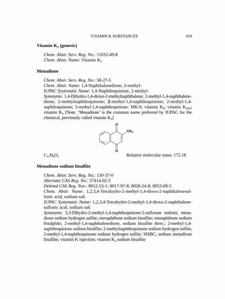
Vitamin K comprises a group of substances, which are widespread in nature and arean essential co-factor in humans in the synthesis of several proteins that play a role inhaemostasis and others that may be important in calcium homeostasis. The K vitaminsall contain the 2-methyl-1,4-naphthoquinone (menadione) moiety, and the variousnaturally occurring forms differ in the alkyl substituent at the 3-position. Phylloquinone(vitamin K1) is 2-methyl-3-phytyl-1,4-naphthoquinone and is widely found in higherplants, including green leafy vegetables, and in green and blue algae. The menaquinones(formerly vitamin K2) have polyisoprenyl substituents at the 3-position and are producedby bacteria. The compound menadione (formerly vitamin K3) lacks an alkyl group at the3-position but can be alkylated in vivo in some species. Several synthetic water-solublederivatives, such as the sodium diphosphate ester of menadiol and the addition productof menadione with sodium bisulfite, also have commercial applications (NationalResearch Council, 1989; Gennaro, 1995; Weber & Rüttimann, 1996).
1. Exposure Data
1.1 Chemical and physical data
1.1.1 Nomenclature, structural and molecular formulae and relative molecularmasses
Vitamin K (generic)
Chem. Abstr. Serv. Reg. No.: 12001-79-5Chem. Abstr. Name: Vitamin K
Chem. Abstr. Name: 2-Methyl-3-[(2E,7R,11R)-3,7,11,15-tetramethyl-2-hexadece-nyl]-1,4-naphthalenedioneIUPAC Systematic Name: [R-[R*,R*-(E)]]-2-Methyl-3-(3,7,11,15-tetramethyl-2-hexadecenyl)-1,4-naphthalenedioneSynonyms: Antihaemorrhagic vitamin; 2-methyl-3-phytyl-1,4-naphthoquinone;2-methyl-3-(3,7,11,15-tetramethyl-2-hexadecenyl)-1,4-naphthalenedione;α-phylloquinone; trans-phylloquinone; phylloquinone K1; phytomenadione;phytonadione; phytylmenadione; 3-phytylmenadione; phytylmenaquinone;vitamin K1; vitamin K1(20); 2′,3′-trans-vitamin K1 [Note: The IUPAC recommendsuse of the name ‘phylloquinone’ and the abbreviation ‘K’ (rather than ‘K1’). Bothphylloquinone and vitamin K1 are in common use. The United States Pharmaco-peia uses the name ‘phytonadione’; The European Pharmacopoeia uses the name‘phytomenadione’, which is a synonym occasionally found in the pharmaceuticaland pharmacological literature.]
IUPAC recommends that 2-methyl-3-polyprenyl-1,4-naphthoquinone be referred toas menaquinone-n, previously vitamin K2, n being the number of prenyl residues.Vitamin K2(20) is so named because it contains 20 carbon atoms in the chain. In the biolo-gical literature, vitamin K2 is frequently referred to as menaquinone and is furtherdesignated by the number of isoprene units in the side-chain. For example, vitamin K2(20)is also called menaquinone-4 for the four isoprene units in the side-chain. The compoundoriginally isolated from rotting fish meal and named vitamin K2 was later identified asmenaquinone-7 (2-methyl-3-farnesylgeranyl-geranyl-1,4-naphthoquinone). In the olderliterature, the designation vitamin K2(35) is used for menaquinone-7, but this is no longerused. Menaquinones found in nature have side-chains of 4–13 isoprenoid residues andare usually in the all-trans configuration; however, menaquinones with the cis confi-guration and partially saturated side-chains also exist (Suttie, 1985, 1991; Weber &Rüttimann, 1996; Van Arnum, 1998).
1.1.2 Chemical and physical properties of the pure substances
Phylloquinone
(a) Description: Clear, yellow to amber, very viscous, odourless liquid (Gennaro,1995; Budavari, 1996)
(b) Spectroscopy data: Ultraviolet, infrared, nuclear magnetic resonance (protonand 13C) and mass spectral data have been reported (Hassan et al., 1988).
(c) Solubility: Insoluble in water; sparingly soluble in methanol; soluble in acetone,benzene, chloroform, diethyl ether, dioxane, ethanol, hexane, petroleum etherand other fat solvents and vegetable oils (Budavari, 1996)
(d) Stability: Stable to air and moisture; decomposes in sunlight; unaffected bydilute acids; destroyed by solutions of alkali hydroxides and by reducingagents (Gennaro, 1995; Budavari, 1996)
From Japan Medical Products Trade Association (1996)(a) Description: Yellow crystals or an oily substance (b) Melting-point: 34–38 °C(c) Solubility: Practically insoluble in water; very soluble in diethyl ether, chloro-
form and hexane; freely soluble in isooctane; sparingly soluble in ethanol andisopropanol; slightly soluble in methanol
(d) Stability: Decomposed by light or alkalis
Menadione
(a) Description: Bright-yellow crystals with a very faint acrid odour (Budavari,1996)
and nuclear magnetic resonance (proton [3217]; 13C [6002]) spectral datahave been reported (Sadtler Research Laboratories, 1980; British Pharmaco-poeial Commission, 1993).
(d) Solubility: Insoluble in water; soluble in benzene (1 g/10 mL), ethanol(1 g/60 mL), and vegetable oils (1 g/50 mL); moderately soluble in carbontetrachloride and chloroform (Budavari, 1996)
(e) Stability: Stable in air; decomposed by sunlight; destroyed by alkalis andreducing agents (Budavari, 1996)
(b) Solubility: Soluble in water (~0.5 g/mL); slightly soluble in chloroform andethanol; practically insoluble in benzene and diethyl ether (Gennaro, 1985;Budavari, 1996)
(c) Stability: Discolours and may turn purple under light (Budavari, 1996)
Menadiol
(a) Description: White needles (Budavari, 1996)(b) Melting-point: 168–170 °C (Budavari, 1996)(c) Solubility: Very soluble in acetone and ethanol; slightly soluble in benzene and
chloroform (Budavari, 1996)
VITAMIN K SUBSTANCES 423
Menadiol sodium phosphate (hexahydrate)
(a) Description: White to pinkish, hygroscopic powder with a salty taste(Gennaro, 1995; Budavari, 1996)
(b) Spectroscopy data: Infrared spectral data have been reported (British Phar-macopoeial Commission, 1993).
(c) Solubility: Very soluble in water; practically insoluble in acetone, diethylether, ethanol and methanol (Budavari, 1996)
Acetomenaphthone
(a) Description: Crystalline solid (Budavari, 1996)(b) Melting-point: 112–114 °C (Budavari, 1996)(c) Solubility: Practically insoluble in water; slightly soluble in ethanol; soluble
[6761] and nuclear magnetic resonance (proton [2298]; 13C [2451]) spectraldata have been reported (Sadtler Research Laboratories, 1980).
1.1.3 Technical products and impurities
Commercially available phylloquinone is prepared synthetically and may containnot only 2′,3′-trans-phylloquinone (not less than 75%) but also 2′,3′-cis-phylloquinoneand trans-epoxyphylloquinone (not more than 4.0%). Phylloquinone occurs in natureonly as the 2′,3′-trans-phylloquinone stereoisomer (Weber & Rüttimann, 1996;American Hospital Formulary Service, 1997; Council of Europe, 1997).
Phylloquinone is available as a 5- and 10-mg tablet (chewable), a 2- and10 mg/mL injection solution, a 10- and 20-mg/mL oral solution and a 20-mg/mLemulsion. The tablet may also contain carmellose, carob bean flour, carob gum, cocoabutter, cocoa powder, ethyl cellulose, ethyl vanillin, glucose, glycerol, gum arabic,hard and viscous paraffin, lactose, rice starch, sugar, silicic acid, silicon dioxide, skim-milk powder, sodium cyclamate, talc and titanium dioxide. The injection solution mayalso contain benzyl alcohol, dextrose, glacial acetic acid, glucose, glycocholic acid,hydrochloric acid, macrogol ricinoleate, phenol, phosphatidylcholine from soyabeans, polyethoxylated fatty acid derivative (castor oil), polysorbate 80, propyleneglycol, sodium acetate, sodium hydroxide and water. A widely used injectable formu-lation, Konakion®, formerly contained a polyethoxylated castor oil as an emulsifyingagent, but has been reformulated as a mixed micellar preparation, Konakion MM®,containing glycocholic acid, lecithin and buffered to pH 6. The oral solution may alsocontain benzoic acid, glycocholic acid, hydrochloric acid, lecithin, macrogol ricino-leate, methyl 4-hydroxybenzoate, propyl 4-hydroxybenzoate, sodium hydroxide andwater. The emulsion may also contain polysorbate 80, purified water and sorbic acid.
IARC MONOGRAPHS VOLUME 76424
Phylloquinone is also available as a component (200 μg) of a multivitamin lyophi-lized, sterile powder intended for reconstitution and dilution in intravenous infusions,as a component (0.075 mg) of an effervescent multivitamin tablet, and as a component(5.5 μg) of a multivitamin infant formula (Gennaro, 1995; American Hospital Formu-lary Service, 1997; Canadian Pharmaceutical Association, 1997; British MedicalAssociation/Royal Pharmaceutical Society of Great Britain, 1998; Editions du Vidal,1998; LINFO Läkemedelsinformation AB, 1998; Rote Liste Sekretariat, 1998;Thomas, 1998; US Pharmacopeial Convention, 1998).
Trade names for phylloquinone include AquaMEPHYTON, AquaMephyton,AquaMephyton R, Combinal K1, Hymeron, Kanakion, Kanavit, Kaywan, Kephton,Kinadion, K1 Delagrange, Konakion, Konakion MM, Menadion ‘Dak’, Mephyton,Monodion, Synthex P, Vitacon, Vita-K1, Vitamina K1 Biol, Vitamine K1 Roche andVitamin K1 (CIS Information Services, 1998; Royal Pharmaceutical Society of GreatBritain, 1999; Swiss Pharmaceutical Society, 1999).
Menaquinone-4 is available in Japan as 5- and 15-mg capsules and as a 2-mg/mLsyrup. The capsules may also contain ethyl parahydroxybenzoate, propyl paraoxy-hydroxybenzoate, sodium lauryl sulfate and FD&C Yellow No. 6 (Sunset Yellow). Thesyrup may also contain polyoxyethylene hydrogenated castor oil 60, propylene glycol,ethyl parahydroxybenzoate, sodium benzoate and flavouring (Japan Medical ProductsTrade Association, 1996).
Trade names for menaquinone-4 include Glakay and Kaytwo (Japan MedicalProducts Trade Association, 1996).
Menadione is available as a 2-, 5- and 10-mg tablet and as a 2- and 10-mg/mLinjection (in oil). Menadione sodium bisulfite is available as a 10-mg tablet and as a5- and 10-mg/mL and 72-mg/10 mL injection (Gennaro, 1985).
Trade names for menadione sodium bisulfite include Austrovit-K, Golagen K,Hemoklot, Hetrogen K, Hetrogen K Premix, Hykinone, Ido-K, K-Thrombin, K-Trombina, Kalzon, Kareon, Kavitamin, Kavitan, Kavitol, Kawitan, Klotogen, Libavit K,Nuvit K, Vikaman, Vikasol, Vitaminum K and Zimema K (Swiss PharmaceuticalSociety, 1999).
Menadiol sodium phosphate (as the hexahydrate) is available as a 5-mg and 10 mg(equivalent of menadiol phosphate) tablet and as 5- and 10-mg/mL and 75-mg/2 mLinjections (Gennaro, 1995; British Medical Association/Royal Pharmaceutical Societyof Great Britain, 1998; US Pharmacopeial Convention, 1998).
VITAMIN K SUBSTANCES 425
Acetomenaphthone is available in a chilblain formula tablet containing 30 mg nico-tinamide and 5 mg acetomenaphthone and as a component (10 mg) of a multivitamininjection solution, which may also contain butyl hydroxyanisole, butyl hydroxytoluene,peanut oil, medium-chain triglycerides and olive oil (Rote Liste Sekretariat, 1998;Thomas, 1998).
Trade names for menadiol sodium phosphate hexahydrate include Kappadione,Kativ (injection), Kipca water soluble, Naphthidone, Procoagulo, Synkavit, Synka-Vit, Synkavite, Synkayvite and Thylokay (Swiss Pharmaceutical Society, 1999).
Trade names for acetomenaphthone include Adaprin, Davitamon-K, Davitamon-K-oral, Kapathrom, Kapilin, Kapilon, Kappaxan, Kativ powder, Kayvite, Pafavit, Pro-kayvit Oral and Vitavel K.
1.1.4 Analysis
Several international pharmacopoeias specify infrared (IR) and ultraviolet (UV)absorption spectrophotometry with comparison to standards as the methods for iden-tifying phylloquinone; UV absorption spectrophotometry and liquid chromatography areused to assay its purity. Phylloquinone is identified in pharmaceutical preparations by IRand UV absorption spectrophotometry and liquid chromatography; liquid chromato-graphy is used to assay for its content (British Pharmacopoeial Commission, 1993; USPharmacopeial Convention, 1994; Society of Japanese Pharmacopoeia, 1996; Council ofEurope, 1997). AOAC International (1996) has developed a liquid chromatographicmethod with UV detection for the determination of phylloquinone in ready-to-feed milk-based infant formulae.
As a result of its high selectivity and sensitivity, high-performance liquid chroma-tography (HPLC) is the method of choice for the determination of phylloquinone andmenaquinones in blood, tissues, milk and foods. Various procedures for extraction andpreliminary purification, normal or reversed-phase HPLC and UV, electrochemicaland fluorescence detection (both after electrochemical or chemical reduction and afterphotochemical decomposition) of the various vitamin K substances have beendescribed. The limit of detection of phylloquinone is 25–500 pg, depending on thedetection method used. Similar values, which vary according to the length of the side-chain, apply to the menaquinones. HPLC methods are also available for the determi-nation of menadione and water-soluble derivatives in feedstuffs, premixes and vitaminconcentrates (Weber & Rüttimann, 1996).
Alternative methods are thin-layer chromatography, high-performance thin-layerchromatography and gas chromatography. The spectrophotometric, fluorimetric andcolorimetric methods previously used without chromatographic purification of thesamples to be analysed are frequently less sensitive and less specific than HPLC, forinstance allowing no distinction between phylloquinone and menaquinones (Weber &Rüttimann, 1996).
IARC MONOGRAPHS VOLUME 76426
Several international pharmacopoeias specify IR absorption spectrophotometrywith comparison to standards and colorimetry as the methods for identifying menadiolsodium phosphate hexahydrate; potentiometric titration with ceric sulfate is used toassay its purity. In pharmaceutical preparations, menadiol sodium phosphate is iden-tified by IR absorption spectrophotometry and colorimetry; potentiometric titrationwith ceric sulfate and UV absorption spectrophotometry are used to assay for itscontent (British Pharmacopoeial Commission, 1993; US Pharmacopeial Convention,1994; Council of Europe, 1997).
Several international pharmacopoeias specify IR and UV absorption spectro-photometry with comparison to standards as the methods for identifying menadione;titration with ammonium and cerium nitrate or ceric sulfate is used to assay its purity.Visible (635 nm) absorption spectrophotometry is used to assay for its content inpharmaceutical preparations (British Pharmacopoeial Commission, 1993; US Phar-macopeial Convention, 1994; Council of Europe, 1997).
1.2 Production
Although the predominant commercial form of phylloquinone is the synthetic race-mate, natural phylloquinone is accessible either by extraction from a natural source orfrom condensation of menadione with natural phytol. The stability of phylloquinone toheat made possible the use of commercially dehydrated alfalfa meal, for example, as anatural source (Hassan et al., 1988). The synthesis and spectral properties of all fourstereoisomers of (E)-phylloquinone have been described and their biological potenciesdetermined. When natural phylloquinone was used as a standard in bioassays, it wasconcluded that all four stereoisomers have essentially identical activity (Van Arnum,1998).
The first syntheses and structural elucidation of phylloquinone were published in1939 almost simultaneously by four groups. The starting materials were menadione ormenadiol as the aromatic component and natural phytol or one of its derivatives. Abreakthrough in commercial synthesis was achieved in the 1950s, when it was foundthat monoacylated menadiols (e.g. the monoacetate or the monobenzoate) could beused advantageously in the alkylation step and that natural phytol could be replacedby isophytol, which is easy to synthesize (Weber & Rüttimann, 1996).
In the Isler-Lindlar method, excess menadiol monobenzoate is condensed with iso-phytol in the presence of boron trifluoride etherate as a catalyst. The alkylation productis obtained as a 70:30 trans/cis mixture. The trans form can be enriched by recrys-tallization. The trans-enriched alkylation product (trans:cis 9:1) is saponified withpotassium hydroxide and oxidized to phylloquinone with oxygen (Weber & Rüttimann,1996).
The industrial synthesis of menaquinones parallels that of phylloquinone andinvolves as a key step alkylation of monosubstituted menadione with an appropriate(all-trans) polyisoprenyl derivative. Considerably more work has been done on
VITAMIN K SUBSTANCES 427
fermentative approaches to menaquinones than for phylloquinone. Menaquinones ofvarying chain lengths, from C5 to C65, have been produced and isolated from bacteria.Menaquinone-4 is produced and used extensively in Japan (Van Arnum, 1998).
Menadione can be prepared by oxidizing 2-methylnaphthalene with chromic acidor hydrogen peroxide (Weber & Rüttimann, 1996). A process based on biotechno-logical techniques has been reported in Japan (Van Arnum, 1998).
Menadione sodium bisulfite can be prepared by reacting menadione with sodiumbisulfite. The reaction may be visualized as consisting of the typical addition ofsodium bisulfite to a ketone, forming the R(OH)(SO3Na) compound, which thenrearranges at the expense of one degree of unsaturation of the quinoid nucleus. Thecompound readily regenerates menadione on treatment with mild alkali and behavesas a typical ketone–sodium bisulfite addition compound (Gennaro, 1985; Van Arnum,1998).
Menadiol sodium phosphate can be prepared by reducing menadione to the diol,followed by double esterification with hydriodic acid, metathesis of the resulting 1,4-diiodo compound with silver phosphate and neutralization of the bis(dihydrogenphosphate) ester with sodium hydroxide (Gennaro, 1995).
Information available in 1999 indicated that phylloquinone was manufacturedand/or formulated in 41 countries, menadione in 26 countries, menadione sodiumbisulfite in 21 countries, menadiol and menadiol sodium phosphate (as the hexa-hydrate) in two countries each and acetomenaphthone in seven countries (CIS Infor-mation Services, 1998; Royal Pharmaceutical Society of Great Britain; 1999; SwissPharmaceutical Society, 1999).
1.3 Use
1.3.1 Physiological function
The only established biochemical role for vitamin K is as a cofactor in a unique post-translational chemical modification in which selective glutamate (Glu) residues oncertain specialized calcium-binding proteins are transformed to γ-carboxyglutamate(Gla) residues (Suttie, 1991; Shearer, 1997). The modification is catalysed by a micro-somal enzyme called γ-glutamyl or vitamin K-dependent carboxylase, which is presentin most tissues. The best-known vitamin K-dependent proteins are those synthesized inthe liver, which play a role in the maintenance of normal haemostasis. They comprisefour proteins (II, VII, IX and X) that promote coagulation and two proteins (C and S)that act in the regulatory feedback control of coagulation. Vitamin K-dependent proteins,of uncertain function, are also known to occur in a variety of other tissues such as bone,kidney, pancreas, placenta, spleen and lungs. They include the bone protein osteocalcin(also called bone Gla protein) and matrix Gla protein; there is growing evidence thatthese proteins may be important for bone health and other regulatory functions incalcium metabolism. In those proteins with well-established functions, such as
IARC MONOGRAPHS VOLUME 76428
coagulation proteins, the Gla groups are essential for the biological activity (Thijssen &Drittij-Reijnders, 1996; Shearer, 1997).
Naturally occurring phylloquinone and menaquinones all γ-carboxylate thevitamin K-dependent coagulation proteins. Synthetic forms of menadione (and relatedwater-soluble salts) that lack a side-chain at the 3-position have biological activityin vivo only after side-chain alkylation, which results in the specific synthesis ofmenaquinone-4 (Suttie, 1991; see also section 4).
1.3.2 Supplementation and therapy
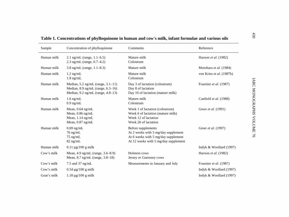
Vitamin K is given as a supplement to prevent or cure vitamin K deficiency whenthe endogenous vitamin K supply from the diet is likely to become or has proven to beinsufficient. Neonates are born with very limited vitamin K stores, but most infants donot show relevant hypoprothrombinaemia at birth (von Kries et al., 1987a, 1988; vonKries, 1991). Biochemical signs of vitamin K deficiency are common during the firstweek of life, however, unless sufficient amounts of vitamin K are ingested. The naturaldiet of newborns is human milk, which contains vitamin K at concentrations of0.69–9.2 ng/mL (see Table 1). [The Working Group noted that some of the high valuesin the Table may reflect methodological problems with analysis and milk collection.]Bleeding, the classical clinical manifestation of vitamin K deficiency, is extremely rareon the first day of life, and the typical time of onset is during the first week, withbleeding from mucous membranes, the umbilicus, following circumcision, and rarely,into the central nervous system (von Kries et al., 1988; von Kries, 1991). This condi-tion was originally called ‘classical haemorrhagic disease of the newborn’; the presentnomenclature is ‘classical vitamin K deficiency bleeding’ (Sutor et al., 1999).
During the first three months of life, exclusively breast-fed infants remain at riskfor vitamin K deficiency bleeding. In many of these infants, the bleeding episode,which is often intracranial haemorrhage, is the first perceived symptom of an under-lying cholestatic disease. In 10–30% of the cases, however, no underlying disease canbe found (von Kries et al., 1988).
After the first three months of life, vitamin K deficiency is almost completelyconfined to patients with cholestatic diseases (congenital or acquired obstruction ofthe bile duct), malabsorption syndromes or cystic fibrosis (Houwen et al., 1987; vanden Anker & Sinaasappel, 1993; O’Brien et al., 1994; Kowdley et al., 1997; Nowaket al., 1997; see also section 1.3.4).
1.3.3 For prevention of vitamin K deficiency in newborns and early infancy
The use of vitamin K prophylaxis since the 1950s has varied widely over time,between countries and within countries between institutions. The predominantpatterns were to give either selective intramuscular prophylaxis only to infantspresumed to be at special risk for bleeding (mainly premature and low-birth-weight
VITAMIN K SUBSTANCES 429
IARC M
ON
OG
RAPH
S VO
LUM
E 76430
Table 1. Concentrations of phylloquinone in human and cow's milk, infant formulae and various oils
Sample Concentration of phylloquinone Comments Reference
Human milk 2.1 ng/mL (range, 1.1–6.5)2.3 ng/mL (range, 0.7–4.2)
Mature milkColostrum
Haroon et al. (1982)
Human milk 3.8 ng/mL (range, 1.1–8.3) Mature milk Motohara et al. (1984)
Cow’s milk 7.5 and 37 ng/mL Measurements in January and July Fournier et al. (1987)
Cow’s milk 0.54 μg/100 g milk Indyk & Woollard (1997)
Goat’s milk 1.18 μg/100 g milk Indyk & Woollard (1997)
VITA
MIN
K SU
BSTAN
CES431
Table 1 (contd)
Sample Concentration of phylloquinone Comments Reference
Formula 79–118 ng/mL
118–256 ng/mL
19–69 ng/mL
Milk-substituted formulae with soya oil butwithout added vitamin K1Milk-substituted formulae with variousvegetable oils and with added vitamin K1Milk-based formulae with various vegetableoils but without added vitamin K1
Schneider et al. (1974)
Formula Mean, 4.4 ng/mL
Mean, 11.5 ng/mL
Unsupplemented infant formula containingonly milkfatUnsupplemented infant formula containingonly vegetable oils
Haroon et al. (1982)
Formula ~72–166 ng/mL~125–146 ng/mL~129–175 ng/mL
Ready-to-feedConcentratePowder
Bueno & Villalobos(1983)
Formula 30–225 ng/mL (trans isomer); 2.8–25ng/mL (cis isomer; 9.3–11% of total)120–211 ng/mL (trans isomer); 7.2–31ng/mL (cis isomer; 6.0–15% of total)90–195 ng/mL (trans isomer)
Ready-to-feed liquids
Concentrated liquids
Powders
Hwang (1985)
Formula 0.87 μg/g0.95 μg/g
Powder (milk-based)Powder (soya protein-based)
Schneiderman et al.(1988)
IARC M
ON
OG
RAPH
S VO
LUM
E 76432
Table 1 (contd)
Sample Concentration of phylloquinone Comments Reference
0.65 μg/100 g (range, 0.30–1.19)2.91 μg/100 g (range, 1.63–4.18)6.70 μg/100 g9.03 μg/100 g (range, 8.86–9.19)9.13 μg/100 g (range, 6.49–11.77)15.0 μg/100 g15.5 μg/100 g (range, 12.1–18.7)55.5 μg/100 g (range, 37.2–82.1)141 μg/100 g (range, 114–188)193 μg/100 g (range, 139–290)
Combined averageCombined average
Combined averageCombined average
Combined averageCombined averageCombined averageCombined average
Ferland & Sadowski(1992)
infants and those delivered surgically) or general prophylaxis for all infants. In thelatter case, vitamin K was given either intramuscularly or orally.
Several preparations of fat-soluble vitamin K have been in use. In the early 1950s,water-soluble menadiol sodium phosphate was widely used, until haemolysis due to highdoses of this preparation in neonates was identified (Meyer & Angus, 1956). In mostcountries, phylloquinone has been used since that time, although in some third-worldcountries water-soluble menadione sodium bisulfite still seems to be used (Sharma et al.,1995). Because it is technically difficult to dissolve phylloquinone, only a limited numberof preparations became available. The Roche preparation (Konakion®) in which Cremo-phor (polyethoxylated castor oil) is used as an emulsifying vehicle has been widelyavailable in Europe and North America. The manufacturer has recently replaced theCremophor preparation by a new mixed micellar preparation Konakion–MM® (BritishMedical Association/Royal Pharmaceutical Society of Great Britain, 1998). In Japan, anoral preparation of menaquinone-4 is used instead of phylloquinone (Hanawa, 1992).
Almost all cases of vitamin K deficiency bleeding can be prevented by intramuscularadministration of 1 mg of vitamin K at birth (von Kries & Hanawa, 1993). Clinical obser-vations and laboratory investigations have also clearly shown that a single oral dose ofvitamin K protects against classical vitamin K deficiency bleeding (Clark & James, 1995)but is less effective for prevention of this condition later in life (Tönz & Schubiger, 1988;Ekelund, 1991). Without vitamin K prophylaxis, the incidence of late vitamin Kdeficiency bleeding in Europe was estimated to be 40–100 per million livebirths, whereasin Asia the condition appears to be considerably more common (Hanawa, 1992; Chooet al., 1994).
Since intramuscular vitamin K prophylaxis has proven effective against late defi-ciency bleeding, 1 mg of vitamin K at birth was recommended in most westerncountries (von Kries, 1991). After reports of a potential association between vitaminK prophylaxis and the risk for childhood cancer (Golding et al., 1990, 1992), severalcountries switched to oral prophylaxis regimens with repeated doses of phylloquinone(Hill, 1994; Doran et al., 1995; Hansen & Ebbesen, 1996; Cornelissen et al., 1997).The optimal oral dose regimen remains to be established (von Kries, 1999).
1.3.4 Cholestatic and malabsorption syndromes
Vitamin K deficiency is observed in patients with cholestatic jaundice, cysticfibrosis, primary biliary cirrhosis and other diseases. In most cases, however, vitamin Kdeficiency is detectable only by measuring the plasma concentrations of vitamin K orwith sensitive biochemical markers of vitamin K deficiency (Cornelissen et al., 1992;O’Brien et al., 1994; Kowdley et al., 1997). Bleeding is observed only rarely. Additionalrisk factors, such as therapy with antibiotics that interfere with vitamin K metabolism,may cause bleeding in patients with cystic fibrosis (Nowak et al., 1997). Some patientswith this disease are given vitamin K supplements, although there are no uniform recom-mendations (Durie, 1994).
IARC MONOGRAPHS VOLUME 76434
1.3.5 Vitamin supplementation to overcome side-effects of drugs that interferewith vitamin K metabolism
An important indication for vitamin K supplementation is the side-effects of drugsthat interfere with its metabolism. Mothers on antiepileptic drugs, for example, are athigh risk of delivering an infant with manifest vitamin K deficiency (Cornelissenet al., 1993a) and intracranial bleeding (Renzulli et al., 1998).
Hypoprothrombinaemia may be caused by some cephalosporins, especially thosecontaining an N-methylthiotetrazole side-chain, and may require vitamin K supple-mentation (Breen & St Peter, 1997).
1.3.6 Vitamin K therapy
(a) Overdosage of vitamin K antagonistsThe coumarin derivatives acenocoumarol, phenprocoumon and warfarin are among
the most commonly used oral anticoagulants (Keller et al., 1999). The clinicalsymptom of overdosage of these drugs is bleeding. A tendency to bleed is alsoincreased by individual susceptibility to one of these anticoagulants, interference withother drugs or poor dietary intake of vitamin K. The biochemical indicator for over-dosage is an excessive prolongation of the prothrombin time. Minor bleeding is mostcommonly managed by temporarily discontinuing treatment and by giving vitamin Kto counteract the effects of the coumarin derivative. In the case of major bleeding, espe-cially intracranial haemorrhages, higher doses of vitamin K and use of prothrombincomplex concentrates are recommended to induce immediate reversal of anticoagu-lation (Pindur et al., 1999). In the past, the oral or intravenous dose of phylloquinoneused to counteract supratherapeutic anticoagulation was 10–50 mg (Fetrow et al.,1997). Much lower doses have been proposed recently. In asymptomatic patients, a1-mg oral dose of vitamin K was shown to reduce the international normalized ratioeffectively (Crowther et al., 1998). Low subcutaneous doses of phylloquinone are aneffective alternative to intravenous administration of phylloquinone in the treatment ofwarfarin-induced hypoprothrombinaemia (Fetrow et al., 1997).
(b) Prevention of intracranial haemorrhage in very-low-birth-weight,premature infants
The effect of high doses of vitamin K given to women at imminent risk of earlypreterm parturition has been studied with the primary aim of preventing periventricularhaemorrhage and the associated neurological injury in the infant. A first meta-analysisof the trials came to the conclusion, however, that it is ineffective (Thorp et al., 1995).
1.3.7 Other uses
Menadione is of industrial importance as an intermediate in the synthesis of phyllo-quinone, and salts of its bisulfite adduct are used as stabilized forms in the animal feed
VITAMIN K SUBSTANCES 435
industry. Commercially significant forms are menadione sodium bisulfite and mena-dione dimethyl pyrimidinol (Van Arnum, 1998).
Menaquinone-4 has been used in Japan at high doses for the treatment of osteo-porosis (Shearer, 1997).
1.4 Occurrence
Phylloquinone is widely distributed in higher plants and in some blue–green algae.It is present in many foods, especially leafy green vegetables and some vegetable oils.Table 2 shows the concentrations in some common foods (Booth et al., 1995; Sheareret al., 1996; Booth & Suttie, 1998).
The Total Diet Study of the US Food and Drug Administration is conducted period-ically to monitor the safety and nutritional quality of the US food supply by assessingthe levels of nutrients and contaminants in daily diets. It is based on the collection andanalysis of 265 core foods. Intakes are estimated from the concentrations of individualnutrients and contaminants in the core foods and the mean consumption of the foods ineach population group. The quantitative contributions of specific foods to the phyllo-quinone intake of the total population are presented in Table 3. Table 4 gives the esti-mated daily intake in 1990 for 14 categories of age and sex (Booth et al., 1996).
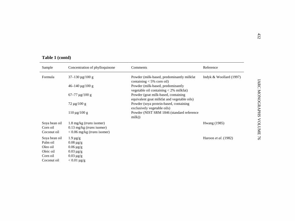
Phylloquinone has been determined by several analytical methods in human milk,in cow’s milk, in many brands of infant formula and in the oils that have been addedto infant formulas for many years. Some of the concentrations found in each of thesesources are presented in Table 1.
Menaquinones are synthesized by bacteria. They have a more restricted distribu-tion in the diet than phylloquinone, and nutritionally significant amounts probablyoccur only in animal liver and some fermented foods, including cheese. Menaquinonesare also synthesized by specific inhabitants of the human gut microflora. The majorintestinal forms are MK-10 and MK-11 produced by Bacteroides, MK-8 by Entero-bacteria, MK-7 by Veillonella genus and MK-6 by Eubacterium lentum (Shearer et al.,1996). The total concentration of menaquinones in human distal colonic contents isabout 20 μg/g dry weight, with MK-10 predominating (Conly & Stein, 1992; Shearer,1995). It seems likely that menaquinones synthesized by the gut microflora make asignificant contribution to human tissue stores and are used by the hepatic vitamin K-dependent carboxylase, but the extent of this contribution remains uncertain (Shearer,1995; Suttie, 1995).
1.5 Regulations and guidelines
Phylloquinone is listed (as phytomenadione or phytonadione) in the British,Chinese, Czech Republic, European, French, German, International, Japanese, Swissand US pharmacopoeias (Royal Pharmaceutical Society of Great Britain, 1999; SwissPharmaceutical Society, 1999).
IARC MONOGRAPHS VOLUME 76436
VITAMIN K SUBSTANCES 437
Table 2. Phylloquinone content of common foodsa
0.1–1.0 μg/100 g 1–10 μg/100 g 10–100 μg/100 g 100–1000 μg/100 g
Avocado [1]Banana [0.1]Beef, steak [0.8]Bread, white [0.4]Chicken, thigh [0.1]Coconut oil [0.5]Cod, fresh, fillet [< 0.1]Cornflakes [< 0.1]Flour, white [0.8]Grapefruit [< 0.1]Ham, tinned [0.1]Maize [0.3]Mangoes [0.5]Melon, yellow [0.1]Melon, water [0.3]Milk, cows [0.6]Mushrooms [0.3]Oranges [< 0.1]Parsnips [< 0.1]Peanuts, roasted [0.4]Pilchards, in brine [0.6]Pineapple [0.2]Pork, chop, lean [< 0.1]Potatoes [0.9]Rice, white [0.1]Rice, brown [0.8]Salmon, tinned, in brine [0.1]Sausage, pork or beef [0.2]Spaghetti [0.2]Tuna, tinned, in brine [0.3]Turnips [0.2]Yoghurt [0.8]
Apple pie [11]Apples [6]Aubergines [6]Baked beans [3]Barley [7]Beef, corned [7]Beef, minced [2]Bilberries [4]Bran, wheat [10]Bread [3]Bread, wholemeal [2]Butter [7]Carrots [6–10]Cheeses, various [2–6]Chocolate, plain [2]Corn oil [3]Courgettes [3]Cranberries [2]Cream, double [6]Dates, fresh [6]Doughnuts [10]Egg yolk [2]Eggs [2]Figs, fresh [3]French fries [5]Grapes, black [8]Grapes, green [9]Hamburger and bun [4]Hot dog and bun [3]Lasagna [5]Leeks [10]Liver, lamb [7]Liver, ox [4]Macaroni with cheese [5]Nectarines [3]Oats [10]Palm oil [8]Peaches, fresh [4]Pears [6]Peppers, green [6]Peppers, red [2]Pizza [4]Plums, red [8]Potatoes [1]Raisins [4]Rhubarb [4]Safflower oil [3]Strawberries [3]Sunflower oil [6]Swedes [2]Tomatoes [6]Wheat [8]
From Shearer et al. (1996); Booth & Suttie (1998)a Numbers in brackets are actual levels measured. The phylloquinone content of oil-based preparations varies widelydepending on the source of the oil used.
The Food and Drug Administration (1999) requires that all infant formulae sold inthe USA contain a minimum of 4 μg/100 kcal (0.2 mg/kg) vitamin K; and that anyvitamin K added should be in the form of phylloquinone.
Menadione is listed in the Austrian, Belgian, British, Dutch, European, French,German, International, Italian, Portuguese, Swiss and US pharmacopoieas, and mena-dione sodium bisulfite is listed in the Belgian, International, Swiss and US pharmaco-poeias (Royal Pharmaceutical Society of Great Britain, 1999; Swiss PharmaceuticalSociety, 1999). Menadiol sodium phosphate is listed in the British, Czech Republicand US pharmacopoeias (Swiss Pharmaceutical Society, 1999).
2. Studies of Cancer in Humans
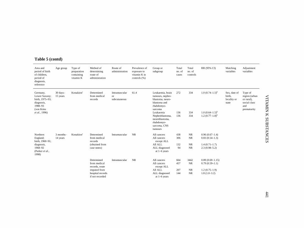
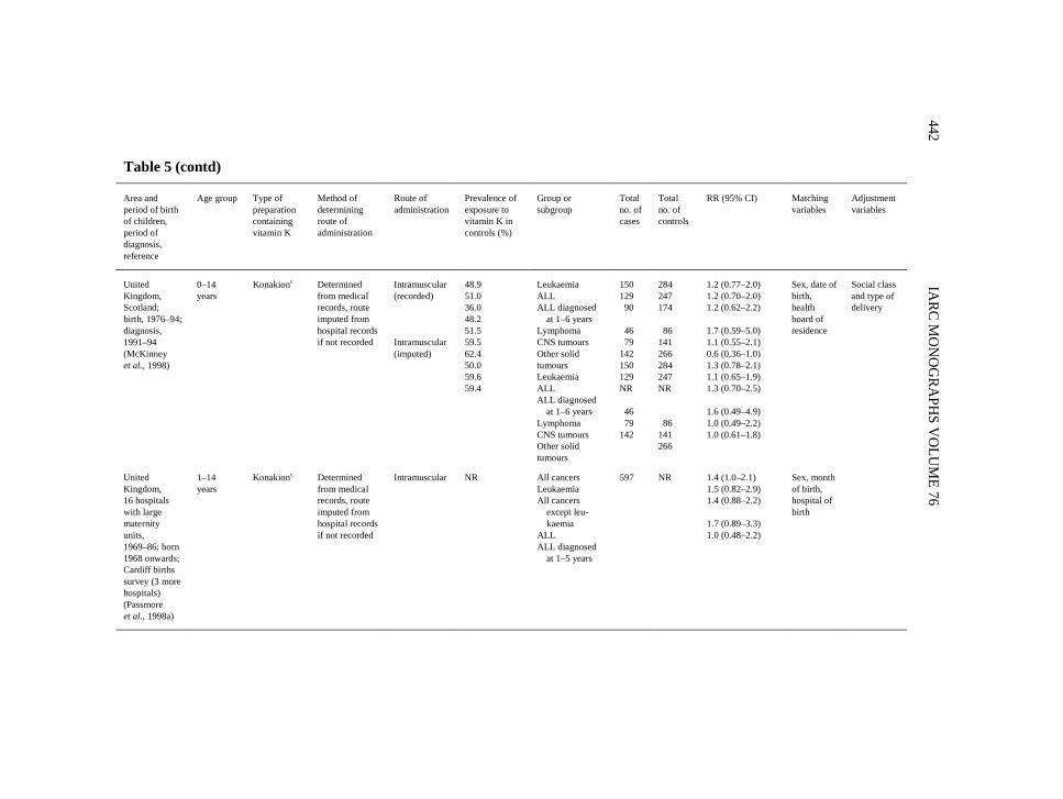
The association between childhood cancer and vitamin K administered during theperinatal period with a view to preventing haemorrhagic disease of the newborn hasbeen investigated in a number of studies (summarized in Table 5). The prophylacticuse of vitamin K in newborns has varied with time, geographical location and amonghospitals within countries. Some hospitals during some periods have had a selectivepolicy based on the indications low birth weight, prematurity and operative delivery.
IARC MONOGRAPHS VOLUME 76438
Table 3. Contribution of certain food groups to total adult intake (%) ofphylloquinone in the USA, stratified by age and sex
From Booth et al. (1996)a Percentages in columns may not add up to 100% as values were rounded to the nearest 0.1.
The hypothesis that vitamin K might be a risk factor for childhood cancer was gener-ated on the basis of the results of a cohort study of 16 193 infants delivered in GreatBritain in one week of April 1970, who were followed up at ages five and 10. The 33cases included in the study were in patients who had died from cancer or were identifiedthrough cancer registration as having a cancer diagnosed before the age of 10. Anunexpected statistically significant association was found between childhood cancer andadministration of any drug during the first week of life (Golding et al., 1990), and 16 ofthe 18 patients who had received drugs during the first week of life had receivedvitamin K. Within the cohort, a comparison was made between the 33 cases and 99controls matched with the cases for the age of the mother at the time of the birth of thechild, parity, social class, marital status at delivery and whether the birth was single ormultiple. Statistically significant associations were identified not only with drug admin-istration during the first week of life, but also with antenatal X-rays, antenatal smoking,non-term delivery and use of pethidine or pethilorfan (a pethidine-containing drug)during labour. Only two of the 33 cases had fewer than two of these risk factors, whereas
VITAMIN K SUBSTANCES 439
Table 4. Estimated and recommended meandietary intakes of phylloquinone in the USA,stratified by age and sex
Phylloquinone intake (μg/day)Population group
Estimatea Recommended
Infants 6-month-old infants 77 10
Children 2-year-old children 6-year-old children 10-year-old children 14–16-year-old girls 14–16-year-old boys
2446455264
15203045–5545–65
Younger adults 25–30-year-old women 25–30-year-old men 40–45-year-old women 40–45-year-old men
59667186
65806580
Older adults 60–65-year-old women 60–65-year-old men > 70-year-old women > 70-year-old men
76808280
65806580
From Booth et al. (1996)a From Total Diet Study
IARC M
ON
OG
RAPH
S VO
LUM
E 76440
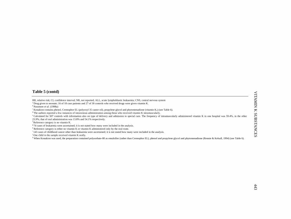
Table 5. Studies on childhood cancer and vitamin K administered during the perinatal period
Area andperiod of birthof children,period ofdiagnosis,reference
Age group Type ofpreparationcontainingvitamin K
Method ofdeterminingroute ofadministration
Route ofadministration
Prevalence ofexposure tovitamin K incontrols (%)
Group orsubgroup
Totalno. ofcases
Totalno. ofcontrols
RR (95% CI) Matchingvariables
Adjustmentvariables
Great Britain;birth, 1970;diagnosis,1970–80(Golding et al.,1990)
5 and10 years
NR NR Oral, intra-venous, intra-muscular
31.2a
(for drug toneonate)(28.1 forvitamin K toneonate)
All cancers 33 99(96 withdata ondrugintake)
2.6 (1.3–5.2)a
(drug to neonate)Maternalage, parity,socialclass,maritalstatus,multiplicity
Social class,smokingduringpregnancy,X-ray inpregnancy,term deliveryand pethidinein labour
Case–control studies
UnitedKingdom,Bristol; birth,1965–87;diagnosis,1971–91(Golding et al.,1992)
0–14yearsb
Konakionc Recorded inmedical recordsor imputed onthe basis ofyear of birth,type of deliveryand whether ornot infantadmitted tospecial careRecorded inmedical records
Konakionc Determinedfrom medicalrecords, routeimputed fromhospital recordsif not recorded
Intramuscular NR All cancersLeukaemiaAll cancers except leu- kaemiaALLALL diagnosed at 1–5 years
597 NR 1.4 (1.0–2.1)1.5 (0.82–2.9)1.4 (0.88–2.2)
1.7 (0.89–3.3)1.0 (0.48–2.2)
Sex, monthof birth,hospital ofbirth
VITA
MIN
K SU
BSTAN
CES443
Table 5 (contd)
RR, relative risk; CI, confidence interval; NR, not reported; ALL, acute lymphoblastic leukaemia; CNS, central nervous systema Drug given to neonate; 16 of 18 case patients and 27 of 30 controls who received drugs were given vitamin K.b Passmore et al. (1998a)c Konakion contains phenol, Cremophor EL (polyoxyl 35 castor oil), propylene glycol and phytomenadione (vitamin K1) (see Table 6).d The authors reported a few instances of intravenous administration among those who received vitamin K intramuscularly.e Calculated for 507 controls with information also on type of delivery and admission to special care. The frequency of intramuscularly administered vitamin K in one hospital was 59.4%, in the other23.9%; that of oral administration was 13.8% and 54.1% respectively.f Reference category is no vitamin K.g 74 cases of leukaemia were ascertained; it is not stated how many were included in the analysis.h Reference category is either no vitamin K or vitamin K administered only by the oral route.i 143 cases of childhood cancer other than leukaemia were ascertained; it is not stated how many were included in the analysis.j One child in the sample received vitamin K orally.k When Konakion was used, the preparation contained polysorbate-80 as emulsifier (rather than Cremophor EL), phenol and propylene glycol and phytomenadione (Rennie & Kelsall, 1994) (see Table 6).
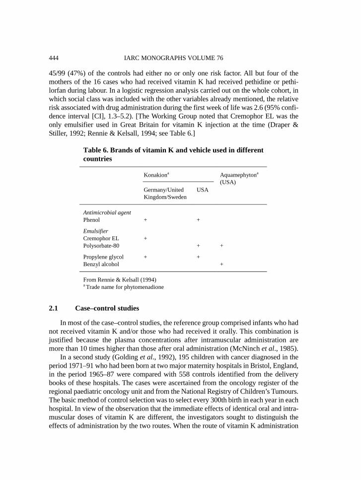
45/99 (47%) of the controls had either no or only one risk factor. All but four of themothers of the 16 cases who had received vitamin K had received pethidine or pethi-lorfan during labour. In a logistic regression analysis carried out on the whole cohort, inwhich social class was included with the other variables already mentioned, the relativerisk associated with drug administration during the first week of life was 2.6 (95% confi-dence interval [CI], 1.3–5.2). [The Working Group noted that Cremophor EL was theonly emulsifier used in Great Britain for vitamin K injection at the time (Draper &Stiller, 1992; Rennie & Kelsall, 1994; see Table 6.]
2.1 Case–control studies
In most of the case–control studies, the reference group comprised infants who hadnot received vitamin K and/or those who had received it orally. This combination isjustified because the plasma concentrations after intramuscular administration aremore than 10 times higher than those after oral administration (McNinch et al., 1985).
In a second study (Golding et al., 1992), 195 children with cancer diagnosed in theperiod 1971–91 who had been born at two major maternity hospitals in Bristol, England,in the period 1965–87 were compared with 558 controls identified from the deliverybooks of these hospitals. The cases were ascertained from the oncology register of theregional paediatric oncology unit and from the National Registry of Children’s Tumours.The basic method of control selection was to select every 300th birth in each year in eachhospital. In view of the observation that the immediate effects of identical oral and intra-muscular doses of vitamin K are different, the investigators sought to distinguish theeffects of administration by the two routes. When the route of vitamin K administration
IARC MONOGRAPHS VOLUME 76444
Table 6. Brands of vitamin K and vehicle used in differentcountries
Konakiona
Germany/UnitedKingdom/Sweden
USA
Aquamephytona
(USA)
Antimicrobial agentPhenol + +
EmulsifierCremophor EL +Polysorbate-80 + +
Propylene glycol + +Benzyl alcohol +
From Rennie & Kelsall (1994)a Trade name for phytomenadione
was not recorded in the neonatal notes, a route was imputed on the basis of year of birth,the type of delivery and whether or not the infant was admitted to special care; theimputed route was identified in the absence of knowledge of case or control status. Onthe basis of 180 cases (92% of those for which notes were available) and 544 controls(98% of those for which notes were available), the relative risk (adjusted for hospital andyear of delivery) for childhood cancer associated with intramuscular vitamin K was 2.2(95% CI, 1.1–4.4) when compared with no vitamin K and 1.2 (95% CI, 0.5–2.7) for oralvitamin K. In view of the absence of an association with oral vitamin K in these data,the authors conducted a subsequent analysis in which the reference group was definedto include infants who had not received vitamin K or who had received it orally. Therelative risk for leukaemia associated with intramuscular vitamin K was 2.7 (95% CI,1.3–5.2) and that for other types of childhood cancer was 1.7 (95% CI, 1.0–2.8). Thus,there was no clear difference in the association by type of childhood cancer. When theanalysis was confined to records in which the route was clearly stated, the odds ratio forall childhood cancer was 2.0 (95% CI, 1.2–3.3). These results could not be accountedfor by other factors associated with the administration of intramuscular vitamin K, suchas type of delivery or admission to a special care unit. Data were collected on 319variables for all controls and for 111 cases of cancer ascertained from the oncologyregister of the regional paediatric oncology unit; these data were not obtained for theremaining 84 cancer cases. Of these variables, the presence of rubella antibody, resusci-tation by intermittent positive pressure and paediatric estimate of gestation were statis-tically significant at the 1% level, which is what would be expected by chance. Adjust-ment for these and other variables reported to be associated with childhood cancer orknown to be indicators for administering intramuscular vitamin K had little effect on theodds ratio for childhood cancer associated with vitamin K. Nineteen of the cases werediagnosed in the first year of life, and the possibility was considered that these cancersmight have been present before the child was born and could therefore not have beeninitiated by an injection of vitamin K; however, the association persisted after exclusionof these 19 cases from the analysis. When the analysis was restricted to subjects whowould have been followed for at least 10 years, by considering only those born in theperiod 1971–80, the relative risk for all childhood cancer associated with intramuscularvitamin K was 1.9 (95% CI, 1.1–3.4), similar to that assessed for all subjects. [TheWorking Group noted, as acknowledged by the authors, a large number of instances inwhich the information on potentially confounding variables was not available, forexample on smoking in pregnancy. Medical records are not necessarily reliable sourcesof information about pregnancy and childbirth (Hewson & Bennett, 1987; Oakley et al.,1990), and this, together with the fact that potential confounding was assessed only fora subset of cases, constitutes a limitation of the study. The relationship between the typeof delivery and intramuscular administration of vitamin K differed markedly betweenthe two maternity hospitals in Bristol in which the case and control subjects in the studyhad been born (Carstensen, 1992; Draper & Stiller, 1992). The association with child-hood cancer is largely accounted for by data from one of the hospitals in which virtually
VITAMIN K SUBSTANCES 445
all of the control infants who received intramuscular vitamin K had been born by anassisted delivery. This raises the issue as to whether bias arose in control selection in thathospital.]
A study in the USA was reported by Klebanoff et al. (1993) which was based onfollow-up to the age of seven or eight years of 54 795 liveborn children of womenenrolled between 1959 and 1966 in 12 centres contributing to the National Collabo-rative Perinatal Project. Neonates whose cancer was diagnosed or strongly suspectedduring the first day of life were excluded because vitamin K could not have been afactor in those cases. Vitamin K was administered in the delivery room or the nursery,and information about the administration was recorded with other events during andafter delivery by observers who were not involved in the clinical care of the mother orthe infant. Cancer was diagnosed in 48 of 54 795 liveborn children after the first dayof life. For each case, five controls were selected and matched with the index case onlength of follow-up. In spite of the prospective recording by the observers, the data onvitamin K administration were not recorded unambiguously for 43 infants; a review ofhospital records without knowledge of case or control status resulted in data for 25(58%) of these. The exposure status was unknown for four case children. The relativerisk for all childhood cancer associated with vitamin K was 0.84 (95% CI, 0.41–1.7),and that for leukaemia was 0.47 (95% CI, 0.14–1.6; based on 15 cases). In the USA,only two brands, Aquamephyton and Konakion, have been approved for use (seeTable 6). Konakion in the USA contains polysorbate-80 rather than Cremophor EL asan emulsifier and phenol as an antrimicrobial agent. In the study of Klebanoff et al.(1993), the relative risk for total childhood cancer associated with the two brandstogether was 0.6, whereas that for children who had received the phenol-containingpreparation alone was 0.7. In this study, only one child had received vitamin K orally.
von Kries et al. (1996) carried out a case–control study of children born in 162obstetric hospitals in Lower Saxony (Germany) during the period 1975–93 when onlyone vitamin K preparation, Konakion, the same as that used in the United Kingdom,was licensed for neonatal vitamin K prophylaxis. Of a total of 218 children withleukaemia identified as eligible, information on vitamin K prophylaxis was obtained for136 (62%). For each leukaemia case, one control was selected from the municipalitywhere the patient lived at the time of diagnosis (local control), and a second one (statecontrol) from a municipality selected at random in Lower Saxony by means of apopulation-weighted sampling scheme. These controls were matched with cases by sexand date of birth. Case and control families were contacted initially by being sent aquestionnaire. If a control family refused to collaborate in the study or did not returnthe questionnaire within three months, another control family was invited; controlfamilies that returned the questionnaire after more than three months were alsoincluded. Thus, a total of 305 local and 308 state controls were invited to participate.Information on vitamin K prophylaxis was obtained for 174 (57%) of the local controlsand 160 (52%) of the state controls. As the study was performed as part of a population-based case–control study to explore possible causes of childhood leukaemia in Lower
IARC MONOGRAPHS VOLUME 76446
Saxony, a third control group for the leukaemia study was identified which comprisedcases of brain tumours, nephroblastoma, neuroblastoma and rhabdomyosarcoma. Nopopulation-based controls were selected for these cases, but they were used as addi-tional cases in the study of vitamin K. Of a total of 246 potentially eligible cases of thistype, information on vitamin K prophylaxis was obtained for 136 (55%). Data onvitamin K prophylaxis were abstracted from the birth report with no knowledge of thecase or control status of each child. Information on the dose and route of vitamin Kprophylaxis was obtained from the birth record or in the delivery book for 72% of the272 cases of leukaemia and other cancers and 64% of the 334 controls. When thisinformation was not available, the index child was assumed to have had the same expo-sure to vitamin K as the child nearest to the index infant in the delivery book with thesame route of delivery and same perinatal morbidity (nine cases and six controls).When this could not be established, staff who worked in the delivery unit at the timewhen the index child was born were asked what kind of vitamin K prophylaxis theindex infant would have received, given the birth weight and route of delivery (63 casesand 109 controls). Finally, similar information was sought from medical staff who didnot work in the delivery unit at the time the index child was born (four cases and fourcontrols). In the comparison with local controls (n = 107), the risk for leukaemia(n = 107) associated with intramuscular or subcutaneous administration of vitamin Krelative to that for oral or no vitamin K prophylaxis was 1.2 (95% CI, 0.68–2.25). Inthe comparison with state controls (n = 160; leukaemia cases = 136), the relative riskwas 0.82 (95% CI, 0.50–1.4). When the control groups were pooled (n = 334), therelative risk was close to unity (136 leukaemia cases), and the relative risk for braintumours, nephroblastoma, neuroblastoma and rhabdomyosarcoma combined (n = 136)associated with vitamin K prophylaxis was 1.2 (95% CI, 0.77–1.8). When the analyseswere repeated for subjects for whom vitamin K prophylaxis had been documented inbirth records or delivery books, the results were almost unchanged, except in thecomparison of leukaemia cases with local controls, which gave a relative risk of 2.0(95% CI, 0.69–6.0). When the analyses were repeated for parenteral prophylaxis versusno prophylaxis, most of the relative risks were slightly decreased. The risk of the sub-group of cases of leukaemia in children aged 1–6 years was analysed as this was consid-ered to be a relatively homogeneous subgroup, most of the cases having common acutelymphoblastic leukaemia. [The Working Group noted that it is not clear whether thedecision to make this subgroup analysis was specified in the original study protocol orwas made post hoc.] The risk relative to both control groups combined was 1.2(95% CI, 0.69–2.15), in the comparison with state controls it was 0.99 (95% CI,0.52–1.9) and in the comparison with local controls it was 2.3 (95% CI, 0.94–5.5).There was no difference between cases and controls in the source of information onvitamin K prophylaxis. The increased relative risk in the comparison with localcontrols could not be explained by any of the potential confounders. It would beexpected that the policy of administration of vitamin K would be more likely to besimilar for cases and local controls than for cases and state controls. Therefore, the
VITAMIN K SUBSTANCES 447
relative risk would be expected to be closer to unity in the comparison between casesand local controls than in the other comparison, whereas the opposite was observed.The non-significantly increased risk relative to local controls may be a chance result insubgroup analysis with multiple testing, as acknowledged by the authors.
In a case–control study of childhood leukaemia based on births in three hospitalsin England (Cambridge, Oxford and Reading), no association with intramuscularvitamin K, either as determined from hospital records (91 cases, 171 controls) or asimputed from hospital policy (132 cases, 264 controls), was found. In addition, noassociation was found specifically for acute lymphoblastic leukaemia (Ansell et al.1996). Subsequently, Roman et al. (1997) reported a more detailed analysis of data onleukaemia and non-Hodgkin lymphoma diagnosed before the age of 30 years insubjects whose obstetric records were stored in the same three hospitals. Ninety-twoper cent (132/143) of the cases of leukaemia were diagnosed at age 14 or less; thesecases and their controls were included in the report of Ansell et al. (1996). There wasno association between leukaemia and intramuscular vitamin K administration eitherrecorded in the notes (relative risk, 1.2; 95% CI, 0.7–2.1) or imputed from informationabout hospital policy (relative risk, 1.2; 95% CI, 0.5–2.4). In view of the finding ofvon Kries et al. (1996), acute lymphoblastic leukaemia diagnosed between the ages of1–6 years was considered; the relative risk associated with recorded administration(based on hospital notes) was 0.6 (95% CI, 0.3–1.4), and that based on hospital policywas again 0.6 (95% CI, 0.2–1.7).
Parker et al. (1998) identified 1432 children born in northern England between 1960and 1991 from the regional Children’s Malignant Disease Registry, in whom cancer wasdiagnosed in 1968–92 when they were aged between three months and 14 years whilestill resident in the region. The birth records of 701 of these children could not be traced,usually because the maternity unit had retained only its most recent records or becausethe unit had closed and the records could not be located. Thirty children who had beengiven vitamin K orally at birth and 16 cases in multiple births were excluded. Thecontrols were selected by taking the fourth, eighth and 12th birth before and after theindex birth from birth or admission registers for the hospital of birth of the index child.Towards the end of the study, the number of controls per case was reduced from six tothree because of time constraints. When the birth notes for control children could not belocated, or when the child selected was found to be on the Malignant Disease Register,the next possible control was selected. The fact of intramuscular administration ofvitamin K or non-administration of vitamin K was recorded in the maternity unit recordsfor 438 of 685 cases (case notes). [The Working Group noted that the correspondingproportion for controls was not specified.] There was no association between intra-muscular vitamin K administration and either all cancers or all cancers other than acutelymphoblastic leukaemia. The relative risk for acute lymphoblastic leukaemia associatedwith vitamin K administration based on case notes was 1.4 (95% CI, 0.71–1.7; 132cases). Two secondary analyses were conducted to consider cases typical of the peakincidence of leukaemia in early childhood. When the 51 children in the case note
IARC MONOGRAPHS VOLUME 76448
analysis who had T-cell leukaemia or for whom subtype characterization was notavailable were excluded, the relative risk for the 81 cases of non-T-cell lymphoblasticleukaemia was 1.8 (95% CI, 0.82–3.9). In an analysis of 94 children aged 1–6 years atdiagnosis, the relative risk was 2.3 (95% CI, 0.98–5.2). In all of these analyses, adjustedrelative risks were calculated separately for the specified potential confoundingfactors—sex, gestation, birth weight, opiates during labour, assisted delivery, signs ofasphyxia at birth, admission to special care or neonatal blood transfusions. Except foradjustment for assisted delivery, admission to special care or opiate exposure in labour,none of these changed any of the relative risks by more than 10%. Adjustment forassisted delivery or admission to special care caused a larger rise in the relative risk. Therelative risk for acute lymphoblastic leukaemia diagnosed at ages 1–6 was 2.4 (95% CI,1.0–5.7) after adjustment for exposure to opiates and 3.6 (95% CI, 1.3–9.7) afteradjustment for assisted delivery based on case note analysis. As in many of the otherstudies, information on hospital policy was obtained in order to impute exposure whenthis was unclear from medical records. This information was obtained by a researchmidwife and neonatal staff in each unit in the region and by a paediatrician from currentand recently retired medical staff, and this independently obtained information was thencross-validated. When inconsistencies were identified, the case notes were sampled todetermine what policy had actually been followed. This enabled a further 226 cases tobe included at the analysis; 21 cases were excluded because the policy of the local unitcould not be ascertained. The relative risks were similar to but somewhat lower thanthose in the analysis based exclusively on subjects for whom data on vitamin K exposurewas obtained only from medical records. [The Working Group noted that it was unclearwhich hypotheses about subgroups had been pre-specified. Bias may have arisen fromthe fact that while a large proportion of cases had to be excluded there was a mechanismfor adding controls when a control record was unobtainable. Availability of recordsmight have associations with both perinatal health problems and subsequent develop-ment of childhood cancer.]
McKinney et al. (1998) carried out a case–control study on childhood cancer inScotland using data abstracted from 76 hospital records. A total of 500 cases of cancerdiagnosed in children aged 0–14 years during the period 1991–94 while resident inScotland were identified. Controls matched on age, sex and health board of residencewere randomly selected from among all eligible children registered for primary carewithin each health board. A total of 1338 eligible controls was identified. A total of 460mothers of cases (92%) and 861 mothers of controls (64%) were interviewed, andmedical notes were abstracted for 440 cases and 802 controls. The data set for statisticalanalysis was restricted to matched sets, and information was lost for 23 cases withoutmatched controls and 25 controls without a matched case. Therefore, 417 cases and 777controls were included in the matched case–control analysis. Vitamin K was recorded asgiven or definitely not given only when this was mentioned in the notes. Similarly, theroute of administration was classified as intramuscular, oral or not recorded. None of therelative risks reported for leukaemias, acute lymphoblastic leukaemia, lymphomas,
VITAMIN K SUBSTANCES 449
central nervous system tumours or other solid tumours, either crude or adjusted for socialclass and type of delivery, was statistically significantly different from unity. Theadjusted relative risk for leukaemia associated with vitamin K given intramuscularly(recorded) in the neonatal period was 1.2 (95% CI, 0.77–2.0) and that for acutelymphoblastic leukaemia was 1.2 (95% CI, 0.70–2.0). In view of the findings of Parkeret al. (1998, see above), the subset of acute lymphoblastic leukaemia diagnosed inchildren aged 1–6 years (90 cases, 174 controls) was also analysed, and the adjustedrelative risk was found to be 1.2 (95% CI, 0.62–2.2). As nothing about vitamin K hadbeen written in the medical records for a substantial proportion of children (37% of casesand 35% of controls), the authors also sought to impute exposure on the basis of hospitalpolicies. Information on the vitamin K policies of hospitals in which over 500 infantswere delivered annually was validated by abstraction of a sample of medical records andthrough consultations with hospital pharmacies and senior labour room midwives. For100 (24%) cases and 191 (25%) controls, no hospital policy was available for any impu-tation. The relative risks for the specific diagnostic categories associated with intra-muscular vitamin K administration in the neonatal period either as recorded in medicalrecords or imputed from hospital policy were very similar to those calculated forsubjects for whom only data from medical records were included. The adjusted relativerisk for leukaemia was 1.3 (95% CI, 0.78–2.1), that for acute lymphoblastic leukaemiawas 1.1 (95% CI, 0.65–1.9) and that for acute lymphoblastic leukaemia in children aged1–6 years was 1.3 (95% CI, 0.70–2.5). Very few subjects were recorded as having orimputed to have been given vitamin K orally in the neonatal period (12 cases, 2.9%; and33 controls, 4.3%).
Passmore et al. (1998a) identified cases of childhood cancer diagnosed at ages up to14 years in persons who were resident in Great Britain and had been born in 16 hospitalswith large maternity units in 1968 or later and diagnosed by the end of 1986 from theNational Registry of Childhood Tumours (excluding retinoblastoma, Down syndrome orneurofibromatosis). The 16 hospitals were selected on the basis of a survey whichshowed that they had a selective policy for the use of vitamin K prophylaxis. Of 1092cases initially identified as born in these hospitals, 523 were born in the years for whicha policy was known and for whom the medical records were found. Four controlsmatched on sex, month of birth and hospital of birth were selected randomly from theseregisters. Medical records departments were asked to locate the records for each case andfor one control. Initially, two out of each of the four potentially eligible controls wereselected randomly for location by the medical records department. If the recordsdepartment was unable to locate the notes of either of these, details were supplied of theother two. Controls with illegible records, twins, stillbirths and neonatal deaths wereexcluded. In addition, infants with severe neural tube defects or a birth weight of lessthan 1000 g were excluded, as they were unlikely to have survived to the age at whichthe case patient developed cancer. For these, an alternative control was selected by usingthe next suitable birth in the hospital birth register. [The numbers of control replacementswere not specified.] A second group of cases from the same period was chosen from
IARC MONOGRAPHS VOLUME 76450
records of the National Registry of Childhood Tumours in order to identify cases ofcancer among children included in a survey of more than 100 000 births in SouthGlamorgan, Wales. For each case, two controls matched for sex, month of birth andhospital were selected, applying the same set of exclusions. Medical records weresought for all cases and controls, and information on vitamin K administration takenfrom these records was supplemented by data from the birth survey, which was availablefor most but not all of the period of study. This added three further hospitals to the study,all of which had selective policies of vitamin K administration, and 74 cases. In thecombined data (16 maternity units in England and Wales and the three hospitals includedin the survey in South Glamorgan), the relative risk for childhood cancer of all typesassociated with intramuscular vitamin K administration was 1.4 (95% CI, 1.0–2.1). Inthe data for the 16 maternity units in England and Wales, the relative risk was 1.2(95% CI, 0.77–1.9), while in the data from South Glamorgan, the relative risk was 2.1(95% CI, 1.1–4.1). For the combined data and for the data from South Glamorgan, modeof delivery (forceps, vacuum extraction, breech or caesarean) was a statistically signifi-cant confounding variable, and adjustment for this reduced the relative risks to 1.1 forthe combined data and 1.3 for the South Glamorgan data. In the combined data, therelative risk for leukaemia was 1.5 (95% CI, 0.82–2.85), that for acute lymphoblasticleukaemia was 1.7 (95% CI, 0.89–3.3) and that for acute lymphoblastic leukaemiadiagnosed at ages 1–5 years was 1.0 (95% CI, 0.48–2.2). Again, adjustment for mode ofdelivery reduced the relative risks. [The Working Group noted that the substantiallylower relative risk for the 1–5 year-old group than for all ages combined implies that theeffect for children of other ages is higher than that for this group, in contrast to the obser-vations of von Kries et al. (1996) and Parker et al. (1998).] The relative risk for non-leukaemia cancers was 1.4 (95% CI, 0.88–2.2) in the combined data and 2.4 (95% CI,1.1–5.4) in the data from South Glamorgan. In the South Glamorgan data, none of thepotential confounders that were adjusted for reduced the magnitude of the relative risk.[The Working Group noted that in the absence of an effect in the data from the 16maternity units in England and Wales, the South Glamorgan finding may reflect anunidentified bias or be a chance finding.]
[The Working Group noted that in the subgroup analyses of acute lymphoblasticleukaemia diagnosed at 1–6 years carried out by Parker et al. (1998) and 1–5 years byPassmore et al. (1998a), adjustment for mode of delivery had contrasting effects. Inthe study of Passmore et al. it attenuated the relative risk associated with vitamin K,while in the study of Parker et al. the relative risk was increased.]
2.2 Ecological studies
These studies are summarized in Table 7.Ekelund et al. (1993) investigated the association between childhood cancer and
intramuscular administration of vitamin K in a study in Sweden based on linkage of themedical birth registry to the national cancer registry. The study was restricted to full-
VITAMIN K SUBSTANCES 451
IARC M
ON
OG
RAPH
S VO
LUM
E 76452
Table 7. Ecological studies on childhood cancer and vitamin K administered intramuscularly during the perinatalperiod as Konakiana
Area and period ofbirth of children,period of diagnosis,reference
Age group Method ofdetermining routeof administration
Prevalence ofexposure inall children(%)
Group orsubgroup
Total no.of cases
No. of patients RR (95% CI) Referencecategory
Sweden;full-term non-instrumentaldeliveries;birth, 1973-89(follow-up, 1992);birth, 1982–89(Ekelund et al.,1993)
30 days–17 years
30 days–9 years
Imputed on thebasis of hospitalpolicy
78.4
66.2
All cancersLeukaemiaAll cancersLeukaemia
2287 708 722 250
Nos of patientsgiven vitamin Kintramuscularlyand orally1 357 7341 357 734 655 454 655 454
1.0 (0.88–1.2)b
0.90 (0.70–1.2)b
1.1 (0.88–1.4)1.2 (0.69–2.1)
Vitamin Korally
Denmark, 1945–54,1975–84;(Olsen et al., 1994)
1–12 years Imputed fromrecommendedpractice as: novitamin K forbirths 1945–54;intramuscularadministration forbirths 1975–84
NR
All cancersLeukaemia
NR No. of patientsgiven vitamin Kintramuscularlyand not givenvitamin K1 421 8081 421 808
1.3 (1.2–1.4)1.0 (0.9–1.1)at age 13
No vitamin K
RR, relative risk; CI, confidence interval; NR, not reporteda Konakion contains phenol, Cremophor EL (polyoxyl 35 castor oil), propylene glycol and phytomenadione (see Table 6).b Adjusted for year of birth
term infants (gestation, 37–42 weeks) who had survived and who were born in 1973–89after a delivery without use of forceps or vacuum extraction. The infants were followedup to 1 January 1992. Cancers diagnosed within 30 days of birth were regarded ascongenital and were excluded from the analysis. Routines for administration ofvitamin K were obtained from all 95 maternity hospitals and validated for a subset of102 children with cancer and 100 control children randomly selected from among thosewho, according to the information on routine exposure, received intramuscular vitaminK, and 94 children with cancer and 100 control children from among those who shouldhave received oral vitamin K. The doses of vitamin K given in Sweden were similar tothose given in the United Kingdom, and the same preparation was used (phylloquinone,Konakion, see Table 6). When the method of administration of vitamin K was recorded,it agreed with the stated routine method of administration in 92% of the 235 cases forwhich individual information could be found. The relative risk for all childhood cancerassociated with a hospital policy of intramuscular administration of vitamin K ascompared with oral administration was 1.0 (95% CI, 0.88–1.2, after stratification foryear of birth). The relative risk for leukaemia was 0.90 (95% CI, 0.70–1.2).
Olsen et al. (1994) compared the cumulative risk of childhood cancer amongchildren aged 1–15 years who were born during the period 1945–54 (n = 835 430), inwhich no vitamin K was administered, those aged 1–15 years born during the period1960–69 (n = 797 472), in which pregnant women received oral vitamin K, and thoseaged 1–13 years born during the period 1975–84 (n = 586 378), in which virtually allnewborns received vitamin K intramuscularly. There was a small increase in risk forall tumour types combined, due mainly to lymphoma in boys and neuroblastoma inboys and girls. There was no trend for childhood leukaemia. The preparation was thesame as that used in the United Kingdom (Draper & McNinch, 1994).
In addition to the case–control study in northern England described above, Parkeret al. (1998) compared the incidence of acute lymphoblastic leukaemia diagnosed inchildren aged up to 14 years who were born in hospital units in which all infantsreceived vitamin K, with those born in units where less than a third received this prophy-laxis. As described above, information on hospital policy was obtained separately andindependently by two people and then cross-validated. In units with a policy of selectiveprophylaxis, less than 30% of infants received intramuscular vitamin K at birth, whilein units offering universal prophylaxis, sampling of case notes showed that more than95% of babies received vitamin K. The risk for acute lymphoblastic leukaemia inchildren born in hospitals with a policy of universal prophylaxis relative to those bornin hospitals with a policy of selective prophylaxis was 0.95 (95% CI, 0.78–1.2). Therelative risk of the subgroup diagnosed at 1–6 years was 1.05 (95% CI, 0.82–1.35). [TheWorking Group noted that the cases included in this analysis overlapped with thoseincluded in the case–control study, so that the results are not independent].
Passmore et al. (1998b) carried out a similar comparison of cancers of all typesother than retinoblastoma or associated with Down syndrome or neurofibromatosisdiagnosed in children aged 1–14 years who were born in 94 hospital units in Great
VITAMIN K SUBSTANCES 453
Britain. Information on hospital policy for neonatal vitamin K was obtained during thecase–control studies of Passmore et. al. (1998a) and Ansell et al. (1996), describedabove, for 30 hospitals in Scotland from members of the Scottish Neonatal Networkand from paediatricians for 41 of a further 80 hospitals in England and Wales in whichmore than 25 children who subsequently developed cancer had been born in the period1968–85. The observed numbers of cases in hospitals with universal and selectivepolicies were compared with the numbers expected on the basis of national rates.Separate analyses were carried out for births in hospitals that followed one policythroughout the period of study and births in hospitals in which the policy changedduring the period of study. A large number of observed:expected ratios werecalculated. The ratio for all cancers was 0.97, that for leukaemia at 1–14 years was1.03, and that for acute lymphoblastic leukaemia at 1–5 years was 1.01 for hospitalswith a consistent, non-selective policy. The ratio tended to be smaller in hospitals witha selective policy than in those offering universal prophylaxis. The only statisticallysignificant (p < 0.05, two-tailed test) departure from unity indicated a lower risk forcancer other than leukaemia among children born in hospitals offering universalprophylaxis that those born in hospitals consistently offering selective prophylaxis inScotland. [The Working Group noted that the cases included in this analysisoverlapped with those in the case–control studies of Parker et al. (1998) and Ansellet al. (1996), so that the results are not independent.]
3. Studies of Cancer in Experimental Animals
No reports of studies specifically designed to investigate the carcinogenicity ofvitamin K substances were available to the Working Group. One study on the initiatingeffects of menadione in an assay of liver foci in rats was available (Denda et al., 1991)but could not be evaluated owing to methodological limitations.
4. Other Data Relevant to an Evaluation of Carcinogenicity and its Mechanisms
The studies summarized in this section should be considered in the light of thedifferences between naturally occurring forms of vitamin K that have a lipophilic side-chain at the 3-position of the 2-methyl-1,4-naphthoquinone (menadione) ring structure(phylloquinone and menaquinones) and the synthetic forms which lack this side-chain(menadione and its water-soluble derivatives). Lack of this side-chain results inprofound differences in the absorption, tissue distribution and metabolism of naturalK vitamins. Importantly, the lack of a lipophilic side-chain is the reason for theincreased chemical reactivity and greater toxicity of menadione when compared with
IARC MONOGRAPHS VOLUME 76454
phylloquinone and menaquinones. In the strict sense, menadione is a provitamin K,because it is biologically active for the synthesis of vitamin K-dependent proteins onlyafter conversion to the naturally occurring menaquinone-4 (four prenyl units) in vivo.
4.1 Absorption, distribution, metabolism and excretion
4.1.1 Humans
(a) Intestinal absorption and plasma transport in adultsThe major dietary form of vitamin K is phylloquinone (Shearer et al., 1996). It is
absorbed chemically unchanged from the proximal intestine after solubilization intomixed micelles composed of bile salts and the products of pancreatic lipolysis. Inhealthy adults, the efficiency of absorption of phylloquinone in its free form is about80% (Shearer et al., 1974), but the efficiency of absorption from green leafy vege-tables such as spinach is < 10% (Gijsbers et al., 1996).
Within the intestinal mucosa, phylloquinone is incorporated into chylomicrons, issecreted into the lymph and enters the blood via the lacteals (Shearer et al., 1970,1974). After a phylloquinone-containing meal, the plasma concentration peaks between3 and 6 h (Shearer et al., 1970; Lamon-Fava et al., 1998). Once in the circulation,phylloquinone is rapidly cleared at a rate consistent with its continuing association withchylomicrons and the chylomicron remnants that are produced by lipoprotein lipasehydrolysis at the surface of capillary endothelial cells. During the postprandial phaseand after an overnight fast, more than half of the circulating phylloquinone is asso-ciated with triglyceride-rich lipoproteins, and the remainder is carried by low-densityand high-density lipoproteins (Kohlmeier et al., 1996; Lamon-Fava et al., 1998).Although phylloquinone is the major circulating form of vitamin K, menaquinone-7 ispresent in plasma at lower concentrations and has a similar lipoprotein distribution tophylloquinone. While phylloquinone in blood is derived exclusively from the diet, it isnot known what proportion of circulating menaquinones such as menaquinone-7derives from the diet or the intestinal flora (Shearer et al., 1996).
(b) Plasma pharmacokinetics of phylloquinone in adults The plasma clearance of an intravenous dose of 1 mg [3H]phylloquinone during
the first 6 h resolved approximately into two exponential functions, the first with ahalf-time of 20–24 min and the second with a half-time of 121–150 min (Sheareret al., 1972). The curves for clearance up to 12 h after an intravenous injection of a10-mg dose of phylloquinone (Konakion MM) were similar to those after 1 mg andwere consistent with a two-compartment (sometimes three-compartment) model inwhich the log-linear terminal phase over 3–12 h had a half-time of about 3 h(Soedirman et al., 1996). A gradual slowing of the clearance rate was seen after thefirst 6 h (Shearer et al., 1972, 1974), as was also found in a study of the clearance ofpharmacological doses of 10–60 mg by Øie et al. (1988), who reported that the log-
VITAMIN K SUBSTANCES 455
linear terminal elimination phase was not reached before 8–12 h and that the averagehalf-time was 14 h (range, 8–22 h). This slowing of the clearance rate may beexplained by the complexity of the plasma transport of phylloquinone, in which theproportion of phylloquinone associated with low-density and high-density lipo-proteins increases progressively (Lamon-Fava et al., 1998).
The plasma disposition of oral doses of 5–60 mg phylloquinone (Konakion orAquaMephyton) is similar to that found after a more physiological dose (≤ 1 mg), withpeak plasma concentrations at 4–6 h followed by a rapid clearance phase (Sheareret al., 1974; Park et al., 1984; Øie et al., 1988; Hagstrom et al., 1995). After an oraldose of 10 or 50 mg Konakion, the plasma concentration declined from the peakabsorptive level at a similar log-linear rate as that seen after intravenous adminis-tration, with a terminal half-time of about 2 h for measurements up to 9–12 h (Parket al., 1984). The absorption of oral preparations of phylloquinone shows inter- andintra-individual variation and, for doses of Konakion ranging from 10 to 60 mg, thebioavailability was 10–63% (Park et al., 1984) and 3.5–60% (Øie et al., 1988).
The pharmacokinetics of phylloquinone after an intramuscular dose is completelydifferent, showing sustained, slow release from the muscle site over many hours andmarked inter-individual variation (Hagstrom et al., 1995; Soedirman et al., 1996). Thepharmacokinetics may also be influenced by the solubilizing agent. The systemic avai-lability of intramuscularly injected Konakion MM, which is a mixed-micellar solutionof phylloquinone in natural solubilizers, the bile acid glycocholic acid and the phos-pholipid lecithin (Schubiger et al., 1997; see Table 6), was irregular and < 65% in 20%of subjects (Soedirman et al., 1996). After intramuscular injection of phylloquinone(AquaMephyton R), most of the substance was carried by low-density and high-density lipoproteins instead of by triglyceride-rich (very-low-density) lipoproteins asfound after oral administration (Hagstrom et al., 1995).
(c) Plasma pharmacokinetics of phylloquinone in neonatesThe pharmacokinetics of phylloquinone during the early clearance phase up to 6 h
in neonates (of low birth weight) after intravenous injection was very similar to thatof adults (Shearer et al., 1972), declining bi-exponentially with median half-times of23 and 109 min (Sann et al., 1985).
An early study of the plasma disposition of 1 mg Konakion given orally or intra-muscularly at birth showed wide inter-individual differences during the first 24 h,especially after oral administration (McNinch et al., 1985). The peak plasma concen-tration after an oral dose occurred after 4 h; the median concentration was 73 ng/mL,which fell to 23 ng/mL after 24 h. The plasma concentration after administration of1 mg of Konakion intramuscularly exceeded those after oral administration at alltimes, and after 24 h the median was 444 ng/mL. Physiologically, these concentrationscompare with adult endogenous levels of about 0.5 ng/mL (Shearer, 1992).
In a comparison of the plasma concentrations of Konakion and Konakion MM inexclusively breast-fed infants at 24 h and 4 and 24 days after a single oral dose of 2 mg
IARC MONOGRAPHS VOLUME 76456
at birth (Schubiger et al., 1997), the mixed-micellar Konakion MM preparation resultedin higher median concentrations at all times, suggesting greater bioavailability. Thelargest difference was seen after four days, with median concentrations of 41 ng/mLKonakion MM and 12 ng/mL Konakion. By 24 days, the concentrations in both groupswere mainly within the adult physiological range (0.3–0.4 ng/mL). An earlier study bythe same group (Schubiger et al., 1993) had shown that a single oral dose of 3 mgKonakion MM resulted in higher plasma concentrations than a single dose of 1.5 mg ofthe same preparation given intramuscularly after four days. In this study, however, theplasma concentrations after 24 days were significantly higher after intramuscularinjection, consistent with the hypothesis of the depot effect of intramuscular phyllo-quinone (Loughnan & McDougall, 1996; see also section 4.1.1(f)).
Stoeckel et al. (1996) pointed out that the terminal elimination plasma half-timeof phylloquinone in neonates is probably longer than that in adults. They calculatedfrom published studies that a realistic estimate of the terminal plasma half-time inneonates was 26–193 h (median, 76 h), as compared with 8–22 h (median, 14 h) inadults after intravenous administration (Øie et al., 1988). This longer terminal half-time may reflect the poorly developed organ systems of neonates and a reducedcapacity to metabolize and excrete vitamin K (Stoeckel et al., 1996).
(d) Plasma pharmacokinetics of menaquinone-4 Oral preparations of menaquinone-4 are used in Japan for the prophylaxis of vitamin
K deficiency bleeding. The plasma profile of an oral dose of this preparation in five-day-old infants appeared to be similar to that of phylloquinone; after a 4-mg dose, a peakconcentration of about 100 ng/mL was achieved after 3–4 h, before declining to about30 ng/mL by 12 h (Shinzawa et al., 1989). The half-time of menaquinone-4 was notcalculated.
(e) Adult tissue reserves and distribution of vitamin K Dietary vitamin K is delivered to the liver and possibly other tissues, including
bone marrow, in the form of chylomicron remnants (Kohlmeier et al., 1996). The liverhas often been assumed to be a major depot for vitamin K because it is the site ofsynthesis of the vitamin K-dependent coagulation proteins. Measurements of phyllo-quinone in livers obtained at autopsy from 32 adults in the United Kingdom revealedhepatic concentrations ranging from 1.1 to 21 ng/g wet tissue [2.4–47 pmol/g], with amedian concentration of 5.5 ng/g [12 pmol/g]. The corresponding total liver stores ofphylloquinone were 1.7–38 μg [3.8–85 pmol/g], with a median total store of 7.8 μg[17 pmol/g] (Shearer et al., 1988). Similar hepatic concentrations of phylloquinonewere found in a smaller number of analyses of post-mortem samples from adults inJapan (10 ng/g) (Uchida & Komeno, 1988) and in The Netherlands (11 ng/g) (Thijssen& Drittij-Reijnders, 1996). The limited ability of the liver to store vitamin K is illus-trated by the observation that the phylloquinone reserves are about 40 000-fold lowerthan those of vitamin A despite a daily dietary intake of vitamin K (∼100 μg) which is
VITAMIN K SUBSTANCES 457
only about 10-fold lower than that of vitamin A (~1000 μg). The distribution of thevarious forms of vitamin K in the liver is quite different from that in plasma in that themajor transport form, phylloquinone, represents the minority of total hepatic stores(about 10%); the remainder comprises bacterial menaquinones, mainly menaquinones-6–13 (Shearer et al., 1988; Shearer, 1992; Shearer et al., 1996). The pattern ofindividual menaquinones in the liver varies considerably between individuals (Sheareret al., 1988; Uchida & Komeno, 1988; Thijssen & Drittij-Reijnders, 1996), perhapsreflecting their origin from the intestinal microflora (Shearer et al., 1996). Thisproposal is supported by the finding that two menaquinones, -10 and -11, which aremajor forms in most liver samples (Uchida & Komeno, 1988; Thijssen & Drittij-Reijnders, 1996), are known to be synthesized by Bacteroides species which are predom-inant members of the human intestinal flora (Conly & Stein, 1992); yet menaquinone-10 and menaquinone-11 do not make appreciable contributions to normal diets (Sheareret al., 1996).
Phylloquinone is also present in other human tissues. The concentration in the heart(~5 ng/g) [~10 pmol/g] is comparable to those in the liver, and even higher concen-trations (~13 ng/g) [~25 pmol/g] are found in the pancreas, but lower concentrations(< 1 ng/g) [< 2 pmol/g] were detected in brain, kidney and lung. These tissues do notappear to contain appreciable concentrations of menaquinones except for the short-chain menaquinone-4. Particularly high concentrations of menaquinone-4 relative tophylloquinone are present in the kidney, brain and pancreas. Although these and othertissues contain the enzymes of the vitamin K epoxide cycle (see Figure 1) and carry outvitamin K-dependent carboxylation of protein precursors, this would not appear toaccount for the tissue-specific accumulation of menaquinone-4 and may suggest ahitherto unrecognized physiological role for menaquinone-4 in certain tissues (Shearer,1992; Thijssen & Drittij-Reijnders, 1996). Indeed, menaquinone-4 may arise by tissuesynthesis from phylloquinone itself (Davidson et al., 1998).
Osteocalcin is a major vitamin K-dependent bone protein synthesized by osteo-blasts and therefore requires a source of vitamin K for γ-glutamyl carboxylation. Bothtrabecular and cortical bone contain ample reserves of vitamin K, with phylloquinonepredominating and smaller amounts of shorter-chain menaquinones (Hodges et al.,1993; Shearer, 1997). With the absence of the typical hepatic forms menaquinones-10–13, the vitamin K content of bone resembles that of other extrahepatic tissues.
(f) Tissue stores and blood concentrations in neonates and infants Information on liver stores (the site of synthesis of vitamin K-dependent clotting
proteins) in infants and their response to vitamin K prophylaxis is limited (Shearer et al.,1988; Guillaumont et al., 1993). The endogenous stores of vitamin K in the liver of thenewborn differ both quantitatively and qualitatively from those of adults because theconcentrations and total reserves of phylloquinone are lower than those of adults(Shearer et al., 1988) and because bacterial menaquinones are undetectable (Sheareret al., 1988; Guillaumont et al., 1993). The endogenous hepatic concentrations of
IARC MONOGRAPHS VOLUME 76458
phylloquinone ranged from 0.3 to 6.0 ng/g (median, 1.4 ng/g) in preterm infants andfrom 0.1 to 8.8 ng/g (median, 1.0 ng/g) in term infants. The median hepatic con-centration of 1 ng/g in term infants is equivalent to a total liver pool of about 0.1 μgphylloquinone, whereas the concentration is 5.5 ng/g and the pool 7.8 μg in adult liver.In infants who had received 0.5 or 1 mg phylloquinone at birth by intramuscularinjection, these liver reserves were raised by some two to three orders of magnitude
VITAMIN K SUBSTANCES 459
Figure 1. Cyclic metabolism of vitamin K for conversion of glutamate (Glu)residues to γγ-carboxy glutamate (Gla) residues in vitamin K-dependent proteins
Adapted from Shearer (1992) and Suttie (1987)PIVKA-II, protein induced by vitamin K absence factor II The active form of vitamin K needed for carboxylation is the reduced form, vitamin K quinol. Thecarboxylation reaction is driven by a vitamin K-dependent carboxylase activity (1) coupled to vitamin K-epoxidase activity (1) which simultaneously converts vitamin K quinol to vitamin K 2,3-epoxide. VitaminK 2,3-epoxide is reduced back to the quinone by vitamin K epoxide reductase (2A). The cycle is completedby the reduction of recycled vitamin K quinone by vitamin K reductase activity (2B). The activities of bothvitamin K epoxide (2A) and vitamin K reductase (2B) are dithiol-dependent (dithiol and disulfide denotereduced and oxidized dithiols) and are inhibited by coumarin anticoagulants such as warfarin. Exogenousvitamin K may enter the cycle via an NAD(P)H-dependent vitamin K reductase activity (3) which is notinhibited by warfarin.
within 24 h. Hepatic phylloquinone concentrations may remain elevated for severalweeks after injection: in two infants known to have received 1 mg phylloquinone by theintramuscular route and who survived 13 and 28 days, the total hepatic stores were 24and 15 μg, respectively (Shearer et al., 1988). Guillaumont et al. (1993) measuredhepatic concentrations in post-mortem liver samples obtained within the first 48 h ofdeath from infants who had received 2 mg phylloquinone intravenously or orally (insome cases combined with extra intravenous or oral doses of 1, 5 or 10 mg). In threenewborns who survived < 24 h, the hepatic concentrations of phylloquinone ranged from63 to 94 μg/g (total liver stores, 2800–7300 μg), which were four orders of magnitudehigher than the endogenous concentrations of 0.002–0.008 μg/g (total liver stores,0.1–0.9 μg). Between 24 and 48 h, the hepatic concentrations in 10 infants had fallen toa median of 8.4 μg/g (total liver stores, 550 μg), and in one infant who survived for fivedays it was 2.9 μg/g (110 μg). The quite rapid fall in hepatic stores presumably reflectsthe relatively rapid metabolism and excretion of vitamin K via the urine and bile(Shearer et al., 1974). The lower hepatic concentration after intramuscular injection(Shearer et al., 1988) compared with intravenous injection (Guillaumont et al., 1993) isconsistent with the idea that phylloquinone injected intramuscularly is released relativelyslowly from the injection site (Loughnan & McDougall, 1996).
The reduced hepatic reserves of vitamin K in the human neonate are best explainedby the existence of a barrier to placental uptake or transfer. This suggestion was origi-nally made on the basis of the large concentration gradient of physiological concen-trations of phylloquinone between maternal and cord blood plasma and the inefficientmaternal–fetal transfer of pharmacological doses administered as an intravenous injec-tion to the mother just before delivery (Shearer et al., 1982). The poor placental transportof phylloquinone has been confirmed by others (Mandelbrot et al., 1988; Yang et al.,1989). There is now general agreement that the cord plasma concentration of phyllo-quinone is < 50 pg/mL [110 pmol/L] and that the average maternal–fetal concentrationgradient is within the range 20:1 to 40:1 (Shearer, 1992).
Few longitudinal studies have been conducted of plasma concentrations in infantswho were not given vitamin K prophylaxis. In one such study, cord plasma concen-trations were compared for breast-fed and formula-fed infants and in blood on days 3, 7and 28 after birth (Pietersma-de Bruyn et al., 1990). In entirely breast-fed infants, theblood concentration rose from undetectable (< 20 pg/mL) at birth to mean values of 0.76,0.49 and 0.49 ng/mL [1.7, 1.1 and 1.1 pmol/mL] on days 3, 7 and 28, respectively. Ininfants fed a milk formula containing 68 ng/mL phylloquinone, the plasma concentrationrose steadily, with mean values of 1.4, 3.1 and 4.4 ng/mL [3.2, 6.8, and 9.9 pmol/mL] ondays 3, 7 and 28, respectively. In another group of infants, Pietersma-de Bruyn et al.(1990) found that phylloquinone was undetectable in cord blood and in venous bloodtaken at 30 min but became measurable in venous blood after 12 h in 30% of infants(range, 0.04–0.40 ng/mL) and after 24 h in 60% of infants (range, 0.04–0.63 ng/mL).
A more detailed longitudinal comparison of plasma concentrations in breast-fed andformula-fed infants at 6, 12 and 26 weeks was made by Greer et al. (1991). This study
IARC MONOGRAPHS VOLUME 76460
is of special interest because the intakes of phylloquinone were also estimated at eachtime by measuring the vitamin K content of the milk and the volume of milk ingested(by weighing the infant). Such an assessment of the intake of phylloquinone depends onboth the analytical accuracy of the measurements in breast milk and validation of themilk collection and sampling technique; both have proved problematical. The study ofGreer et al. (1991) seems to have met the requisite criteria, and, although the concen-trations were at the lower end of published values, they were in the same range as thosein a carefully designed longitudinal study of the phylloquinone content of breast milkover the first five weeks of lactation (1–2 ng/mL) (von Kries et al., 1987b). The results,summarized in Table 8, illustrate the extreme differences in intakes between breast-fedand formula-fed infants, which are also reflected in the plasma concentrations. Theplasma concentrations in the formula-fed infants agree with those found by Pietersma-de Bruyn et al. (1990) after 28 days (4.5 ng/mL), and suggest that they plateau at aroundone month. The concentrations in entirely breast-fed infants aged one month and beyondtend, as in this study, to be at the lower end of the normal range in adults (~0.15–1.0 ng/mL; mean, ~0.5 ng/mL), even when the infants have received prophylaxis duringthe first week of life (Cornelissen et al., 1992; Schubiger et al., 1993, 1997). In contrast,the plasma concentrations in formula-fed infants are about 10-fold higher than theaverage values in adults (Pietersma-de Bruyn et al., 1990; Greer et al., 1991).
(g) Hepatic catabolismThe liver plays an exclusive role in the metabolic transformations leading to the
elimination of vitamin K from the body. After intravenous doses of 45 μg to 1 mg[3H]phylloquinone, about 20% of the radiolabel was excreted in the urine within threedays, and 35–50% was excreted as metabolites in the faeces via the bile (Shearer
VITAMIN K SUBSTANCES 461
Table 8. Dietary intakes and plasma concentrations ofphylloquinone in breast-fed and formula-fed infants aged0–6 months in the USA
From Greer et al. (1991)a The average breast-milk concentrations were 0.86, 1.1 and 0.87 μg/L(ng/mL) at 6, 12 and 26 weeks, respectively.b All infants were fed a formula containing 55 μg/L (ng/mL) phyllo-quinone.
et al., 1974). Rapid depletion of hepatic reserves of phylloquinone was also seen insurgical patients placed on a low-phylloquinone diet (Usui et al., 1990). These resultssuggest that the body stores of vitamin K are replenished constantly.
The route of hepatic catabolism leading to urinary excretion of vitamin K proceedsby oxidative degradation of the phytyl side-chain, probably involving the sameenzymes used for ω-methyl and β-oxidation of fatty acids, steroids and prostaglandins.Two major metabolites or aglycones have been identified, which are carboxylic acidswith five- and seven-carbon atom side-chains and are excreted in the urine asglucuronide conjugates (McBurney et al., 1980). The biliary metabolites have not beenclearly identified but are initially excreted as water-soluble conjugates and becomelipid-soluble during their passage through the gut, probably through deconjugation bythe gut flora. There is no evidence that the body stores of vitamin K are conserved byenterohepatic circulation. Vitamin K itself is too lipophilic to be excreted in the bile,and the side-chain-shortened carboxylic acid metabolites are not biologically active.
(h) Vitamin K-epoxide cycleIn all tissues and cells found to carry out vitamin K-dependent carboxylation, the
reaction has been shown to be intimately linked to a metabolic sequence known as thevitamin K-epoxide cycle. This cycle and the associated enzyme activities are shown inFigure 1. Its function seems to be to serve as a salvage pathway to conserve tissuereserves of vitamin K. In the course of γ-glutamyl carboxylation, vitamin K quinol istransformed into vitamin K epoxide, and the epoxide product is recycled in two steps;firstly by vitamin K epoxide reductase activity to produce vitamin K quinone andsecondly by quinone reductase activity to produce the co-enzyme vitamin K quinol.Both these activities are thiol-dependent and are probably effected by the same enzyme(Suttie, 1987).
An important property of the dithiol-dependent epoxide and quinone reductase istheir sensitivity to certain antagonists, especially those based on 4-hydroxycoumarin(e.g. warfarin) or indandione structures, which have long been used as oral anti-coagulants. It is now clear that their anticoagulant action is based on their ability toinhibit epoxide reductase activity and block the recycling of the vitamin. The dithiol-dependent quinone reductase is also sensitive to warfarin, but the activity of a secondquinone reductase catalysed by an NAD(P)H-dependent enzyme is less sensitive towarfarin inhibition and provides an alternative pathway for the reduction of vitamin Kquinone to quinol in the presence of warfarin and other oral anticoagulant drugs(Shearer, 1992).
(i) Menadione and related water-soluble derivativesNo studies appear to have been conducted on the absorption, distribution, meta-
bolism or excretion of menadione and related compounds in humans. Water-soluble saltsof menadione (vitamin K3) were introduced for vitamin K prophylaxis in newborns inthe early 1940s and, until their use was almost entirely superseded by phylloquinone in
IARC MONOGRAPHS VOLUME 76462
the early 1960s, there were no suitable techniques for measuring menadione, its salts ortheir metabolites other than by radioisotopic techniques.
4.1.2 Experimental systems
(a) Absorption The route and mechanism of absorption of menadione is different from that of
natural K vitamins such as phylloquinone. Jaques et al. (1954) fed [14C]menadione torats and measured the radiolabel in faeces, bile, lymph and urine. They deduced thatall the absorbed menadione was transported exclusively via the portal vein to the liver,unlike phylloquinone which is transported by the lymphatic pathway. Also unlikephylloquinone, menadione participated in rapid entero-hepatic circulation afterexcretion in the bile. Mezick et al. (1968) suggested that, while the portal route isimportant in rats, menadione could also be transported via the lymphatic system.Direct evidence for some lymphatic transport was found by experiments in dogs,showing that about 10% of the absorbed menadione was recovered in thoracic ductlymph. In studies with bile exclusion, the absorption of menadione in rats was foundnot to be dependent on bile, as would be expected if menadione is absorbed predomi-nantly via the portal vein.
(b) Tissue distribution, metabolism and excretionEarly experiments with [14C]menadione in mice showed rapid clearance from the
intramuscular injection site of doses of 0.1 and 1.0 mg (about 4–40 mg/kg bw) withinthe first hour and excretion in the urine. Radiolabel was initially detectable in blood,but the concentrations later declined. No significant accumulation was seen in tissues.Small amounts of activity were sometimes detected in liver, lung and kidney, but nosignificant amounts were found in skin, bone or muscle (Solvonuk et al., 1952). Acomparison of the tissue distribution of [14C]menadione and [14C]phylloquinone inrats after intravenous administration of a pharmacological dose (5 mg/kg bw) showeda much higher (24-fold) concentration of radiolabel in the livers of animals givenphylloquinone than in those given menadione, and a fivefold greater accumulation ofphylloquinone was found in the spleen. As in the studies of Solvonuk et al. (1952), noorgan-specific accumulation of radiolabel was found in rats given labelled menadione,the highest proportions of radioactivity being found in urine and faeces (Taylor et al.,1957). The rapid, extensive excretion of [14C]menadione in the urine was confirmedby Losito et al. (1968) who found that rats excreted about 70% of an intravenous dosein the urine within 24 h compared with only about 10% of a dose of phylloquinone.They also showed that the urinary excretion of menadione (again unlike phyllo-quinone) was not dependent on an intact liver, as hepatectomized rats excreted thesame amount of the dose (70%) as normal rats.
Rats given intracardial injections of a more physiological total dose (10 μg[30 μg/kg bw]) of high-activity 6,7-[3H]menadione showed a pattern of excretion and
VITAMIN K SUBSTANCES 463
tissue distribution similar to that of pharmacological doses, with recovery of 78–83% ofthe label in the urine after 18 h (Taggart & Matschiner, 1969). A similar pattern was seenin rats given an intraperitoneal injection of about 2 μg of the water-soluble salt menadioldiphosphate; 17 h later, some 43% of the radiolabel had been excreted in urine and about4% in faeces. The compound was not concentrated in any tissue but was distributedthroughout all body organs, and the distribution was the same in vitamin K-replete and-deficient animals. This water-soluble compound underwent rapid conversion to lipid-soluble forms, and the compound and its metabolites were found generally to be asso-ciated with the membranous fractions of cells (Thierry & Suttie, 1969).
Three major metabolites of menadione have been isolated from urine. After oraladministration of menadione to rabbits, Richert (1951) isolated the sulfated compound2-methyl-4-hydroxy-1-naphthyl sulfate and noted increased excretion of glucuronicacid. Hoskin et al. (1954) resolved three metabolites from rat urine, of which the majorproduct was tentatively identified as 2-methyl-1,4-dihydroxynaphthalene-1,4-diglucu-ronide and another as the monosulfate conjugate found by Richert (1951). A third,minor metabolite appears to be a phosphate conjugate (Hart, 1958).
Losito et al. (1967) showed in an isolated perfused rat liver system that menadioneglucuronide or sulfate conjugates are excreted but that the glucuronide is confined tobile and the sulfate to the perfusing blood. In rats in vivo, Losito et al. (1968) separatedthree major urinary metabolites, two of which were identified as the same glucuronideand sulfate conjugates as those found in their liver perfusion system (Losito et al.1967). The chromatographic pattern in hepatectomized rats was different, but themajor peak was shown to be a glucuronide conjugate, showing that animals have thecapacity for extrahepatic conjugation of menadione with glucuronic acid (Losito et al.,1968).
(c) Conversion of menadione to menaquinone-4The vitamin K activity of menadione and its water-soluble salts depends on its
specific metabolic conversion to menaquinone-4 (Suttie, 1985, 1991). The early evi-dence that both menadione and phylloquinone could be converted in birds and rats hasbeen reviewed (Martius, 1967). The enzymic alkylation of menadione to menaquinone-4 was subsequently confirmed by more sophisticated techniques both in vivo in rats(Taggart & Matschiner, 1969) and in vitro in chick liver homogenates (Dialameh et al.,1970). The greatest alkylating activity was found in the microsomal fraction and wassix to seven times higher in chick liver microsomes than in rat liver microsomes(Dialameh et al., 1970).
IARC MONOGRAPHS VOLUME 76464
4.2 Toxic effects
4.2.1 Humans
(a) PhylloquinoneReports of acute toxicity associated with pharmaceutical preparations of vitamin K
as phylloquinone are rare and are often attributed to the vehicle of solubilization orother component of the preparation rather than to vitamin K itself. Adverse events asso-ciated with two products (Konakion and Konakion MM, currently representing about50% of the market share worldwide) were monitored in a post-marketing surveillanceprogramme, and the results were analysed and reviewed by Pereira and Williams(1998). During the period 1974 to July 1995, an estimated 635 million adults and 728million children were prescribed Konakion or Konakion MM, and only 404 adverseevents in 286 subjects were reported. Of these, the majority (96%) were associated withthe older, Cremophor EL-based Konakion, which accounted for 95% of sales duringthis period. ‘Skin, hair and nail disorders’ were the most common adverse effects,accounting for about 25% of those reported. Rare cutaneous reactions to anothervitamin K preparation, AquaMephyton, have been reported and are suspected to beimmunologically mediated (Sanders & Winkelmann, 1988). This preparation containsa polyoxyethylated fatty acid derivative as the emulsifying agent (Rich & Drage,1982).
The most serious reaction to vitamin K is anaphylactoid reactions after parenteraladministration, but evidence that this effect is due to the polyethoxylated castor oilemulsifier (non-ionic detergent) Cremophor EL (polyethyleneglycolglycerol rico-noleate) rather than vitamin K is twofold. Firstly, during the last 12 months of post-marketing surveillance (1994–95), 14 serious adverse events were reported from anestimated 21 million individuals receiving the Cremophor EL-based Konakion butnone from the 13 million who received Konakion MM (Pereira & Williams, 1998).Secondly, anaphylactoid reactions in humans have been reported with other drugssolubilized with Cremophor EL, and there is experimental evidence in dogs thatCremophor EL and its components cause histamine release and hypotensive reactions(Lorenz et al., 1982). The mixed-micellar Konakion MM preparation in which thevitamin K is solubilized by the naturally occurring components glycocholic acid andphosphatidylcholine appears to have far fewer anaphylactoid properties, only oneprobable anaphylactoid reaction having been reported in an estimated 66 millionadults and 1–2 million infants and children who received this preparation (Pereira &Williams, 1998). Severe complications resulting in cardiopulmonary arrest werereported after intravenous injection of AquaMephyton (Rich & Drage, 1982).
(b) MenadioneThe potential toxicity of preparations of menadione and its water-soluble
derivatives to newborn infants is well established and has been reviewed (Vest, 1966).
VITAMIN K SUBSTANCES 465
The toxic reactions commonly include haemolytic symptoms evidenced by increasedreticulocyte counts and Heinz body formation. In severe cases, overt haemolyticanaemia with haemoglobinuria may occur. The increased erythrocyte breakdown maylead to hyperbilirubinaemia and kernicterus. These effects are clearly dose-dependent,as premature infants given 30 mg of menadiol sodium phosphate had higher serumbilirubin concentrations, more Heinz bodies, lower haemoglobin concentrations andlower erythrocyte counts than those given 1 mg. The toxic reactions are more pro-nounced and may lead to severe haemolysis in premature infants and in infants with acongenital defect of glucose 6-phosphate dehydrogenase.
An explanation for the haemolytic toxicity of menadione is provided by studiesshowing the high reactivity of the 3-position of menadione with sulfhydryl compounds.Canady and Roe (1956) showed that when menadione is added to blood, it combinesdirectly with blood proteins, probably by forming a thio ether at the 3-position. A laterstudy showed that menadione reacts with both the haem groups and the β-93 thiolgroups of haemoglobin and that it oxidizes the haem groups of oxyhaemoglobin,resulting in the formation of methaemoglobin (Winterbourn et al., 1979).
With elucidation of the toxic properties of menadione in newborn infants and, in the1960s, the industrial synthesis of natural K vitamins, use of menadione for vitamin Kprophylaxis in the newborn was discontinued in most countries (Vest, 1966).
4.2.2 Experimental systems
(a) PhylloquinoneIsraels et al. (1983) suggested that vitamin K compounds may have a regulatory
function in the metabolism of benzo[a]pyrene and possibly other compounds that aremetabolized through the mixed-function oxidase system. This suggestion stemmedfrom their studies with menadione, which was shown to inhibit the conversion ofbenzo[a]pyrene to its more polar metabolites in rat liver microsomes in vitro. The inhi-bition showed a plateau (25% of control) at a concentration of 100 μmol/L[17 μg/mL]. With phylloquinone, no inhibition to polar metabolites was evident atconcentrations up to 50 μmol/L [8.6 μg/mL], but at concentrations of 50–200 μmol/L[34 μg/mL] the inhibition increased rapidly, and at 500 μmol/L [86 μg/mL] the degreeof inhibition was similar to that produced by menadione. The authors concluded thatmenadione acted as an electron acceptor. The weaker effect of phylloquinone at lowerconcentrations is perhaps due to its much greater lipophilicity and reduced penetrationand solubility in microsomal membranes as compared with menadione; thisexplanation would also be consistent with the absence of a difference in solubility athigher concentrations of phylloquinone. In a later paper, Israels et al. (1985) foundthat microsomal metabolism of benzo[a]pyrene to polar metabolites in vitro wasactually increased when the concentration of phylloquinone was reduced to 25 μmol/L[11.3 μg/mL] but, as in their earlier paper, was decreased at a concentration of200 μmol/L [90 μg/mL].
IARC MONOGRAPHS VOLUME 76466
In studies of the effects of menadione and phylloquinone on tumorigenesis in micein vivo, the rate of tumour appearance and the death rate of mice given an intraperitonealinjection of benzo[a]pyrene were slowed by menadione but increased by phylloquinone.In parallel studies, tumorigenesis was inhibited in mice treated with the vitamin Kantagonist warfarin and in mice made vitamin K-deficient by dietary deprivation. Inthese experiments, the compounds were given either before or both before and afterbenzo[a]pyrene (Israels et al., 1983).
(b) MenadioneMenadione also causes haemolytic anaemia in animals. The results of studies
conducted in the 1940s were confirmed by Munday et al. (1991), who gave menadione(in 2% Tween 80) to Sprague-Dawley rats at a single dose of 750 μmol/kg bw per day[equivalent to about 100 mg/kg bw per day] for six consecutive days. This dose resultedin significant increases in splenic weight and decreased blood packed cell volume andhaemoglobin concentration. Heinz bodies were observed in stained erythrocytes. Therewas no evidence that menadione caused haemaglobinaemia, suggesting that the haemo-lysis is not intravascular but is due to the destruction of damaged erythrocytes by cellsof the reticuloendothelial system. Haemolysis was the only toxic change identified inrats dosed with menadione.
Melgar et al. (1991) examined the toxicity of menadione by giving Sprague-Dawley rats gradually increasing oral doses of menadione for six weeks, starting at5 mg/kg bw per day and increasing to 20 mg/kg bw per day in the third week and40 mg/kg bw per day in the fifth week of treatment. This dose regime was generallywell tolerated with no relevant haematological changes, although there was a signifi-cant increase in spleen weight.
Many studies have been reported of the cytotoxicity of menadione in isolated andcultured cells of several types, including isolated rat hepatocytes (Mirabelli et al.,1988; Shertzer et al., 1992; Toxopeus et al., 1993), rat renal epithelial cells (Brownet al., 1991), bovine heart microvascular endothelial cells (Kossenjans et al., 1996),Chinese hamster V79 cells (Ochi, 1996) and human hepatoma and leukaemia celllines (Chiou et al., 1998). The cytotoxicity of menadione has also been studied in iso-lated rat platelets (Chung et al., 1997).
A characteristic finding in isolated rat hepatocytes treated with menadione is theappearance of numerous protrusions in the plasma membrane, known as blebs. Mena-dione produced a dose- and time-dependent increase in the frequency of cytoskeletalabnormalities; protein thiol oxidation seems to be intimately related to the appearanceof surface blebs (Mirabelli et al., 1988).
VITAMIN K SUBSTANCES 467
4.3 Reproductive and prenatal effects
4.3.1 Humans
No formal investigations of the safety of vitamin K in pregnancy have been found,although it has been proposed that vitamin K deficiency causes congenital malfor-mations (Menger et al., 1997). In a study of the efficacy of vitamin K for the preventionof the vitamin K deficiency induced by antiepileptic drugs, 16 women receivingantiepileptic drugs known to induce hepatic microsomal enzymes were treated orallywith phylloquinone (Konakion) at 10 mg/day from the 36th week of pregnancy untildelivery (mean, 29 days; range, 10–46). A control group of 20 epileptic women onsimilar antiepileptic drugs did not receive supplemental vitamin K. No adverse effectswere observed in the infants of women given vitamin K supplementation. The medianmaternal plasma concentration of phylloquinone was raised 60-fold and the cord bloodconcentration was raised 15-fold, for a median maternal:cord blood ratio of 44(Cornelissen et al., 1993b).
4.3.2 Experimental systems
The offspring of mice treated with phylloquinone by injection had cleft lip andexencephaly (Schardein, 1993). Six pregnant Sprague-Dawley rats were dosed with10 mg/kg bw phylloquinone (Konakion) daily on days 9–20 of gestation, and thefetuses were delivered on day 21 and examined for external malformations and thepresence of haemorrhages only. No adverse effects were noted when compared with agroup of five untreated controls (Howe & Webster, 1990). [The Working Group notedthe small numbers of animals and the restricted fetal examination.]
Oral administration of menadione to groups of 10 pregnant Wistar rats throughoutgestation at a dose of 0.15, 15 or 150 mg/day [approximately 0.6, 60 or 600 mg/kg bwper day] had no adverse effect on maternal body-weight gain, pregnancy rate or littersize, but the fetuses showed slightly retarded growth and delayed ossification at thehigh dose. No abnormalities were observed (Kosuge, 1973).
4.4 Genetic and related effects
4.4.1 Humans
Cornelissen et al. (1991) observed no difference in sister chromatid exchange orchromosomal aberration frequency in peripheral blood lymphocytes from six neonatesgiven intramuscular phylloquinone prophylaxis and in those from six controlneonates. The blood was taken 24 h after an intramuscular dose of 1 mg, at which timethe plasma concentrations of phylloquinone ranged from 115 to 1150 ng/mL (mean,536 ng/mL), compared with about 0.15 ng/mL in the control neonates.
IARC MONOGRAPHS VOLUME 76468
Pizer et al. (1995) used the glycophorin A mutation assay to assess the risk forsomatic mutations of NO and NN variant red cells of 64 infants aged 10 days to sixmonths heterozygous for the MN blood group, who had received either oral, intra-muscular or intravenous phylloquinone prophylaxis at birth. All three groups showeda lower variant frequency than a reference group of children aged 1–15 years. Forethical reasons, there was no control group of infants who had not received vitamin Kprophylaxis, and the conclusion was therefore limited to a lack of association betweenthe route of vitamin K administration and somatic mutation.
4.4.2 Experimental systems
Limited data are available on the genetic and related effects of phylloquinone andmenaquinones (Table 9). Phylloquinone did not induce mutation in Salmonella typhi-murium. It enhanced the frequency of sister chromatid exchange in cultured humanmaternal lymphocytes at concentrations that are relevant in vivo, and a similar increasein sister chromatid exchange frequency was observed in cultured lymphocytes fromhuman placental blood. In fetal sheep that received a catheter in the femoral vein 10–15days before term, phylloquinone significantly increased the frequency of sisterchromatid exchange in peripheral blood lymphocytes sampled 24 h later.
Menaquinone-4 but not phylloquinone inhibited osteoclastic bone resorption byinducing osteoclast apoptosis (Kameda et al., 1996). Menaquinone-4 and its derivativesalso induced apoptosis in various human leukaemic cell lines (Yaguchi et al., 1997).
In preincubation protocols with Ames Salmonella tester strains, menadione did notinduce reverse mutation in strains TA100, TA102, TA1535, TA1537, TA1538 orTA2638 in the presence or absence of an exogenous metabolic activation system. It wasmutagenic in TA98 with metabolic activation and in TA2637 with or without acti-vation. Menadione also induced mutation in strain TA104, but only with metabolicactivation by purified NADPH–cytochrome P450 reductase; in another study it wasmutagenic in this strain without activation. Menadione did not induce reverse mutationin Escherichia coli WP2/pKM101 or WP2uvrA/pkM101 in the absence of metabolicactivation.
In tests with derivatives of E. coli WP2s (uvrA trpE) that are defective in 7,8-dihydro-8-oxoguanine DNA glycosylase activity (mutM) or MutY glycosylase activityon an A:7,8-dihydro-8-oxoguanine mispair (mutY) or give an adaptive response tooxidative stress by superoxide (soxRS), to compare the mutability of various reactiveoxygen-generating compounds, menadione was not mutagenic; however, it was muta-genic in two strains of E. coli WP2 that contain deficiencies in the oxyR function.Menadione induced forward mutation to L-arabinose resistance (AraR) in E. coli K-12strains with diminished concentrations of superoxide dismutase and induced a SOSresponse in PQ37.
This agent induced concentration-dependent single-strand and double-strandDNA breaks in a human breast cancer MCF-7 cell line, in cultured rat hepatocytes, in
VITAMIN K SUBSTANCES 469
IARC M
ON
OG
RAPH
S VO
LUM
E 76470Table 9. Genetic and related effects of phylloquinone and menadione
ResultaTest system
Withoutexogenousmetabolicsystem
Withexogenousmetabolicsystem
Doseb
(LED or HID)Reference
PhylloquinoneSalmonella typhimurium TA98, TA100, TA2637, reverse mutation – – 100 μg/plate Tikkanen et al. (1983)Sister chromatid exchange, human peripheral blood lymphocyes in vitro + NT 0.45 Israels et al. (1987)Sister chromatid exchange, fetal sheep peripheral blood lymphocytes in vivo + 1 mg/animal Israels et al. (1987)
MenadioneEscherichia coli K12, forward mutation, arabinose resistance (+)c NT 43 μg/plate Prieto-Alamo et al.
Escherichia coli WP2/pKM101, WPSuvrA/pkM101, reverse mutation –d NT 30 μg/plate Blanco et al. (1998)Escherichia coli WP2/pKM101, WP2uvrA/pKM101, reverse mutation – NT 300 μg/plate Watanabe et al. (1998)Salmonella typhimurium TA102, TA2638, reverse mutation – NT 300 μg/plate Watanabe et al. (1998)Salmonella typhimurium TA102, TA1535, TA1537, TA1538, reverse mutation – – NR Hakura et al. (1994)Salmonella typhimurium TA2637, reverse mutation + + NR Hakura et al. (1994)Salmonella typhimurium TA98, reverse mutation – + NR Hakura et al. (1994)Salmonella typhimurium TA97, TA100, TA104, reverse mutation + – NR Hakura et al. (1994)Salmonella typhimurium TA104, reverse mutation NT + 0.17 Chesis et al. (1984)Salmonella typhimurium TA100, reverse mutation – – 140 μg/plate Tikkanen et al. (1983)Salmonella typhimurium TA98, reverse mutation – (+) 140 μg/plate Tikkanen et al. (1983)Salmonella typhimurium TA2637, reverse mutation – + 80 Tikkanen et al. (1983)Drosophila melanogaster, genetic crossing-over or recombination (white–ivory assay)
– 10 mmol/Lin feed
Ferreiro et al. (1997)
lacI Mutation, rat embryonic fibroblasts (λlacI-transfected) + NT 0.85 Andrew et al. (1999)DNA single-strand breaks, rat primary hepatocytes in vitro + NT 4.3 Morrison et al. (1984)
VITA
MIN
K SU
BSTAN
CES471
Table 9 (contd)
ResultaTest system
Withoutexogenousmetabolicsystem
Withexogenousmetabolicsystem
Doseb
(LED or HID)Reference
Menadione (contd)DNA single-strand breaks, rat primary hepatocytes in vitro + NT 1.7 Morrison et al. (1985)DNA single-strand breaks, rat primary hepatocytes in vitro + NT 8.5 Morgan et al. (1992)DNA fragmentation, rat hepatocytes in vitro + NT 4.3 Fischer-Nielsen et al.
(1995)Cell transformation, BALB/c 3T3 cells (followed by TPA treatment) +e NT 0.5 Sakai et al. (1995)DNA single-strand breaks, human breast cancer MCF-7 cells in vitro +f NT 0.85 Ngo et al. (1991)DNA single-strand breaks, human primary fibroblasts in vitro + NT 3.4 Morrison et al. (1985)DNA strand breaks (alkaline single-cell gel electrophoresis assay, comet), human lymphocytes in vitro
+ NT 0.17 Woods et al. (1997)
DNA single-strand breaks, human chronic myeloid leukaemic K562 cells in vitro
+ NT 2.6 Morgan et al. (1992);Morgan (1995)
MenaquinoneEscherichia coli PQ37, SOS response + NT 50 μg/plate Cook et al. (1991)
TPA, 12-O-tetradecanoylphorbol 13-acetatea +, positive; (+), weak positive; –, negative; NT, not testedb LED, lowest effective dose; HID, highest ineffective dose; in-vitro tests, μg/mL; NR, not reportedc LEDs in E. coli K12 lacking superoxide dismutase or catalase were ∼25 and ∼5 times lower, respectively.d Response in cells deficient in OxyR function (WP2 oxyR/pKM101 and WP2uvrA oxyR/pKM101) was positive at 10 μg/plate.e Menadione acted as an initiator.f DNA double-strand breaks were induced at 4.3 μg/mL.
human fibroblasts, in human chronic myeloid leukaemic K562 cells and in a single-cell gel electrophoresis assay to measure DNA strand breaks in human lymphocytes atdoses as low as 1 μmol/L. At concentrations of 15–100 μmol/L, menadione inducedextensive DNA fragmentation in human chronic myeloid leukaemic K562 cells whichcould be measured in alkaline elution assays. At these doses, no oxidative stressappeared to occur in these cells.
Cantoni et al. (1991) reported that hydrogen peroxide produced during the meta-bolism of menadione does not contribute to the cytotoxic action of the quinone. Inisolated rat hepatocytes, menadione induced DNA fragmentation consistent with apop-tosis. These effects occurred in the absence of 8-oxo-2′-deoxyguanosine production, andthe authors concluded that oxidative modification of DNA bases was unlikely to beinvolved (Fischer-Nielsen et al., 1995). Menadione induced protein-linked DNA breaksin the presence of purified human DNA topoisomerase II but not DNA topoisomerase I(Frydman et al., 1997), and it seems likely that DNA topoisomerase II poisoning isinvolved in DNA breakage by menadione at the lower concentrations, at which oxygenstress does not occur.
Menadione induced morphological transformation of BALB/c 3T3 cells, but onlywhen tested in the presence of the tumour promotor 12-O-tetradecanoylphorbol 13-acetate.
Andrew et al. (1999) found that menadione enhanced the spontaneous mutationfrequency and induced a novel mutation spectrum of lacI genes recovered from a ratembryonic fibroblast line transfected with a λ-phage shuttle vector, in both the tradi-tional plaque assay and a positive selection assay.
4.5 Mechanistic considerations
(a) PhylloquinoneOn the basis of studies of microsomal metabolism in vitro and studies in rats and
mice in vivo, Israels et al. (1983, 1985) suggested that vitamin K may be mutagenic byaffecting the mixed-function oxidase system which metabolizes benzo[a]pyrene.Phylloquinone at a high concentration (200 μmol/L) inhibited the conversion of benzo-[a]pyrene to its more polar metabolites, a property it shares with menadione. Paradoxi-cally, at a lower concentration of phylloquinone (25 μmol/L), but not with menadione,the metabolism of benzo[a]pyrene was increased. In this system, therefore, whereasmenadione consistently acts as a potential inhibitor of carcinogenesis, phylloquinonecould either potentiate or inhibit it, depending on the concentration. The overall weakerinhibitory effect of phylloquinone could be due to the low solubility of this lipophiliccompound, but it is difficult to explain the mechanism of the enhanced metabolism ofbenzo[a]pyrene at lower concentrations of phylloquinone.
In studies in vivo, Israels and co-workers found that menadione and vitamin K defi-ciency (nutritional or induced by the vitamin K antagonist, warfarin) both inhibited the
IARC MONOGRAPHS VOLUME 76472
rates of benzo[a]pyrene-induced tumour appearance and death, whereas phylloquinoneincreased the rate of carcinogenesis. They concluded that vitamin K deficiency confersa protective effect against benzo[a]pyrene-induced tumour formation. They sub-sequently tendered the hypothesis that the low vitamin K status of normal newbornsconfers a biological advantage by reducing the risk of mutagenic events during a periodof rapid cell proliferation (Israels et al., 1987; Saxena et al., 1997).
Vervoort et al. (1997) reported that metabolic cycling of vitamin K compounds viathe vitamin K cycle (Figure 1) confers potent antioxidant activity against lipid peroxi-dation. They concluded that the antioxidant effect is probably due to radical chain-breaking by vitamin K quinol and that dietary intake of vitamin K may strengthencellular defences against oxidative stress.
(b) MenadioneIn many of the studies of the cytotoxicity of menadione in cultured cells and blood
platelets, menadione was used as a model compound for induction of cellular damageeither by arylating protein-bound and soluble thiols or by inducing oxidative stress. Therelative importance of these two mechanisms is difficult to determine. The toxicity mayresult directly from binding of menadione to a critical protein thiol (such as a membranecation transporter) or indirectly from binding to and decreasing concentrations ofreduced glutathione, thereby predisposing the cell to oxidative stress. An alternativemechanism whereby menadione may produce oxidative stress is by redox cycling,which ultimately results in the production of reactive oxygen species. Oxidative stressresults when the production of reactive oxygen species exceeds the antioxidant defencemechanisms, which in turn may result in cellular injury and death through a variety ofmechanisms. In human cancer cells, menadione-induced cell degeneration was consid-ered to result mainly from lipid peroxidative damage rather than from other mechanismssuch as a depleted glutathione content (Chiou et al., 1998).
It has been proposed that menadione causes mutations by generating activeoxygen species from semiquinone radicals (e.g. Chesis et al., 1984; Smith et al., 1987;Hakura et al., 1994; Morgan et al., 1998). Semiquinones can generate superoxideanion, which itself produces other active species, such as hydrogen peroxide andhydroxyl radical, through enzyme- and metal-catalysed reactions (Chesis et al., 1984).
It now seems likely that menadione has an additional mode of action as a mutagen,by acting as a poison of DNA topoisomerase II enzymes. This could well be respon-sible for the DNA breakage, chromosomal aberrations and apoptosis observed inmammalian cells under conditions that did not lead to oxidative stress (e.g. Sawadaet al., 1987; Fischer-Nielsen et al., 1995; Morgan, 1995). Cells in culture can, however,convert menadione to menaquinone-4, and there is already evidence that this plays arole in apoptosis.
VITAMIN K SUBSTANCES 473
5. Summary of Data Reported and Evaluation
5.1 Exposure data
The term ‘vitamin K’ refers to a group of 2-methyl-1,4-naphthoquinone derivativeswhich can fulfil an essential co-factor function in humans in the biosynthesis of anumber of calcium-binding proteins, some of which are essential for haemostasis. Innature, vitamin K occurs as phylloquinone in plants and as menaquinones produced bybacteria. The major dietary sources of vitamin K are green leafy vegetables and certainvegetable oils. Clinically, vitamin K is used primarily to prevent or cure deficiency-related bleeding in newborns and patients with malabsorption syndromes and to reversethe anticoagulative effects of vitamin K antagonists.
5.2 Human carcinogenicity data
An association between childhood leukaemia and vitamin K prophylaxis given bythe intramuscular route was found in two reports but was not confirmed in a number ofstudies in various countries. A major limitation of most of the studies is that the fact ofintramuscular administration of vitamin K was difficult to establish retrospectively fora substantial proportion of subjects, although the results of the analyses based onindividual records and on imputed hospital policies for vitamin K administration aresimilar. In the studies in which a suggestion of an association was observed, selectionbias may have accounted for the result. The possibility cannot be entirely excluded ofa small increase in the risk for acute lymphoblastic leukaemia occurring at ages aroundthose of the peak incidence in childhood in children given intramuscular administrationof vitamin K.
The few studies that investigated oral administration of vitamin K found noincrease in the relative risk for leukaemia.
5.3 Animal carcinogenicity data
No adequate study on the carcinogenicity of vitamin K substances was availableto the Working Group.
5.4 Other relevant data
Phylloquinone and menaquinones are absorbed from food into the lymphaticsystem and carried by triglyceride-rich lipoproteins in the blood. Menaquinonessynthesized by the gut microflora may also be absorbed. Phylloquinone is rapidlycleared from the circulation by the liver, metabolized to metabolites with shortened side-chains and excreted in the bile and urine. In animals, menadione is absorbed predomi-nantly by the portal route, does not accumulate in specific organs and is extensively
IARC MONOGRAPHS VOLUME 76474
excreted unchanged in the urine. A fraction of menadione is converted in tissues tomenaquinone-4.
Phylloquinone rarely has toxic effects, and the few serious immunological compli-cations observed have been attributed to the vehicle of solubilization. Menadione maycause haemolytic anaemia and induce cellular damage by arylating protein-bound andsoluble thiols or by inducing oxidative stress.
No adverse effects have been reported in mothers or infants after administrationof vitamin K during pregnancy, whereas vitamin K deficiency is teratogenic. Thesafety of vitamin K in pregnancy has not been adequately studied experimentally.
Neither phylloquinone nor menaquinones have been adequately studied for muta-genicity. Menadione acts as a bacterial mutagen in several specific strains of Salmo-nella typhimurium and Escherichia coli. In mammalian cells, menadione leads toDNA breakage, and there are isolated reports of chromosomal aberrations and sisterchromatid exchange.
5.5 Evaluation
There is inadequate evidence in humans for the carcinogenicity of vitamin Ksubstances.
There is inadequate evidence in experimental animals for the carcinogenicity ofvitamin K substances.
Overall evaluation
Vitamin K substances are not classifiable as to their carcinogenicity to humans(Group 3).
6. References
American Hospital Formulary Service (1997) AHFS Drug Information® 97, Bethesda, MD,American Society of Health-System Pharmacists, pp. 2834–2836
Andrew, S.E., Hsiao, L., Milhausen, K. & Jirik, F.R. (1999) Comparison of selectable andplaque assay systems to detect menadione- and UV-induced lacI mutations in mammaliancells. Mutat. Res., 427, 89–97
van den Anker, J.N. & Sinaasappel, M. (1993) Bleeding as presenting symptom of cholestasis.J. Pernatol., 13, 322–324
Ansell, P., Bull, D. & Roman, E. (1996) Childhood leukaemia and intramuscular vitamin K:Findings from a case–control study. Br. med. J., 313, 204–205
AOAC International (1996) AOAC Official Method 992.27. trans-Vitamin K1 (phylloquinone)in ready-to-feed milk-based infant formula. In: Official Methods of Analysis of AOACInternational, 16th Ed., 4th rev., Gaithersburg, MD [CD-ROM edition]
VITAMIN K SUBSTANCES 475
Blanco, M., Urios, A. & Martínez, A. (1998) New Escherichia coli WP2 tester strains highlysensitive to reversion by oxidative mutagens. Mutat. Res., 413, 95–101
Booth, S.L & Suttie, J.W. (1998) Dietary intake and adequacy of vitamin K1. J. Nutr., 128,785–788
Booth, S.L., Sadowski, J.A. & Pennington, J.A.T. (1995) Phylloquinone (vitamin K1) contentin the US Food and Drug Administration’s Total Diet Study. J. agric. Food Chem., 43,1574–1579
Booth, S.L., Pennington, J.A.T. & Sadowski, J.A. (1996) Food sources and dietary intakes ofvitamin K-1 (phylloquinone) in the American diet: Data from the FDA Total Diet Study. J.Am. diet. Assoc., 96, 149–154
Breen, G.A. & St Peter, W.L. (1997) Hypoprothombinemia associated with cefmetazole. Ann.Pharmacother., 31, 180–184
British Medical Association/Royal Pharmaceutical Society of Great Britain (1998) BritishNational Formulary, No. 36, London, pp. 422–423
British Pharmacopoeial Commission (1993) British Pharmacopoeia 1993, London, HerMajesty’s Stationery Office, Vols I & II, pp. 409–411, 516–517, 999–1000, 1059–1060,S79–S80
Brown, P.C., Dulik, D.M. & Jones, T.W. (1991) The toxicity of menadione (2-methyl-1,4-naphthoquinone) and two thioether conjugates studied with isolated renal epithelial cells.Arch. Biochem. Biophys., 285, 187–196
Bueno, M.P. & Villalobos, M.C. (1983) Reverse phase high pressure liquid chromatographicdetermination of vitamin K1 in infant formulas. J. Assoc. off. anal. Chem., 66, 1063–1066
Canadian Pharmaceutical Association (1997) CPS Compendium of Pharmaceuticals andSpecialties, 32nd Ed., Ottawa, Ontario, p. 1730
Canady, W.J. & Roe, J.H. (1956) Studies on the reaction of menadione with blood anddenatured proteins. J. biol. Chem., 220, 571–582
Canfield, L.M., Martin, G.S. & Sugimoto, K. (1988) Vitamin K in human milk. In: Suttie, J.W.,ed., Current Advances in Vitamin K Research, New York, Elsevier, pp. 499–504
Cantoni, O., Fiorani, M., Cattabeni, F. & Bellomo, G. (1991) DNA breakage caused by hydrogenperoxide produced during the metabolism of 2-methyl-1,4-naphthoquinone (menadione)does not contribute to the cytotoxic action of the quinone. Biochem. Pharmacol., 42,S220–S222
Carstensen, J. (1992) Intramuscular vitamin K and childhood cancer (Letter to the Editor). Br.med. J., 305, 709–710
Chesis, P.L., Levin, D.E., Smith, M.T., Ernster, L. & Ames, B.N. (1984) Mutagenicity ofquinones: Pathways of metabolic activation and detoxification. Proc. natl Acad. Sci. USA,81, 1696–1700
Chiou, T.-J., Chou, Y.-T. & Tzeng, W.-F. (1998) Menadione-induced cell degeneration isrelated to lipid peroxidation in human cancer cells. Proc. natl Sci. Counc., ROC, Part B:Life Sci., 22, 13–21
Choo, K.E., Tan, K.K., Chuah, S.P., Ariffin, W.A. & Gururaj, A. (1994) Haemorrhagic diseasein newborn and older infants: A study in hospitalized children in Kelantan, Malaysia. Ann.trop. Paediatr., 14, 231–237
IARC MONOGRAPHS VOLUME 76476
Chung, J.-H., Seo, D.-C., Chung, S.-H., Lee, J.-Y. & Seung, S.-A. (1997) Metabolism and cyto-toxicity of menadione and its metabolite in rat platelets. Toxicol. appl. Pharmacol., 142,378–385
CIS Information Services (1998) Worldwide Bulk Drug Users Directory 1997/98 Edition,Dallas, TX [CD-ROM]
Clark, F.I. & James, E.J. (1995) Twenty-seven years of experience with oral vitamin K1 therapyin neonates. J. Pediatr., 127, 301–304
Conly, J.M. & Stein, K. (1992) Quantitative and qualitative measurements of K vitamins inhuman intestinal contents. Am. J. Gastroenterol., 87, 311–316
Cook, A., Stovicek, R., D’Odorico, A., Tkac, A. & Bilton, R.F. (1991) Menaquinone mediatedfree radical generation: A possible mutagenic mechanism. Biochem. Soc. Trans., 19, 426S
Cornelissen, M., Smeets, D., Merkx, G., De Abreu, R., Kollée, L. & Monnens, L. (1991)Analysis of chromosome aberrations and sister chromatid exchanges in peripheral bloodlymphocytes of newborns after vitamin K prophylaxis at birth. Pediatr. Res., 30, 550–553
Cornelissen, E.A.M., Kollée, L.A.A., De Abreu, R.A., van Baal, J.M., Motohara, K.,Verbruggen, B. & Monnens, L.A.H. (1992) Effects of oral and intramuscular vitamin Kprophylaxis on vitamin K1, PIVKA-II, and clotting factors in breast fed infants. Arch. Dis.Child., 67, 1250–1254
Cornelissen, E.A.M., van Lieburg, A.F., Motohara, K. & van Oostrom, C.G. (1992) Vitamin Kstatus in cystic fibrosis. Acta paediatr., 81, 658–661
Cornelissen, M., Steegers-Theunissen, R., Kollée, L., Eskes, T., Vogels-Mentink, G., Motohara,K., De Abreu, R. & Monnens, L. (1993a) Increased incidence of neonatal vitamin Kdeficience resulting from maternal anticonvulsant therapy. Am. J. Obstet. Gynecol., 168,923–928
Cornelissen, M., Steegers-Theunissen, R., Kollée, L., Eskes, T., Motohara, K. & Monnens, L.(1993b) Supplementation of vitamin K in pregnant women receiving anticonvulsanttherapy prevents neonatal vitamin K deficiency. Am. J. Obstet. Gynecol., 168, 884–888
Cornelissen, M., von Kries, R., Loughnan, P. & Schubiger, G. (1997) Prevention of vitamin Kdeficiency bleeding: Efficacy of different multiple oral dose schedules of vitamin K. Eur.J. Pediatr., 156, 126–130
Council of Europe (1997) European Pharmacopoeia, 3rd Ed., Strasbourg, pp. 1154–1155,1332–1333
Crowther, M.A., Donovan, D., Harrison, L., McGinnis, J. & Ginsberg, J. (1998) Low-dose oralvitamin K reliably reverses over-anticoagulation due to warfarin. Thromb. Haemostasis,79, 1116–1118
Davidson, R.T., Foley, A.L., Engelke, J.A. & Suttie, J.W. (1998) Conversion of dietary phyllo-quinone to tissue menaquinone-4 in rats is not dependent on gut bacteria. J. Nutr., 128,220–223
Denda, A., Sai, K.M., Tang, Q., Tsujiuchi, T., Tsutsumi, M., Amanuma, T., Murata, Y., Nakae,D., Maruyama, H., Kurokawa, Y. & Konishi, Y. (1991) Induction of 8-hydroxydeoxy-guanosine but not initiation of carcinogenesis by redox enzyme modulations with orwithout menadione in rat liver. Carcinogenesis, 12, 719–726
Dialameh, G.H., Yekundi, K.G. & Olson, R.E. (1970) Enzymatic alkylation of menaquinone-0to menaquinones by microsomes from chick liver. Biochim. biophys. Acta, 223, 332–338
VITAMIN K SUBSTANCES 477
Doran, O., Austic, N.C. & Taylor, B.J. (1995) Vitamin K administration in neonates: Survey ofcompliance with recommended practices in the Dunedin area. N.Z. J. Med., 108, 337–339
Draper, G. & McNinch, A. (1994) Vitamin K for neonates: The controversy. Br. med. J., 308,867–868
Draper, G.J. & Stiller, C.A. (1992) Intramuscular vitamin K and childhood cancer (Letter to theEditor). Br. med. J., 305, 709
Durie, P.R. (1994) Vitamin K and the management of patients with cystic fibrosis. Can. med.Assoc. J., 151, 933–936
Editions du Vidal (1998) Dictionnaire Vidal 1998, 74th Ed., Paris, OVP, pp. 1989–1990Ekelund, H. (1991) Late haemorrhagic disease in Sweden 1987–89. Acta paediatr. scand., 80,
966–968Ekelund, H., Finnström, O., Gunnarskog, J., Källén, B. & Larsson, Y. (1993) Administration of
vitamin K to newborn infants and childhood cancer. Br. med. J., 307, 89–91Ferland, G. & Sadowski, J.A. (1992) Vitamin K1 (phylloquinone) content of edible oils: Effects
of heating and light exposure. J. agric. Food Chem., 40, 1869–1873Fetrow, C.W., Overlock, T. & Leff, L. (1997) Antagonism of warfarin-induced hypoprothrom-
binemia with use of low-dose subcutaneous vitamin K1. J. clin. Pharmacol., 37, 751–757Fischer-Nielsen, A., Corcoran, G.B., Poulsen, H.E., Kamendulis, L.M. & Loft, S. (1995) Mena-
dione-induced DNA fragmentation without 8-oxo-2′-deoxyguanosine formation in isolatedrat hepatocytes. Biochem. Pharmacol., 49, 1469–1474
Food & Drug Administration (1999) Food and drugs. US Code Fed. Regul., Title 21, Part107.100, Subpart D, pp. 186–187
Fournier, B., Sann, L., Guillaumont, M. & Leclercq, M. (1987) Variations of phylloquinoneconcentration in human milk at various stages of lactation and in cow’s milk at variousseasons. Am. J. clin. Nutr., 45, 551–558
Frydman, B., Marton, L.J., Sun, J.S., Neder, K., Witiak, D.T., Liu, A.A., Wang, H.-M., Mao, Y.,Wu, H.-Y., Sanders, M.M. & Liu, L.F. (1997) Induction of DNA topoisomerase II-mediatedDNA cleavage by beta-lapachone and related naphthoquinones. Cancer Res., 57, 620–627
Gennaro, A.R. (1995) Remington: The Science and Practice of Pharmacy, 19th Ed., Easton,PA, Mack Publishing Co., Vol. II, pp. 1114–1115
Gijsbers, B.L.M.G., Jie, K.-S.G. & Vermeer, C. (1996) Effect of food composition on vitaminK absorption in human volunteers. Br. J. Nutr., 76, 223–229
Golding, J., Paterson, M. & Kinlen, L.J. (1990) Factors associated with childhood cancer in anational cohort study. Br. J. Cancer, 62, 304–308
Golding, J., Greenwood, R., Birmingham, K. & Mott, M. (1992) Childhood cancer, intra-muscular vitamin K, and pethidine given during labour. Br. med. J., 305, 341–346
Greer, F.R., Marshall, S., Cherry, J. & Suttie, J.W. (1991) Vitamin K status of lactating mothers,human milk, and breast-feeding infants. Pediatrics, 88, 751–756
Greer, F.R., Marshall, S.P., Foley, A.L. & Suttie, J.W. (1997) Improving the vitamin K statusof breastfeeding infants with maternal vitamin K supplements. Pediatrics, 99, 88–92
Guillaumont, M., Sann, L., Leclercq, M., Dostalova, L., Vignal, B. & Frederich, A. (1993)Changes in hepatic vitamin K1 levels after prophylactic administration to the newborn. J.pediatr. Gastroenterol. Nutr., 16, 10–14
IARC MONOGRAPHS VOLUME 76478
Hagstrom, J.N., Bovill, E.G., Soll, R.F., Davidson, K.W. & Sadowski, J.A. (1995) The pharmaco-kinetics and lipoprotein fraction distribution of intramuscular vs. oral vitamin K1 supplemen-tation in women of childbearing age: Effects on hemostasis. Thromb. Haemostasis, 74,1486–1490
Hakura, A., Mochida, H., Tsutsui, Y. & Yamatsu, K. (1994) Mutagenicity and cytotoxicity ofnaphthoquinones for Ames Salmonella tester strains. Chem. Res. Toxicol., 7, 559–567
Hanawa, Y. (1992) Vitamin K deficiency in infancy: The Japanese experience. Acta paediatr.jpn., 34, 107–116
Hansen, K.B. & Ebbesen, F. (1996) Neonatal vitamin K prophylaxis in Denmark: Three years’experience with oral administration during the first three months of life compared with oneoral administration at birth. Acta pediatr., 85, 1137–1139
Haroon, Y., Shearer, M.J., Rahin, S., Gunn, W.G., McEnery, G. & Barkhan, P. (1982) Thecontent of phylloquinone (vitamin K1) in human milk, cows’ milk and infant formula foodsdetermined by high-performance liquid chromatography. J. Nutr., 112, 1105–1117
Hart, K.T. (1958) Study of hydrolysis of urinary metabolites of 2-methyl-1,4-naphthoquinone.Proc. Soc. exp. Biol. Med., 97, 848–851
Hassan, M.M.A., Mossa, J.S. & Taragan, A.H.U.K. (1988) Analytical profile of phytonadione.In: Florey, K., ed., Analytical Profiles of Drug Substances, New York, Academic Press,Vol. 17, pp. 449–531
Hewson, D. & Bennett, A. (1987) Childbirth research data: Medical records or women’sreports? Am. J. Epidemiol., 125, 484–491
Hill, R.J. (1994) The uptake of the third oral vitamin K dose in general practice. N.Z. Med. J.,107, 177–178
Hodges, S.J., Bejui, J., Leclercq, M. & Delmas, P.D. (1993) Detection and measurement ofvitamins K1 and K2 in human cortical and trabecular bone. J. Bone Min. Res., 8, 1005–1008
Hoskin, F.C.G., Spinks, J.W.T. & Jaques, L.B. (1954) Urinary excretion products of menadione(vitamin K3). Can. J. Biochem. Physiol., 32, 240–250
Houwen, R.H.J., Bouquet, J. & Bijleveld, C.M.A. (1987) Bleeding as the first symptom of extra-hepatic biliary atresia. Eur. J. Pediatr., 146, 425–426
Howe, A.M. & Webster, W.S. (1990) Exposure of the pregnant rat to warfarin and vitamin K1:An animal model of intraventricular hemorrhage in the fetus. Teratology, 42, 413–420
Hwang, S.-M. (1985) Liquid chromatographic determination of vitamin K1 trans- and cis-isomers in infant formula. J. Assoc. off. anal. Chem., 68, 684–689
Indyk, H.E. & Woollard, D.C. (1997) Vitamin K in milk and infant formulas: Determinationand distribution of phylloquinone and menaquinone-4. Analyst, 122, 465–469
Israels, L.G., Walls, G.A., Ollmann, D.J., Friesen, E. & Israels, E.D. (1983) Vitamin K as aregulator of benzo(a)pyrene metabolism, mutagenesis, and carcinogenesis. Studies withrat microsomes and tumorigenesis in mice. J. clin. Invest., 71, 1130–1140
Israels, L.G., Ollmann, D.J. & Israels, E.D. (1985) Vitamin K1 as a modulator of benzo(a)-pyrene metabolism as measured by in vitro metabolite formation and in vivo DNA-adductformation. Int. J. Biochem., 17, 1263–1266
Israels, L.G., Friesen, E., Jansen, A.H. & Israels, E.D. (1987) Vitamin K1 increases sisterchromatid exchange in vitro in human leukocytes and in vivo in fetal sheep cells: Apossible role for ‘vitamin K deficiency’ in the fetus. Pediatr. Res., 22, 405–408
VITAMIN K SUBSTANCES 479
Japan Medical Products Trade Association (1996) Japan Pharmaceutical Reference (JPR).Products and Administration in Japan, 4th Ed., Tokyo, pp. 161–164, 181–186
Jaques, L.B., Millar, G.J. & Spinks, J.W.T. (1954) The metabolism of the K vitamins. Schweiz.med. Wochenschr., 84, 792–796
Kameda, T., Miyazawa, K., Mori, Y., Yuasa, T., Shiokawa, M., Nakamaru, Y., Mano, H., Hakeda,Y., Kameda, A. & Kumegawa, M. (1996) Vitamin K2 inhibits osteoclastic bone resorptionby inducing osteoclast apoptosis. Biochem. biophys. Res. Commun., 220, 515–519
Kato, T., Watanabe, M. & Ohta, T. (1994) Induction of the SOS response and mutations byreactive oxygen-generating compounds in various Escherichia coli mutants defective inthe mutM, mutY or soxRS loci. Mutagenesis, 9, 245–251
Keller, C., Matzdorff, C. & Kemkes-Matthes, B. (1999) Pharmacology of warfarin and clinicalimplication. Semin. Thromb. Hemostasis, 25, 13–16
Klebanoff, M.A., Read, J.S., Mills, J.L. & Shiono, P.H. (1993) The risk of childhood cancerafter neonatal exposure to vitamin K. New Engl. J. Med., 329, 905–908
Kohlmeier, M., Salomon, A., Saupe, J. & Shearer, M.J. (1996) Transport of vitamin K to bonein humans. J. Nutr., 126, 1192S–1196S
Kossenjans, W., Rymaszewski, Z., Barankiewicz, J., Bobst, A. & Ashraf, M. (1996) Menadione-induced oxidative stress in bovine heart microvascular endothelial cells. Microcirculation,3, 39–47
Kosuge, Y. (1973) [Study of developmental pharmacology on vitamin K3. Part 1. Effect ofvitamin K3 on the rat fetus.] Folia pharmacol. jpn., 69, 285–291 (in Japanese)
Kowdley, K.V., Emond, M.J., Sadowski, J.A. & Kaplan, M.M. (1997) Plasma vitamin K1 levelis decreased in primary biliary cirrhosis. Am. J. Gastroenterol., 92, 2059–2061
von Kries, R. (1991) Neonatal vitamin K—Prophylaxis for all. Br. med. J., 303, 1083–1084von Kries, R. (1999) Oral versus intramuscular phytomenadione: Safety and efficacy compared.
Drug Saf., 21, 1–6von Kries, R. & Hanawa, Y. (1993) Neonatal vitamin K prophylaxis. Report of Scientific and
Standardization Subcommittee on Perinatal Haemostasis. Thromb. Haemostasis, 69,293–295
von Kries, R., Becker, A. & Göbel, U. (1987a) Vitamin K in the newborn: Influence of nutri-tional factors on acarboxy-prothrombin detectability and factor II and VII clotting activity.Eur. J. Pediatr., 146, 123–127
von Kries, R., Shearer, M., McCarthy, P.T., Haug, M., Harzer, G. & Göbel, U. (1987b) VitaminK1 content of maternal milk: Influence of the stage of lactation, lipid composition, andvitamin K1 supplements given to the mother. Pediatr. Res., 22, 513–517
von Kries, R., Shearer, M.J. & Gobel, U. (1988) Vitamin K in infancy. Eur. J. Pediatr., 147,106–112
von Kries, R., Göbel, U., Hachmeister, A., Kaletsch, U. & Michaelis, J. (1996) Vitamin K andchildhood cancer: A population based case–control study in Lower Saxony, Germany. Br.med. J., 313, 199–203
Lamon-Fava, S., Sadowski, J.A., Davidson, K.W., O’Brien, M.E., McNamara, J.R. & Schaefer,E.J. (1998) Plasma lipoproteins as carriers of phylloquinone (vitamin K1) in humans. Am.J. clin. Nutr., 67, 1226–1231
LINFO Läkemedelsinformation AB (1998) FASS 1998 Läkemedel i Sverige, Stockholm,pp. 694–695
IARC MONOGRAPHS VOLUME 76480
Lorenz, W., Schmal, A., Schult, H., Lang, S. Ohmann, C., Weber, D., Kapp, B., Lüben, L. &Doenicke, A. (1982) Histamine release and hypotensive reactions in dogs by solubilizingagents and fatty acids: Analysis of various components in Cremophor EL and developmentof a compound with reduced toxicity. Agents Actions, 12, 64–80
Losito, R., Owen, C.A., Jr & Flock, E.V. (1967) Metabolism of [14C]menadione. Biochemistry,6, 62–68
Losito, R., Owen, C.A., Jr & Flock, E.V. (1968) Metabolic studies of vitamin K1-14C and mena-dione-14C in the normal and hepatectomized rats. Thromb. Diath. Haemorrh., 19, 383–388
Loughnan, P.M. & McDougall, P.N. (1996) Does intramuscular vitamin K1 act as anunintended depot preparation? J. paediatr. Child Health, 32, 251–254
Mandelbrot, L., Guillaumont, M., Leclercq, M., Lefrère, J.J., Gozin, D., Daffos, F. & Forestier,F. (1988) Placental transfer of vitamin K1 and its implications in fetal hemostasis. Thromb.Haemostasis, 60, 39–43
Martius, C. (1967) Chemistry and function of vitamin K. In: Seegers W.H., ed., Blood ClottingEnzymology, New York, Academic Press, pp. 551–575
McBurney, A., Shearer, M.J. & Barkhan, P. (1980) Preparative isolation and characterization ofthe urinary aglycones of vitamin K1 (phylloquinone) in man. Biochem. Med., 24, 250–267
McKinney, P.A., Juszczak, E., Findlay, E. & Smith, K. (1998) Case–control study of childhoodleukaemia and cancer in Scotland: Findings for neonatal intramuscular vitamin K. Br. med.J., 316, 173–177
McNinch, A.W., Upton, C., Samuels, M., Shearer, M.J., McCarthy, P., Tripp, J.H. & Orme,R.L.’E. (1985) Plasma concentrations after oral or intramuscular vitamin K1 in neonates.Arch. Dis. Child., 60, 814–818
Melgar, M.J., Anadon, A. & Bello, J. (1991) Effects of menadione on the cardiovascularsystem. Vet. hum. Toxicol., 33, 110–114
Menger, H., Lin, A.E., Toriello, H.V., Bernert, G. & Spranger, J.W. (1997) Vitamin K deficiencyembryopathy: A phenocopy of the warfarin embryopathy due to a disorder of embryonicvitamin K metabolism. Am. J. med. Genet., 72, 129–134
Meyer, T.C. & Angus, J. (1956) The effect of large doses of ‘Synkavit’ in the newborn. Arch.Dis. Child., 31, 212–215
Mezick, J. A., Tomkins, R.K. & Cornwell, D.G. (1968) Absorption and intestinal lymphatictransport of 14C-menadione. Life Sci., 7, 153–158
Mirabelli, F., Salis, A., Marinoni, V., Finardi, G., Bellomo, G., Thor, H. & Orrenius, S. (1988)Menadione-induced bleb formation in hepatocytes is associated with the oxidation of thiolgroups in actin. Arch. Biochem. Biophys., 264, 261–269
Morgan, W.A. (1995) DNA single-strand breakage in mammalian cells induced by redoxcycling quinones in the absence of oxidative stress. J. biochem. Toxicol., 10, 227–232
Morgan, W.A., Hartley, J.A. & Cohen, G.M. (1992) Quinone-induced DNA single strand breaksin rat hepatocytes and human chronic myelogenous leukaemic K562 cells. Biochem.Pharmacol., 44, 215–221
Morgan, W.A., Kaler, B. & Bach, P.H. (1998) The role of reactive oxygen species in adriamycinand menadione-induced glomerular toxicity. Toxicol. Lett., 94, 209–215
Morrison, H., Jernström, B., Nordenskjöld, M., Thor, H. & Orrenius, S. (1984) Induction ofDNA damage by menadione (2-methyl-1,4-naphthoquinone) in primary cultures of rathepatocytes. Biochem. Pharmacol., 33, 1763–1769
VITAMIN K SUBSTANCES 481
Morrison, H., Di Monte, D., Nordenskjöld, M. & Jernström, B. (1985) Induction of celldamage by menadione and benzo(a)pyrene-3,6-quinone in cultures of adult rat hepatocytesand human fibroblasts. Toxicol. Lett., 28, 37–47
Motohara, K., Matsukura, M., Matsuda, I., Iribe, K., Ikeda, T., Kondo, Y., Yonekubo, A.,Yamamoto, Y. & Tsuchiya, F. (1984) Severe vitamin K deficiency in breast-fed infants. J.Pediatr., 105, 943–945
Munday, R., Smith, B.L. & Fowke, E.A. (1991) Haemolytic activity and nephrotoxicity of 2-hydroxy-1,4-naphthoquinone in rats. J. appl. Toxicol., 11, 85–90
National Research Council (1989) Recommended Dietary Allowances, 10th Ed., WashingtonDC, National Academy Press, pp. 107–114
Ngo, E.O., Sun, T.-P., Chang, J.-Y., Wang, C.-C., Chi, K.-H., Cheng, A.-L. & Nutter, L.M.(1991) Menadione-induced DNA damage in a human tumor cell line. Biochem. Pharmacol.,42, 1961–1968
Nowak, D., Chudzik, J., Pietras, T. & Bialasiewicz, P. (1997) Severe haemorrhagic diathesis inan adult patient with cystic fibrosis after long-term antibiotic treatment of pulmonaryinfection. Monaldi Arch. Chest Dis., 52, 343–345
Oakley, A., Rajan, L. & Robertson, P. (1990) A comparison of different sources of informationabout pregnancy and childbirth. J. biosoc. Sci., 22, 477–487
O’Brien, D.P., Shearer, M.J., Waldron, R.P., Horgan, P.G. & Given, H.F. (1994) The extent ofvitamin K deficiency in patients with cholestatic jaundice: A preliminary communication.J. R. Soc. Med., 87, 320–322
Ochi, T. (1996) Menadione causes increases in the level of glutathione and in the activity of γ-glutamylcysteine synthetase in cultured Chinese hamster V79 cells. Toxicology, 112, 45–55
Øie, S., Trenk, D., Guentert, T.W., Mosberg, H. & Jähnchen, E. (1988) Disposition of vitaminK1 after intravenous and oral administration to subjects on phenprocoumon therapy. Int. J.Pharm., 48, 223–230
Olsen, J.H., Hertz, H., Blinkenberg, K. & Verder, H. (1994) Vitamin K regimens and incidenceof childhood cancer in Denmark. Br. med. J., 308, 895–896
Park, B.K., Scott, A.K., Wilson, A.C., Haynes, B.P. & Breckenridge, A.M. (1984) Plasmadisposition of vitamin K1 in relation to anticoagulant poisoning. Br. J. clin. Pharmacol., 18,655–662
Parker, L., Cole, M., Craft, A.W. & Hey, E.N. (1998) Neonatal vitamin K administration andchildhood cancer in the north of England: Retrospective case–control study. Br. med. J.,316, 189–193
Passmore, S.J., Draper, G., Brownbill, P. & Kroll, M. (1998a) Case–control studies of relationbetween childhood cancer and neonatal vitamin K administration. Br. med. J., 316, 178–184
Passmore, S.J., Draper, G., Brownbill, P. & Kroll, M. (1998b) Ecological studies of relationbetween hospital policies on neonatal vitamin K administration and subsequent occurrenceof childhood cancer. Br. med. J., 316, 184–189
Pereira, S.P. & Williams, R. (1998) Adverse events associated with vitamin K1: Results of aworldwide postmarketing surveillance programme. Pharmacoepidemiol. Drug Saf., 7,173–182
Pietersma-de Bruyn, A.L.J.M., van Haard, P.M.M., Beunis, M.H., Hamulyák, K. & Kuijpers,J.C. (1990) Vitamin K1 levels and coagulation factors in healthy newborns till 4 weeksafter birth. Haemostasis, 20, 8–14
IARC MONOGRAPHS VOLUME 76482
Pindur, G., Mörsdorf, S., Schenk, J.F., Krischek, B., Heinrich, W. & Wenzel, E. (1999) The over-dosed patient and bleeding with oral anticoagulation. Semin. Thromb. Hemostasis, 25, 85–88
Pizer, B., Boyse, J., Hunt, L. & Mott, M. (1995) Neonatal vitamin K administration and in vivosomatic mutation. Mutat. Res., 347, 135–139
Prieto-Alamo, M.J., Abril, N. & Pueyo, C. (1993) Mutagenesis in Escherichia coli K-12mutants defective in superoxide dismutase or catalase. Carcinogenesis, 14, 237–244
Rennie, J.M. & Kelsall, A.W.R. (1994) Vitamin K prophylaxis in the newborn—again. Arch.Dis. Child., 70, 248–251
Renzulli, P., Tuchschmid, P., Eich, G., Fanconi, S. & Schwobel, M.G. (1998) Early vitamin Kdeficiency bleeding after maternal phenobarbital intake: Management of massive intra-cranial haemorrhage by minimal surgical intervention. Eur. J. Pediatr., 157, 663–665
Rich, E.C. & Drage, C.W. (1982) Severe complications of intravenous phytonadione therapy.Postgrad. Med., 72, 303–306
Richert, D.A. (1951) Studies on the detoxification of 2-methyl-1,4-naphthoquinone in rabbits.J. biol. Chem., 189, 763–768
Roman, E., Ansell, P. & Bull, D. (1997) Leukaemia and non-Hodgkin’s lymphoma in childrenand young adults: Are prenatal and neonatal factors important determinants of disease? Br.J. Cancer, 76, 406–415
Rote Liste Sekretariat (1998) Rote Liste 1998, Frankfurt, Rote Liste Service GmbH, pp. 84-171, 84-177, 84-180
Royal Pharmaceutical Society of Great Britain (1999) Martindale, The Extra Pharmacopoeia,13th Ed., London, The Pharmaceutical Press [MicroMedex Online: Health Care Series]
Sadtler Research Laboratories (1980) Sadtler Standard Spectra, 1980 Cumulative Index,Philadelphia, PA, p. 881
Sakai, A., Miyata, N. & Takahashi, A. (1995) Initiating activity of quinones in the two-stagetransformation of BALB/3T3 cells. Carcinogenesis, 16, 477–481
Sanders, M.N. & Winkelmann, R.K. (1988) Cutaneous reactions to vitamin K. J. Am. Acad.Dermatol., 19, 699–704
Sann, L., Leclercq, M., Frederich, A., Bourgeois, J., Bethenod, M. & Bourgeay-Causse, M.(1985) Pharmacokinetics of vitamin K1 in low-birth-weight neonates. Dev. Pharmacol.Ther., 8, 269–279
Sawada, M., Sofuni, T., Hatanaka, M. & Ishidate, M., Jr (1987) Induction of chromosomeaberrations in active oxygen-generating systems. 4. Studies with hydrogen peroxide-resistant cells in culture (Abstract No. 52). Mutat. Res., 182, 376
Saxena, S.P., Fan, T., Li, M., Israels, E.D. & Israels, L.G. (1997) A novel role for vitamin K1in a tyrosine phosphorylation cascade during chick embryogenesis. J. clin. Invest., 99,602–607
Schardein, J.L. (1993) Chemically Induced Birth Defects, 2nd Ed., New York, Marcel Dekker,p. 552
Schneiderman, M.A., Sharma, A.K., Mahanama, K.R.R. & Locke, D.C. (1988) Determinationof vitamin K1 in powdered infant formulas, using supercritical fluid extraction and liquidchromatography with electrochemical detection. J. Assoc. off. anal. Chem., 71, 815–817
VITAMIN K SUBSTANCES 483
Schubiger, G., Tönz, O., Grüter, J. & Shearer, M.J. (1993) Vitamin K1 concentration in breast-fed neonates after oral or intramuscular administration of a single dose of a new mixed-micellar preparation of phylloquinone. J. pediatr. Gastroenterol. Nutr., 16, 435–439
Schubiger, G., Grüter, J. & Shearer, M.J. (1997) Plasma vitamin K1 and PIVKA-II after oraladministration of mixed-micellar or Cremophor EL-solubilized preparations of vitamin K1to normal breast-fed newborns. J. pediatr. Gastroenterol. Nutr., 24, 280–284
Sharma, R.K., Marwaha, N., Kumar, P. & Narang, A. (1995) Effect of oral water solublevitamin K on PIVKA-II levels in newborns. Indian Pediatr., 32, 863–867
Shearer, M.J. (1992) Vitamin K metabolism and nutriture. Blood Rev., 6, 92–104 Shearer, M.J. (1995) Fat-soluble vitamins. Vitamin K. Lancet, 345, 229–234Shearer, M.J. (1997) The roles of vitamins D and K in bone health and osteoporosis prevention.
Proc. Nutr. Soc., 56, 915–937Shearer, M.J., Barkhan, P. & Webster, G.R. (1970) Absorption and excretion of an oral dose of
tritiated vitamin K1 in man. Br. J. Haematol., 18, 297–308 Shearer, M.J., Mallinson, C.N., Webster, G.R. & Barkhan, P. (1972) Clearance from plasma and
excretion in urine, faeces and bile of an intravenous dose of tritiated vitamin K1 in man.Br. J. Haematol., 22, 579–588
Shearer, M.J., McBurney, A. & Barkhan, P. (1974) Studies on the absorption and metabolismof phylloquinone (vitamin K1) in man. In: Harris, R.S., Munson, P.L., Diczfalusy, E. &Glover, J., Vitamins and Hormones, Advances in Research and Applications, New York,Academic Press, Vol. 32, pp. 513–542
Shearer, M.J., Rahm, S., Barkhan, P. & Stimmler, L. (1982) Plasma vitamin K1 in mothers andtheir newborn babies. Lancet, ii, 460–463
Shearer, M.J., McCarthy, P.T., Crampton, O.E. & Mattock, M.B. (1988) The assessment ofhuman vitamin K status from tissue measurements. In: Suttie J.W., ed., Current Advancesin Vitamin K Research, New York, Elsevier, pp. 437–452
Shearer, M.J., Bach, A. & Kohlmeier, M. (1996) Chemistry, nutritional sources, tissuedistribution and metabolism of vitamin K with special reference to bone health. J. Nutr.,126 (Suppl. 4), 1181S–1186S
Shertzer, H.G., Låstbom, L., Sainsbury, M. & Moldéus, P. (1992) Menadione-mediatedmembrane fluidity alterations and oxidative damage in rat hepatocytes. Biochem.Pharmacol., 43, 2135–2142
Shinzawa, T., Mura, T., Tsunei, M. & Shiraki, K. (1989) Vitamin K absorption capacity and itsassociation with vitamin K deficiency. Am. J. Dis. Child., 143, 686–689
Smith, P.F., Alberts, D.W. & Rush, G.F. (1987) Menadione-induced oxidative stress in hepa-tocytes isolated from fed and fasted rats: The role of NADPH-regenerating pathways.Toxicol. appl. Pharmacol., 89, 190–201
Society of Japanese Pharmacopoeia (1996) The Japanese Pharmacopoeia JP XIII, 13th Ed.,Tokyo, p. 569
Soedirman, J.R., De Bruijn, E.A., Maes, R.A.A., Hanck, A. & Grüter, J. (1996)Pharmacokinetics and tolerance of intravenous and intramuscular phylloquinone (vitaminK1) mixed micelles formulation. Br. J. clin. Pharmacol., 41, 517–523
Stoeckel, K., Joubert, P.H. & Grüter, J. (1996) Elimination half-life of vitamin K1 in neonatesis longer than is generally assumed: Implications for the prophylaxis of haemorrhagicdisease of the newborn. Eur. J. clin. Pharmacol., 49, 421–423
Sutor, A.H., von Kries, R., Cornelissen, M., McNinch, A.W. & Andrew, M. (1999) Vitamin Kdeficiency bleeding (VKDB) in infancy. Thromb. Haemostasis, 81, 456–461
Suttie, J.W. (1985) Vitamin K. In: Diplock, A.T., ed., Fat-soluble Vitamins. Their Biochemistryand Applications, Lancaster, PA, Technomic Publishing Co., pp. 225–233, 295–311
Suttie, J.W. (1987) Recent advances in hepatic vitamin K metabolism and function.Hepatology, 7, 367–376
Suttie, J.W. (1991) Vitamin K. In: Machlin, L.J., ed., Handbook of Vitamins, 2nd Ed., NewYork, Marcel Dekker, pp. 145–194
Suttie, J.W. (1995) The importance of menaquinones in human nutrition. Ann. Rev. Nutr., 15,399–417
Swiss Pharmaceutical Society, ed. (1999) Index Nominum, International Drug Directory, 16thEd., Stuttgart, Medpharm Scientific Publishers [MicroMedex Online: Health Care Series]
Taggart, W.V. & Matschiner, J.T. (1969) Metabolism of menadione-6,7-3H in the rat.Biochemistry, 8, 1141–1146
Taylor, J.D., Millar, G.J. & Wood, R.J. (1957) A comparison of the concentration of C14 in thetissues of pregnant and nonpregnant female rats following the intravenous administrationof vitamin K1-C14 and vitamin K3-C14. Can. J. Biochem. Physiol., 35, 691–697
Thierry, M.J. & Suttie, J.W. (1969) Distribution and metabolism of menadiol diphosphate inthe rat. J. Nutr., 97, 512–516
Thijssen, H.H.W. & Drittij-Reijnders, M.J. (1996) Vitamin K status in human tissues: Tissue-specific accumulation of phylloquinone and menaquinone-4. Br. J. Nutr., 75, 121–127
Thorp, J.A., Gaston, L., Caspers, D.R. & Pal, M.L. (1995) Current concepts and controversiesin the use of vitamin K. Drugs, 49, 376–387
Tikkanen, L., Matsushima, T., Natori, S. & Yoshihira, K. (1983) Mutagenicity of naturalnaphthoquinones and benzoquinones in the Salmonella/microsome test. Mutat. Res., 124,25–34
Tönz, O. & Schubiger, G. (1988) [Neonatal vitamin K prophylaxis and vitamin K deficiencyhemorrhage in Switzerland 1986–1988.] Schweiz. med. Wochenschr., 118, 1747–1752 (inGerman)
Toxopeus, C., van Holsteijn, I., Thuring, J.W.F., Blaauboer, B.J. & Noordhoek, J. (1993) Cyto-toxicity of menadione and related quinones in freshly isolated rat hepatocytes: Effects onthiol homeostasis and energy charge. Arch. Toxicol., 67, 674–679
Uchida, K. & Komeno, T. (1988) Relationships between dietary and intestinal vitamin K,clotting factor levels, plasma vitamin K, and urinary Gla. In: Suttie J.W., ed., CurrentAdvances in Vitamin K Research, New York, Elsevier, pp. 477–492
US Pharmacopeial Convention (1994) The 1995 US Pharmacopeia, 23rd Rev./The NationalFormulary, 18th Rev., Rockville, MD, pp. 946–948, 1224–1226
US Pharmacopeial Convention (1998) USP Dispensing Information, Vol. I, Drug Informationfor the Health Care Professional, 18th Ed., Rockville, MD, pp. 2984–2986
VITAMIN K SUBSTANCES 485
Usui, Y., Tanimura, H., Nishimura, N., Kobayashi, N., Okanoue, T. & Ozawa, K. (1990)Vitamin K concentrations in the plasma and liver of surgical patients. Am. J. clin. Nutr.,51, 846–852
Van Arnum, S.D. (1998) Vitamin K. In: Kroschwitz, J.I. & Howe-Grant, M., eds, Kirk-OthmerEncyclopedia of Chemical Technology, 4th Ed., New York, John Wiley & Sons, Vol. 25,pp. 269–283
Vervoort, L.M.T., Ronden, J.E. & Thijssen, H.H.W. (1997) The potent antioxidant activity ofthe vitamin K cycle in microsomal lipid peroxidation. Biochem. Pharmacol., 54, 871–876
Vest, M. (1966) Vitamin K in medical practice: Pediatrics. Vitam. Horm., 24, 644–663Watanabe, K., Sakamoto, K. & Sasaki, T. (1998) Comparisons on chemically-induced mutation
among four bacterial strains, Salmonella typhimurium TA102 and TA2638, andEscherichia coli WP2/pKM101 and WP2 uvrA/pKM101: Collaborative study II. Mutat.Res., 412, 17–31
Weber, F. & Rüttimann, A. (1996) Vitamins. 5. Vitamin K. In: Elvers, B. & Hawkins, S., eds,Ullmann’s Encyclopedia of Chemical Technology, 5th Ed., Weinheim, VCH Verlags-gesellschaft, Vol. A27, pp. 488–506
Winterbourn, C.C., French, J.K. & Claridge, R.F.C. (1979) The reaction of menadione withhaemoglobin. Mechanism and effect of superoxide dismutase. Biochem. J., 179, 665–673
Woods, J.A., Young, A.J., Gilmore, I.T., Morris, A. & Bilton, R.F. (1997) Measurement ofmenadione-mediated DNA damage in human lymphocytes using the comet assay. FreeRadic. Res., 26, 113–124
Yaguchi, M., Miyazawa, K., Katagiri, T., Nishimaki, J., Kizaki, M., Tohyama, K. & Toyama,K. (1997) Vitamin K2 and its derivatives induce apoptosis in leukemia cells and enhancethe effect of all-trans retinoic acid. Leukemia, 11, 779–787
Yang, Y.-M., Simon, N., Maertens, P., Brigham, S. & Liu, P. (1989) Maternal–fetal transport ofvitamin K1 and its effect on coagulation in premature infants. J. Pediatr., 115, 1009–1013