This article was downloaded by: [McMaster University] On: 17 April 2013, At: 14:30 Publisher: Taylor & Francis Informa Ltd Registered in England and Wales Registered Number: 1072954 Registered office: Mortimer House, 37-41 Mortimer Street, London W1T 3JH, UK Chemistry and Ecology Publication details, including instructions for authors and subscription information: http://www.tandfonline.com/loi/gche20 Vitellogenesis in the deep-sea shark Centroscymnus coelolepis Luca Tosti a , Roberto Danovaro a , Antonio Dell'Anno a , Ike Olivotto a , Stefano Bompadre b , Simona Clò c & Oliana Carnevali a a Department of Marine Science, Polytechnic University of Marche, Via Brecce Bianche, 60131, Ancona, Italy b Institute of Biomedical Science, Polytechnic University of Marche, Via Brecce Bianche, 60131, Ancona, Italy c ICRAM, via dei Casalotti 300, Rome, Italy Version of record first published: 25 Jan 2007. To cite this article: Luca Tosti , Roberto Danovaro , Antonio Dell'Anno , Ike Olivotto , Stefano Bompadre , Simona Clò & Oliana Carnevali (2006): Vitellogenesis in the deep-sea shark Centroscymnus coelolepis , Chemistry and Ecology, 22:4, 335-345 To link to this article: http://dx.doi.org/10.1080/02757540600812016 PLEASE SCROLL DOWN FOR ARTICLE Full terms and conditions of use: http://www.tandfonline.com/page/terms-and- conditions This article may be used for research, teaching, and private study purposes. Any substantial or systematic reproduction, redistribution, reselling, loan, sub-licensing, systematic supply, or distribution in any form to anyone is expressly forbidden. The publisher does not give any warranty express or implied or make any representation that the contents will be complete or accurate or up to date. The accuracy of any instructions, formulae, and drug doses should be independently verified with primary sources. The publisher shall not be liable for any loss, actions, claims, proceedings, demand, or costs or damages whatsoever or howsoever caused arising directly or indirectly in connection with or arising out of the use of this material.
Transcript
This article was downloaded by: [McMaster University]On: 17 April 2013, At: 14:30Publisher: Taylor & FrancisInforma Ltd Registered in England and Wales Registered Number: 1072954 Registeredoffice: Mortimer House, 37-41 Mortimer Street, London W1T 3JH, UK
Chemistry and EcologyPublication details, including instructions for authors andsubscription information:http://www.tandfonline.com/loi/gche20
Vitellogenesis in the deep-sea sharkCentroscymnus coelolepisLuca Tosti a , Roberto Danovaro a , Antonio Dell'Anno a , IkeOlivotto a , Stefano Bompadre b , Simona Clò c & Oliana Carnevalia
a Department of Marine Science, Polytechnic University ofMarche, Via Brecce Bianche, 60131, Ancona, Italyb Institute of Biomedical Science, Polytechnic University ofMarche, Via Brecce Bianche, 60131, Ancona, Italyc ICRAM, via dei Casalotti 300, Rome, ItalyVersion of record first published: 25 Jan 2007.
To cite this article: Luca Tosti , Roberto Danovaro , Antonio Dell'Anno , Ike Olivotto , StefanoBompadre , Simona Clò & Oliana Carnevali (2006): Vitellogenesis in the deep-sea sharkCentroscymnus coelolepis , Chemistry and Ecology, 22:4, 335-345
To link to this article: http://dx.doi.org/10.1080/02757540600812016
PLEASE SCROLL DOWN FOR ARTICLE
Full terms and conditions of use: http://www.tandfonline.com/page/terms-and-conditions
This article may be used for research, teaching, and private study purposes. Anysubstantial or systematic reproduction, redistribution, reselling, loan, sub-licensing,systematic supply, or distribution in any form to anyone is expressly forbidden.
The publisher does not give any warranty express or implied or make any representationthat the contents will be complete or accurate or up to date. The accuracy of anyinstructions, formulae, and drug doses should be independently verified with primarysources. The publisher shall not be liable for any loss, actions, claims, proceedings,demand, or costs or damages whatsoever or howsoever caused arising directly orindirectly in connection with or arising out of the use of this material.
Chemistry and EcologyVol. 22, No. 4, August 2006, 335–345
Vitellogenesis in the deep-sea shark Centroscymnus coelolepis
LUCA TOSTI†, ROBERTO DANOVARO†, ANTONIO DELL’ANNO†, IKE OLIVOTTO†,STEFANO BOMPADRE‡, SIMONA CLÒ§ and OLIANA CARNEVALI*†
†Department of Marine Science, Polytechnic University of Marche, Via Brecce Bianche 60131Ancona, Italy
‡Institute of Biomedical Science, Polytechnic University of Marche, Via Brecce Bianche 60131Ancona, Italy
§ICRAM, via dei Casalotti 300, Rome, Italy
(Received 16 March 2006; in final form 26 April 2006)
At present, information on the reproductive physiology of Centroscymnus coelolepis, which is oneof most important and widespread deep-sea shark species, is completely lacking. In this study, weinvestigated vitellogenesis, a key step in the reproduction biology of fishes. Specimens of C. coelolepiswere collected at 2850 m depth in the Western Mediterranean Sea. The size of the collected sharks(range: 35.5–65.0 cm TL) was much lower than those typically reported for the Atlantic and PacificOceans. The marked distinctiveness of Mediterranean and Atlantic/Pacific populations was reflectedby the achievement of sexual maturity at a smaller size in Mediterranean specimens. The examinationof cytoplasmatic components of oocytes indicated that vitellogenin uptake in the ovary startedwhen oocytes reached 14 mm in diameter. Only reproductive females displayed a significant rela-tionship between plasmatic vitellogenin and gonadal development, suggesting that vitellogenesisin C. coelolepis is a discontinuous process. Oestradiol levels were tightly coupled with gonadaldevelopment, underlining the importance of this hormone in controlling vitellogenesis. All thesefindings suggest that vitellogenesis in this yolk-sac viviparous shark might occur with similarmechanisms of oviparous vertebrates.
Keywords: Deep-sea shark; Vitellogenesis; Physiology of reproduction
1. Introduction
Sharks, like all elasmobranchs, are particularly vulnerable to overexploitation due to theirreproductive characteristics (e.g. low fecundity and high age at maturity), which are closerto those of mammals than to teleostean fishes [1]. There is increasing evidence that intensiveexploitation of shark populations is generating a significant decline of these large predatorsin marine environments worldwide [2]. This fact, coupled with their high longevity and lowfecundity, especially for the deep-sea species, increases our concern for their future exploitation[3]. A key element for the sustainable management of shark populations is the knowledge oftheir reproductive cycle. For instance, it is known that sexual maturity of deep-sea sharks is
delayed, but our understanding of the reasons of such adaptation is extremely limited [4].Despite the overall distribution of deep-sea sharks and their potential role as top predators inregulating deep-sea food webs, information dealing with their mating systems and reproductivemechanisms is practically unknown.
The Portuguese dogfish (Centroscymnus coelolepis, Bocage and Capello, 1864) is an econo-mically exploited species and one of the most widespread deep-sea sharks with a worldwidedistribution [3, 5]. As such, C. coelolepis represents an optimal model for gathering informa-tion on reproductive mechanisms of deep-sea elasmobranchs, which are subjected to extremeenvironmental conditions (e.g. high pressure and low temperature) in their natural habitat.
In teleosts, the main yolk components derive from vitellogenin (VTG), a hepatic high-molecular-weight glycol-lipo-phospho-protein, whose synthesis is controlled by oestradiol[6–8]. This allows oocyte growth during the meiotic maturation process. VTG is incorporatedin growing oocytes, where it is proteolytically cleaved into smaller yolk proteins (i.e.lipovitellins and phosvitins), which are involved in early embryo development and thusreproductive success [6, 9–14]. Previous studies, carried out on Squalus acanthias, showed anincrease in theVTG level in the plasma during oocyte maturation and subsequent accumulationin the ovary [15, 16]. These results suggested that VTG and derived proteins play an impor-tant role also in the embryonic development of elasmobranchs. Despite this, the mechanismsof VTG synthesis, ovary incorporation, and molecular transformations in elasmobranchs arecompletely unknown.
In this study, in order to elucidate the role of VTG as biomarker of the ovarian maturationin the deep-sea sharks, we investigated the physiology of reproduction of the C. coelolepis.For this purpose, we characterized plasma VTG and yolk proteins of C. coelolepis oocytesat different maturity stages in relation to hormone levels, and gonado-somatic and hepato-somatic indices. These results provide new elements to improve our knowledge about thefemale reproductive biology of deep-sea sharks.
2. Materials and methods
2.1 Study area and sampling
Seventeen specimens of C. coelolepis were collected in November 2001 at two sites of theWestern Mediterranean Sea (39◦ 25.33′N 06◦ 04.32′E and 38◦ 14.64′N 07◦ 09.99′E) locatedat 2850 m depth. Specimens were collected using two traps along a single mooring line lyingon the bottom sediments for 24–72 h. Both traps were truncated-conical and constructed of a2 mm metallic mesh. The traps were 2.5 m in length and 1 m in diameter, tapering to 0.5 m atthe smaller end, and with a single funnel opening of 0.4 m diameter in the larger base. Each trapcontained baits (consisting of squids, horse-mackerel, and commercial fish food enriched withfish blood) and artificial starlight. A 300 kg dead weight was used to sink the line of mooredtraps, whereas eight glass spheres (Mod. Vitrovex, 432 mm, net buoyancy 27.2 kg each) wereused to recover the traps after deployments. An acoustic release from Ocean Technologies(Mod. 661CS) was used to release the weight on the bottom. Once on board, all animals weredivided by sex and classified for maturity stages, which were determined on the basis of themorphological characteristics of sexual apparatus (i.e. uteri width, oviduct characteristics, andegg diameter) described for all elasmobranchs [17].
Once on board, blood samples were collected from the heart of the animals (1.0–1.5 ml).The blood was placed into tubes containing EDTA (1.5 mg) and aprotenin (3000 KU) andcentrifuged at 1500 × g for 10 min at 4◦C. Plasma was then recovered and stored at −80◦Cuntil analysis. Moreover, the whole ovary and different classes of oocytes were collected from
Dow
nloa
ded
by [
McM
aste
r U
nive
rsity
] at
14:
30 1
7 A
pril
2013
Vitellogenesis in deep-sea sharks 337
pre-reproductive and reproductive females. All these samples were stored at −80◦C untilanalysis.
2.2 Gonado-somatic index and hepato-somatic index
The gonado-somatic index (GSI) was determined in all collected females as the ratio of gonadto body weight and allowed three different reproductive stages (i.e. immature, pre-reproductiveand reproductive females) to be distinguished. The hepato-somatic index (HSI) was determinedas the ratio of liver to body weight. Both indexes have already been applied to other sharkpopulations [18].
2.3 Electrophoresis analysis of plasma and yolk protein
Plasma samples were diluted 1:1 (v:v) with denaturing sample buffer (64 mM dithiothreitol,1.6 mM EDTA, 0.01% Bromophenol Blue, 1% SDS, 0.1 M Tris–HCl pH 6.8, and 10%glycerol), heated in a water bath at 100◦C for 5 min and electrophoretically analysed.Electrophoresis was carried out using 7.5% polyacrilamide gels under denaturing conditions[19]. High- and low-molecular-weight proteins (Bio-Rad) were used as standard.After electro-phoresis, proteins were stained with Coomassie Blue, and their molecular mass in the differentbands was determined by the log–log method described by Lambin (1978) [20].
For yolk-protein analyses, oocytes were homogenized in 4 volumes of 0.1 M sodiumphosphate buffer pH 6.8, containing AEBSF (amino-ethhyl-benzen-sulphonyl-fluoride),trypsin, leupeptin, and aprotinin inhibitors, and homogenized for 5–10 s at 2000 rpmusing a glass–Teflon Potter-Elvehjem homogenizer. Homogenates were transferred tomicro-centrifuge tubes and centrifuged at 10 000 × g for 25 min at 4◦C. Supernatant wascollected, diluted 1:1 (v:v) with denaturing sample buffer, and electrophoretically analysed asdescribed above.
2.4 Western blot analyses
Plasma and yolk samples, analysed by SDS-PAGE electrophoresis, were electro-blotted ontoa Bio-Rad nitrocellulose membrane using a Bio-Rad mini trans-blot electrophoretic transfercell. Electrophoretic transfer was carried out at 250 mA for 2 h at 4◦C using 25 mM Tris base,192 mM glycine and 20% methanol as electrode solution. After transfer, the nitro-cellulosemembrane was incubated for 1 h in 5% Nonidet P-40 and subsequently incubated overnightat 4◦C using a Tris-buffered saline solution (TBS 20 mM, NaCl, 150 mM Tris, 1 mM EDTAand 0.05% Nonidet P-40 pH 7.4) containing powdered skimmed milk (3%) for blockingthe free binding sites. Membrane was then added with diluted 1/1000 anti-VTG from seabream (Sparus aurata), carp (Cyprinus Carpio), and frog (Rana esculenta), incubated for 2 hat room temperature, and washed several times in Tris-buffered saline solution containingTween 20 (0.05% final concentration). The membrane was incubated again for 1 h witha non-labelled secondary antibody (α-rabbit-IgG) diluted 1/2000 and subsequently rinsedthree times. Finally, the membrane was incubated for 1 h with peroxidase-anti-peroxidasediluted 1/3000, and analysed by auto-radiography (using the ECL + PLUS kit purchasedfrom Amersham).
2.5 Chromatographic analysis
Twenty-five microlitres of plasma obtained from reproductive and pre-reproductive femalesand from males sharks (as a negative control) were analysed by HPLC equipped with a
Dow
nloa
ded
by [
McM
aste
r U
nive
rsity
] at
14:
30 1
7 A
pril
2013
338 L. Tosti et al.
Resource Q 1 ml column. Samples were eluted using a linear gradient from 0.07 to 0.5 M NaClat a constant flow rate of 1 ml min−1. Eluted sequential fractions were collected each minuteusing a Bio-Rad fraction collector, and proteins were subsequently precipitated using methanoland chloroform (4:1 v:v). Samples were centrifuged, re-suspended with 0.1 M sodium phos-phate (diluted with sample buffer 1:2 v:v), and boiled for 5 min. Finally, the different fractionswere analysed by SDS-PAGE electrophoresis as described above.
2.6 Hormone analysis
Plasma samples from females, were extracted with ether, and steroid hormones weredetermined by radioimmununoassay (RIA) according to Polzonetti-Magni et al. [21]. Thesensitivities were 7 pg ml−1, 8 pg ml−1, and 5 pg ml−1 for progesterone, oestradiol, and testo-sterone, respectively. Steroid antisera were provided by Dr G. Bolelli (physiopathology ofreproduction service, University of Bologna, Italy). Tritium-labelled steroids were purchasedfrom Amersham (Amersham, UK), and authentic steroids from Sigma (St. Louis, MO).
3. Results
Shark length ranged from 35.5 to 65.0 cm TL. Among 17 captured sharks, 12 were females,and five were males. Among males, three were at stage C and two at stage A–B. Eleven of thefemales were at stage A and B: in particular, among these, eight were immature with oviducts
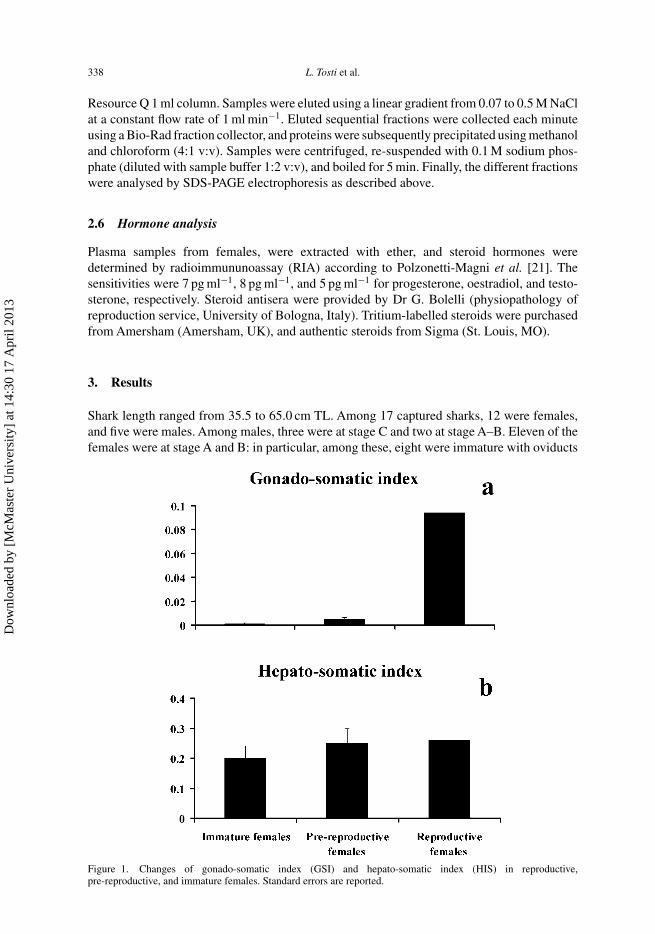
Figure 1. Changes of gonado-somatic index (GSI) and hepato-somatic index (HIS) in reproductive,pre-reproductive, and immature females. Standard errors are reported.
Dow
nloa
ded
by [
McM
aste
r U
nive
rsity
] at
14:
30 1
7 A
pril
2013
Vitellogenesis in deep-sea sharks 339
not developed, and three were pre-reproductive with a maximum oocyte diameter of 4, 6,and 11 mm, respectively. Only one was reproductive (stage C), with an oocyte diameter of atleast 47 mm.
The gonado-somatic index of females of C. coelolepis changes significantly in animalsduring immature and pre-reproductive periods (P < 0.05, Mann–Whitney U test; figure 1a).
Figure 2. Macroscopic characteristics of the ovaries from females at different phases of reproduction: (a) immaturefemale, (b) pre-reproductive female, displaying oocyte at different stage of maturation (asynchronous ovary), and(c) reproductive female.
Dow
nloa
ded
by [
McM
aste
r U
nive
rsity
] at
14:
30 1
7 A
pril
2013
340 L. Tosti et al.
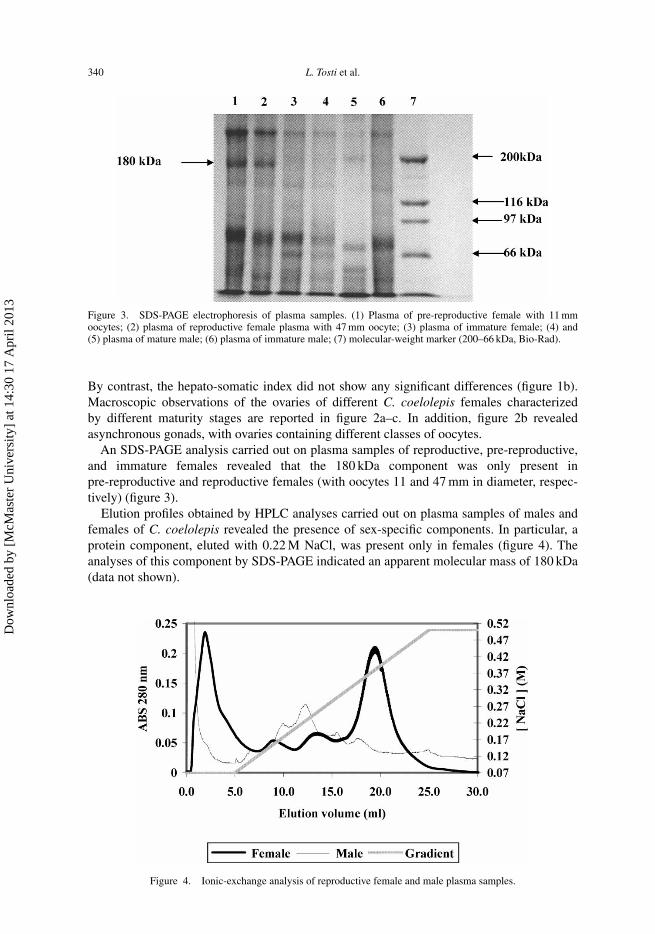
Figure 3. SDS-PAGE electrophoresis of plasma samples. (1) Plasma of pre-reproductive female with 11 mmoocytes; (2) plasma of reproductive female plasma with 47 mm oocyte; (3) plasma of immature female; (4) and(5) plasma of mature male; (6) plasma of immature male; (7) molecular-weight marker (200–66 kDa, Bio-Rad).
By contrast, the hepato-somatic index did not show any significant differences (figure 1b).Macroscopic observations of the ovaries of different C. coelolepis females characterizedby different maturity stages are reported in figure 2a–c. In addition, figure 2b revealedasynchronous gonads, with ovaries containing different classes of oocytes.
An SDS-PAGE analysis carried out on plasma samples of reproductive, pre-reproductive,and immature females revealed that the 180 kDa component was only present inpre-reproductive and reproductive females (with oocytes 11 and 47 mm in diameter, respec-tively) (figure 3).
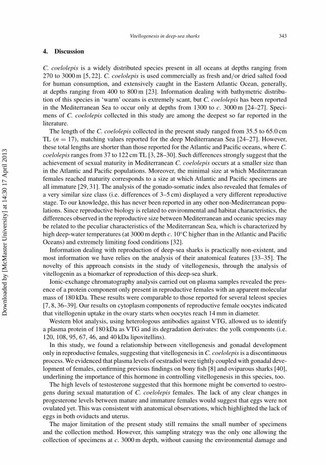
Elution profiles obtained by HPLC analyses carried out on plasma samples of males andfemales of C. coelolepis revealed the presence of sex-specific components. In particular, aprotein component, eluted with 0.22 M NaCl, was present only in females (figure 4). Theanalyses of this component by SDS-PAGE indicated an apparent molecular mass of 180 kDa(data not shown).
Figure 4. Ionic-exchange analysis of reproductive female and male plasma samples.
Dow
nloa
ded
by [
McM
aste
r U
nive
rsity
] at
14:
30 1
7 A
pril
2013
Vitellogenesis in deep-sea sharks 341
Figure 5. SDS-PAGE electrophoresis of yolk proteins. (1) 4 mm pre-vitellogenic oocytes; (2) 6 mm pre-vitellogenicoocytes; (3) 11 mm pre-vitellogenic oocyte; (4) 14 mm vitellogenic oocytes; (5) 47 mm vitellogenic oocytes;(6) molecular-weight marker (200–31 kDa, Bio-Rad).
SDS-PAGE analysis carried out on yolk components of reproductive and pre-reproductivefemales characterized by gonads at different maturation stages revealed that vitellogeninuptake starts when the oocytes reach a diameter of 14 mm (figure 5). Moreover, neutralcomponents traceable to lipovitellins, commonly present in vitellogenic oocytes, weredetectable, showing an apparent molecular mass of 120, 104, 97, 66, 46, 38, and 31 kDa.
In addition, by Western blot analysis, using heterologous antibodies against VTG, bothlipovitellin components and the 180 kDa plasma protein showed a good cross-reaction; amongall the heterologous antibodies used, the anti-VTG raised against Rana esculenta showed abetter cross-reaction with the major yolk components (figure 6).
Results on the progesterone, testosterone, and oestradiol concentrations in plasma samplesare shown in figure 7a and c. Plasmatic progesterone levels were very low and did not showany significant differences among females at different maturity stages (Mann–Whitney U
Figure 6. Western blotting analysis of yolk and plasma samples using frog antibody anti-VTG. (1) Yolk fromimmature female; (2) yolk from reproductive female; (3) plasma from reproductive female; (4) plasma of immaturefemale.
Dow
nloa
ded
by [
McM
aste
r U
nive
rsity
] at
14:
30 1
7 A
pril
2013
342 L. Tosti et al.
Figure 7. Concentrations of progesterone (a), testosterone (b), and oestradiol (c) in plasma samples collected fromimmature, pre-reproductive, and reproductive females. Standard errors are reported.
test). Testosterone plasmatic levels of immature females were about 60-fold higher than inpre-reproductive females (P < 0.01, Mann–Whitney U test). Levels of oestradiol increasedin relation to ovary maturation, with values in immature females significantly lower than inpre-reproductive females (P < 0.01, Mann–Whitney U test).
Dow
nloa
ded
by [
McM
aste
r U
nive
rsity
] at
14:
30 1
7 A
pril
2013
Vitellogenesis in deep-sea sharks 343
4. Discussion
C. coelolepis is a widely distributed species present in all oceans at depths ranging from270 to 3000 m [5, 22]. C. coelolepis is used commercially as fresh and/or dried salted foodfor human consumption, and extensively caught in the Eastern Atlantic Ocean, generally,at depths ranging from 400 to 800 m [23]. Information dealing with bathymetric distribu-tion of this species in ‘warm’ oceans is extremely scant, but C. coelolepis has been reportedin the Mediterranean Sea to occur only at depths from 1300 to c. 3000 m [24–27]. Speci-mens of C. coelolepis collected in this study are among the deepest so far reported in theliterature.
The length of the C. coelolepis collected in the present study ranged from 35.5 to 65.0 cmTL (n = 17), matching values reported for the deep Mediterranean Sea [24–27]. However,these total lengths are shorter than those reported for the Atlantic and Pacific oceans, where C.coelolepis ranges from 37 to 122 cm TL [3, 28–30]. Such differences strongly suggest that theachievement of sexual maturity in Mediterranean C. coelolepis occurs at a smaller size thanin the Atlantic and Pacific populations. Moreover, the minimal size at which Mediterraneanfemales reached maturity corresponds to a size at which Atlantic and Pacific specimens areall immature [29, 31]. The analysis of the gonado-somatic index also revealed that females ofa very similar size class (i.e. differences of 3–5 cm) displayed a very different reproductivestage. To our knowledge, this has never been reported in any other non-Mediterranean popu-lations. Since reproductive biology is related to environmental and habitat characteristics, thedifferences observed in the reproductive size between Mediterranean and oceanic species maybe related to the peculiar characteristics of the Mediterranean Sea, which is characterized byhigh deep-water temperatures (at 3000 m depth c. 10◦C higher than in the Atlantic and PacificOceans) and extremely limiting food conditions [32].
Information dealing with reproduction of deep-sea sharks is practically non-existent, andmost information we have relies on the analysis of their anatomical features [33–35]. Thenovelty of this approach consists in the study of vitellogenesis, through the analysis ofvitellogenin as a biomarker of reproduction of this deep-sea shark.
Ionic-exchange chromatography analysis carried out on plasma samples revealed the pres-ence of a protein component only present in reproductive females with an apparent molecularmass of 180 kDa. These results were comparable to those reported for several teleost species[7, 8, 36–39]. Our results on cytoplasm components of reproductive female oocytes indicatedthat vitellogenin uptake in the ovary starts when oocytes reach 14 mm in diameter.
Western blot analysis, using heterologous antibodies against VTG, allowed us to identifya plasma protein of 180 kDa as VTG and its degradation derivates: the yolk components (i.e.120, 108, 95, 67, 46, and 40 kDa lipovitellins).
In this study, we found a relationship between vitellogenesis and gonadal developmentonly in reproductive females, suggesting that vitellogenesis in C. coelolepis is a discontinuousprocess. We evidenced that plasma levels of oestradiol were tightly coupled with gonadal deve-lopment of females, confirming previous findings on bony fish [8] and oviparous sharks [40],underlining the importance of this hormone in controlling vitellogenesis in this species, too.
The high levels of testosterone suggested that this hormone might be converted to oestro-gens during sexual maturation of C. coelolepis females. The lack of any clear changes inprogesterone levels between mature and immature females would suggest that eggs were notovulated yet. This was consistent with anatomical observations, which highlighted the lack ofeggs in both oviducts and uterus.
The major limitation of the present study still remains the small number of specimensand the collection method. However, this sampling strategy was the only one allowing thecollection of specimens at c. 3000 m depth, without causing the environmental damage and
Dow
nloa
ded
by [
McM
aste
r U
nive
rsity
] at
14:
30 1
7 A
pril
2013
344 L. Tosti et al.
disturbance associated with deep-sea trawling. Thus, the present results should be consideredas a starting-point for further investigations on the reproductive biology of deep-sea sharks.
Overall, our results suggested that vitellogenesis in this aplacental viviparous shark mightoccur via mechanisms similar to those in oviparous vertebrates. These results open newresearch perspectives on the reproductive biology of deep-sea sharks and highlight the needof further comparative investigations between oceanic and Mediterranean specimens of C.coelolepis, for understanding better the role of environmental factors in controlling thereproductive process of deep-sea sharks.
Acknowledgements
This work was carried out as part of the programme ADIOS, ‘Atmospheric Deposition andImpact of pollutants, key elements and nutrients on the Open Mediterranean Sea’, financiallysupported by the EC under contract no. EVK3-CT-2000-00035.
References
[1] J.A. Musick, G. Burgess, G. Cailliet, M. Camhi, S. Fordham. Management of sharks and their relatives(Elasmobranchii). Fisheries, 25, 9–13 (2000).
[2] J.K. Baum, R.A. Myers, D .G. Kehler, B. Worm, S.J. Harley, P.A. Doherty. Collapse and Conservation of SharkPopulations in the Northwest Atlantic. Science, 299, 389–392 (2003).
[3] J.D.M. Gordon. Management considerations of deep-water shark fisheries. In Shotton, R. Case studies of themanagement of elasmobranch fisheries. FAO Fish. Tech. Pap., 378, 774–818 (1999).
[4] E. Cortés. Life history patterns and correlates in sharks. Rev. Fish. Sci., 8(4), 299–344 (2000).[5] L.J.V. Compagno. Checklist of living elasmobranchs. In Sharks, Skates, and Rays: the Biology of Elasmobranch
Fishes, W.C. Hamlett (Ed.), pp. 471–498. Johns Hopkins University Press, Baltimore, MD (1999).[6] S.M. Ho. Endocrinology of vitellogenesis. In Hormones and Reproduction in Fishes, Amphibians and Reptiles,
D.O. Norris and R.E. Jones (Eds), pp. 145–171, Plenum, New York (1987).[7] M. Kishida, T.R. Anderson, T.L. Specker. Induction by beta-estradiol of vitellogenin in stringed bass Morone
saxantilis: characterization and quantification in plasma and mucus. Gen. Comp. Endocrinol., 88, 29–39 (1992).[8] G. Mosconi, O. Carnevali, R. Carletta, M. Nabissi, A.M. Polzonetti-Magni. Gilthead seabream Sparus aurata
vitellogenin: Purification, partial characterization, and validation of an enzyme-linked immunosorbent assay(ELISA). Gen. Comp. Endocrinol., 119, 252–261 (1998).
[9] R. Wallace. Vitellogenesis and oocyte growth in non-mammalian vertebrates. Dev. Biol., 1, 127–177 (1985).[10] K. Selman, R.A. Wallace. Cellular aspects of oocyte growth in teleosts. Zool. Sci., 6, 211–231 (1989).[11] O. Carnevali, G. Mosconi, K. Yamamoto, T. Kobaiashi, S. Kikuyama, A.M. Polzonetto-Magni. ‘In vitro’ effects
of mammalian and anphibian prolactins on hepatic vitellogenin syntesis in Rana esculenta. J. Endocrinol., 137,383–389 (1993).
[12] O. Carnevali, G. Mosconi. In vitro induction of vitellogenesis synthesis in Rana esculenta: Role of the pituitary.Gen. Comp. Endocrinol., 86, 352–358 (1992).
[13] T.P. Mommsen, P. Walsh. Vitellogenesis and oocyte assembly, In Fish Physiology Vol. XIA, W.S. Hoar andD.J. Randall (Eds), pp. 347–406, Academic Press, New York (1998).
[14] O. Carnevali, G. Mosconi,A. Cambi, S. Ridolfi, S. Zanuy,A.M. Polzonetti-Magni. Changes of lysosomal enzymeactivities in sea bass (Dicentrarchus labrax) eggs and developing embryos. Aquaculture, 202, 249–256 (2001).
[15] J.C.A. Craik. Plasma levels of vitellogenin in the elasmobranch Scyliorhinus canicula L. Comp. Biochem.Physiol., 60B, 9–18 (1978a).
[16] J.C.A. Craik. Kinetics studies of Vitellogenin metabolism in the elasmobranch Scyliorhinus canicula L. Comp.Biochem. Physiol., 61A, 355–61 (1978b).
[17] M. Stehmann. Quick and dirty tabulation of stomach content and maturity stage for stakes rajidae, squaloid andother viviparous and ovoviviparous species of sharks. Am. Elasmobranch Soc. Newslett., 3, 5–9 (1987).
[18] L.O. Lucifora, R.C. Menni, A.H. Escalante. Reproductive ecology and abundance of the sand tiger shark,Carcharias taurus, from the south-western Atlantic. ICES J. Mar. Sci., 59, 553–561 (2002).
[19] R. Wallace, K. Selman. Major protein changes during vitellogenesis and maturation of Fundulus oocytes.Dev. Biol., 68, 171–182 (1985).
[20] P. Lambin. Reliability of molecular weight determination of proteins by polyacrylamide gradient gelelectrophoresis in the presence of sodium dodecyl-sulfate. Anal. Biochem., 85, 114–25 (1978).
[21] A.M. Polzonetti-Magni, V. Botte, L. Bellini-Cardellini, A. Gobbetti, A. Castro. Plasma sex hormones and post-reproductive period in green frog Rana esculenta complex. Gen. Comp. Endocrinol., 54, 372–377 (1984).
[22] L.J.V. Compagno. FAO species catalogue. Sharks of the world. An annotated and illustrated catalogue of sharkspecies known to date, Part I. Fish. Synop. FAO, 4, 125, 249 (1984).
Dow
nloa
ded
by [
McM
aste
r U
nive
rsity
] at
14:
30 1
7 A
pril
2013
Vitellogenesis in deep-sea sharks 345
[23] J.D.M. Gordon, O.A. Bergstad, I. Figueiredo, G. Menezes. The deep-water fisheries of the Northeast Atlantic:I. description and current trends. J. Northwest Atlantic Fish Sci., 31, 137–150 (2003).
[24] M. Grey. The distribution of fishes found below a depth of 2000 m. Fieldiana Zool., 36, 75–183 (1956).[25] M. Torchio, M. Michelangeli. Prima segnalazione in acque italiane di uno squalide del genere Centroscymnus.
Natura. Milano. (1971).[26] G. Alberelli, N. Drago. Indagini preliminari sulla fauna batiale (Mare di Sardegna). Atti VIII Congresso AIOL,
Bregant D., Fanzutti, G.P. (Eds) 8, 53–58 (1991).[27] M. Carrassòn, C. Stefanescu, J.E. Cartes. Diets and bathymetric distributions of two bathyal sharks of the Catalan
deep sea (Western Mediterranean). Mar. Ecol. Progr. Ser., 82, 21–30 (1992).[28] K. Yano, S. Tanaka. Some biological aspects of the deep sea squaloid shark Centroscymuns from Suruga bay,
Japan. Bull. Japan.Soc. Scient. Fish., 50, 249–256 (1984).[29] M. Girard, M.H. Du Buit. Reproductive biology of two deep-water sharks from the British Isles, Centroscymnus
coelolepis and Centrophorus squamosus (Chondrichthyes, Squalidae). J. Mar. Biol. Assoc. UK, 79, 923–931(1999).
[30] M. Kjerstad, I. Fossen, M. Willemsen. Utilisation of deep-sea sharks at hatton bank in the North Atlantic. J.Northwest Atlantic Fish Sci., 31, 333–338 (2003).
[31] K.Yano, S. Tanaka. Size at maturity, reproductive cycle, fecundity, and depth segregation of the deep sea squaloidshark Centroscymnus owstoni and C. coelolepis in Suruga bay. Japan. Nippon Suisan Gakkaishi, 54, 167–174(1988).
[32] R. Danovaro, A. Dell’Anno, M. Fabiano, A. Pusceddu, A. Tselepides. Deep-sea ecosystem response to climatechanges: the Eastern Mediterranean case study. Trends Ecol. Evol., 16, 505–510 (2001).
[33] M.W. Clarke, P.L. Connolly, J.J. Bracken. Aspects of reproduction of the deep water sharks Centroscymnuscoelolepis and Centrophorus squamosus from west of Ireland and Scotland. J. Mar. Biol. Assoc. UK, 81,1019–1029 (2001).
[34] P. Crozier. Distribution and reproductive biology of two deep-water squalid sharks, Centroscymnus coelolepis(Portuguese Dogfish) and Centrophorus squamosus (Leafscale Gulper Shark), in the Rockall Trough Area ofthe Northeast Atlantic. Scientific Council Research Documents NAFO. 01/105, 5 (2001).
[35] S. Clo, M. Dalu, R. Danovaro, M. Vacchi,. Segregation of the Mediterranean population of Centroscymnuscoelolepis (Chondrichthyes: Squalidae): a description and survey (Elasmobranch Fisheries). Scientific CouncilResearch Documents. NAFO. 02/83, 3 (2002).
[36] P.J. Babin. Plasma lipoprotein and apoprotein distribution as a function of density in the rainbow trout (Salmogairdneri). J. Biochem., 246, 425–429 (1987).
[37] C. Silversand, C. Haux. Isolation of turbot Scophthalmus maximus vitallogenin by high-performance anion-exchange chromatography. J. Chromatogr., 478, 387–397 (1989).
[38] E. Mananos, S. Zanuy, F. Le Menn, M. Carrello, J. Nunez. Seabass Dicentrarchus labrax L. vitellogenin. I.Induction, purification and partial characterization. Comp. Biochem. Physiol., 107B, 205–216 (1994).
[39] T. Matsubara, T. Wada, A. Hara. Purification and establishment of ELISA for vitellogenin of Japanese sardineSardinops melanostictus. Comp. Biochem. Physiol., 109B, 545–555 (1994).
[40] J.P. Sumpter, J.M. Dodd. The annual reproductive cycle of the female lesser spotted dogfish, Scyliorhinuscanicula L., and its endocrine control. J. Fish. Biol., 15, 687–695 (1979).