Page 1
1
Vocal Cord MedializationMedialization
LaryngoplastyCarolyn Waddington RN MSN FNP CORLN
The Methodist HospitalHouston, TX
SOHN, Boston, 2010
Objectives
Describe the history of the first treatments for vocal paralysis
Discuss the epidemiology of vocal cord paralysis
identify normal anatomy and physiology of the larynx and distinguish when abnormal findings are present.
explain the various methods of treatment for vocal cord paralysis to assist the patient ’s decision making.
Illustrate the nursing care appropriate for the patient to assist hem through the continuum of care
History of Vocal Cord Paralysis
Epidemiology
Anatomy of the Larynx
Function of the Larynx
Evaluation of Vocal Cord Paralysis
treatment options
nursing care
Overview History1855 Garcia presented mirror laryngoscopy
1857 development of laryngology by czermak & turck
1859 Turck demonstrated vc paralysis
late 1800s - in us, knight & elsberg described vc paralysis
1937 - jacksons described galvanic current application
1977 - zealer et al resurrected concept electrical pacing
1911 - brunings introduced injection techniques
1950-1960s - arnold improved techniques
1924 -ballance introduced reinnervation while tucker and crumley
1915 - payr introduced medialization framework surgery
1970s - isshiki advanced laryngeal framework work
history Vocal cord insufficiency
vocal cord paralysis
vocal cord paresis
vocal cord insufficiency
Page 2
2
CausesInadvertent injury during surgery - thyroid, carotid, lung, esophagus, heart or large vessels - RLN; head & neck - SLN
Complication from endotracheal intubation - RLN
Blunt neck or chest trauma
tumors of the skull base, neck, and chest
viral infections - vagus nerve or branches; RLN or SLN
central neurological conditions
aging
medications
Idiopathic
Cause Unilateral % Bilateral %
Surgery 24 26
Idiopathic 20 13
Malignancy 25 17
Trauma 11 11
Neurologic 8 13
Intubation 8 18
Other 5 5
Benninger et al., Evaluation and Treatment of the Unilateral Paralyzed Vocal Fold. Otolaryngol Head Neck Surg1994;111-497-508
Etiology
Signs & Symptoms
Voice Changes
airway problems
swallowing problems
psychosocial
Signs & Symptoms - voice changes
Hoarseness - croaky or rough voice
breathy voice - airy voice, change in pitch
effortful phonation - extra effort on speaking
air wasting - excessive air pressure required to produce usual voice
diplophonia - voice is like a gargle
Signs & symptoms -airway problems
Shortness of breath with exertion
Stridor - noisy breathing
ineffective or poor cough
signs & symptoms -swallowing problems
choking or coughing when swallowing
food sticking in the throat
Page 3
3
signs & symptoms - psychosocial
inability to be heard
singers or professional speakers
constant strain
inability to ear or drink confortably
exercise intolerance
anatomy & physiology
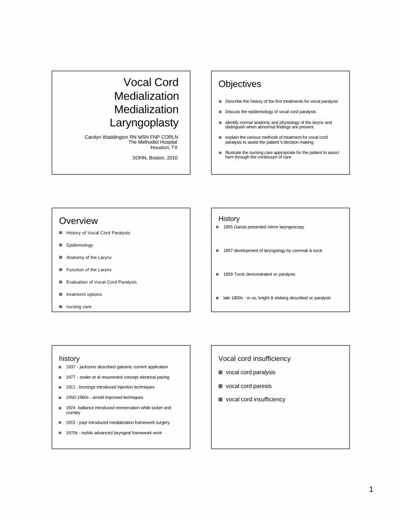
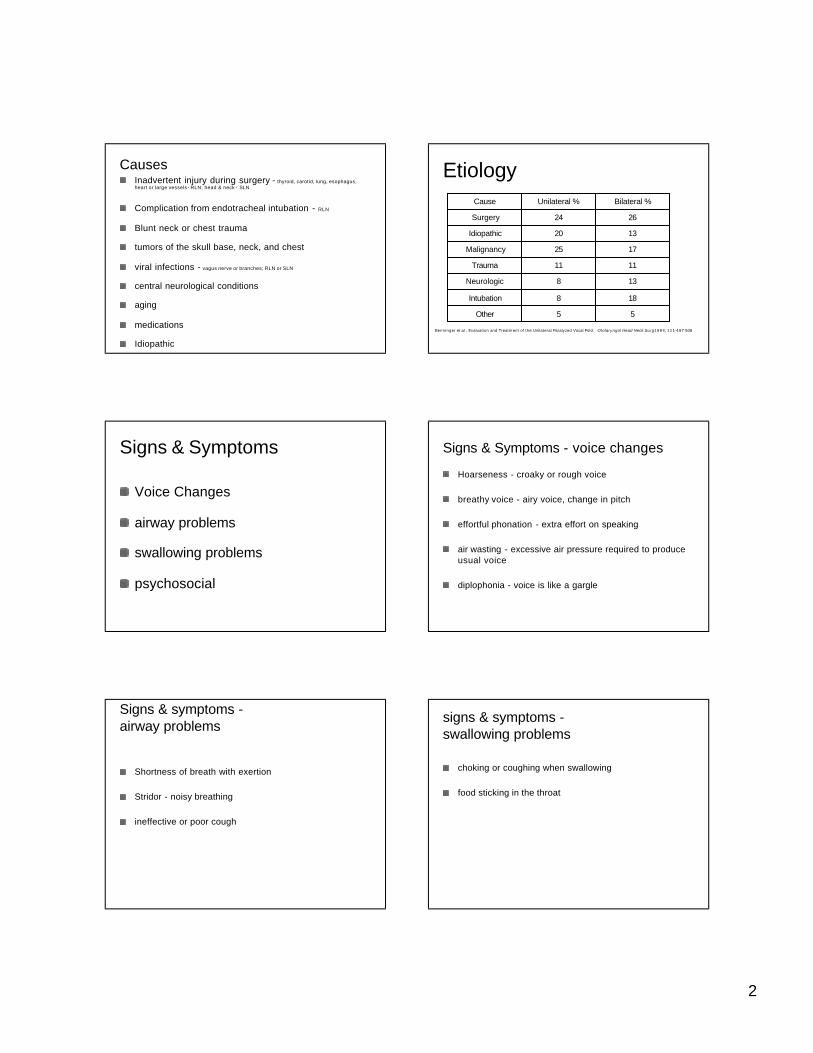
Anatomy of the Larynx - Cartilages Anatomy of the Larynx - Cartilages
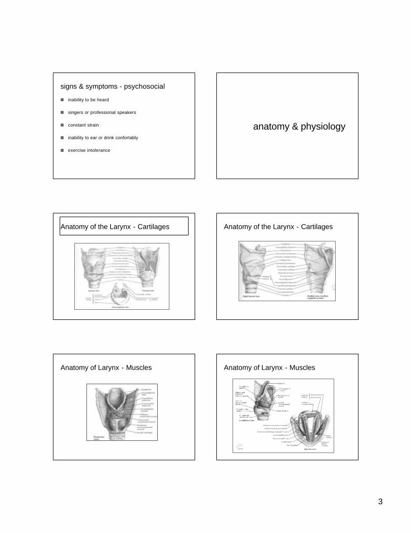
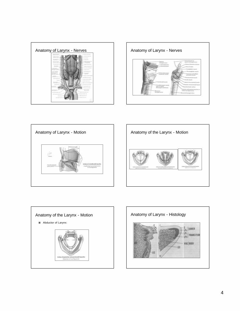
Anatomy of Larynx - Muscles Anatomy of Larynx - Muscles
Page 4
4
Anatomy of Larynx - Nerves Anatomy of Larynx - Nerves
Anatomy of Larynx - Motion Anatomy of the Larynx - Motion
Abductor of Larynx:
Anatomy of the Larynx - Motion Anatomy of Larynx - Histology
Page 5
5
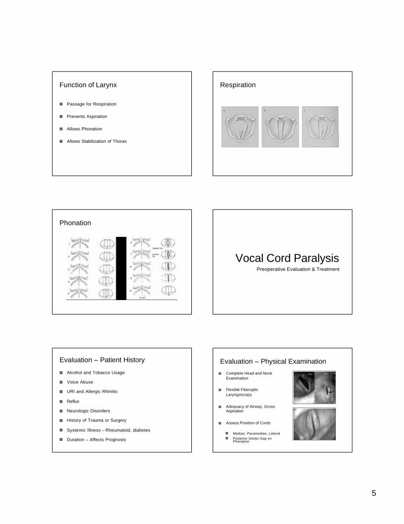
Passage for Respiration
Prevents Aspiration
Allows Phonation
Allows Stabilization of Thorax
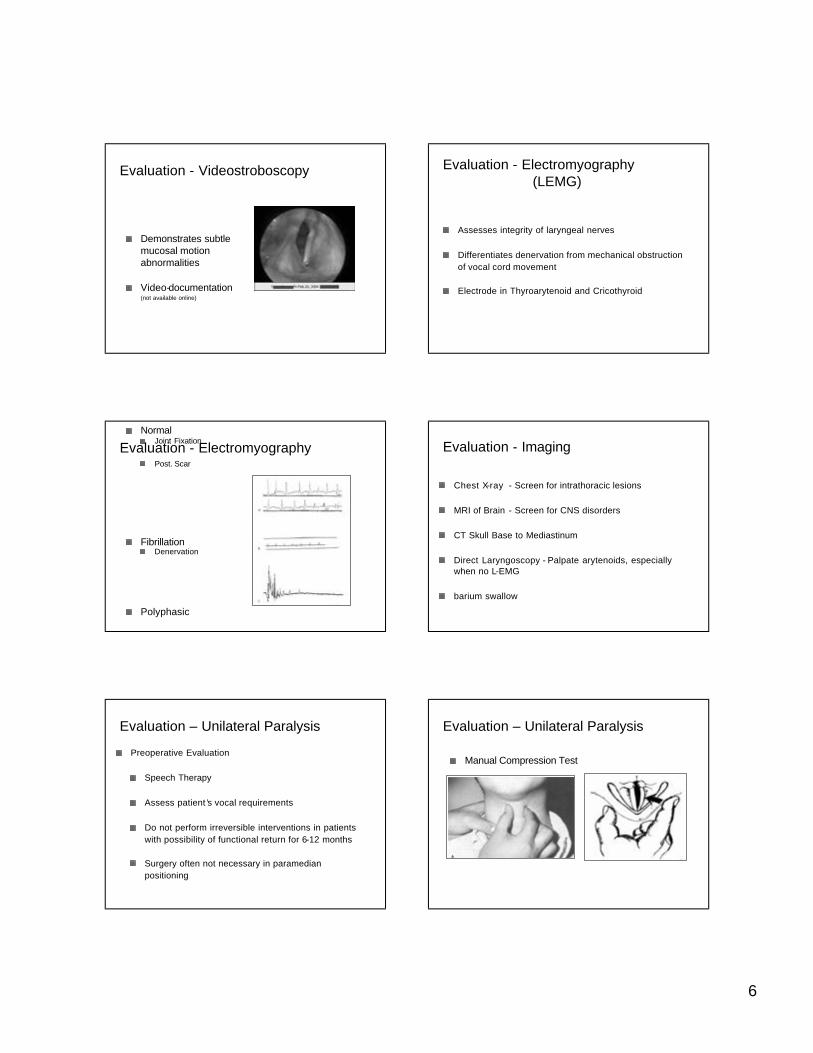
Function of Larynx Respiration
Phonation
Vocal Cord ParalysisPreoperative Evaluation & Treatment
Evaluation – Patient History
Alcohol and Tobacco Usage
Voice Abuse
URI and Allergic Rhinitis
Reflux
Neurologic Disorders
History of Trauma or Surgery
Systemic Illness – Rheumatoid, diabetes
Duration – Affects Prognosis
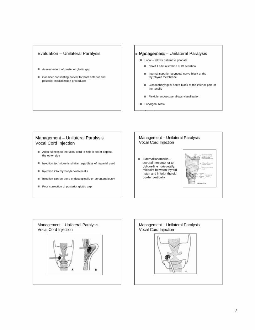
Complete Head and Neck Examination
Flexible Fiberoptic Laryngoscopy
Adequacy of Airway, Gross Aspiration
Assess Position of Cords
Median, Paramedian, LateralPosterior Glottic Gap on Phonation
Evaluation – Physical Examination
Page 6
6
Demonstrates subtle mucosal motion abnormalities
Video-documentation (not available online)
Evaluation - Videostroboscopy Evaluation - Electromyography(LEMG)
Assesses integrity of laryngeal nerves
Differentiates denervation from mechanical obstruction of vocal cord movement
Electrode in Thyroarytenoid and Cricothyroid
NormalJoint Fixation
Post. Scar
FibrillationDenervation
Polyphasic
Synkinesis
Evaluation - Electromyography
Chest X-ray - Screen for intrathoracic lesions
MRI of Brain - Screen for CNS disorders
CT Skull Base to Mediastinum
Direct Laryngoscopy - Palpate arytenoids, especially when no L-EMG
barium swallow
Evaluation - Imaging
Preoperative Evaluation
Speech Therapy
Assess patient ’s vocal requirements
Do not perform irreversible interventions in patients with possibility of functional return for 6-12 months
Surgery often not necessary in paramedian positioning
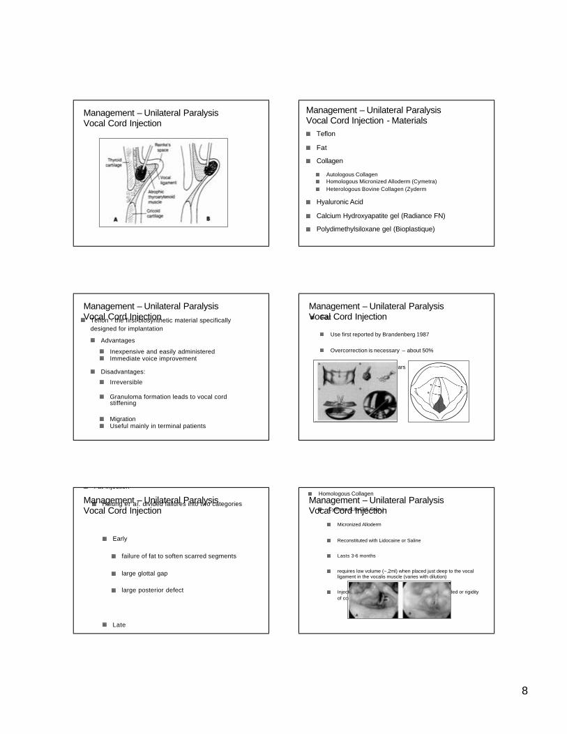
Evaluation – Unilateral Paralysis Evaluation – Unilateral Paralysis
Manual Compression Test
Page 7
7
Assess extent of posterior glottic gap
Consider consenting patient for both anterior and posterior medialization procedures
Evaluation – Unilateral Paralysis Type of Anesthesia
Local – allows patient to phonate
Careful administration of IV sedation
Internal superior laryngeal nerve block at the thyrohyoid membrane
Glossopharyngeal nerve block at the inferior pole of the tonsils
Flexible endoscope allows visualization
Laryngeal Mask
General
Management – Unilateral Paralysis
Adds fullness to the vocal cord to help it better appose the other side
Injection technique is similar regardless of material used
Injection into thyroarytenoid/vocalis
Injection can be done endoscopically or percutaneiously
Poor correction of posterior glottic gap
Management – Unilateral ParalysisVocal Cord Injection
External landmarks –several mm anterior to oblique line horizontally, midpoint between thyroid notch and inferior thyroid border vertically
Management – Unilateral ParalysisVocal Cord Injection
Management – Unilateral ParalysisVocal Cord Injection
Management – Unilateral ParalysisVocal Cord Injection
Page 8
8
Management – Unilateral ParalysisVocal Cord Injection
Teflon
Fat
Collagen
Autologous CollagenHomologous Micronized Alloderm (Cymetra)Heterologous Bovine Collagen (Zyderm
Hyaluronic Acid
Calcium Hydroxyapatite gel (Radiance FN)
Polydimethylsiloxane gel (Bioplastique)
Management – Unilateral ParalysisVocal Cord Injection - Materials
Teflon - the first biosynthetic material specifically designed for implantation
Advantages
Inexpensive and easily administeredImmediate voice improvement
Disadvantages:
Irreversible
Granuloma formation leads to vocal cord stiffening
MigrationUseful mainly in terminal patients
Management – Unilateral ParalysisVocal Cord Injection Fat
Use first reported by Brandenberg 1987
Overcorrection is necessary – about 50%
Resorption in months to years
Management – Unilateral ParalysisVocal Cord Injection
Fat Injection
Hsiung et al. divided failures into two categories
Early
failure of fat to soften scarred segments
large glottal gap
large posterior defect
Late
Management – Unilateral ParalysisVocal Cord Injection
Homologous Collagen
Cymetra (LifeCell Corp.)
Micronized Alloderm
Reconstituted with Lidocaine or Saline
Lasts 3-6 months
requires low volume (~.2ml) when placed just deep to the vocal ligament in the vocalis muscle (varies with dilution)
Injection into superficial lamina propria must be avoided or rigidity of cord will occur
Management – Unilateral ParalysisVocal Cord Injection
Page 9
9
Heterologous Collagen
Zyderm
Bovine collagen
May cause immune reaction in 1-2% of cases
Does not last as long as micronized alloderm (Cymetra)
Management – Unilateral ParalysisVocal Cord Injection
• (Radiance FN; BioForm)
Composed of small spherules of CaHydroxyapatite
No granuloma formation
Currently under study
Polydimethylsiloxane gel
• (Bioplastique; Bioplasty)
Widely used in Europe, not approved for U.S.
Management – Unilateral ParalysisVocal Cord Injection
Variety of materials used for implants
Autologous Cartilage
Silastic
Hydroxyapatite
Gore-Tex
Titanium
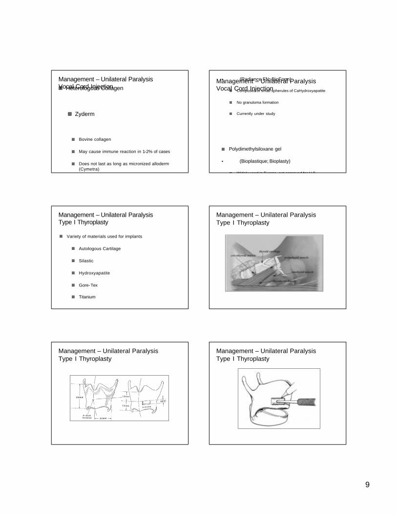
Management – Unilateral ParalysisType I Thyroplasty
Management – Unilateral ParalysisType I Thyroplasty
Management – Unilateral ParalysisType I Thyroplasty
Management – Unilateral ParalysisType I Thyroplasty
Page 10
10
Management – Unilateral ParalysisType I Thyroplasty
Management – Unilateral ParalysisType I Thyroplasty
Management – Unilateral ParalysisType I Thyroplasty
Advantages:
Permanent, but surgically reversible
No need to remove implant if vocal function returns
Excellent at closing anterior gap
Disadvantages:
More invasive
Poor closure of posterior glottic gap
Management – Unilateral ParalysisType I Thyroplasty
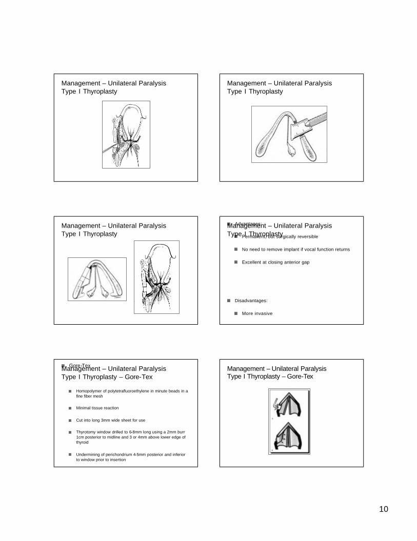
Gore-Tex
Homopolymer of polytetrafluoroethylene in minute beads in a fine fiber mesh
Minimal tissue reaction
Cut into long 3mm wide sheet for use
Thyrotomy window drilled to 6-8mm long using a 2mm burr 1cm posterior to midline and 3 or 4mm above lower edge of thyroid
Undermining of perichondrium 4-5mm posterior and inferior to window prior to insertion
Management – Unilateral ParalysisType I Thyroplasty – Gore-Tex
Management – Unilateral ParalysisType I Thyroplasty – Gore-Tex
Page 11
11
Extrusion/Displacement (Intraoperative vs Postop)
Misplacement – most often superior
Infection
Undercorrection – important to overcorrect by 1-2mm
Controversies
Location of graft placement
Status of inner perichondrium
Management – Unilateral ParalysisType I Thyroplasty
Many variations have been proposed to address the posterior gap
When arytenoid is displaced, the implant is permanent because of scarring in the CA joint
Hong et al :
Management – Unilateral ParalysisType I Thyroplasty –Variations
(these movies may not be available online)
Management – Unilateral ParalysisResults
Arytenoid Adduction
First described by Ishiki with modifications by Zeitels and others
Addresses posterior glottic gap by pulling arytenoid into adducted position
Difficult to predict which patients will benefit preoperatively.
Most advocate use in combination with anterior medialization
Management – Unilateral ParalysisArytenoid Adduction
Management – Unilateral ParalysisArytenoid Adduction
Management – Unilateral ParalysisArytenoid Adduction
Page 12
12
Endoscopic Approaches
Suture Placed to Cricoid Cartilage
Simulates action of lateral cricoarytenoid
Zeitels Modification – Arytenopexy
Management – Unilateral ParalysisArytenoid Adduction –Modifications
Results in synkynetic tone of vocal cord
Ansa to Recurrent Laryngeal Nerve
Ansa to Omohyoid to Thyroarytenoid
Management – Unilateral ParalysisReinnervation
Anatomy
•TVC positioned at about ½ vertical height of the anterior thyroid cartilage and is anterior to the oblique line
Causes of Vocal Cord Paralysis
•Iatrogenic (Surgery and intubation #1)
Evaluation
Conclusions – Key Points nursing care - pre-operative
professional occupation
comorbidities
medication list
anxiety level
nursing care - intra-operative
patient assessment
procedure scheduled
laterality, implants
anesthesia planned
nursing care - post-operative
airway
voice rest
swallowing
follow up
Page 13
13
Questions???
Thank you