Prone positioning has been used as a treatment option for patients with acute lung injury oracute respiratory distress syndrome (ARDS) since the early 1970s. Prone position and extendedprone position ventilation have been shown to increase end-expiratory lung volume, alveolarrecruitment, and oxygenation in patients with severe hypoxemic and acute respiratory failure.Prone positioning is not a benign procedure and there are potential risks (complications) that canoccur to both the patient and health care worker. Notable complications that can arise includethe following: unplanned extubation, lines pulled, and tubes kinked; and back and other injuriesto personnel. Prone positioning is a viable, inexpensive therapy for the treatment of severe ARDS.This maneuver consistently improves systemic oxygenation in 70% to 80% of patients with ARDS.With the utilization of a standardized protocol and a trained and dedicated critical care staff, pronepositioning can be performed safely. Key words: acute lung injury, acute respiratory distresssyndrome, ARDS, extended prone position ventilation, prone positioning
N UMEROUS METHODS of improving ven-tilation and preventing further lung in-
jury are used in patients with acute respiratory
Author Affiliations: Progressive Care (Ms Dirkes),Surgical Intensive Care and Rapid Response (MsDickinson), and Surgical Intensive Care (Ms Havey),University of Michigan, Ann Arbor, University ofMichigan School of Nursing, Ann Arbor (MssDickinson and O’Brien); and University of MichiganHospitals and Health Centers, Ann Arbor (Ms O’Brien).
Mss Dirkes and Dickinson share first authorship.
The authors thank Craig Meldrum for his assistance ingathering the data for evaluating their prone position-ing protocol.
They also thank the staff of the SICU who have madethe prone protocol initiative safe and effective for thepatients.
The authors have disclosed that they have no signif-icant relationships with, or financial interest in, anycommercial companies pertaining to this article.
Correspondence: Sharon Dickinson, RN, MSN, CNS-BC, ANP, CCRN, University of Michigan Surgical In-tensive Care/Rapid Response, University of MichiganHospital and Heath Centers, Box 0076, 1500 E MedicalCenter Dr, Ann Arbor, MI 48176 ([email protected]).
DOI: 10.1097/CNQ.0b013e31823b20c6
distress syndrome (ARDS). Present treatmentof ARDS is largely supportive in nature, includ-ing the use of small tidal volumes, low plateau
[AQ1]
pressures, and respiratory rates.1 Correctionof life-threatening hypoxia and improvement
[AQ2]
in respiratory mechanics and lung volumesare the main goals of treatment. Prone posi-tioning is one relatively simple and inexpen-sive method to improve oxygenation and re-cruit alveoli, and should be considered earlyin the management of ARDS when atelecta-sis and lung edema predominate and alveolarrecruitment is still possible.2-4 To understandthe influence of prone positioning on ARDS,it is helpful to understand the nature of thedisease.
ACUTE RESPIRATORYDISTRESS SYNDROME
Acute Respiratory Distress Syndrome is de-fined as a condition affecting critically ill pa-tients resulting from either direct or indirectinjury to the lungs.4 The injury may have beencaused by surgery, trauma, sepsis, or otherserious illnesses. The resulting lung injury
2 CRITICAL CARE NURSING QUARTERLY/JANUARY–MARCH 2012
results in severe hypoxemia that is often re-fractory to conventional treatment. Despitethe implementation of supportive treatmentmeasures, mortality rates for ARDS continueto be high, ranging from 35% to 45% depend-ing on the center.5
In ARDS, the alveoli of the lungs be-come damaged, resulting in increased cap-illary permeability and hyaline membraneformation.6 The increased permeability of thealveolar-capillary barrier favors edema forma-tion. The balance between edema formationand edema clearance is critical for the patientto recover from lung injury. The accumula-tion of protein-rich fluid inside the alveolicontains macrophages and neutrophils, bothof which release proinflammatory and anti-inflammatory cytokines. This fluid in the alve-oli causes impaired oxygenation and createsthe hallmark of bilateral infiltrates on chest x-ray and decreased lung compliance, originallydescribed by Ashbaugh et al in 1967.7 Thissequence of events can activate the coagula-tion cascade and promotes cellular responsesleading to microvascular injury and multior-gan failure.8
It is beyond the scope of this article to re-view the entire lung structure and functionbut understanding the structure of the lungswith regard to aeration and blood flow assistsin fully understanding the effect of position-ing on ventilation. The bronchioles containsmooth muscle, which allow the dilation orconstriction of the lungs based on the bod-ies need for oxygen. These terminate in alve-oli, which are thin walled sacs responsible foroxygen and carbon dioxide exchange in theblood. The alveoli can be thought of as tinybubbles whose surface is bathed with capil-lary blood. The effectiveness of the diffusionof gas in the alveoli requires minimizing thedistance that gasses must move by diffusionbetween alveolar air and the capillary blood.The surface area for gas exchange is large,about 60 to 80 m2, about the size of a tenniscourt.9
Since the bronchi and bronchioles have lesssurface area and are not the functional units ofgas exchange, they contain less blood flowing
through them than the alveoli. When a patienthas ARDS, the anterior alveoli in a supine pa-tient are more distended than the posterioralveoli. When the lungs become edematous,the lung begins to collapse under its ownweight, squeezing out gas from the depen-dent regions causing compression atelectasis.In 1988, Gattinoni10 studied computed tomo-graphic scans of normal subjects and those [AQ3]
with ARDS, finding that gravity played an im-portant role in ventilation and lung weight inARDS. In patients with ARDS, the lung had anexcess weight on average of 184% of the ex-pected normal weight due to the increase inintravascular lung water.
In addition, the weight of the heart on thedependent region of the lung has a signifi-cant influence of the aeration of the lung.11
In patients with ARDS, the cardiac mass isincreased, resulting in further alveolar col-lapse in the dependent regions of the lung.12
These 2 things, along with displacement ofthe abdomen, contribute significantly to alve-olar collapse.
The patients with ARDS are often sedatedand paralyzed.13 Therefore, the weight ofthe abdominal contents, no longer opposedby the diaphragm causes a displacement ofthe abdominal contents toward the poste-rior regions of the diaphragm contributing tobasal atelectasis.14 Ventilation in the supineposition, in the patient with ARDS at zeropositive end expiratory pressure (PEEP) is dis-tributed preferentially to the upper lung. Dur-ing mechanical ventilation, the lung continu-ously collapses and inflates in its dependentparts at low levels of PEEP. At higher PEEP, theamount of collapsed tissue at end expirationis decreased.15 However, with the increasedweight of the lungs and the compression ofthe heart and diaphragm, the supine patienthas large regions of dependent, atelectaticlung.16
Perfusion in the ARDS patient is influencedby gravity, hypoxic vasoconstriction, and ex-trinsic vessel compression17 Recent researchsuggests that perfusion in ARDS is more in thedependent, atelectatic regions.17 Review of allthese factors indicates then, that in ARDS, the
lungs are edematous, heavy, and compressedin the dependent regions in a supine patient.Even though the perfusion is greatest gravi-tationally, in the dependent regions, the ven-tilation is significantly less, thereby causingventilation perfusion mismatch.
As previously mentioned, ARDS can resultfrom either direct or indirect lung injury.Pelosi et al1 stated that patients suffering fromprimary ARDS (direct injury) with consolida-tions seem to be less likely to be responsiveto the recruitment effects of prone ventilationthan those who suffer from secondary ARDS(indirect injury). The alveolar collapse seen inpatients with secondary ARDS is widespreadas opposed to being concentrated in one areaas in seen in primary ARDS.1 The differencesin the patterns of atelectasis could potentiallyallow a greater number of alveoli to be re-cruited by positioning the patient with sec-ondary ARDS in the prone position.1
PRONE POSITIONING IN ARDS
In the last 25 years, there have been severalreports using prone positioning as a tool toimprove respiratory function in patients withARDS. Bryan18 first described the use of pronepositioning in 1974 and suggested that anes-thetized and paralyzed patients in the proneposition should exhibit a better expansion ofthe dorsal lung regions with consistent im-provement in oxygenation. In 1976, Pheil andBrown19 showed that the prone position im-proved oxygenation in 5 patients without ad-verse effects. In a study by Gattinoni et al,20
the use of prone positioning improved oxy-genation in more than 70% of the instances inwhich it was used, and about 70% of the effectoccurred during the first hour of pronation.Pelosi et al1 suggest the prone position itselfmay exert a protective effect on the mechan-ically ventilated injured lung. Patients withARDS are often exposed to high levels of PEEPand oxygen which are well-known to con-tribute to progressive lung damage in mechan-ically ventilated patients, and can contributeto the development of atelectasis and edema,thereby contributing to the impairment of
gas exchange.21 The prone position may de-crease ventilator-induced lung injury by allow-ing a lower FiO2 and lower airway pressuresto achieve adequate oxygenation.22,23
Although an improvement in oxygenation isan expected outcome with prone ventilation,Protti et al24 suggested that oxygen and car-bon dioxide responses to prone position areindependent and, in fact, a decrease in thePaCO2 rather than an increase in PaO2/FiO2, issignificantly associated with lung recruitabil-ity. Gattinoni et al25 retrospectively analyzedthe 225 patients from the prone arms of pre-vious studies and found a strong associationbetween the PaCO2 response and outcome butno association with PaO2/FiO2 response. ThePaCO2 responders seem to have a higher po-tential to be recruited with prone positioningthan with nonresponders, revealing a differ-ence in underlying lung pathologies.
Routine use of the prone position is notalways warranted, but prone position ventila-tion is a safe, effective recruiting modality toimprove oxygenation and is a salvage optionfor patients with severe ARDS.22,26-28
When a patient is turned prone, researchhas shown that there is a movement of lungdensities from dorsal to ventral regions, anda more homogenous distribution of alveolarinflation occurs.29 This could be explained byredistribution of the lung weight, the cardiacmass no longer resting on the lungs, and re-distribution of abdominal contents. Proningcauses recruitment of the most atelectatic re-gions of the lungs by relieving external com-pression forces, thus improving ventilation-perfusion matching without subjecting thelungs to high airway pressures.30 It is believedthat in the prone position, ventilation shouldredistribute from ventral regions (which arecollapsed in the prone position) to dorsal re-gions, which are recruited in the prone po-sition. Since the dorsal regions are heavierand more fluid filled in the supine position,changing to a prone position would changethe position of the fluid by gravity.
Prone positioning has been advocated asa rescue maneuver in severe ARDS in mul-tiple studies.19,25,31-34 At a minimum, it
4 CRITICAL CARE NURSING QUARTERLY/JANUARY–MARCH 2012
also can enhance the mobilization of secre-tions, thus optimizing the effectiveness ofchest physiotherapy35 and improve ventila-tion to previously atelectatic regions. In ad-dition, prone positioning has been shownto reduce the risk of ventilator-associatedpneumonia.36,37
Previous studies on prone positioning in-cluded several limitations. Small sample size,initiation of positioning, length of timeproned, type of proning, and use of lung pro-tective ventilation in conjunction with pron-ing were identified as limitations.20,38,39,40
Overall, there appeared to be no differ-ence in improved survival using the proneposition.2,41-44 However, the use of intermit-tent prone positioning has been documentedto significantly improve oxygenation in 60%to 70% of acute lung injury patients withARDS.25,45 Sud et al,44 in a 2008 system-[AQ4]
atic review and meta-analysis of the effect ofprone positioning on clinical outcomes of pa-tients with acute hypoxemic respiratory fail-ure, concluded that despite an improvementin oxygenation and reduced risk for venti-lator associated pneumonia associated withmechanical ventilation in the prone position,overall survival rates were not improved. Al-saghir and Martin reported in a meta-analysisthat prone positioning did not improve over-all mortality, except in possibly the moreseverely ill patients.26 However, there wasno difference in the length of ventilator daysor incidence of ventilator associated pneumo-nia between the prone and supine groups.In 2010, another systematic review and meta-analysis reinforced the aforementioned mor-tality findings, stating that prone positioningreduces mortality in those with severe hypox-emia, defined by a baseline PaO2/FiO2 less than100 mm Hg, but not in those with less severehypoxemia.46 The prone position, therefore,may be useful as a rescue strategy in patientswith severe hypoxemia.
WHEN TO PRONE AND HOW LONG?
Taccone et al40 randomized 342 patientswith moderate (PaO2/FiO2 ratio between 100
and 200 mm Hg) and severe (PaO2/FiO2 ra-tio less than 100 mm Hg) ARDS to either asupine or prone group. The prone group waspositioned on their abdomens for at least 20hours per day, until the resolution of acuterespiratory failure or the end of the 28-daystudy period. Findings from this study sug-gest a nonsignificant 10% difference in mor-tality favoring the prone group, and a signif-icantly greater proportion of patients in theprone group experienced at least 1 complica-tion than in the supine group. Of note, the rateof complications was almost 3 times greaterin the prone group than in the supine group,and this rate was found to be significantly cor-related with the number of days each pronegroup patient and supine group patient re-mained in the study.
Romero et al47 prospectively studied 15 pa-tients diagnosed with severe ARDS and placedthem in the prone position for at least 48hours or until they reached an oxygenationindex of 10 or less in 2 successive measure-ments. The authors found a statistically signifi-cant improvement in PaO2/FiO2 and oxygena-tion index, reduction of PaCO2, and plateaupressure, and increment of the static com-pliance with prolonged prone ventilation; allof which continued to improve while pa-tients remained in the prone position and re-mained unchanged when the patients werereturned to the supine position.47 They sum-marize that the improvement in oxygenation,ventilation, and static compliance probably re-flect recruitment of alveolar units and deadspace reduction.47 Ultimately, Romero et al47
describe prolonged prone ventilation as safeand easy to implement when performed bytrained staff within an established protocoland suggest that prolonged prone ventilationcould be considered part of a protective ven-tilatory strategy.
Mancebo et al39 hypothesized that proneventilation would decrease mortality if imple-mented early in the course of ARDS, if ap-plied for most of the day, and if maintained forprolonged period of time. A total of 136 pa-tients were evaluated, 60 randomized to thesupine group and 76 to the prone group. A
nonsignificant decrease in intensive care unit[AQ5]
mortality was observed and a similar trendwas noted for hospital mortality. Twenty-eight complications were noted. Multiplelogistic regression analysis revealed that 3variables were independently associated withincreased risk of death: simplified Acute Physi-ology score II at inclusion (is a validated AcutePhysiology tool that uses 17 variables: 12 phys-iological variables and 3 underlying diseasevariables to estimate the risk of death withouthaving a primary diagnosis),48 the number ofdays elapsed between ARDS diagnosis and in-clusion and random allocation to supine po-sition. Limitations of the study included thatthe study was stopped secondary to decreasedpatient enrollment and it was underpowered.Despite these limitations, the authors suggestthat prolonged prone ventilation is both safeand feasible and may potentially reduce mor-tality in patients with severe ARDS when ap-plied early in the disease course and for a pro-longed duration.
A literature review by Rowe21 suggested ifa patient begins to decompensate when be-ing returned to the supine position, they mayneed to be placed back in the prone posi-tion for longer than 12 hours or longer thanthe length of time generally regarded as ac-ceptable at a given institution. Finally, Abrouget al41 sought to address optimal proning du-ration and found a trend toward an interactionbetween longer proning duration and reduc-tion in mortality in their meta-analysis, and ul-timately suggested that long-prone durationsshould be applied.
OPTIONS FOR PRONING
There are several options for proning. Thesimplest is the use of a flat sheet and nursingstaff to complete a rotation in a standard bed(Figures 1 and 2). This method requires 4 staff[F1]
[F2] members, 2 positioned on each side of the bedto manage the lines and tubes. Step 1, startwith a flat sheet placed under the patient, andpull the patient to one side of the bed (we gen-erally turn the patient toward the ventilator).The flat sheet, step 2, is then placed around
the patient’s arm, on the side you are turningthe patient toward. A second flat sheet, step3, is placed on the bed and tucked under thepatient. This sheet will pull through as youturn the patient prone, step 4. Lastly, step 5,straighten the sheet, adjust lines and tubes,and place the patient’s arms in the swimmersposition, one arm positioned up toward thehead and the second arm at the patient’s side.To supine the patient, reverse the process.This technique is simple and easy to perform,but most importantly, it allows full access tothe patient. Other options include the use ofa proning device such as the Vollman PronePositioner (Hill Rom Services Inc, Batesville,Indiana; Figure 3). This device requires 2 to [F3]3 staff members, is easy to use, and is rela-tively inexpensive. Other devices include aspecialty bed known as the Rotoprone Ther-apy System (KCI USA Inc, San Antonio, Texas;Figure 4). This bed requires that the patient [F4]be “packed” in and secured before turning. Itprovides the advantages that it is automatedand can be done by one staff member. How-ever, for procedures such as extracorporealmembrane oxygenation or continuous renalreplacement therapy the external bloodlinesmust be lengthened to extend from the bedto the machines. Quick access to the patientis a concern using this device. Also, the daily [AQ6]
expense of the Rotoprone is significant, andmay limit its use in many centers.
ment, the suitability of the patient should beevaluated. We have successfully proned pa-tients with multiple lines and those on extra-corporeal membrane oxygenation and contin-uous renal replacement therapy after carefulassessment and planning.
Existing contraindications (Table 1) may [T1]
[AQ8]preclude a patient from being placed in theprone position. Potential contraindications in-clude hemodynamic instability, mean arterialpressure less than 60 or systolic blood pres-sure less than 90 (regardless of fluid resusci-tation or inotropes), recent cardiopulmonary
6 CRITICAL CARE NURSING QUARTERLY/JANUARY–MARCH 2012
Figure 1. The 5-step method to prone a patient using a regular bed, flat sheet, and 4 staff members,University of Michigan Surgical Intensive Care technique. A, step 1, with a flat sheet, pull the patient toone side of the bed using 4 staff members. B, step 2, place the flat sheet around the arm that will pullthrough (side you are turning toward). C, step 3, a second flat sheet is placed on the bed and tucked underthe patient. D, step 4, using the sheet turn the patient over and position them prone. The arm and sheetwill pull across the bed. E, step 5, discard the sheet that was used to supine patients. Straighten lines andtubes.
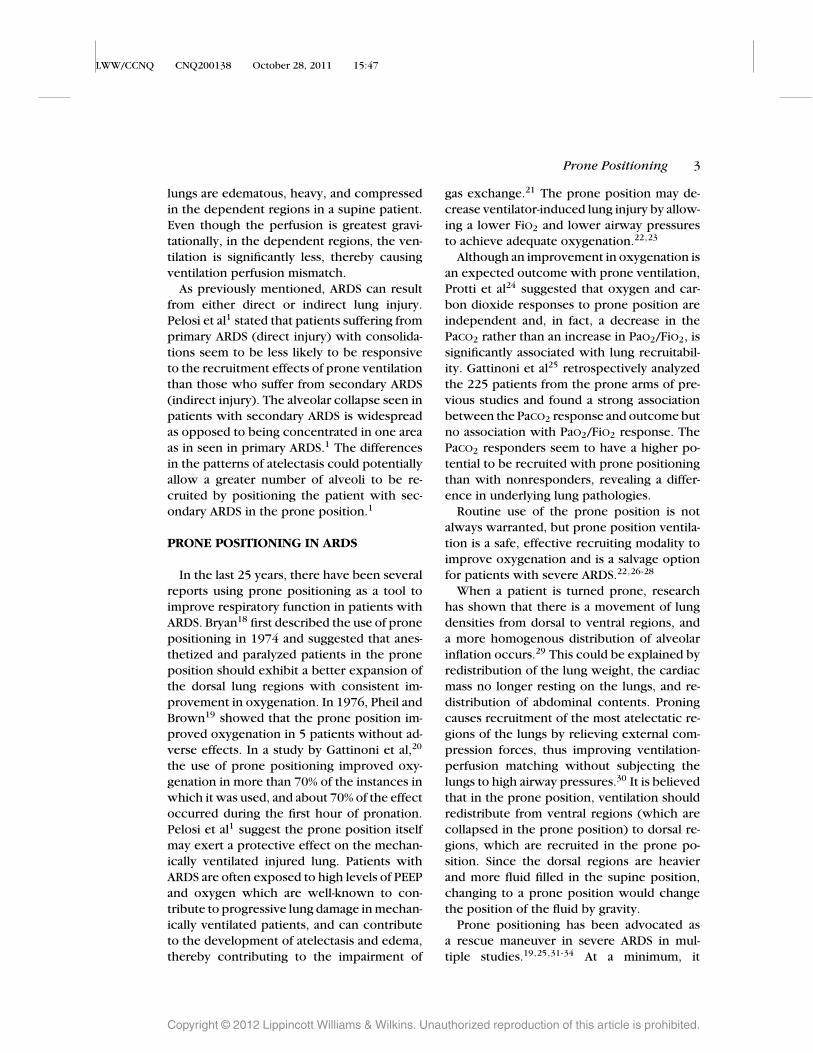
Figure 2. The 5-step method to supine a patient using a flat sheet and 4 staff members, University ofMichigan Surgical Intensive Care technique. A, step 1, using a flat sheet, pull the patient to one side of thebed. B, step 2, place the flat sheet around the arm that will pull through (side you are turning toward).C, step 3, a second flat sheet is placed on the bed and tucked under the patient. This sheet will pullthrough as you are turning the patient. D, step 4, using the sheet, turn the patient over and position themprone. The arm and sheet will pull across the bed. E, step 5, discard the sheet that was used to supinepatients. Straighten lines and tubes.
8 CRITICAL CARE NURSING QUARTERLY/JANUARY–MARCH 2012
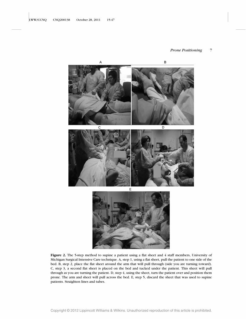
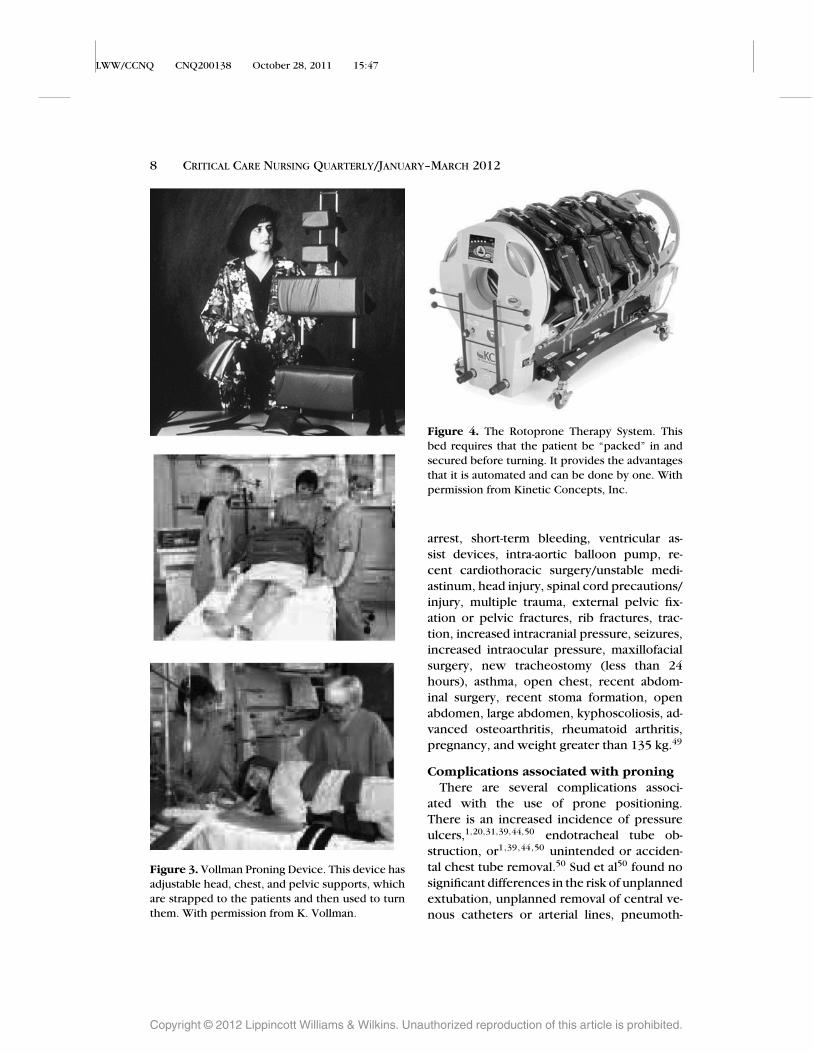
Figure 3. Vollman Proning Device. This device hasadjustable head, chest, and pelvic supports, whichare strapped to the patients and then used to turnthem. With permission from K. Vollman.
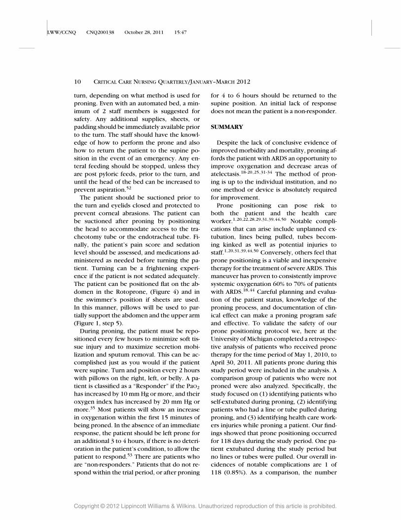
Figure 4. The Rotoprone Therapy System. Thisbed requires that the patient be “packed” in andsecured before turning. It provides the advantagesthat it is automated and can be done by one. Withpermission from Kinetic Concepts, Inc.
arrest, short-term bleeding, ventricular as-sist devices, intra-aortic balloon pump, re-cent cardiothoracic surgery/unstable medi-astinum, head injury, spinal cord precautions/injury, multiple trauma, external pelvic fix-ation or pelvic fractures, rib fractures, trac-tion, increased intracranial pressure, seizures,increased intraocular pressure, maxillofacialsurgery, new tracheostomy (less than 24hours), asthma, open chest, recent abdom-inal surgery, recent stoma formation, openabdomen, large abdomen, kyphoscoliosis, ad-vanced osteoarthritis, rheumatoid arthritis,pregnancy, and weight greater than 135 kg.49
Complications associated with proningThere are several complications associ-
ated with the use of prone positioning.There is an increased incidence of pressureulcers,1,20,31,39,44,50 endotracheal tube ob-struction, or1,39,44,50 unintended or acciden-tal chest tube removal.50 Sud et al50 found nosignificant differences in the risk of unplannedextubation, unplanned removal of central ve-nous catheters or arterial lines, pneumoth-
oraces, or cardiac arrests. Other complica-tions include transient oxygen desaturation,arrhythmias, hypotension, vomiting, acciden-tal loss of central venous catheters, accidentalextubation, and accidental loss of thoracic orabdominal drains.1 Facial edema was noted inseveral studies that improved after the patientwas returned to the supine position.22,28,39
Conjunctival hemorrhage, vascular cathetermalfunction during continuous renal replace-ment therapy, dislodgement of a PA catheter[AQ10]
resulting in cardiac arrest (patient was resusci-tated), indwelling bladder catheter and naso-gastric tube displacement, kinking of thoracictube, and one unplanned extubation were as-sociated with a study examining prolongedproning periods.39
Experienced team
An experienced team of staff members(nurses, respiratory therapist, intensive careunit technicians, and physicians) are re-quired when performing this intervention.50
Evidence-based practice guidelines should bein place for bedside nurses on critical careunits that use prone positioning, including in-dications and contraindications, preprone as-sessment and safety practices, strategies forplacing the patient in the prone position, as-sessment guidelines for monitoring the pa-tient response to the prone positioning, andlimb positioning in the prone position.46 Es-tablishing and carefully evaluating differentguidelines for prone positioning patients withARDS is essential before implementing a stan-dard protocol.23
Procedure
Arterial blood gases, baseline FiO2, SpO2,and vital signs should be initially assessedto compare to proned values and any im-provements in oxygenation. Measurement ofthe PaO2/FiO2 ratio should be done to as-sess the severity of lung injury. A P/F ratio [AQ11]
of less than 200 or less is a finding char-acteristic of ARDS. A P/F ratio of 300 orless is more characteristic of acute lung in-jury, not ARDS.51 According to the Universityof Michigan prone protocol, there are somebasic requirements to complete before ini-tiating prone positioning (Table 2). Preoxy- [T2]genate the patient before turning with FiO2
of 1.0. The central lines and tracheotomyor endotracheal tube and any other linesshould be secured and tracked during theturn. Adequate staff should be available for the
Table 2. Preprone Position Requirements forSafe Prone Positioning the University of Michi-gan Surgical Intensive Care Checklist
• Preoxygenate the patient with FiO2 1.0.• Secure the endotracheal tube and lines.• Correct number of staff members to assist in
the turn and monitor the turn.• Adequate number supplies to turn (pads for
bed, sheet, protection for the patient, orspecialty bed).
• Experienced staff with working knowledge ofhow to perform the turn and how to supinethe patient in the event of an emergency.
10 CRITICAL CARE NURSING QUARTERLY/JANUARY–MARCH 2012
turn, depending on what method is used forproning. Even with an automated bed, a min-imum of 2 staff members is suggested forsafety. Any additional supplies, sheets, orpadding should be immediately available priorto the turn. The staff should have the knowl-edge of how to perform the prone and alsohow to return the patient to the supine po-sition in the event of an emergency. Any en-teral feeding should be stopped, unless theyare post pyloric feeds, prior to the turn, anduntil the head of the bed can be increased toprevent aspiration.52
The patient should be suctioned prior tothe turn and eyelids closed and protected toprevent corneal abrasions. The patient canbe suctioned after proning by positioningthe head to accommodate access to the tra-cheotomy tube or the endotracheal tube. Fi-nally, the patient’s pain score and sedationlevel should be assessed, and medications ad-ministered as needed before turning the pa-tient. Turning can be a frightening experi-ence if the patient is not sedated adequately.The patient can be positioned flat on the ab-domen in the Rotoprone, (Figure 4) and inthe swimmer’s position if sheets are used.In this manner, pillows will be used to par-tially support the abdomen and the upper arm(Figure 1, step 5).
During proning, the patient must be repo-sitioned every few hours to minimize soft tis-sue injury and to maximize secretion mobi-lization and sputum removal. This can be ac-complished just as you would if the patientwere supine. Turn and position every 2 hourswith pillows on the right, left, or belly. A pa-tient is classified as a “Responder” if the PaO2
has increased by 10 mm Hg or more, and theiroxygen index has increased by 20 mm Hg ormore.35 Most patients will show an increasein oxygenation within the first 15 minutes ofbeing proned. In the absence of an immediateresponse, the patient should be left prone foran additional 3 to 4 hours, if there is no deteri-oration in the patient’s condition, to allow thepatient to respond.53 There are patients whoare “non-responders.” Patients that do not re-spond within the trial period, or after proning
for 4 to 6 hours should be returned to thesupine position. An initial lack of responsedoes not mean the patient is a non-responder.
SUMMARY
Despite the lack of conclusive evidence ofimproved morbidity and mortality, proning af-fords the patient with ARDS an opportunity toimprove oxygenation and decrease areas ofatelectasis.18-20,25,31-34 The method of pron-ing is up to the individual institution, and noone method or device is absolutely requiredfor improvement.
Prone positioning can pose risk toboth the patient and the health careworker.1,20,22,28,29,31,39,44,50 Notable compli-cations that can arise include unplanned ex-tubation, lines being pulled, tubes becom-ing kinked as well as potential injuries tostaff.1,20,31,39,44,50 Conversely, others feel thatprone positioning is a viable and inexpensivetherapy for the treatment of severe ARDS. Thismaneuver has proven to consistently improvesystemic oxygenation 60% to 70% of patientswith ARDS.18,44 Careful planning and evalua-tion of the patient status, knowledge of theproning process, and documentation of clin-ical effect can make a proning program safeand effective. To validate the safety of ourprone positioning protocol we, here at theUniversity of Michigan completed a retrospec-tive analysis of patients who received pronetherapy for the time period of May 1, 2010, toApril 30, 2011. All patients prone during thisstudy period were included in the analysis. Acomparison group of patients who were notproned were also analyzed. Specifically, thestudy focused on (1) identifying patients whoself-extubated during proning, (2) identifyingpatients who had a line or tube pulled duringproning, and (3) identifying health care work-ers injuries while proning a patient. Our find-ings showed that prone positioning occurredfor 118 days during the study period. One pa-tient extubated during the study period butno lines or tubes were pulled. Our overall in-cidences of notable complications are 1 of118 (0.85%). As a comparison, the number
of overall incidences while NOT proning(extubations/line pulls) during the study pe-riod was 91 of 6997 (1.30%). No employeeinjuries were noted secondary to proning apatient. On the basis of the current litera-ture and the conclusions from our study, wewould suggest that the use of prone position-
ing is an effective strategy for the treatmentof severe hypoxemia in patients’ with ARDS.Prone positioning of patient’s with ARDS us-ing a standardized protocol does not result inan increased incidence of lines pulls and ex-tubations and finally proning does not resultin increased injuries to health care workers.
REFERENCES
1. Pelosi P, Brazzi L, Gattinoni L. Prone position inacute respiratory distress syndrome. Eur Respir J.2002;20:1017-1028.
2. Alsaghir AH, Martin CM. Effect of prone positioningin patients with acute respiratory distress syndrome:a meta-analysis. Crit Care Med. 2008;36(2):603-609.
3. Powers J. The five P’s spell positive outcomes forARDS patients. Am Nurse Today. 2007;2(3):34-38.[AQ12]
4. Powers J. Use of prone positioning with ARDS. CritConnect. 2011;10(2):8-9.
5. Phua J, Badia JR, Adhikari NKJ, et al. Has mortalityfrom acute respiratory distress syndrome decreasedover time? Am J Respir Crit Care Med. 2009;179:220-227.
6. Ware LB, Matthay MA. The acute respiratory distresssyndrome. N Engl J Med. 2000;342:1334-1349.
8. Dernaika TA, Keddissi JI, Kinasewitz GT. Update onARDS: beyond the low tidal volume. Am J Med Sci.2009;337(5):360-367.
9. Moffett DF, Moffett SB, Schauf C. Human Physiology:Foundations and Frontiers, 2nd ed. St Louis, MO:Mosby Inc; 1993:454.
10. Gattinoni L, Pesenti A, Bombino M, et al. Relation-ships between lung computed tomographic density,gas exchange and PEEP in acute respiratory failure.Anesthesiology. 1988;69:824-832.
11. Hyatt RE, Bar-Yisay E, Abel MD. Influence of the hearton the vertical gradient of transpulmonary pressurein dogs. J Applied Physiol. 1985;58:52-57.
12. Malbouisson LM, Busch CJ, Puybasset L, et al. Role ofthe heart in the loss of aeration characterizing lowerlobes in acute respiratory distress syndrome. Am JRespir Crit Care Med. 2000;161:2005-2012.
13. Otis AB, McKerrow CB, Bartlett RA, et al. Mechanicalfactors in distribution of pulmonary ventilation. J ApplPhysiol. 1956;8(4):427-443.[AQ14]
14. Froese AB, Bryan AC. Effects of anaesthesia and paral-ysis on diaphragmatic mechanics in man. Anesthesi-ology. 1974;41:242-255.
15. Gattinoni L, Pelosi P, Croitti S, Valenza F. Effects ofpositive end-expiratory pressure on regional distribu-tion of tidal volume and recruitment in adult respira-
tory distress syndrome. Am J Respir Crit Care Med.1995;151:1807-1814.
16. Albert RK, Hubmayr RD. The prone position elimi-nates compression of the lungs by the heart. Am JRespir Crit Care Med. 2000;161:1660-1665.
17. Lamm WJE, Graham MM, Albert RK. Mechanismby which prone positioning improves oxygenationin acute lung injury. Am J Respir Crit Care Med.1994;150:184-193.
18. Bryan AC. Comment of a devil’s advocate. Am RevRespir Dis. 1974;110:143-144.
19. Pheil MA, Brown RS. Use of extreme position changesin acute respiratory failure. Crit Care Med. 1976;4:13-14.
20. Gattinoni L, Tognomi G, Pesenti A, et al. Effect ofprone positioning on the survival of patients withacute respiratory failure. N Engl J Med. 2001;345:568-573.
21. Rowe C. Development of clinical guidelines forprone positioning of adult patients. Nurs Crit Care.2004;9(2):50-57.
22. Klein Y, Blackbourne L, Barquist E. Non-ventilatory-based strategies in the management of acute respi-ratory distress syndrome. J Trauma Inj Infect CritCare. 2004;57(4):915-924.
23. Brieburg AN, Aitken L, Reaby L, Clancy RL, Pierce JD.Efficacy and safety of prone positioning for patientswith acute respiratory distress syndrome. J Adv Nurs.2000;32(4):922-929
24. Protti A, Chiumello D, Cressoni M, et al. Relationshipbetween gas exchange response to prone positionand lung recruitability during acute respiratory fail-ure. Intensive Care Med. 2009;35(6):1011-1017.
25. Gattinoni L, Vagginelli F, Carlesso E, et al. Decrease inPaCO2 with prone position is predictive of improvedoutcome in acute respiratory distress syndrome. CritCare Med. 2003;294(2):229-237.
26. Alsaghir AH, Martin CM. Effect of prone positioningin patients with acute respiratory distress syndrome:a meta-analysis. Crit Care Med. 2008;36(2):603-609.
27. Dernaika TA, Keddissi JI, Kinasewitz GT. Update onARDS: beyond the low tidal volume. Am J Med Sci.2009;337(5):360-367. [AQ15]
28. Fan E, Mehta S. High-frequency oscillatory ventilationand adjunctive therapies: inhaled nitric oxide and
12 CRITICAL CARE NURSING QUARTERLY/JANUARY–MARCH 2012
prone positioning. Crit Care Med. 2005;33(3):S182-S187.
29. Mutoh T, Guest RJ, Lamm WJE, Albert RK. Prone po-sition alters the effect of volume overload on regionalpleural pressures and improves hypoxemia in pigs invivo. Am Rev Respir Dis. 1992;146:300-306.
30. Curley MA, Hibbard PL, Fineman LD, et al. Effect ofprone positioning on clinical outcomes in childrenwith acute lung injury. A randomized controlled trial.JAMA. 2005;294:229-237.
31. Taccone P, Pesenti A, Latini R, et al. Prone positioningin patients with moderate and severe acute respira-tory distress syndrome: a randomized controlled trial.JAMA 2009;302:1977-1984.
32. Albert RK, Leasa D, Sanderson M, Robertson HT,Hlastala MP. The prone position improves arte-rial oxygenation and reduces shunt in oleic-acid-induced acute lung injury. Am Rev Respir Dis.1987;135(3):628-633
33. Gattinoni L, Pesenti A, Avalli L, Rossi F, BombinoM. Pressure-volume curve of total respiratory systemin acute respiratory failure. Computed tomographicscan study. Am Rev Respir Dis. 1987;136(3):730-736
34. Pelosi P, Croci M, Ravagnan I, et al. The effects ofbody mass on lung volumes, respiratory mechanics,and gas exchange during general anesthesia. AnesthAnalg. 1998;87(3):654-660.
35. Chatte G, Sab J, Dubois J, et al. Prone position inmechanically ventilated patients with severe acuterespiratory failure. Am J Respir Crit Care Med.1997;155:473-478.
36. Pappert D, Rossaint R, Slama K, et al. Influence ofpositioning on ventilation-perfusion relationships insevere adult respiratory distress syndrome. Chest.1994;106:1511-1516.
37. Brazzi L, Ravagnan I, Pelosi P, et al. Prone position inanesthesia and critical care. Care of the critically ill.1999;15:5-9.[AQ16]
38. Guerin C, Galliard S, Lemasson S, et al. Effects ofsystematic prone positioning in hypoxemic respi-ratory failure: a randomized controlled trial. JAMA.2004;292:2379-2387.
39. Mancebo J, Fernandez R, Blanch L, et al. A multicentertrial of prolonged prone ventilation in severe acuterespiratory distress syndrome. Am J Respir Crit CareMed. 2006;173(11):1233-1239.
40. Taccone P, Pesenti A, Latini R, et al. Prone positioningin patients with moderate and severe acute respira-tory distress syndrome: a randomized controlled trial.JAMA. 2009;302:1977-1984.
41. Abroug F, Ouanes-Besbes L, Elatrous S, Brochard L.The effect of prone positioning in acute respira-
tory distress syndrome or acute lung injury: a meta-analysis. Areas of uncertainty and recommendationsfor research. Intensive Care Med. 2008;34(6):1002-1011.
42. Tiruvoipati R, Bangash M, Manktelow B, Peek GJ.Efficacy of prone ventilation in adult patients withacute respiratory failure: a meta-analysis. J Crit Care.2008;23(1):101-110.
43. Kopterides P, Siempos II, Armaganidis A. Pronepositioning in hypoxemic respiratory failure: meta-analysis of randomized controlled trials. J Crit Care.2009;24(1):89-100.
44. Sud S, Sud M, Friedrich JO, Adhikari NKJ. Effect of me-chanical ventilation in the prone position on clinicaloutcomes in patients with acute hypoxemic respira-tory failure: a systematic review and meta-analysis.CMAJ. 2008;178(8):1153-1161
45. Douglas WW, Rehder K, Beynen FM, et al. Im-proved oxygenation in patients with acute respira-tory failure: The prone position. Am Rev Respir Dis.1977;115:559-566.
46. Johnson K, Meyenburg T. Physiological rationale andcurrent evidence for therapeutic positioning of crit-ically ill patients. AACN Advanced Critical Care.2009;20(3):228-240.
47. Romero CM, Cornejo RA, Galvez LR, et al. Extendedprone position ventilation in severe acute respiratorydistress syndrome: a pilot feasibility study. J Crit Care.2009; 24(1):81-88.
48. Le Gall JR, Lemeshow S, Saulnier F. A new simpli-fied acute physiology score (SAPS II) based on aEuropean/North American multicenter study. JAMA.1993;270:2957-2963.
49. Vollman K. Prone positioning for the ARDS patient.Dimens Crit Care Nurs. 1997;16:184-193
50. Sud S, Freidrich JO, Taccone P, et al. Prone ventilationreduces mortality in patients with acute respiratoryfailure and severe hypoxemia: a systematic reviewand meta-analysis. Intensive Care Med. 2010;36:585-589
51. Voggenreiter G, Aufmkolk M, Stilette R, et al. Pronepositioning improves oxygenation in post-traumaticlung injury- a prospective randomized trial. J Trauma.2004;59(2):333- 343.
52. Mc Clave SA, Martindale RG, Vanek VW, et al. Guide-lines for provision and assessment of nutritional sup-port therapy in the adult critically ill patient: Societyof Critical care Medicine (SCCM) and American Soci-ety for Parenteral and Enteral Nutrition (A.S. P. E. N.).JPEN J Parenter Enteral Nutr. 2009;33;277.
[AQ1]: Please check whether the author affiliations are OK as set. Please do confirm thedepartment names and the inserted city name.
[AQ2]: Please check whether address for correspondence and title page footnotes are OKas set.
[AQ3]: CT has been expanded as “computed tomographic” in text. Please check.[AQ4]: Citation of Sud et al has been corrected. Please check.[AQ5]: ICU has been expanded as intensive care unit. Please check.[AQ6]: Please provide a copy of the letters granting permission for Figures 1 to 4. Also,
provide complete reference details, if any, for Figures 1 and 2. Note that Figures 1and 2 have been divided in to 5 subparts each (A, B, C, D, and E), the respectivesteps have been included in the legend. Please check whether the changes madeare OK.
[AQ7]: Please provide a reference citation (and complete reference details, if any) for “K.Vollman” and “Kinetic Concepts, inc” in the legends of Figure 3 and 4.
[AQ8]: Figure 5 has been converted to Table 1, and Table 1 to Table 2. Please check.[AQ9]: Please define B/P in Table 1.
[AQ10]: Please define PA.[AQ11]: Please define P/F. Does P/F ratio stand for Pao2/Fio2?[AQ12]: Reference 3 has been updated as per information on the Internet. Please check and
confirm the page range.[AQ13]: Author names in reference 7 has been updated as per PubMed. Please check.[AQ14]: Reference 13 has been updated as per PubMed. Please check.[AQ15]: Year number has been inserted from PubMed in reference 27. Please check.[AQ16]: References 37 and 18 could not be verified from the Internet. Please check.