54
ISSN No. 1815-4018 PM&DC No. IP/0059 Vol: 8, No. 3, December 2013 Recognized by PM, DC & HEC
| Date post: | 14-Mar-2019 |
| Category: |
Documents |
| Upload: | vuongkhanh |
| View: | 220 times |
| Download: | 0 times |

ISSN No. 1815-4018PM&DC No. IP/0059
Vol: 8, No. 3, December 2013 Recognized by PM, DC & HEC
PATRON-IN-CHIEFMaj. Gen. (R) Muhammad Zulfiqar Ali Khan, TI (M), SBtManaging Trustee, Islamic International; Medical College Trust
PATRONMr. Hassan Muhammad KhanPro Chancellor Riphah International University
ADVISOR Prof. Dr. Anis AhmedVice Chancellor Riphah International University
CHIEF EDITORMaj.Gen. (R) Masood Anwar, HI (M)Dean Faculty of Health & Medical Sciences (RIU)Principal Islamic International Medical CollegeRiphah International University
MANAGING EDITORSDr. Muhamad Nadeem Akbar KhanDr. Mirza Inam ul Haq
EDITORSProf. Azra Saeed AwanProf. Ulfat BashirProf. M. Ayyaz Bhatti
ASSOCIATE EDITORS Dr. Saadia SultanaDr. Raheela YasmeenDr. Faisal MoeenDr. Shazia QayyumDr. Owais Khalid Durrani
NATIONALLt. Gen. (Retd) Najam Khan HI (M)Brig (Retd) Prof. M. SalimBrig (Retd) Prof. Wahid Bakhsh SajidBrig (Retd) Prof. Ahsan Ahmad AlviCol (Retd) Prof. Abdul Bari Khan Prof. Rehana RanaProf. Samiya Naeema UllahProf. Fareesa WaqarProf. Sohail Iqbal SheikhProf. Muhammad TahirProf. Dr. Azeem AslamProf. Aneeq Ullah Baig Mirza Prof Khalid Farooq DanishBrig (Retd) Dr. Shahid JavedProf. Dr. Aamir Shahzad
EDITORIAL BOARD
Prof. Arif SiddiquiDr. Yawar Hayat KhanDr. Muhamad Azhar SheikhDr. Noman NasirDr. Shazia AliDr. Alya AhmedDr. Zehra Naz
INTERNATIONALDr. Samina Afzal, Nova Scotia, CanadaProf. Dr. Noor Hayati Othman, MalaysiaDr. Adil Irfan Khan, Philadelphia, USADr. Samina Nur, New York, USADr. Naseem Mahmood, Liverpool, UK
MAILING ADDRESS: Chief Editor Islamic International Medical College274-Peshawar Road, RawalpindiTelephone: 111 510 510 Ext. 207
E-mail: [email protected]
i
All rights reserved. No part of this publication
may be produced, stored in a retrieval system
or transmitted in any form or by any means,
electronic, mechanical, photocopying or
otherwise, without the prior permission of the
Editor-in-Chief JIIMC, IIMC, Al Mizan 274,
Peshawar Road, Rawalpindi
ISSN No. 1815-4018 PM&DC No. IP/0059 Recognized by PM, DC & HEC
JIIMC JOURNAL OF ISLAMICINTERNATIONAL MEDICAL COLLEGE

ii
CONTENTS
ORIGINAL ARTICLES
INSTRUCTIONS FOR AUTHORS 115
Volume 8 Number 3 2013
74Comparison of Improvised VentilatingNasal Packs with Vaseline Gauze Packsin Nasal Surgery
Nasirullah Khan, Mirza Khizer Hameed,
Zeeshan Ayub, Muhammad Junaid Alam
78Spectrum of Pediatric Dermatosis andSeasonal Variation
Asma Khalid, Tariq Mehmood
83Acute Lymphoblastic Leukaemia:Clinicohaematological Features,Laboratory Characteristics and PrognosticFactors: A Single Center Experience
Ayesha Nayyar, Suhaib Ahmed
110Clinical Audit and Its Role in the Practiceof Dentistry
Muhammad Humza Bin Saeed,Shakeel Kazmi, Faisal Moeen, Yusuf Bhatti
94Complications of Diode Laser inEndourological Procedures in Co-morbidPatients
Farooq Hameed, Mohammad Imran Zahoor,Javed Aziz, Saadat Hashmi, Abdul Jalil,Abdul Rehman
98Comparison of Self-Assessment With Peerand Student Assessment in Evaluating theOverall Performance of the Faculty
Imran Amjad, Syed Shakil-ur-Rehman,Asghar Khan , Khalid Farooq Danish,Ilyas Babar Awan, Sikandar Ghayas Khan
69Are We Aware of Dengue Fever?A Community Based KAP Survey on DengueFever in Rawalpindi
Farah Rashid Siddiqui, Abdul Qadir Usmani, Iffat Atif,S. Hassan Bin Usman,Syed Hammad Haider
89Selection of Appropriate Artificial MaxillaryCentral Incisor Size Using Dimensions ofHard Palate
Wasiq Riaz, Ayesha Aslam, Muhammad Umer Javed, Azad Ali Azad
EDITORIAL 67
Dengue Fever: A Drain on Health Resources
Masood Anwar
103Emergency Peripartum Hysterectomy inPakistan Railway Teaching HospitalRawalpindi: Eleven Years Review
Saadia Sultana, Muhammad Nadim Akbar Khan,Shamsunnisa Sadia, Noor Mah khan,Fareesa Waqar, Azra Saeed, Umber Jalil
REVIEW ARTICLE
67
Dengue fever is a very hot topic nowadays, not only among health professionals but among politicians as well. And it is rightly so because since 2010, Pakistan has been experiencing an epidemic of dengue fever that has caused 16 580 confirmed cases and 257 deaths in Lahore and nearly 5000 cases and 60 deaths reported from the rest of the country. The three provinces facing the epidemic are Khyber Pakhtunkhwa, Punjab and Sindh. This year Punjab has reported 2300 cases, of which 50% alone are from Rawalpindi District. In spite of allocation of additional funds it has resulted in draining much of the already meager health resources.While handling the epidemic a lot of emphasis was on providing indoor treatment facilities including platelet transfusions to as many patients as possible. While doing so important principals of community medicine for dealing with epidemics were largely ignored because of the political pressure on the hospital administrators and doctors.It is not only Pakistan facing this menace, it is estimated that about 2.5 billion people (over 40% of the world population) are at risk. There are about 50-100 million cases occurring in the population at risk every year. Dengue is transmitted by an Arthropod (mosquito Aedes aegypti ) and caused by a Flavivirus infection, the primary host of which are human. Therefore the emphasis for controlling the disease should
be on the vector control (mainly Aedes aegypti mosquito) and protection from mosquito bite. This is what the WHO recommends. Obviously the responsibility for vector control rests on Civic bodies and the society itself. In a country where leaking fresh water pipes, broken roads and ditches holding rain water, tyres and pottery shops are abundant with no sense of responsibility for regular garbage disposal and mosquito control, there is abundant space for breading of vector. This is added by underground fresh water tanks, most favorite site for Aedes to breed is in almost every house. The money which is spent upon providing treatment facilities, which are expensive, should be diverted towards improving civic services and creating awareness about the preventive measures, which are far less expensive. Prevention is better than treatment as it also reduces morbidity whereas treatment will only reduce mortality.T h e Wo r l d H e a l t h O r g a n i z a t i o n recommends an Integrated Vector Control program consisting of five elements. If these are practiced in an integrated manner, the disease can be prevented. These are:1. Advocacy, social mobilization and
legislation to ensure that public health b o d i e s a n d c o m m u n i t i e s a r e strengthened;
2. Collaboration between the health and other sectors (public and private);
3. An integrated approach to disease control to maximize use of resources;
4. Evidence-based decision making to ensure any interventions are targeted
EDITORIAL
-------------------------------------------------
Masood Anwar
Correspondence:Prof. Masood AnwarDean Faculty of Health and Medical SciencesRiphah International UniversityIslamabad
Dengue Fever: A Drain on Health Resources

68
appropriately; and5. Capacity-building to ensure an
adequate response to the local situation.This should also be remembered that not every person suffering from fever, body aches and thrombocytopenia is suffering from Dengue. Malar ia , especial ly falciparum malaria which is also caused by mosquito bite and which also occurs in about the same season has similar signs and symptoms. It is essential that all blood slides from suspected patient must be seen by a well-trained technician, if not by a haematologist. I have yet to see a case of falciparum malaria which does not have some degree of thrombocytopenia. Another important differential diagnosis is Chickengunya virus infection. It is also caused by bite of Aedes mosquito and has sign and symptoms very similar to Dengue. Therefore appropriate laboratory diagnosis is also important to establish the diagnosis of Dengue fever.Even if the diagnosis is confirmed, patient needs not to be admitted in the hospital and if admitted he needs not to be in an isolation ward. Patients of Dengue are best isolated under a mosquito net in the same ward. WHO in 2009 gave new Dengue case definition to facilitate decision regarding indoor management of patients. These are:A. Dengue without Warning Signs
expanded: When there is fever and two of the following:
! Nausea, vomiting! Rash! Aches and pains! Leukopenia
! Positive tourniquet testB. Dengue with Warning Signs expanded:
Dengue as defined above with any of the following:
! Abdominal pain or tenderness! Persistent vomiting! Clinical fluid accumulation (ascites,
pleural effusion)! Mucosal bleeding! Lethargy, restlessness! Liver enlargement >2 cm! L a b o r a t o r y : i n c r e a s e i n H C T
concurrent with rapid decrease in platelet count
C. Severe Dengue expanded: Dengue with at least one of the following criteria:
! Severe Plasma Leakage leading to:– Shock (DSS)– Fluid accumulation with
respiratory distress! Severe Bleeding as evaluated by
clinician! Severe organ involvement
– Liver: AST or ALT ≡ 1000– CNS: impaired consciousness– Failure of heart and other
organsIn (A) hospital admission is not required. Second stage requires close supervision and medical intervention but admission and vigorous treatment is essential in third case. Following WHO guidelines, both for prevention and treatment with strict monitoring and audit will not only substantially reduce the cost of Dengue control but may completely eliminate the disease.

ABSTRACTObjective: To assess the knowledge, attitude and practices regarding dengue fever and its prevention in RawalpindiStudy Design: A Cross Sectional Survey.Place and Duration of Study: Community of Rawalpindi, from July to Sept. 2012. Materials and Methods: A total of 215 participants were selected through consecutive sampling technique. A structured questionnaire was self administered after informed consent was obtained from all the participants. Knowledge of dengue was measured by asking questions related to disease symptoms and preventive measures. Association between knowledge and awareness at p<0.05 was accepted as significant. Results: It was found that the knowledge of the community regarding Dengue fever was adequate (91%). The respondents' awareness about preventive measures for dengue was also satisfactory (88%). A significant association found between knowledge & awareness of dengue fever and preventive measures (P= 0.01). Mass media was identified as an effective tool in raising awareness. However; adequate knowledge about prevention did not reflect in community practices (P=0.031); factors identified responsible for it, were like water storage for domestic use due to water shortage and excessive load shedding. Conclusion: Local community is well aware about dengue fever and its prevention; however it was found that good knowledge doesn't necessarily lead to good practice. Health educational campaigns should be designed to improve behavior and practices of prevention & control measures against dengue fever.
Key Words: Dengue fever, Viral hemorrhagic fever, Healthcare. Preventive measures.
69
ORIGINAL ARTICLE
5from Srilanka in 1989. Tropical season, peri-urbinization with ill planned and crowded areas and improper waste water management are supposedly responsible for DF in this region. DHF was found in China, Indonesia, Malaysia, Thailand, some studies have reported its epidemics occurred in
6,7,8,9India and Bangladesh. In Pakistan Dengue has been around for the past 20 years. The first documented report
10 was in 1985 whereby Dengue type 2 virus was isolated in a sero-epidemiological study for encephalitis. The first major outbreak was reported in 1994-95, another Epidemic has been witnessed in Karachi following heavy rainfalls in 2006. During the previous two epidemics in Karachi, Dengue fever was more commonly seen in the 20 to 40 years
10,11age group Dengue vector control requires effective
12 participation of the local community.Knowledge, attitude, and practice (KAP) surveys provide a suitable format to evaluate existing programs and to identify effective strategies for behavior and
Introduction Since the beginning of the 21st century, Dengue Fever (DF)/ Dengue Hemorrhagic Fever (DHF) is the emerging most important arboviral disease of humans, occurring in tropical countries of the world where >2.5
1,2billion people are at risk of infection. It is still endemic in 112 countries around the world and DHF has been documented in
3>60 of these countries. At the beginning of the 21st century it is estimated that between 50 -100 million cases of DF and several hundred thousand cases of DHF occurred each year, depending on the epidemic activity. The case fatality rate (CFR) varies among countries, but can be as
4high as 10–15% in some and <1% in others. Dengue fever (DF) is endemic in Southeast Asia. First major epidemic was reported
-------------------------------------------------
Are We Aware of Dengue Fever?A Community Based KAP Survey on Dengue Feverin RawalpindiFarah Rashid Siddiqui, Abdul Qadir Usmani, Iffat Atif, S. Hassan Bin Usman, Syed Hammad Haider
Correspondence:Dr. Farah Rashid SiddiquiAssociate ProfessorYusra Medical & Dental College, Islamabad.Email: [email protected]

70
environmental change in order to control disease effectively. It has been noticed such studies have been relatively rare in dengue
13,14research .The present KAP study was done with the aim of assessing knowledge regarding Dengue fever among general population and to assess, whether knowledge of dengue symptoms and preventive measures contribute to better preventive practices.
A cross sectional survey was conducted amongst the urban community of Rawalpindi during July – September 2012. A total of 215 participants were selected through consecutive sampling technique. A structured quest ionnaire was sel f administered after informed consent was obtained from all the participants. Knowledge of dengue was measured by asking questions related to disease symptoms and preventive measures. Regarding practices, questions were asked about the use of preventive measures against dengue fever. Knowledge of symptoms was defined as the respondent mentioning at least two of the following s y m p t o m s : f e v e r , h e a d a c h e , nausea/vomiting, rash, bleeding, shock, or muscular pain. Similarly, the criteria was set that the participants had knowledge of preventive measures if mentioned at least three of the following measures: using a mosquito net, using mosquito repellents, sprays, coils, changing and covering stored water and safe disposal of garbage. Preventive practice was defined as using at least one of the following measures; using mosquito repellent, bed net or mosquito coils, screening on windows/doors, covering stored water for domestic use, checking the flower pots and coolers.
Overall the level of awareness about dengue fever was 91% and awareness about preventive measures was 88% which was
Materials and Methods
Results
found out after interviewing 215 participants. The study population was mainly comprised of adults; Mean age of the population was 28 + 5 years; 66% female and 34% male; 67% of the participants were literate and 33% illiterate. Table I; showed the details of demographic features of the study population and KAP in relation with age, gender, education and socioeconomic status. KAP has been categorized on the basis of the responses in to Poor (one or no correct answer), Fair (at least 2 correct answers), Good (3 > 3 correct answers) about knowledge of symptoms, preventive measures and preventive practices against dengue fever.About mode of transmission of dengue, 99% of the participants knew that Dengue fever is transmitted through mosquitoes. Regarding knowledge about symptoms of dengue, 89%persons mentioned one symptom (fever), 72% persons specified 2 symptoms (fever, headache), 64% told 3 symptoms of dengue (fever, headache & muscular pain) and 24% specified 4 symptoms (fever, headache, muscular pain and bleeding). Majority of the participants 89% reported that the knowledge and awareness of dengue fever was gained by mass media, TV, radio, internet, pamphlets and newspapers. Regarding Knowledge about preventive measures of dengue fever majority of the participants 89% were aware of at least one m e t h o d o f p re v e n t i o n ( m o s q u i t o coil/spray/repellent), 80% knew about 2 p r e v e n t i v e m e a s u r e s ( m o s q u i t o coil/spray/repellent and bed nets), 75% were aware of 3 preventive measures (mosquito coil/spray/repellent, bed nets and safe disposal of garbage), although very few participants 18% were aware of covering and changing clean stored water. The association between knowledge of dengue and awareness about its preventive measures found statistically significant (p = 0.01)

Low-middle(10-20,000RS)
71
When the participants were asked about the preventive practices they have adopted 55% of them were practicing mosquito coil/spray/repellent on & off, 12% bed nets, 10% safe garbage disposal and only 3% covering stored water for domestic use, checking the flower pots and coolers; 20% of the participants were not practicing any preventive measures. This shows adequate
level of awareness about dengue symptoms and preventive measures wasn't successful in changing the practices of the community as preventive practices were poor as compare to knowledge, this finding is statistically significant (p = 0.03).
DiscussionAlthough the level of dengue knowledge and awareness about preventive measures
Table No I: KAP (%ages) in relation to Demographic Features (n=215)
of local community was satisfactory, however results of this study showed that this knowledge and awareness wasn't effectively put into practice. The personal preventive practices against dengue control weren't at satisfactory level. The focus should be now to motivate community to adopt the preventive practices against dengue.Previous studies have reported conflicting
results regarding the effects of knowledge on dengue prevention practices. Some studies have shown that dengue knowledge was associated with an effective use of
15,16,17preventive measures against the disease and a reduced number of development sites
18for vector larvae. Other studies found a significant reduction in the vector
Upper-middle(>31,000RS)

72
infestation index after community-based 18,19,20prevention campaigns. However, 21 22studies in Puerto Rico, Brazil, and
23Trinidad en Tobago that found little or no correlation between knowledge of dengue and levels of preventive measures adopted by the communities, findings of these studies are in line with our results.
Our results indicated a weak association
between dengue knowledge and preventive
practices adopted by the community. Better
knowledge does not necessarily lead to
better practice, presumably because it is
difficult to change a person's behavior due
to multiple social and cultural issues like
water storages practices, sleeping outdoor
due to load shedding, affordability and lack
of resources to adopt preventive measures
like covering windows with nets, large
container with lids etc.
Adequate knowledge of preventive
measures in our study could improve the
preventive practices. Mass media play a
vital role in emphasizing preventive
practices like reducing the numbers of
unprotected containers. This suggests that
more emphasis should be put on practical
ways to prevent dengue in educational
campaigns. Although in our study it was not
directly associated with better practice
however, adequate knowledge of symptoms
is important to recognize the severity of
dengue at an early stage which can lead to
proper case management and saves lives. Conclusion It is concluded that the local community is well aware about dengue fever and its prevention; however it was found that good knowledge doesn't necessarily lead to good practice. Health educational campaigns should be designed to improve behavior
and practices of prevention & control m e a s u r e s a g a i n s t d e n g u e f e v e r. Intersectoral collaboration is needed between different sectors of life like educational, religious and Municipal Corporation for stressing on adopting preventive measures and distributing low cost preventive material against dengue. Closing the gap between knowledge and practice will remain an important challenge for public health to dengue control.
1. Gubler DJ. Dengue and dengue hemorrhagic
fever; its history and resurgence as a global
public health problem. In Dengue and Dengue
Hemorrhagic Fever (Gubler, D.J.and Kuno, G.,
eds), pp. 1–22, (1998) CAB International Press.
2. Gubler, D.J. and Meltzer, M. The impact of
dengue/dengue hemorrhagic fever on the
developing world. Adv. Virus Res.1999;53:35-
7 0 3 . W o r l d H e a l t h O r g a n i z a t i o n
S t rengthening implementa t ion o f the
global strategy for dengue f e v e r / d e n g u e
hemorrhagic fever prevention and control.
Report of the Informal Consultation, 18–20
October (2000),WHO, Geneva.
4. Ratageri VH, Shepur TA, Wari Pk, Chavan SC,
Mujahid IB, Yergolkar PN. Clinical profile and
outcome of dengue fever cases. Indian J Pediator
2005; 72:705-6.
5. Vijayakumar TS, Chandy S, Satish N, Abraham
M, Abraham P, Sridhavan G. Is Dengue
emerging as a major public health problem?
Indian J Med Res 2005 121:100-7.
6. Srivastava VK, Suri S, Bhasin A, Srivastava L,
Bharadwaj M .An epidemic of dengue
hemorrhagic fever and Dengue shock syndrome
in Delhi: a clinical study. Annl Trop Paediatr
1990, 10: 329-34.
7. Kabilan L, Balasurbramanian S, Keshava SM,
Satyanavayana K. The 2001 dengue epidemic in
Chennai. Indian J Pediatr 2005; 72: 919-23.
8. Ratho RK, Mishra B, Kaur J, Kakkar N, Sharma
K. An outbreak of Dengue fever in periurban
slums of Chandigarah, India, with special
References

73
reference to entomological and climatic factor.
India J Med Sci 2005; 59: 518-26
9. Abu Bakar, Nazmul Ahsan HAM, Ahsan M,
Mamun AA, Kavin SR. Emergence of Dengue in
Bangladesh. Pak Armed Forces Med J 2004; 54;
147-50.
10. Qureshi JA, Notta NJ, Salahuddin N, Zaman V,
Khan JA. An epidemic of Dengue fever in
Karachi. associated clinical manifestations. J Pak
Med Assoc 1997, 47: 178-81.
11. Ansari JK, Siddiq M, Hussain T, Baig I, Tariq
WZ. Outbreak of Dengue Haemorrhagic Fever
in Karachi. Pak Armed Forces Med J 2001; 51: 94
-8.
12. Winch P, Kendall C, Gubler D, Effectiveness of
community participation in vector-borne
disease control. Health Policy 2007; 7: 342–51.
13. Guha-Sapir D, Schimmer B,. Dengue fever: new
paradigms for a changing epidemiology. Emerg
Themes Epidemiol 2005; 2: 1.
14. Tram TT, Anh NT, Hung NT, Lan NT, Cam LT,
Chuong NP, et al. Heegaard ED. The impact of
` health education on mother's knowledge,
attitude and practice (KAP) of dengue
haemorrhagic fever. Dengue Bull 2003; 27:
174–80.
15. Swaddiwudhipong W, Lerdlukanavonge P,
Khumklam P, Koonchote S, Nguntra P,
Chaovakiratipong C. A survey of knowledge,
attitude and practice of the prevention of
dengue hemorrhagic fever in an urban
community of Thailand. Southeast Asian J Trop
Med Public Health 2006; 23: 207–11.
16. Ayyamani UA, Ying GC, San O G. A knowledge
attitude and practice (KAP) study on
dengue/dengue haemorrhagic fever and the
Aedes mosquitoes. Med J Malaysia. 2007; 41:
108–15.
17. Van Benthem BHB, Khantikul N, Panart PJ,
Kessels J, Somboon P, Oskam L. Knowledge and
use of prevention measures related to dengue in
nothern Thailand. Trop Med Int Health. 2009; 7:
993–1000.
18. Chiaravalloti Neto F, Fiorin AM, Conversani
DT, Cesarino MB, Barbosa AA, Dibo MR et. a l .
Controle do vetor do dengue e participação da
comunidade em Catanduva, São Pauo,
Brasil.Cad Saude Publica 2003;19:1739-49.
19. Fernandez E, Lagos I, Sherman C. Advances in
the Aedes aegypti community-based control
project in El Progreso, Honduras. J Am Mosq
Control Assoc 2007; 9: 449.
20. Espinoza-Gómez F, Hernández-Suárez CM,
Coll-Cárdenas R. Educational campaign versus
malathion spraying for the control of Aedes
aegypti in Colima, Mexico. J Epidemiol
Community Health 2002; 56: 148–52.
21. Winch PJ, Leontsini E, Rigau-Perez JG, Ruiz-
P e r e z M , C l a r k G G , G u b l e r D J .
Community-based dengue prevention
programs in Puerto Rico: impact on knowledge,
behavior, and residential mosquito infestation.
Am J Trop Med Hyg 2002;67:363-70.
22. Degallier N, Vilarinhos PT, de Carvalho MS,
K n o x M B , C a e t a n o J J r . P e o p l e ' s
knowledge and practice about dengue, its
vectors, and control means in Brasilia (DF),
Brazil: its relevance with entomological factors. J
Am Mosq Control Assoc 2000;16: 114–23.
23. Rosenbaum J, Nathan MB, Ragoonanansingh R,
Rawlins S, Gayle C, Chadee DD, et. al.
Community participation in dengue prevention
and control: a survey of knowledge, attitudes,
and practice in Trinidad and Tobago. Am J Trop
Med Hyg 1995;53:111-7.

74
ORIGINAL ARTICLE
ABSTRACTObjective: To study the outcome of using Improvised ventilating nasal packs compared with Vaseline gauze packs in nasal surgery.Study Design: A comparative study.Place and Duration of Study: Department of ENT, Combined Military Hospital Rawalpindi, from July 2011 to December 2012.Materials and Methods: One hundred and twenty patients undergoing nasal surgery were divided into two groups of sixty each. After surgery, Group A was packed with Improvised Ventilating nasal packs and Group B with Vaseline gauze nasal packs. Effects of nasal packs in both the groups were studied and compared in terms of control of bleeding, comfort level while in place, and discomfort level while packs were being removed. Results: Patient comfort level was significantly better in Group A as compared to Group B, while there was no significant difference in post operative bleeding control among the two groups. Discomfort level while packs were being removed, was also similar among the two groups.Conclusion: Ventilating nasal packs provide a better alternative to conventional nasal packs in terms of patient comfort after nasal surgery, while they are as good in providing bleeding control.
Keywords: Improvised nasal packs, nasal packing, ventilating nasal packs.
from a few selected cases of septoplasty, where haemostasis can be achieved by stitching or fibrin glue, or other haemostatic agents, majority of cases require nasal packing as nasal packing provides
3tamponade effect. It has been a long journey in search of an ideal nasal pack that not only controls bleeding, but also causes minimal discomfort in terms of nasal breathing, good sleep and minimal pain and bleeding during its removal. Traditional nasal packing methods using Vaseline ribbon gauze or paraffin mesh may cause nasal obstruction, sleep disturbance, mouth dryness and adhesions formation due to the mucosal
4abrasions caused by them. As these traditional packs do little in terms of patient comfort, especially patient is forced to breathe through mouth, they often result in an unsmooth recovery from anaesthesia, disturbance in sleep and distress. Hence many innovations of nasal packs have been carried out to maintain nasal breathing so as
5to reduce patients' inconvinience. Ventilating nasal packs allow the patient to breathe through the nose thereby alleviating
Introduction Nasal surgery is one of the corner stone's of o torh ino laryngology. In the USA approximately 600,000 patients underwent ambulatory sinonasal procedures in 2006 for
1various nasal conditions. The foremost problem encountered after nasal surgery is bleeding, as nasal mucosa is one of the most vascular structures of the body being richly supplied both by the internal and external carotid system. Hence post-operative nasal packing is required to control it. Even if this bleeding is mild, it may clot resulting in adhesion formation. If the bleeding is severe, it may result in inhalation as well as swallowing causing aspiration and nausea
2and vomiting respectively. But nasal packing is probably the most dreadful part of the nasal surgery from patients' perspective, as it results in discomfort causing nasal blockage and poor sleep while it is in place, and also causes severe discomfort while it is being removed. Apart -------------------------------------------------
Comparison of Improvised Ventilating Nasal Packs withVaseline Gauze Packs in Nasal SurgeryNasirullah Khan, Mirza Khizer Hameed, Zeeshan Ayub, Muhammad Junaid Alam
Correspondence: Dr. Mirza Khizer HameedENT Deptarment Combined Military Hospital, RawalpindiE-mail: [email protected]

75
the patient's distress, resulting in smooth recovery from anaesthesia and offer better sleep as patient can breathe through nose.A l t h o u g h c o m m e rc i a l l y p re p a re d ventilating packs are available nowadays, but in our part of the world, the huge costs mark a question mark on their cost effectiveness. Locally prepared ventilated nasal packs is not a new concept but has never been studied in our setup. Therefore we carried out a prospective study to compare the improvised ventilating nasal packs with traditional gauze packs to see their effects in terms of post operative bleeding control, patients comfort while the packing was in place, and discomfort while removing the nasal packs.
This study was carried out in ENT Department, Combined Military Hospital Rawalpindi from July 2011 to December 2012. A total of 120 patients undergoing nasal surgery were included in the study. Patients were randomly divided into two groups A and B. Group A consisted of patients who were postoperatively packed with improvised ventilating nasal packs, and group B patients were packed with tradi t ional Vasel ine gauze packs . Improvised ventilating nasal packs consisted of 9 cm long size 5 French endotracheal tube on which Vaseline gauze was wrapped so as to give a cylindrical nasal pack with a breathing passage. They were secured by placing loose Vaseline gauze around them. The traditional Vaseline gauze pack consisted of 4 to 5 sheets of Vaseline gauze rolled on it to form a cylindrical nasal pack. The packs were removed 24 hours after surgery.Patients were observed in three parameters:1. Bleeding judged by any soakage/
change of pack2. Comfort level judged by comfortable
sleep/ disturbed sleep3. Discomfort on pack removal, judged by
pain/ bleeding.
Materials and Methods
The results were analyzed using SPSS 12.
In this study one hundred and twenty patients were included. There were 31 females and 89 males in the study and ages varied from 18 to 55 years.Mean for age in group A was 38 years (SD 7.5) and in group B was 41 years (SD 5.3).Difference in bleeding control was found not to be significant using chi square test (P value > 0.05) as shown in Table I. Difference in comfort level was significantly better in Group A (Improvised Ventilating Pack) with P value< 0.05 as shown in Table II.Difference in discomfort levels on pack removal was not significant with P value>0.05 as shown in Table III.
Results
Table I: Bleeding episodes in patients (n=120)
Table II: Patient Comfort Level (n=120)
Table III: Pain on pack removal (n=120)

76
DiscussionNasal packing is routinely carried out primarily to control post operative bleeding, although some surgeons do not believe in
6this concept. Nasal packing currently being used consist of either Vaseline gauze packs, finger glove stalls, or ribbon gauze packing. These packs though effective in stopping post operative bleeding but are extremely uncomfortable due to the fact that the patient is unable to breathe through the nose. Furthermore these packs cause headache, throat dryness and local
7discomfort.This study showed an excellent bleeding control in both these groups, probably bleeding control is more due to better packing technique rather than the nasal packing and the packing material.In our study we found that our improvised ventilating packs were superior to conventional Vaseline gauze packs in terms of patient comfort as they reduced patients' inconvenience due to active nasal breathing.
8Similar results were shown by Kim et al. But in other studies ventilating nasal packs are not found superior in maintaining
9eustachian tube function.The ability to have a patent airway after nasal surgery is of the utmost importance as it provides a natural way of breathing, where as a blocked nose as in conventional nasal packs causes throat dryness and headache.In this study, discomfort in terms of pain and bleeding on removal of pack was not significant among both the groups. Probably it was because of the material of the packing, as some packing materials like merocel packs cause much pain and bleeding when
10removed. Regarding materials to be used for nasal packing, biodegradable synthetic polyurethane foam has also found to be much superior as it causes less pain and
11bleeding.Commercially available ventilating packs
like Rapid Rhino are available but when compared to Improvised nasal packs the price is enormous. The ability to pack a patient's nostril helps the patient to breathe normally even though the patient has undergone nasal surgery.
Ventilating nasal packs provide a better alternative to conventional nasal packs in terms of patient comfort after nasal surgery, while they are as good in providing bleeding control.
1. Bhattacharyya N. Ambulatory sinus and nasal
surgery in the United States: Demographics and
perioperative outcomes. Laryngoscope 2010;
120: 635–8.
2. Cruise AS, Amonoo-Kuofi K, Srouji I,
Kanagalingam J, Georgalas C, Patel NN et al. A
randomized trial of Rapid Rhino Riemann and
Telfa nasal packs following endoscopic sinus
surgery. Clin. Otolaryngol 2006; 31: 25-32.
3. Dhanasekar G, Simmen D, Briner HR. Breathing
straws. JLaryngol Otol 2010; 124 : 73–4.
4. Son KM, Yang JY, Kim GB. The effect of
nasal packing with rolled silastic sheet after
closed reduction of nasal bone fracture. J Korean
Soc Plast Reconstr Surg 2011; 38 :602–8.
5. Rhee SC, Kim JS. A simple method of fabricating
nasal packing armed with ventilation tube. J
Craniofac Surg 2008; 19: 1385–6.
6. Orlandi RR, Lanza DC. Is Nasal Packing
Necessary Following Endoscopic Sinus
Surgery?. The Laryngoscope 2004; 114:
1541–4.
7. Baig MN, Malik AA, Ajmal M, Ashfaq AH.
Comparison of quilting of perichondrial flaps
with routine nasal packing in patients
undergoing septoplasty. Rawal Med J 2012; 37
: 187-90.
8. Kim HY, Kim SR, Park JH, Han YS. The
Usefulness of Nasal Packing with Vaseline
Gauze and Airway Silicone Splint after Closed
Reduction of Nasal Bone Fracture. Arch Plast
Surg 2012; 39 : 612–7.
Conclusion
References

77
9. Karahatay S, Birkent H, Demir D, Ceyhan A,
Satar B. The effects of ventilated and non
ventilated nasal packs on Eustachian tube
function: nine step inflation-deflation tests
results. Rhinology 2006; 44 :197-200.
10. Acioglu E, Edizer DT, Yigit O, Onur F, Alkan Z.
Nasal septal packing: which one? Eur Arch
Otorhinolaryngol 2012; 269: 1777–81.
11. Moon SH, Baek SO, Jung SN, Seo BF, Lee DC,
Kwon H. Efficacy of biodegradable synthetic
polyurethane foam for packing nasal bone
fractures. J Craniofac Surg 2012; 23: 1848-50.

78
ORIGINAL ARTICLE
ABSTRACTObjective: To determine the pattern of skin disorders seen among children attending a Medical College Hospital.Study Design: A descriptive Study .Place and Duration of Study: The study was conducted at Dermatology Dept. Pakistan Railway Hospital from Dec 2011 to July 2012. Materials and Methods: All children 13 years and below attending the Dermatology OPD with skin diseases were included between the period of December 2011 to July 2012. A detailed history was taken; thorough clinical examination was done and was supported by investigations wherever necessary. The diseases were tabulated based on the various groups and results were analysed.Results: A total of 2357 cases (boys 1037; girls 1320) with different dermatosis were included in the study.. Most of the disorders were seen between 1 to 5 years of age. The most common dermatoses were bacterial infections (26.21 %) and infestations( 13.70% ) followed by viral and fungal infections ( 11.96% , 11.41%). Seasonal variation among childhood dermatosis were also noted during summer and winter. Total of 996 patients were included in the study. Most common dermatosis seen among children during summer were bacterial infections (41.16 %) followed by miliaria ( 12.55), viral and fungal infections (11%) napkin dermatitis (10.84) and infestations ( 9.63). During winter most common dermatosis seen were infestations (26.26 %), seborrheic dermatitis ( 24.45%) bacterial and fungal infections and pityriasis alba ( 9.31 %). Among other dermatosis seen were papular urticaria, vitiligo, alopecia areata, papulosquamous disorders, acne and genetic disorders (0.76 %). Conclusion: In the present setting bacterial infections and infestations are the most common pediatric dermatoses followed by viral and fungal infections and eczematous eruptions.
Key words: Dermatosis, season, pediatric dermatosis.
are chronic and recurrent and thus require 5,6
more frequent follow-up. Different types of dermatosis have psychological impact on the child and parents. Dermatologic conditions in children also pose a special dilemma to primary care physicians and
7,8 pediatricians. To efficiently plan the health services for a given community, it is mandatory to have a fair idea about the
9,10 existing ailments in the region.The pattern of skin diseases is known to differ in different countries of the world and in different regions of the same country. It's a common knowledge that type and amount of disease in any community are affected
11directly or indirectly by climate. Also, different degrees of exposure to external factors may give rise to differential prevalence of dermatoses among infants,
11,12 toddlers and children. The literature is scanty on pattern of skin diseases in children in this part of the globe. Therefore present study was undertaken to identify the pattern of common dermatoses in this important age group.
IntroductionSkin diseases are common in children and a re e n c o u n t e re d f re q u e n t l y. T h e presentation and spectrum of diseases
1among children are unique. Children with skin diseases are attended by pediatricians
2 , 3 a n d d e r m a t o l o g i s t s w o r l d w i d eDermatological problems constitute at least 30 % of all outpatient visits to a pediatrician and 30 % of all visits to a dermatologist
4involve children. One study reported that more than 65% of children consult a physician for a skin problem by 5 years of age and various other studies have reported the incidence of cutaneous disorders in
1,5 children to be 9 to 37 %.Some of the skin ailments in children are transitory and require only a single or a few visits to the dermatologist, whereas others -------------------------------------------------
Spectrum of Pediatric Dermatosis and Seasonal VariationAsma Khalid, Tariq Mehmood
Correspondence: Dr. Asma Khalid Assistant Prof. DermatologyPakistan Railway HospitalIslamic International Medical College, Rawalpindi E-mail: [email protected]

79
Materials and Methods
Results
The study was conducted at Department of Dermatology Pakistan Railway Hospital Rawalpindi. All the children 13 years and below attending the Dermatology outpatient department with cutaneous manifestations between the period of December 2011 to July 2012 were included in the study. A detailed history was taken; thorough clinical examination was done and was supported by investigations wherever necessary.A total of 2357 consecutive patients were enrolled in the study. Each child's name, age, sex, and diagnosis were recorded on a proforma. Informed consent was taken from each patient. The following parameters were studied: age distribution, distribution of dermatosis according to their percentage frequency, frequency and pattern of skin diseases in different age groups, and categorization of the dermatosis under specific groups. Another parameter studied was seasonal variation among childhood dermatosis during winter and summer. Majority of patients were diagnosed clinically and special diagnostic tests were conducted in 2.6% of patients. The most common diagnostic test used was KOH mount and skin biopsy was done in 2 patients. It is generally preferred that biopsy should be discouraged as a routine procedure in children and should be used only in complicated dermatosis where clinical diagnosis is difficult.Categorization of the dermatosis was done under various groups and results were analyzed using Microsoft excel.
A total of 2357 patients were enrolled in the study. Table I shows the age and sex distribution. There were 1037 (44%) male and 1320 (56%) female patients. The ages of the patients ranged from neonates to 13-year-old. (Table I)
To compare the pattern of dermatoses in different age groups within the pediatric population, these patients were divided into three broad age categories. These included infants (<1 years), other age group comprised children 1–5 years of age and third age group comprised of children 5-13 years of age. The largest patient population was from 1 to 5 years, they comprised 42.3% of the total number of patients. Patients more than 5 years of age constituted 35.59 %, while children less than 1 year of age constituted around 20.66 % of the total patients studied. (Table II). Pattern of dermatosis and their frequencies were seen in different age groups. To simplify the data, some of the dermatosis were grouped under a broad category, for example, fungal infections covered all forms of dermatophytic infections (tinea capitis, corporis, etc). Dermatosis most frequently seen were bacterial infections (26.2 %) and infestations (13.7%). Table II lists all the dermatosis in descending order of frequency. The first three dermatosis constituted about 50% of the total cases. Among other dermatosis seen were psoriasis, vitiligo, urticaria, alopecia areata, naevi, acne and genetic disorders (0.76%).Data regarding seasonal variation in childhood dermatosis was also recorded. Seasonal variation was seen during summer and winter. A total of 996 patients were studied for dermatosis during summer from May to July 2012. Data was studied during winter from December 2012 to February 2013. Total of 773 patients were seen during winter and data was collected regarding different dermatosis. (Table III)During summer most common dermatosis seen were infections followed by miliaria, napkin dermatitis, infestations and pityriasis alba. During winter commonest dermatosis were infestations (scabies), followed by bacterial infection, seborrheic dermatitis and pityriasis alba.(Table III).

80
DiscussionSkin diseases in children are encountered frequently and their characterization is essential for the preparation of academic,
11research and health plans. The pattern of skin diseases in any geographic area are affected directly or indirectly by climate, external environment, dietary habits and
11,12socioeconomic status.In the present study the most common dermatosis seen were infections and infestations comprising about 63.2 % of patients. Various studies have reported them occurring in the range of 35.6 % to 85.2
13,14%. Bacterial infections were most frequent ( 2 6 . 2 1 % ) i n t h e c a t e g o r y o f infections/infestations. Various studies have reported them occurring in range of 11.4 to 54 % showing the variable trends in
15,16 different populations. Scabies was common among infestations and it highlights the varying trends with a higher prevalence from studies from Africa, China,
16,17India and lower prevalence from the West
18 showing improved level of hygiene.Among the fungal infections tinea capitis was most common, similar to some other
19 studies. Among viral infections viral warts were the most common. They were more prevalent in school children, which is probably related to an increase in outdoor
19,20,21and sports activities in this age group.The high incidence of infection and infestations could possibly be due to poverty, overcrowding, under nutrition, poor hygiene and lack of health education. Hot and humid climate of this region could have favoured higher incidence of infections.Among eczemas seborrheic dermatitis was the commonest form (9.67 %). However, many times it becomes difficult to differentiate atopic dermatitis in infancy from infantile seborrheic dermatitis so they were classified together. That is in
22accordance with another study (13 %).
Table I: Demographic profile of study patients(n=2357)
Table II. Frequency and Pattern of Dermatosis inDifferent Age Groups (n=2357)
Table III. Seasonal Variation in ChildhoodDermatosis (n=2357)

81
Seasonal variation among childhood dermatosis were also noted during summer and winter. Total of 1769 patients were included in the study. Most common dermatosis seen among children during summer were infections ( bacterial, viral and fungal) followed by miliaria ( 12.55). Among infections impetigo was most common during the summer. High temperature and humidity of summer season favors rapid proliferation of pyogenic bacteria, hence high prevalence of bacterial skin infections. Other dermatosis seen commonly in summers were napkin dermatitis (10.84 %) and infestations (9.63 %). This is in accordance with other
21,22 studies. During winter most common dermatosis seen were infestations (26.26 %), seborrheic dermatitis ( 24.45%) bacterial and fungal infections and pityriasis alba ( 9.31 %). Among other dermatosis seen were papular urticaria, vitiligo, alopecia areata, papulosquamous disorders, acne and genetic disorders (0.76 %). The first large epidemiologic survey of skin diseases was conducted in 1974 with an analysis of 10,000 patients from South
23 Africa. In the western world, skin problems among children contribute to about one-third of all consultations in pediatricians' offices. A few similar studies have been performed previously from other regions
24,25,26and from Pakistan.Most pediatric dermatologic diagnoses do not require investigations as evidenced by our study where only a few of dermatoses were investigated. Skin scraping for KOH was the most common investigation carried out in our study .Of the patients referred from the other departments, a majority were from pediatricians (82 %) followed by surgery and other departments.In summary, this study has shown that majority of skin diseases seen in our setup are from a few categories, mainly infections, infestations and various eczematous
disorders. The percentage frequency of various dermatoses not only represents the distribution of skin diseases within a region but gives a fair basis on which to decide future health plans, health education, and research activities.Prospective epidemiologic surveys carried out in outpatient clinics form an important aid in understanding the spectrum of skin diseases in the region and form a basis for planning the future health care, s. Only a few surveys of a similar kind in the pediatric age group are available in the literature. Our study revealed a preponderance of infectious dermatosis and infestations that one would expect in a tropical pediatric dermatology clinic.Therefore, it seems necessary to ensure that the dermatologic education of medical students, primary care physicians, and pediatr ic ians focuses on accurate recognition, diagnosis, and management of these common skin diseases.
To conclude, skin diseases have great psychological impact and children, being more sensitive and vulnerable, are affected more severely. In order to plan better health care for children, it is mandatory to have a fair idea about the existing ailments in the region. In the present study we have attempted to acquire sufficient information regarding the skin ailments in our region. More surveys of a similar kind are required from different regions in order to study the spectrum of pediatric dermatology problems.
1. Foley P, Zuo Y, Plunkett A, Marks R. Arch
Dermatol. The Frequency of Common Skin
Conditions in Preschool-Age Children in
Australia Atopic Dermatitis. Arch Dermatol
2001; 137:293-300.
2. Sardana K, Mahajan S, Sarkar R, Mendiratta V,
Bhushan P, Koranne RV et al. The Spectrum of
Conclusion
References

82
Skin Diseases Among Indian Children. Pediatric
Dermatology 2009 ; 26: 6–13.
3. Banerjee S, Gangopadhyay DN, Jana S,
Chanda M. Seasonal variation in pediatric
dermatoses. Indian J Dermatol 2010; 55: 44 – 6.
4. Darmstadt GL, Lane AT. Impetigo: An
Overview. Pediatric Dermatology 1994; 11: 293-
303.
5. Nanda SA, Al-Hasawi F, Alsaleh QA. A
Prospective Survey of Pediatric Dermatology
Clinic Patients in Kuwait: An Analysis of 10,000
Cases. Pediatric Dermatology 1999; 16: 6-11.
6. Hogan PA. Common skin, hair and nail
problems during the first six months of life. Aust
Fam Physician 1995;24:1830- 41.
7. Goh CL, Akarapanth R. Epidemiology of skin
diseases among children in a referral skin clinic
in Singapore. Pediatr Dermatol 1994;
11:125–8.
8. Figuerosa JI, Fuller LC, Abraha A. The
prevalence of skin disease among school
children in rural Ethiopia: a preliminary
assessment of dermatologic needs. Pediatr
Dermatol 1996;13: 378–81.
9. Fung WK, Lo KK. Prevalence of skin disease
among school children and adolescents in a
student health service center in Hong Kong.
Pediatr Dermatol 2000;17: 440–6.
10. Hon KL, Leung TF, Wong Y. Skin diseases in
Chinese children at a pediatric dermatology
center. Pediatr Dermatol 2004;21: 109–13.
11. Negi KS, Kandpal SD, Parsad D. Pattern of skin
diseases in children in Garhwal region of Uttar
Pradesh. Indian Pediatr 2001;38:77–80.
12. Karthikeyan K, Thappa DM, Jeevankumar B.
Pattern of pediatric dermatoses in a referral
center in south India. Indian Pediatr 2004;41:
373–7.
13. Negi KS, Kandpal SD, Prasad D. Pattern of skin
diseases in children in Garwal region of Uttar
Pradesh. Indian Pediatr 2001;38: 77–80.
14. Sharma RC, Mendiratta V. Clinical profile of
cutaneous infections and infestations in
pediatric age group. Indian J Dermatol
1999;44:174–8.
15. Wenk C, Itin PH. Epidemiology of pediatric
dermatology and allergology in the region of
Aargau, Switzerland. Pediatr Dermatol 2003; 20:
482–6.
16. Hayden GF. Skin diseases encountered in a
pediatric clinic. A one-year prospective study.
Am J Dis Child 1985; 139: 36–8.
17. Jain N, Khandpur S. Pediatric dermatoses in
India. Indian J Dermatol Venereol Leprol 2010;
76:451–62.
18. Dogra S, Kaur I. Childhood psoriasis. Indian J
Dermatol Venereol Leprol 2010; 76:357–65.
19. Henderson CA. The prevalence of atopic
eczema in two different villages in rural
Tanzania. Br J Dermatol 1995; 133:50.
20. Greaves MW. Chronic urticaria in childhood.
Allergy 2000;55:309–20.
21. Handa S, Kaur I. Vitiligo: clinical findings in
1436 patients. J Dermatol 1999; 26:653–7.
22. Findlay GH, Vismer HF, Sophianos T. The
spectrum of pediatric dermatology. Analysis of
10,000 cases. Br J Dermatol 1974; 91:379-87.
23. Al-Fouzan AS & Nanda A. A survey of
childhood psoriasis in Kuwait. Pediatr
Dermatol 1994; 11:116 -9.
24. Javed M, Jairamani C. Pediatric dermatology: an
audit at Hamdard University Hospital, Karachi.
J Pak Assoc Dermatol 2006;16:93–6.
25. Sharma VK, Kumar B, Dawn G. A clinical study
of childhood alopecia areata in Chandigarh,
India. Pediatr Dermatol 1996;13: 372-- 7.
26. Gosh SK, Saha DK, Roy AK. A clinico etiological
study of dermatoses in pediatric age group.
Indian J Dermatol 1995;40: 29–31.

ABSTRACTObjective: To study clinico-haematological features, Laboratory results and prognostic factors in patients of acute lymphoblastic leukaemia. Study Design: Descriptive study. Place and Duration of Study: This study included all newly diagnosed cases of acute lymphoblastic Leukaemia coming to Armed Forces Institute of Pathology Rawalpindi from Jun 2008-Feb2010. Materials and Methods: The detailed clinical history with physical findings were charted on the proforma. About 3ml blood from each patient was taken in EDTA container. The blood was analyzed on Haematology analyzer Sysmex KX 21. Quality control was maintained by running normal and abnormal controls. Bone marrow aspiration was done at the time of diagnosis. Five push smears were made from each case; 2 for leishman stain, one for Sudan black B, one for periodic cid schiff, and one for acid phosphatase. Results: The common clinical features in children were pallor (100%), fever (93%), hepatomegaly (70%), splenomegaly(64%), lymphadenopathy (58%), bleeding manifestations (27%) and bone pain(9%). Pallor(100%) and fever(89%) were also common manifestations in adults.Initial high white cell count (> 50x109/l) was observed in 9 (12%) patients. Three patients showed hyperleucocytosis (> 100x109/l). Haemoglobin < 8gm/dl was seen in 30(11%)patients and platelet count less than 20x109/l was observed in 8(10.8%) cases. About 9 (12%)patients showed pancytopenia.According to French-American-British (FAB) criteria ALL-L1 was the commonest FAB type (81%), followed by L2 (16%) and L3 (3%) in children while ALL L2 was high among adult age group . Conclusion: We found that ALL is a frequent childhood hematological malignancy in our setting and is more prevalent in males both in children and adults. ALL- L I type being more common than other types of ALL. Considering the prognostic factors of age, WBC count, lymphadenopathy, T immunophenotyping an FAB classification; most of our patients constitute a better prognostic group. Key words: ALL, clinicohaematological features, lab findings, prognostic factors.
83
ORIGINAL ARTICLE
1meninges, gonads and thymus. Acute lymphoblastic leukaemia is mainly a
4childhood malignancy. It affects both children and adults with peak incidence
5between 2-5 year and a rise again after 50 6years of age. Younger patients especially
those younger than age 50years have a better 7prognosis than older patients. ALL in elders
is a rare disease.1Acute lymphoblastic leukaemia is still the most common cause of
8death in children suffering from cancer”.
It was a descriptive study conducted on seventy four patients of ALL selected on the basis of non probability purposive sampling. All newly diagnosed patients of ALL were included in the study. The subjects of study were 74 cases of ALL. All of the cases came to Armed Forces
Materials and Methods
IntroductionAcute lymphoblastic leukaemia is a malignant disorder of lymphoid progenitor
1ce l l s . I t resu l t s f rom neoplas t i c transformation of lymphoid stem cell due to altered genome of stem cells. There is lack of differentiation beyond blast stage and progressive accumulation of leukaemic
2blasts in the bone marrow with resultant suppression of normal haematopoiesis leading to anemia, thrombocytopenia and
3neutropenia. The lymphoblasts also accumulate in various extramedullary sites, especially the liver, spleen , lymph nodes,
-------------------------------------------------
Acute Lymphoblastic Leukaemia: ClinicohaematologicalFeatures, Laboratory Characteristics and Prognostic Factors:A Single Center ExperienceAyesha Nayyar, Suhaib Ahmed
CorrespondenceDr. Ayesha NayyarAsst. Prof. HematologyPakistan Railway Hospital Islamic International Medical CollegeE.mail: [email protected]

84
Institute of Pathology Rawalpindi for bone marrow aspiration and were diagnosed by standard morphologyi.e blast cells having high N/C ratio, moderately open nuclear chromatin, 0-2 inconspicuous nucleoli and scanty or absence of cytoplasmic granules) & cytochemical methods i.e blast cells showing SBB negativity, acid phosphatase and periodic acid Schiff positivity).Demographic data including name, age, sex, telephone no. was recorded. Clinical examination for liver, spleen, lymph nodes enlargement, bleeding manifestations and bone pains was recorded. Hematological parameters including Total leucocyte count, Haemoglobin and Platelets count were also recorded. Blood counts were performed on sysmax KX 21. Percentage of blasts in peripheral blood and bone marrow at the time of diagnosis was charted on the proforma.
A total of 74 patients of acute lymphoblastic leukaemia were studied The age of patients with ALL ranged between 1 and 80 years. The total no. of children were 45(60%) and adult were 29(40%). The percentage of patients between 1-14 years is 43%. The mean age for children (<15 yrs) was 5.68+3.32 and the mean age for adults was 36.12+17.9.(TableI)There were 45(61%) males and the females were 29 (39%) cases.R e g a rd i n g C h i l d re n , m a l e s w e re 28(62%)cases and females were 17(38%).(Fig 1.1) In adults males constituted 17(59%) and females were 12(41%).(Table II)In children Pallor and fever were the two most common presenting features (100 % and 93%) respectively, the next common were hepatomegaly (70%), splenomegaly (64%), lymphadenopathy (58%), bleeding manifestations (27%). Other less common symptom was bone pain which was seen in
Results
9% of cases. (Fig 1)Pallor(100%) and fever(89%) were also common manifestations in adults followed by hepatomegaly (59%), splenomegaly (36%), lymphadenopathy (25%), bleeding manifestations (25%). Bone pain was seen in 9% of adult cases and mediastinal Mass in 2 (3%) cases. (Fig 2)Using FAB criteria, 60(90%) children showed L1 morphology, 12(16%) children showed L2 morphology and 2 (3%) patient had L3 morphology. (Fig 3) While in adults 29(39%) patients showed L1 morphology, 42(56%) patients showed L2 morphology and 4(5%) patient had L3 morphology. (Fig 4)
Table I: Age distribution, no. of patients andmean age of patients with ALL (n=74)
Table II: Gender distribution of patients with ALL(n=74)

85
DiscussionAcute lymphoblastic leukaemia constitutes
912% of all leukaemias. It affects both adults
10 and children and can occur at any age.There has been a gradual increase in the
11 incidence of ALL in the past 25 years.However with improvement in diagnosis and treatment, overall cure rate for children with acute lymphoblastic leukemia has
12 reached 90%.ALL is more common than other acute leukaemias especially in children. Few researchers have made the high percentage of ALL among different types of
13leukaemias in their study groups.The highest incidence of ALL is found in Italy, United States (US), Switzerland, and
14Costa Rica. In the United States there are approximately 2 ,900 chi ldren and adolescents younger than 20 years
15 diagnosed with ALL each year.The peak age in our study was seen between 2-7 years, a later peak between 10-17 years and a slight rise between 21-28 years. Hence as far as age is concerned all of these patients fall in good prognostic group. The age distribution in children and adolescent in our study has been in agreement with other
16 observations. The male preponderance 2:1 has also been well observed by other
Fig 1: Frequencies of symptoms amongchildren of ALL(n=74)
Fig 2: Frequencies of symptoms among adultsof ALL(n=74)
Fig 3: FAB types of ALL in Children.(n=74)
Fig 4: FAB types of ALL in adults.(n=74)

86
17 researchers. The mean age for children was 18 also in agreement with other studies.
Regarding FAB ALL type; approximately 81% of children with L1 morphology fall in good prognostic group while 56% of adults with ALL L2 morphology fall in moderate prognostic group. Clinical features of ALL varies. Generally patients with ALL presents with fever, easy fatiguability, shortness of breath, infections, haemorrhagic manifestations especially
11 oozing from gums and epistaxis. Pallor, petechiae, echymoses, weight loss, h e p a t o s p l e n o m e g a l y a n d lymphadenopathy are common presenting
1signs in these cases. In more than half of the patients hepatomegaly and splenomegaly are present. 1 Less than 10% of patients have symptomatic central nervous system (CNS) involvement and T cell mediastinal mass.
11Testicular involvement is rare in adults.Rarely (5% of cases) bone pain, and limping may be the only presenting symptom which is due to leukaemic infiltration of periosteum or joints, and may cause delay in
19the diagnosis. In our study bone pain was seen in 9% of patients. A minor percentage of patients of ALL presents with pancytopenia and are labelled as subleukaemic leukaemia cases. These patients usually do not have significant visceromegaly; hence mimicking aplastic anaemia. The peripheral blood in these patients usually do not show the presence of blast cells. Therefore they can only be diagnosed by bone marrow aspiration/ trephine biopsy. About 12% of our patients fall in this category which is in agreement
20with a study conducted by Tariq et al. This incidence is higher as compared to western
21study reported by Patthak et al. Childhood ALL cases have much better prognosis than the adults. Infants and children age 10 years and older tend to have a poorer outcome than young children with
22ages 1 - 9 years. Infants with MLL gene
rearrangement have very high (WBC) counts and increased incidence of central nervous involvement with poor outcome.28 Some studies indicate a better prognosis for girls than boys. This may be partly due to
23boys' risks for testicular cancer. The survival of adults with acute lymphoblastic leukemia (ALL) is inferior to
24that of paediatric patient because a higher proportion of adults have unfavourable cytogenetic abnormalities such as t(9;22)
25translocation. Many patients over the age of 60 years do not tolerate intensive chemotherapy,hence the outcome remains
26poor for older patients. Younger patients especially those younger than age 50years have a better prognosis than older patients.7 About 54 (73%) patients in our study fall in age group below 50 years.A WBC count of 50x109/l is used as a cut off
27 limit between better and poor prognosis.Hence People diagnosed with a WBC count below 50,000 tend to do better than people with higher WBC counts. Nine patients in our study showed WBC count >50x109/l. Three patients showed WBC count >100x109/l. Two of our adult patients and one of our patient aged 4 yrs died with WBC c o u n t ; > 5 0 x 1 0 9 / l a n d 1 0 0 x 1 0 9 / l respectively. Two of these patients had ALL-L2 morphology and one patient had L3 morphology. The subtype of T and B cell, also affects the prognosis. Patients with T cell ALL tend to have a better prognosis than those with mature B cell ALL i.e Burkitt
28Leukaemia.
We found that ALL is a frequent childhood hematological malignancy in our setting and is more prevalent in males both in children and adults. In childhood ALL cases ALL –L I is more common than other ALL subtypes. Considering the prognostic factor of age, WBC count, lymphadenopathy, T i m m u n o p h e n o t y p i n g a n d F A B classification; most of our patients constitute
Conclusion

87
a better prognostic group. Another important finding of this study is that about 12% of the patients presented with pancytopenia. This is an ongoing study and includes as a second stage, remission response of our patients to standard induction therapy.
1. Pui CH. Acute lymphoblastic leukemia. In:
Lichtman MA, Kipps TJ, Kaushansky K,
Beutlar E, Saligson U, Parchal JT, editors.
Williams haematology. Seventh edition.
McGraw- Hill: Medical publishing; 2006. p.
1321-42.
2. Teital MA, Pandolfi PP. Molecular Genetics of
Acute Lymphoblastic Leukemia Annu Rev
Pathol 2009; 4: 175-98.
3. Pui CH, Relling MV, Downing JR. Acute
lymphoblastic leukemia. N Engl J Med 2004;
350: 49-62.
4. Ziegler DS, Pozza LD, Waters KD, Marshall GM.
Advances in childhood leukaemia: successful
clinical-trials research leads to individualised
therapy. MJA 2005; 182: 78- 81.
5. Pui CH, Robinson LL ,Look AT. Acute
lymphoblastic leukaemia. Lancet 2008; 371: 1030
– 43
6. Jemal A, Siegel R, Ward E. Cancer statistics. CA
Cancer J Clin 2006; 56: 106-30.
7. Grigoropoulos NF, Petter R, Van 't Veer MB,
Scott MA, Follows GA. Leukaemia update. Part
1: diagnosis and management. BMJ 2013; 28:346.
8. Moppett J, Burke GAA, Steward CG, Oakhill A,
Goulden NJ. The clinical relevance of detection
of minimal residual disease in childhood acute
lymphoblastic leukaemia. J Clin Pathol.2003; 56:
249–53.
9. Hoezler D, Gokbuget N. Adult acute
lymphoblastic leukaemia. In Hoffbrand AV,
Catovsky D, Tuddenham EGD, editors.
Postgraduate haematology.Fifth edition.
Oxford: Blackwell Publishing 2005. p. 525-41.
10. Crazolara R, Bendall L. Emerging treatments in
acute lymphoblastic leukemia. Curr Cancer
Drug Targets2009; 9: 19-31.
References
11. Shah A, Coleman MP. Increasing incidence of
childhood leukaemia: a controversy re-
examined.Br J Cancer 2007; 97: 1009-12.
12. Hunger SP, Lu X, Devidas M. Improved
Survival for Children and Adolescents With
Acute Lymphoblastic Leukemia Between 1990
and 2005: A Report From the Children's
Oncology Group. J ClinOncol 2012; 30: 1663- 9.
13. Kulshresta R and Sah SP. Pattern of occurrence
of leukaemia at a Teaching Hospital in Eastern
Region of Nepal. A six year study. J Nepal Med
Assoc 2009; 48: 35-40.
14. Carlos SQ, Mario V, Patricia V, Melvin C,
Catalina O, Berta V, et al. Molecular and
epidemiological findings of childhood acute
leukemia in Costa Rica. Pediatr Hematol Oncol
2009; 31: 131-5.
15. Dores GM, Devesa SS, Curtis RE, Linet MS,
Morton LM.Acute leukemia incidence and
patient survival among children and adults in
the United States, 2001-2007. Blood 2012; 119: 34-
43.
16. Nawannadi I, AlaoO, BazuayeG, NwaguM,
BorkeM: Clinical and Laboratory characteristics
of patients with leukaemia in south- South
Nigeria. The internet J of oncol 2011; 7: 157-68.
17. Zuhair A. Ali Al- Barazanchi. A.K. Al-Sani.
Nadheera F. Naema. Haematological and
C y t o m o r p h o l o g i c a l S t u d y o f A c u t e
Lymphoblastic Leukemia (ALL)Bahrain Med
Bull 2005; 27: 1-4.
18. Babatunde A, Amivero C, Olatunji P and
Durotoye I. Pattern of Hematological
Malignancies in llorin, Nigeria: A ten year
review. The internet J of Hematol 2009; 5:10.
19. Ganesan P, Thulkar S, Gupta R, Bakhshi S.
Chi ldhood aleukemic leukemia with
hypercalcemia and bone lesions mimicking
metabolic bone disease. J Pediatr Endocrinol
Metab 2009; 22: 463-7
20. Tariq M, Khan NU, Basri R, Amin S. Aetiology of
pancytopenia. Professional Med J 2010; 17: 252-
6.
21. Pathak R, Jha A, Sayami G. Evaluation of bone
marrow in patients with pancytopenia. Journal
of pathology of Nepal 2012; 2: 265-71.

88
22. Inaba H, Greaves M, Mullighan CG. Acute
lymphoblastic leukaemia. Lancet 2013; 381:
1943-55.
23. Pui CH, Gaynon PS, Boyett JM, Chessells JM,
Baruchel A, Kamps W, et al. Outcome of
treatment in childhood acute lymphoblastic
leukaemia with rearrangements of the 11q23
chromosomal region. Lancet 2002; 359: 1909-15.
24. Pui CH, Boyett JM, Relling MV, Rivera GK,
Behm FG. Sex differences in prognosis for
children with acute lymphoblastic leukemia. J
Clin Oncol 1999; 17: 818- 24.
25. Ram R, Wolach O , Vidal L, Gafter-Gvili A,
Shpilberg O, Raanani P. Adolescents and young
adults with acute lymphoblastic leukemia have
a better outcome when treated with pediatric-
inspired regimens: Systematic review and meta-
analysis† Am. J. Hematol 2012; 87: 472–8.
26. Faderl S , Brien SO , Pui CH , Stock W , Wetzler
M, Hoelzer D. Adult acute lymphoblastic
leukemia. Cancer 2010; 116: 1165–76
27. Smith M, Arthur D, Camitta B,Carroll AJ, Crist
W, Gaynon P et al. Uniform approach to
risk classification and treatment assignment for
children with acute lymphoblastic leukemia. J
Clin Oncol 1996; 14: 18-24.
28. Richard A Larson, Richard K Dodge, C. Patrick
Burns, Edward J Lee, Richard M. Stone, Philip
Schulman. A Five-Drug Remission Induction
Regimen With Intensive Consolidation
forAdults With Acute Lymphoblastic
Leukemia: Cancer and Leukemia Group B
Study 8811. Blood 1995; 85: 2025-37.

ABSTRACTObjective:To determine the ratio between width of hamular notches and maxillary central incisors' width at cervical, incisal and contact points.Study Design: Cross–sectional descriptive study.Place and Duration of Study: Department of Prosthodontics, Armed Forces Institute of Dentistry, Rawalpindi from Feb 2010 to Aug 2010.Materials and Methods:Impressions of the maxillary jaw of 125 subjects were made and casts were obtained. A precise caliper was used to make the measurement ofthe widths of the maxillary central incisors at three different levels; the incisal edge (IW), at the level of interdental contact points (ConW) and in the cervical region(CerW). The hamular width (HW) was measured between the most mesial demarcation point of the left and the right hamular notches.The ratios between the hard palate width (HW) and maxillary central incisor widths at all the three levels (IW, ConW, CerW) were calculated.Data was analyzed using SPSS 16. Results: Of the125 subjects, 52 (41.6%) were males and 73 (58.4%) were females while mean age of the subjects was 26.56 years. Ratios HW/CerW, HW/IW and HW/ConWwere calculated as 6.08+0.18mm, 5.9+0.17mm and 5.81+ 0.17mm.Conclusion: The HW can be used as a preliminary method for determining the width of the maxillary central incisor.
Keywords: Incisor width, complete dentures, denture esthetics.
89
ORIGINAL ARTICLE
appropriately sized maxillary anterior 5
teeth.There is no single universally accepted that can be used reliably to help select artificial
6,7 teeth. Many researchers haveadressed the correlation of dimensions of various facial landmarks and the size of a maxillary
8,9,10,11anterior tooth. Levin suggested the “golden proportion” to relate the width of the successive anterior teeth as viewed from
12the labial aspect. Snow proposed the “golden percentage” to evaluate the mesio-distal dimensions of anterior teeth. More recently, Ward gave the concept of the “ recurr ing es the t i c denta l (RED) proportion”. He described RED as the proportion of the successive width of the teeth remaining constant when progressing
12 distally from the midline. Various anatomic measurements have been suggested as guides to determine the correct size of the anterior teeth including the inter-commissural width, bi-zygomatic width, inter-alar width, and inter-pupillary distance.10In previous studies, the size and
IntroductionA harmonious and natural smile is essential
1 in achieving a pleasant face. Esthetics is the primary consideration for patients who seek
2 prosthodontic treatment. For the treatment to be successful, optimal facial esthetics
3must be achieved. The ultimate objective of prosthodontic treatment in anterior segment of the mouth is to create a harmoniously balanced smile with ideal interaction of the
3,4teeth, gingivae, lips and face.F o r d e n t u r e s t o b e e s t h e t i c a l l y acceptable,they should not vary from
4 natural teeth. This makes the selection of artificial teeth significant. Many authors agree that the upper central incisors are the key determinants of anterior dental esthetics. Therefore, one difficult and important aspect of prosthodontic rehabi l i ta t ion i s the se lec t ion of
-------------------------------------------------
Selection of Appropriate Artificial Maxillary Central IncisorSize Using Dimensions of Hard Palate Wasiq Riaz, Ayesha Aslam, Muhammad Umer Javed, Azad Ali Azad
Correspondence:Dr. Muhammad Umer JavedDept. of ProsthodonticsArmed Forces Institute of Dentistry Rawalpindi.Email: [email protected]

90
shape of maxillary central incisor has shownno significant correlation to the shape and dimensions of a patient's soft-tissue
6landmarks. However, studies correlating the dimensions of the hard palate and the maxillary incisors are rare.The anterior portion of maxilla undergoes extensive resorptive changes following
5 tooth extractions. Hamular notches, however, are not subject to resorption after
6 the extraction of teeth. Studies reveal that a close relationship exists between the morphology and dimensions of maxillary central incisors and those of the hard
11palate. In a study by Petricevic N et al, the author correlated some dimensions of hard palate and the maxillary incisors. The various ratios calculated are: hamular width / cervical width of central incisor = 5.71, hamular width / incisal width of central incisor = 5.70, hamular width / contact point
6width of central incisor = 5.51.The aim of this study is to determine the relationship between dimensions of maxillary anterior teeth and those of the hard palate. No recognizable work has yet been done on this subject on the local population. This study will be a step ahead in suggesting a single reliable biometric criteria for the selection of appropriately sized maxillary central incisors. This will enable the clinicians to achieve a dental appearance that is in accordance with overall facial esthetics. It will also give us an insight towards restoring the facial as well as dental esthetics in a more scientific way, thereby satisfying the patients up to their expectations.
This case control study was carried out in Department of Prosthodontics, Armed Forces Institute of Dentistry, Rawalpindi over a period of six months from Feb 2010 to Aug 2010.One hundred and twenty five subjects age between 18 to 35 years with intact anterior teeth and Angle's Class I
Materials and Methods
molar relationship were selected for participation in the study.Subjects with one or more teeth missing (except the third molars), having any restorations or attrition of anterior teeth, any tooth size/ shape abnormalities, marginal periodontitis and gingival recession or had undergone orthodontic treatment were not included in the study.A written consent was obtained from each subject. A round end filling instrument was used to locate the hamular notch precisely and indelible pencil (0.1 mm point) was used for their demarcation.Impressions of the maxillary jaw of each subject were made using irreversible hydrocolloid. Casts were obtained by pouring the recorded impressions in hard stone. A precise caliper (0.1 mm precision) was used to measure distance between the two hamular notches and widths of right and left maxillary central incisors(MCIs) on the dental cast. The measurements were made between incisal edge and the most apical point of marginal gingiva. The widths of the right and the left MCIs were measured at three different levels, at the incisal edge, interdental contact points and between the tips of interdental papilla. Mean for each dimension between right and left maxillary central incisor was calculated to obtain the incisal edge width (IW), the interdental contact point width (ConW) and the cervical width (CerW) of the central incisor of the subject. The hamular width (HW) was measured between the most mesial demarcation point of the left and the right hamular notch.Data was analyzed using SPSS Version 16. Mean±S.D was calculated for age, hamular width,central incisor width at incisal edge, contact point, andcervicallevel. Frequencies and percentages were presented for gender. Ratios (hamular width / cervical width of central incisor, hamular width / incisal width of central incisor, hamular width /
91
contact point width of central incisor) were then calculated.
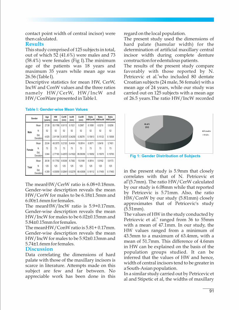
This study comprised of 125 subjects in total, out of which 52 (41.6%) were males and 73 (58.4%) were females (Fig I).The minimum age of the patients was 18 years and maximum 35 years while mean age was 26.56 (Table I).Descriptive statistics for mean HW, CerW, IncW and ConW values and the three ratios namely HW/CerW, HW/IncW and HW/ConWare presented in Table I.
Results
The meanHW/CerW ratio is 6.08+0.18mm. Gender-wise description reveals the mean HW/CerW for males to be 6.18±1.5mm and 6.00±1.6mm for females.The meanHW/IncW ratio is 5.9+0.17mm. Gender-wise description reveals the mean HW/IncW for males to be 6.02±0.15mm and 5.84±0.15mm for females.The meanHW/ConW ratio is 5.81+ 0.17mm. Gender-wise description reveals the mean HW/IncW for males to be 5.92±0.13mm and 5.74±1.6mm for females.
Data correlating the dimensions of hard palate with those of the maxillary incisors is scarce in literature. Attempts made on this subject are few and far between. No appreciable work has been done in this
Discussion
Table I: Gender-wise Mean Values
Fig 1: Gender Distribution of Subjects
regard on the local population.The present study used the dimensions of hard palate (hamular width) for the determination of artificial maxillary central incisor width during complete denture construction for edentulous patients.The results of the present study compare favorably with those reported by N.
6Petricevic et al. who included 80 dentate Croatian subjects (24 male, 56 female) with a mean age of 24 years, while our study was carried out on 125 subjects with a mean age of 26.5 years.The ratio HW/IncW recorded
in the present study is 5.9mm that closely correlates with that of N. Petricevic et
6al (5.7mm). The ratio HW/CerW calculated by our study is 6.08mm while that reported by Petricevic is 5.71mm. Also, the ratio HW/ConW by our study (5.81mm) closely approximates that of Petricevic's study (5.51mm).The values of HW in the study conducted by
6Petricevic et al. ranged from 36 to 55mm with a mean of 47.1mm. In our study, the HW values ranged from a minimum of 43.5mm to a maximum of 63.4mm, with a mean of 51.7mm. This difference of 4.6mm in HW can be explained on the basis of the population groups studied. It can be inferred that the values of HW and hence, width of central incisors tend to be greater in a South-Asian population.In a similar study carried out by Petricevic et al and Stipetic et al, the widths of maxillary
92
central incisor at the cervical, incisal and contact point areas were reported to be 8.26,
116.19 and 8.55 mm respectively. These values compare well with those of our study except for the incisal width which tends to be higher among our study group.Cesario et al. conducted a study on 229 dentate Saudi subjects (120 males, 109 females) mean age 21.16 years,and reportedthe central incisor width to be
15 8.9mm on average. The results match closely with those of our study.A comparable study conducted on 80
9patients by S. Wolfart et al. revealed maxillary central incisor width to be 9.1mm which is slightly larger than the values achieved in our study.Dimensions of the all anterior teeth for most racial groups vary with gender, with men exhibiting wider anterior teeth than women
16,17,18as reported in many studies. It was studied and reported by Gillen et althat in both black and white populations, men hand wider as well as longer maxillary
16 anterior teeth in comparison to women.Similarly, Sterrett et al.postulated the average width and length of the crowns of the maxi l lary anter ior tee th was significantly greater for white males than for
1 7white females . In the s tudy of Hasanreisoglu et al. the averagecrown width and height values for the central incisors and the canines were significantly greater for men as compared to women, with the central incisors being the widest
7teeth in both genders. Hock DA et al. measured the width of the maxillary central incisor in several racial groups and noted variations in most of them, with men, again
13having wider central incisors than women.In our study the mean values for the width of central incisor at the cervical, incisal and contact point areas for males were 8.91, 9.15 and 9.29 mm respectively. For females, the v a l u e s w e re 8 . 2 1 , 8 . 4 4 a n d 8 . 5 9 mmrespectively. This clearly indicates that
the males have larger mesiodistal width of central incisors than females in our study population. These findings are in good agreement with the results of other related
13, 17studies.The widths of maxillary central incisor vary considerably amongst different races. Very few data is available on this subject in local population. There is a requirement of extensive local work on larger scale in order to evaluate orofacial biometric guides in our population.
With the limitations of this study, the following conclusions were drawn:1. Hamular width can serve as a reliable
and helpful landmark in order to determine the maxillary central incisor's dimensions.
2. Maxillary central incisor's cervical (CerW), incisal (IncW) and contact point width (ConW) might be calculated by dividing hamular width (HW) by 6.08, 5.91 and 5.81 respectively.
3. The HW can only be used as a preliminary method for determining the width of the maxillary central incisor. The final decision regarding tooth selection should be made byemploying various guidelines suggested in the literature regarding anterior tooth selection and should be confirmed by consultation with the patient.
1. Paranhos LR, Lima CS, Silva RH, Daruge Junior
E, Torres FC. Correlation between maxillary
central incisor crown morphology and
mandibular dental arch form in normal
occlusion subjects. Braz Dent J 2012; 23: 149-53.
2. Tozum TF, Guncu GN, Yildirim YD. Evaluation
of maxillary incisive canal characteristics
related to dental implant treatment with
computerized tomography: A clinical
multicenter study. J Periodontol 2012; 83: 337-
Conclusions
References

93
43.
3. Paranhos LR, Joias RP, Velasco LG, Berzin F,
Daruge Junior E. Prevalence of the different
maxillary central incisor shapes in individuals
with natural normal occlusion. Braz J Oral Sci
2010; 9: 104-7.
4. Petricevic N, Celebic A, Seper LI, Kovacic I.
Appropriate proportions as guidelines in
selection of anterior denture teeth. Med glas
2008; 5: 103-8.
5. Park YS, Lee SP, Paik KS. The three-dimensional
relationship on a virtual model between the
maxillary anterior teeth and incisive papilla. J
Prosthet Dent 2007; 98: 312-8.
6. Petricevic N, Katunaric M, Mehulic K, Simeon P,
Rener-Sitar K, Celebic A. Selection of
Appropriate Artificial Frontal Teeth Size Using
Dimensions of Hard Palate. Coll Antropol 2006;
30: 573–7.
7. Hasanreisoglu U, Berksun S, Aras K, Arslan I.
An analysis of maxillary anterior teeth: Facial
and dental proportions. J Prosthet Dent 2005; 94:
530-8.
8. Amin WM, Taha ST, Al-Tarawneh SK, Saleh
MW, Ghzawi A. The relationships of the
maxillary central incisors and canines to the
incisive papilla in Jordanians. J Contemp Dent
Pract 2008; 9: 42-51.
9. Wolfart S, Quaas A C, Freitag S, Kropp P, Gerber
W D, Kern M. Subjective and objective
perception of upper incisors. Journal of Oral
Rehabilitation 2006; 33: 489–95.
10. Zlatariæ DK, Kristek E, Celebiæ A. Analysis of
width/length ratios of normal clinical crowns of
the maxillary anterior dentition: correlation
between dental proportions and facial
measurements. Int J Prosthodont 2007; 20: 313-5.
11. Petricevic N, Stipetic J, Antonic R, Borcic J,
Strujic M, Kovacic I, et al. Relations between
Anterior Permanent Teeth, Dental Arches and
Hard Palate. Coll Antropol 2008; 32: 1099–1104.
12. Ali Fayyad M, Jamani KD, Agrabawi J.
Geometric and mathematical proportions and
their relations to maxillary anterior teeth. J
Contemp Dent Pract 2006; 7: 62-70.
13. HockDA. Qualitative and Quantitative Guides
to the Selection and arrangement of the
Maxillary Anterior Teeth. J Prosthet Dent 1992;
1: 106-11.
14. Scandrett FR, Kerber PE, Umrigar ZR. A clinical
evaluation of techniques to determine the
combined width of the maxillary anterior teeth
and the maxillary central incisor. J Prosthet Dent
1982;48: 15-22.
15. Cesario VA Jr, Latta GH Jr: Relationship
between the mesiodislal width of the maxillary
central incisor and interpupillary distance. J
Prosthet Dent 1984; 52: 641-3.
16. Gillen RJ, Schwartz RS, Hilton TJ, Evans DB. An
analysis of selected normative tooth
proportions. Int J Prosthodont 1994; 7 :410-7.
17. Sterret JD, Oliver T, Robinson F, Fortson W,
Knaak B, Russel CM. Width/length ratios of
normal clinical crowns of the maxillary anterior
dentition in man. J Clin Periodontol 1999; 26:
153-7.
18. Sherfudhin H, Abdullah MA, Khan N. A cross-
sectional study of canine dimorphism in
establishing sex identity: comparison of two
statistical methods. J Oral Rehabil 1996; 23: 627-
31.
19. Keng SB, Foong KW. Maxillary arch and central
incisor dimensions of an ethnic Chinese
population in relation to complete denture
prosthodontics.Int Dent J 1996; 46: 103-7.
20. Ghoddousi H, Edler R, Haers P, Wertheim D,
Greenhill D. Comparison of three methods of
facial measurements. Int J Oral Maxillofac Surg
2007; 3: 250-8.
21. Ulhas ET, Shankar PD, Arun NK. Biometric
relationship between intercanthal dimension
and the widths of maxillary anterior teeth. J
Indian prosthod Soc 2007: 123-5.
ABSTRACTObjective: To evaluate the overall faculty performance by comparison of Self-assessment with peer and student assessment. Study Design: A comparative cross sectional survey.Place and Duration of Study: Study was conducted from January to June 2012 in Riphah College of Rehabilitation Sciences, Riphah International University Islamabad. Materials and Methods: This research study was conducted among students of Doctor of physical therapy (DPT), post-professional Doctor of physical Therapy (PPDPT), and Master of Science in speech language pathology (MS-SLP), and faculty members at Riphah College of Rehabilitation Science (RCRS), Riphah International University Islamabad. The total sample size was 730, including 700 students and 30 faculty members. A questionnaire was developed according to likert scale, and after a pilot study on 20 student and 10 faculty member to determine the reliability. The questionnaire was circulated among all the 30 faculty members and 700 students, including 500 undergraduate and 200 post graduate students of all the 3 programs. The data was analyzed and Wilcoxon (Kruskal-Wallis) was applied at 95% level of significance for all the 3 groups. The group “A” included assessment of the performance of the faculty members done by the students, group “B” done by other faculty members, and group “C” included self assessment done by faculty members. The averages were calculated to determine the overall performance of the faculty members as assessed by themselves, other faculty members, and the by students as well, afterwards the averages of 3 groups were compared. Results: the overall performance of the faculty members was graded as 71% (P=0.015) as evaluated by the students, 77% (P=0.009) as evaluated by other faculty members, and 73% (P=0.011) as evaluated by the faculty members through self assessment. Conclusion: It is concluded that there was no significant difference in the performance of the faculty members, as assessed themselves, by the students and the other faculty members.
Key words: Faculty performance, self assessment, students' feedback.
94
ORIGINAL ARTICLE
faculty members outcomes. Though it is not very much popular in the literature concern with medical and rehabilitation fields, but it is the most useful practice in business sector. The performance evaluation technique focuses on multiple perspectives and levels performance leading to results that are considered to be highly convincing and a powerful phenomenon to bring change in behavior. This feedback also known as mult isource feedback, mult i rater assessment, peer evaluation and full-circle appraisal. Peer evaluation provides developmental feedback which is always used to assess competency and behavior r a t h e r t h a n p e r s o n a l i t y a n d
2professionalism.
IntroductionFaculty evaluation is always vital for the Faculty development in academic institutions for further improvement and enhancement. This evaluation process helps the organization to arrange the faculty development programs for further improvement in the performance of the
1faculty members.Accreditation Council for Graduate Medical Education (ACGME) develops variety of assessment ''tools'' for performance of -------------------------------------------------
Comparison of Self-Assessment with Peer and StudentAssessment in Evaluating the Overall Performance of theFaculty
Correspondence:Dr. Syed Shakil-ur-RehmanPrincipal/Assistant ProfessorRiphah College of Rehabilitation SciencesRiphah International University Islamabad
Imran Amjad, Syed Shakil-ur-Rehman, Asghar Khan , Khalid Farooq Danish, Ilyas Babar Awan,Sikandar Ghayas Khan

95
One frequently used method for identifying e d u c a t i o n a l n e e d s i s t h e m a i l e d questionnaire. Generally, educational program planners gather initial information about a particular audience and design an appropriate questionnaire to elicit information from that audience regarding its perceived importance of identified
3topics. Although the literature related to conducting educational needs, assessment is quite plenteous, specific literature related to the tools and methods which are useful in the process is somewhat less abundant. Randol G. Waters, used the modified Borich Model to describe the educational needs of extension in field, faculty and indentifying the faculty development needs. Researchers would recommend the use of this need assessment model in determining educational needs of similar groups of
4clients. This study analyzes the outcomes of peer reviews of faculty members and the outcomes of students' feedback. Before this the general practice was getting feedback from the students or by the faculty member's separately. This particular research includes self assessment by the faculty members themselves, compared with the students'
5feedback.
This comparative cross sectional research survey was conducted among students of Doctor of physical Therapy (DPT), Post-Professional Doctor of physical Therapy (PPDPT), and Master of Science in speech language pathology (MS-SLP), and faculty m e m b e r s a t R i p h a h C o l l e g e o f Rehabilitation Science (RCRS), Riphah International University Islamabad. The faculty performance was evaluated for semester Spring 2012 and for the period of 6 months, from January-June 2012. The total sample size was 730, including 700 students and 30 faculty members.
Materials and Methods
A questionnaire was developed according to likert scale, and after a pilot study on 20 student and 10 faculty member to determine the reliability. The questionnaire was circulated among all the 30 faculty members and 700 s tudents , inc luding 500 undergraduate and 200 post graduate students of all the 3 programs. The details about the study sample are summarized in Table I.The questionnaire has 10 questions, which covered all of the following 10 important domains for performance of the faculty members: 1. Knowledge of the subject2. Up-to-date knowledge3. Communication Skills4. Students' participation in class5. Distribution of material among students6. Punctuality 7. Regularity 8. The use of virtual learning system VLS 9. The use of campus management system
CMS 10. Following Islamic Ethical valuesThe data was analyzed and Wilcoxon (Kruskal-Wallis) was applied at 95% level of significance for all the 3 groups. The group “ A ” i n c l u d e d a s s e s s m e n t o f t h e performance of the faculty members done by the students, group “B” done by other faculty members, and group “C” included self assessment done by faculty members. The averages were calculated to determine the overall performance of the faculty members as assessed by themselves, other faculty members, and the by students as well, afterwards the averages of 3 groups were compared.
A total of 730 students and faculty members participated in this research study; majority (77%) was female. Mean age of the u n d e rg r a d u a t e s t u d e n t s w a s 2 3 , postgraduates 27 and faculty members 30 years. The majority of participants were
Results

96
from under grade (68%), followed by post grade (27%), and faculty members (4%). The background of the students and faculty members were from physical therapy and speech therapy.The overall performance of the faculty
Table I: Summary of Study Sample (n=730)
Table II: Comparison of Overall Faculty Performance (n=730)
No. of
members was graded as 71% (P=0.015) as evaluated by the students, 77% (P=0.009) as evaluated by other faculty members, and 73% (P=0.011) as evaluated by the faculty members through self assessment. Table-II
Nigel K. Ll. Pope conducted a research study on “The impact of stress in self and peer assessment” and published in a research journal the assessment and evaluation in higher education in 2005. They concluded that the peer assessment and evaluation method is very effective for the evaluation
Discussion
the performance of the students and faculty 6
members.Keith Topping carries out a comparative cross-sectional survey on “Assessment b e t w e e n S t u d e n t s C o l l e g e s a n d Universities” and published in research
journal the Review of Educational Research in 1998. This study strongly supports the peer evaluation method for finding students and faculty outcomes at the colleges and
7universities level.Davis and John conducted a comparative cross-sectional research survey on “Comparison of faculty, peer, self, and Nurses Assessment in Obstetrics and Gynecology Residents” and published in a research journal the Obstetrics and Gynecology in 2002. They had evaluated the performance of residents placed in

97
Obstetrics and Gynecology wards, through self assessment, peer assessment and by the nurses. They concluded that there was no significant difference among the all 3
8groups.F. Dochy and colleagues carries out a review research study on “the use of self, peer, and co-assessment in higher education” and published in an international research journal named the Studies in Higher Education in 1999. They considered the peer review method effective and develop recommendat ions for educat ional institutions. They also stated in the conclusion after the completion the research review that peer review method made the s tudents more respons ib le whi le responding the evaluation and assessment
9at the higher education level. Matthew Ohland and colleagues conducted a r e v i e w r e s e a r c h s t u d y o n “ A Comprehensive Assessment of Team Members Effectiveness: Development of Behaviorally Anchored Scale for Self and Peer Evaluation“, and published in a research journal the Academy of Management, Learning, and Education in 2012. They find three studies which supports the effectiveness of peer review method for the evaluation and assessment of
10the effectiveness team members.
It is concluded that there was no significant difference in the performance of the faculty members, as assessed by the students, themselves, and the other faculty members.
1. Darling-Hammond L, Amrein-BeardsleyA.
Conclusion
References
Haertel E & Rothstein J. Evaluating teacher
evaluation. Phi Delta Kappan 2012; 93:8-15.
2. Schmoker M. The madness of teacher
evaluation frameworks. Phi Delta Kappan 2012;
93:70-1.
3. Hill H C, Kapitula L & Umland K. A validity
argument approach to evaluating teacher value-
added scores. American Educational Research
Journal 2012; 48:794-831.
4. Madden T J, Dillon W R & Leak R L.
Students' evaluation of teaching: concerns of
item diagnosticity. Journal of Marketing
Education 2010; 32:264-74.
5. Johnson D W, Johnson R T & Smith K A.
Cooperative learning returns to college
what evidence is there that it works? Change:
the magazine of higher learning 1998; 30: 26- 35.
6. Davis J D. Comparison of faculty, peer, self,
and nurse assessment of obstetrics and
g y n e c o l o g y re s i d e n t s . O b s t e t r i c s &
Gynecology2012; 99: 647-51.
7. Topping K. Peer assessment between students
in colleges and universities. Review of
Educational Research 1998; 68: 249-76.
8. Pope N K L. The impact of stress in self-and
peer assessment. Assessment & Evaluation in
Higher Education 2005; 30: 51-63.
9. Dochy F J R C, Segers M & Sluijsmans D.
The use of self-, peer and co-assessment in
higher education: A review. Studies in Higher
education 1999; 24: 331-50.
10. Ohland M W, Loughry M L, Woehr D J,
Bullard L G, Felder R M, Finelli C J. The
Comprehensive Assessment of Team Member
Effectiveness: Development of a Behaviorally
Anchored Rating Scale for Self-and Peer
Evaluation. Academy of Management Learning
& Education 2012; 11: 609-30.
ABSTRACTObjective: To determine safety of diode laser by studying post operative complications after endourological procedures in co-morbid patients.Study Design: A descriptive study.Place & Duration of Study: Shalamar hospital, Lahore from June 2009 to June 2012.Materials and Methods: We studied post-operative complications (up to 3 months) in 3 groups (prostatic obstruction, bladder growths ,urethral strictures) of total180 patients with ASA III & IV. We assessed hematuria, UTI, abdominal pain, suprapubic discomfort, urinary retention, dysuria, incomplete procedure, cardiac or respiratory compromise, fluid overload, mortality, catheterization times and mean postoperative hospital stayResults: In prostate group, mean age was 70.8±8.6 years and follow-up period was 3 months. Complications were: mild transient haematuria in 65 (100%), creamy urine in 50 (77%), urinary tract infection in 25 (38.4%), dysuria in 16 (24.6%), retreatment required in 06 (9.2%), suprapubic discomfort in 3 (4.5%), TURP syndrome in 1 (1.5%) and significant hemorrhage requiring blood transfusion in 1 (1.5%),. In urethral strictures, the complications were: microscopic hematuria in 80 (100%), urinary tract infection in 52 (65%) suprapubic discomfort in 9 (11.2%), dysuria in 6 (7.4%) mild transient hematuria in 5 (6.2%). In bladder growths, the complications were: mild transient haematuria in 30 (85.7%), creamy urine in 19 (54.2%), suprapubic discomfort in 18 (51.`4%), dysuria in 9 (25.7%), urinary UTI in 8 (22.8%) , ablation performed in two sittings in 1 (2.8%). No mortality in any group.Conclusion: Diode laser is a safe and useful modality in patients with co-morbidities (ASA III and IV).
Key words: Diode laser, endourology, co-morbidity.
98
ORIGINAL ARTICLE
IntroductionElderly patients with coexisting medical conditions undergoing complex or major surgery are high-risk. Range of surgery and patient-related factors including ischaemic heart disease, chronic obstructive pulmonary disease (COPD), advanced age, poor exercise tolerance determine the
1overall risk Bleeding remains a concern in all endoscopic procedures, like morbidity and mortality for transurethral resection of prostate (TURP) have not changed for
2,3 decades.Potential advantages of laser therapy over traditional procedures include appreciably good hemostasis, decreased morbidity, minimal cardiac stress, and shorter hospital -------------------------------------------------
Complications of Diode Laser in Endourological Proceduresin Co-morbid PatientsFarooq Hameed, Mohammad Imran Zahoor, Javed Aziz, Saadat Hashmi, Abdul Jalil, Abdul Rehman
Correspondence:Dr. Farooq HameedConsultant UrologistShalamar Hoapital, LahoreE-mail:[email protected]
4stay.High-powered diode laser systems are
5available for endoscopic procedures. It has a compact size, easy portability, and a potential for lower capital and maintenance
3costs. The current system (Biolitec) allows a continuous wave mode to a flexible and customizable pulsing regime with side or bare end fire fibre . Diode laser has similar wavelength characteristics to the Nd: YAG laser (Neodymium:yttrium-aluminum- garnett), but scatters less in tissue, high simultaneous absorption in water and hemoglobin and it is postulated to combine high tissue ablative properties with good hemostasis with significantly lower energy
3,6consumption . Safety measures are similar to Nd: YAG laser. Clinical data regarding safety of diode laser in endoscopic surgery is not frequently available.
We studied the post operative complications (up to 3 months) in 180 patients between
Materials and Methods

99
June 2009 and June 2012 in Shalamar hospital, Lahore. Depending upon diagnosis, patients were divided into 3 groups (with Prostatic obstruction, bladder growths and urethral strictures) and complications in each group were studied. In all cases of BPH, pharmacological treatment had been tried. Physical examination including digital rectal examination (DRE), Prostate specific antigen (PSA), abdominal ultrasound scan (trans-rectal scan only in case of disagreement between DRE and the abdominal ultrasound scan) were carried out for prostatic obstruction and bladder growth. Retrograde urethrogram was done for urethral strictures. All operations were done in spinal (60 %) or combined spinal/epidural, epidural alone or general anesthesia.) We used 980 nm Diode laser (Biolitec, Germany) with 1000 ìm core optical fibers (side-fire), 600 ìm end fire with a spot diameter of 1 mm. The output power ranged from 50 W to 140 W during the surgery. The mean applied energy was 130 ± 70 kJ. Vapo-resection (Coagulation and resection of bigger prostates, vaporization and sample collection for small lesions) was performed using laseroscope / resectoscope of 26 Fr. The strictures were dealt with by vaporizing the tissue when passing, using the contact technique or non-contact mode depending on the characteristics of the tissue. Saline solution was used as irrigation during ablation and 1.5% Glycine during sample collection. The antibiotic prophylaxis consisted of three intravenous doses of 1g of Cefoperazone /Sulbactam , at induction, at 12 and 24 hrs post operative period respectively. Patients needing anticoagulation were switched over to Enoxaprin 4000 i.u. sub cutaneous injection before the procedure. All procedures were performed by a single experienced surgeon with a dedicated team.In post operative period, patients were
assessed for transient , s ignif icant haemorrhage needing transfusion, microscopic hematuria, urinary tract infection, abdominal or flank pain, suprapubic discomfort, urinary retention, dysuria, incomplete procedure, cardiac or respiratory compromise, fluid overload , mortality, catheterization times and mean postoperative hospital stay. Patients were discharged from the hospital within 48 hours when urine was bloodless.
Patients from all age groups needing endoscopic laser treatment for BPH, bladder growths, and urethral strictures having1. Co-morbidities
a. altered renal function, (Creatinine > 2.0)
b. COPDc. bleeding / clotting disorders with
international normalization ratio (INR) >1.5
d. myocardial dysfunction, ejection fraction up to 30%)
2. American society of Anesthesiologists (ASA) grade III and IV
3. Refused surgical treatment due to high risk
Exclusion criteria:Patients with ASA grade I & IIFollow up:Follow up intervals postoperatively in out patient clinic were within 5 days for removal of catheter, 15 days, one month, 2 month and 3 months. Any clinical event, adverse effects and additional interventions or repeat surgery was noted. For cases of ablation of prostate and bladder growths, follow up intervals were within 5 days for removal of catheter (upto 7 days for some cases of bladder growths), 15 days, one month, 2 month and 3 months.In case of urethral strictures, the catheter was removed depending upon the severity of the condition and were followed upto 3 months.
Inclusion Criteria:
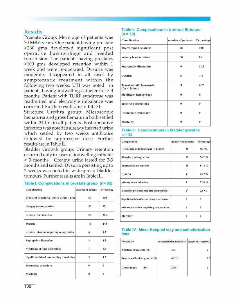
100
ResultsProstate Group: Mean age of patients was 70.8±8.6 years. One patient having prostate >260 gms developed significant post operative haemorrhage and needed transfusion. The patients having prostates >100 gms developed retention within 1 week and were re-operated. Dysuria was moderate, disappeared in all cases by symptomatic treatment within the following two weeks. UTI was noted in patients having indwelling catheter for > 3 months. Patient with TURP syndrome was readmitted and electrolyte imbalance was corrected. Further results are in Table I.Stricture Urethra group: Microscopic hematuria and gross hematuria both settled within 24 hrs in all patients. Post operative infection was noted in already infected urine which settled by two weeks antibiotics followed by suppressive dose. Further results are in Table II.Bladder Growth group: Urinary retention occurred only in cases of indwelling catheter > 3 months. Creamy urine lasted for 2-3 months and settled. Dysuria persisting up to 2 weeks was noted in widespread bladder tumours. Further results are in Table III.
Table I. Complications in prostate group (n= 65)
Table II. Complications in Urethral Stricture(n = 80)
Table III. Complications in bladder growthsn = 35
Table IV. Mean Hospital stay and catheterizationtime

101
DiscussionUse of laser for the treatment of BPH and bladder tumours is the most commonly used alternative to TUR to decrease
7,8 morbidities. For Nd:YAG, laser, it required longer catheterization periods because of the longer time required for expelling the necrotic tissue. Reports of long
9term results with KTP laser are limited. We removed catheter within 3 days because of effective coagulation and hemostasis except for prostatic obstruction with indwelling catheter for > 3months or complicated strictures of posterior and bulbar urethra .Rapid vaporization and hemostasis is
9,10,11possible by diode laser. We easily obtained tissues of prostate and bladder growths for biopsy. The architectural pattern in resected tissue was well m a i n t a i n e d f o r h i s t o p a t h o l o g i c a l evaluation. An ex vivo study showed that diode laser has a higher tissue ablation capacity than KTP laser, and shorter
9operative time. Using vapo-resection technique ,we obtained clean sharp cuts with almost blood-less field. W. Cecchetti et al obtained a bloodless sharp cut and easy vaporization with minimum carbonization and edema in 22 cases. They found diode laser a good compromise between absorption and coagulative effects
11on the tissue .Seitz et al treated 10 patients with BPH with diode laser. Ten patients were followed up at 1 month and 8 patients were followed up at 6 and 12 months. No serious postoperative
12haematuria was reported. We found, mild transient hematuria in almost all patients which settled within 4 hours. Erol et al (2009) studied 47 patients with diode laser prostatectomy. The commonest complication was mild-moderate irritative symptoms (23%) which resolved within the f i rs t two weeks. A late bleeding complication (requiring hospitalisation) was encountered in one patient at 4 weeks.
13Mean operative time was 53 minutes.Chen et al (2010) treated 55 patients of BPH. They reported 10 patients with transient dysuria. Acute urinary retention in two men was resolved by removal of sloughed tissue via TURP. Two patients underwent TURP due to insufficient vaporisation or regrowth of prostatic tissue (reoperation rate 7%).The three studies (Seitz et al; Erol et al; Chen et al) reported no serious intraoperative complications or postoperative haematuria. Lengths of hospital stay were 4.7 (SD 2.3) days in Seitz et al (2007) and 2.8 (SD 1.8) days
12,14 in Chen et al (2010). In our study, almost all patients were discharged within 48 hours after surgery.Clemente Ramos and Luis
15 Miguel evaluated diode laser treatment of BPH, focused on the peri-operative m o r b i d i t y. T h e y f o u n d p r o s t a t e vaporization effective with minimal
15morbidity.The application of lasers in treating urologic disorders has gained widespread clinical a c c e p t a n c e i n m u l t i p l e s u r g i c a l indications.15 Safety has also been demonstrated in patients with large prostates and patients receiving anti
16coagulant therapy or in retention.
The early results showed a virtually bloodless surgery with sharp cut. With acceptable complication rate, diode laser is relatively safe and useful modality in patients with co-morbidities (renal failure, Chronic obstructive pulmonary disease, myocardial dysfunction, ( ASA III & IV).
1 Pearse R M. Perioperative management of the
high-risk surgical patient in Bailey and Love's
short practice of surgery. 25th ed © 2008 Edward
Arnold (Publishers) Ltd, p215
2 Lanzafame RJ. Applications of lasers in
laparoscopic cholecystectomy. J Laparoendosc
Surg 1990;1:33-6.
3 Mulligan ED, Lynch TH, Mulvin D, Greene D,
Conclusion
References

102
Smith JM, Fitzpatrick JM. High-intensity
focused ultrasound in the treatment of benign
prostatic hyperplasia. British Journal of Urology
1997; 79: 177-80.
4 http://tau.sagepub.com/content/1/2/85,
Chung D E, Te A E. New techniques for laser
prostatectomy: an update Therapeutic
Advances in Urology 2009; 1: 85.
5 Nordahl G, Huckele S, Honeck P, Alken P, Knoll
T, Michel MS et al. 980-nm Diode laser: a novel
laser technology for vaporization of the
prostate. European Urology 2007; 52:1723-8.
6 Madersbacher S, Alivizatos G, Nordling J, Sanz
CR, Emberton M, de la Rosette JJ. EAU 2004
guidelines on assessment, therapy and follow-
up of men with lower urinary tract symptoms
suggestive of benign prostatic obstruction (BPH
guidelines). Eur Urol 2004;46:547-54.
7 Bülent Oktay, Hakan Kýlýçarslan, Hasan
Serkan Doðan. Diode laser in the treatment of
benign prostatic enlargement: a preliminary
study, Turkish Journal of Urology 2011;37:25-9
8 Seitz M, Ruszat R, Bayer T, Tilki D, Bachmann A,
Stief C et al. Ex vivo and in vivo investigations of
the novel 1,470 nm diode laser for potential
treatment of benign prostatic enlargement.
Lasers Med Sci 2009;24:419-24.
9 Seitz M, Bayer T, Ruszat R, Tilki D, Bachmann
A, Gratzke C, et al. Preliminary evaluation of a
novel sidefire diode laser emitting light at 940
nm, for the potential treatment of benign
prostatic hyperplasia: ex-vivo and in-vivo
investigations. BJU Int 2009;103:770-5.
10 Wendt-Nordahl G, Huckele S, Honeck P, Alken
P, Knoll T, Michel MS, et al. 980-nm Diode laser:
a novel laser technology for vaporization of the
prostate. Eur Urol 2007;52:1723-8.
11 Cecchetti W, Guazzieri S, Tasca A, Dal Bianco
M, Zattoni3 F, Pagano F, European Biomedical
Optics Week, BiOS Europe '96
12 Seitz M, Sroka R, Gratzke C. The Diode
Laser: A novel side-findding approach for Laser
vaporisation of the human prostate-Inmediate
efficacy and 1 –year follow up. Eur Urol 2007;
52:1717-22.
13 Erol A, Cam K, Tekin A, Memik O, Coban S,
Ozer Y. High power diode laser vaporization of
the prostate: preliminary results of benign
prostatic hyperplasia. The Journal of Urology
2009; 182: 1078-82.
14 Chen CH, Chiang PH, Chuang YC, Lee WC,
Chen YT, Lee WC. Preliminary results of
prostate vaporization in the treatment of benign
prostatic hyperplasia by using a 200-W high-
intensity diode laser. Urology 2010; 75: 658-63.
15 Ramos C, Miguel L. High power diode laser at
980 nm: preliminary results in the treatment of
the benign prostatic hyperplasia. Urology 2011;
70: 734-43.
16 Thomas R.W. Evangelos N. Olivier T. EAU
Guidelines on Laser Technologies, European
Urology 2012; 61: 23-40.

ABSTRACTBackground: Emergency Peripartum Hysterectomy (EPH) is a major surgical procedure. There appears to be a rise in the rate of emergency peripartum hysterectomy (EPH) in the developed world. Peripartum hysterectomy is high risk but a life saving operation. It is usually associated with significant maternal and fetal morbidity and mortality. Obstetricians should identify patients at risk and anticipate the complications, as early intervention and proper management results in optimal outcome.Objective: To determine the incidence, indications, risk factors, complications and management of EPH over the last eleven years. Study Design: A descriptive study.Place and Duration of Study: All cases of EPH performed in the period between January 2001 and December 2011 were included in the study. Study was conducted at Railway Teaching Hospital, Rawalpindi (in northern Pakistan).Materials and Methods: Data of all the cases of EPH cases operated during the study period was collected from the hospital obstetric record. Data of basic demographics, mode of delivery, maternal and fetal outcome along with associated complications was collected and entered into the Statistical Package for Social Sciences version 14.0 (SPSS Inc., Chicago, IL, USA) for further analysis. Indications, pre-op planning, type of operation, emergency decision, blood loss, transfusion, complications, were compared and cross-tabulated. Statistical analysis included: Chi-square and Fisher exact tests, where appropriate, and two-sample t test.Results: Total deliveries were 13560 in 11 years. Twenty Six EPH cases were performed among deliveries, giving an incidence of 1.8 per 1000 deliveries i.e. one in 566 deliveries. The indications were uterine atony (34.61%), ruptured uterus (23.07%) and cervical/vaginal tears (11.53%), placenta praevia (11.53%), invasive placental adhesion [accreta, increta, percreta (11.53%)]. A significant association between previous uterine surgery and abnormal placentation was shown (p=0.02), especially those with previous caesarean (p=0.003). One maternal and six perinatal mortalities were recorded. Four perinatal deaths were of non booked patients, handled by traditional birth attendants/private clinics. Out of two perinatal deaths of booked patients, one was pre-term and other was delivered by forceps. Maternal morbidity was prevalent, including twenty three intensive care admissions, three disseminated intravascular coagulopathies, three bladder injuries, three re-explorations, multiple blood, FFPs & platelet transfusions, two pulmonary embolism & one cardio- respiratory failure. Maternal death was of non booked patient with previous scar, home delivery, uterine rupture and brought to hospital with un-recordable BP and very weak pulse. Conclusions: Incidence of peri-partum hysterectomy is increasing. It is a major operation, and almost always an emergency with significant blood loss. An early decision should be made to save life of the patient and prevent complications. The most important risk factor for peripartum hysterectomy in our patients is hemorrhage, most notably caused by uterine atony, uterine rupture, placenta previa and abnormally adherent placenta.
Keywords: Peripartum hysterectomy, uterine atony, maternal morbidity, perinatal mortality
103
ORIGINAL ARTICLE
UNFPA and the World Bank: 2010. Trends in Maternal Mortality: 1990 to 2008). Approximately one-half of these deaths are preventable.Peripartum hysterectomy is removal of pregnant uterus after 20 weeks gestation at or near time of delivery (performed within 24 hours of a delivery) but within the first 6 weeks postpartum. It is a technically difficult but life saving procedure especially
IntroductionWomen are the pillars of social & economic development. Their health & well being is vital for future generations. An estimated 3,58000 maternal deaths occurred worldwide in 2008. (WHO, UNICEF, -------------------------------------------------
Emergency Peripartum Hysterectomy in Pakistan RailwayTeaching Hospital Rawalpindi: Eleven Years ReviewSaadia Sultana, Muhammad Nadim Akbar Khan, Shamsunnisa Sadia, Noor Mah khan, Fareesa Waqar,Azra Saeed, Umber Jalil
Correspondence:Dr. Saadia SultanaAssociate Prof. Obs/ GynaePakistan Railway Hospital, IIMC-T, RawalpindiE-mail: [email protected]

104
when performed as an emergency. Its complications are 4 times than those of an elective procedure. Intraoperative and postoperative complications are much more than that of gynecological hysterectomy. Its Mortality is 5 times greater than an elective procedure.Life-threatening haemorrhage resulting from uterine rupture and atony has become rare events in the developed world. But it is still a major problem in obstetric care in
1developing countries . Peripartum hysterectomy may be performed in emergency as a last resort to save the life of a woman with persistent bleeding or as planned procedure, often in conjunction with cesarean delivery. It is performed in 0.05 to 0.1 percent of deliveries.It is one of the life saving surgical procedures performed at the Department of Obstetrics and Gynaecology of the Railway Teaching Hospital. Moreover, it is considered one of the most serious complications in obstetrics
2with adverse outcomes for women.The main complications related to emergency peripartum hysterectomy include Disseminated Intravascular
3Coagulation (DIC), transfusions , need for re-laparotomy because of persistent bleeding, febrile morbidity, major surgical
4complications or maternal death.Many studies have estimated an incidence rate of 0.8 and 1.5 per 1,000 deliveries in
5United States. Incidence is higher in developing countries due to decrease compliance of patients and lack of availability and utilization of antenatal care services especially in the rural areas. There also is a rise of EPH in the developing
6 world. Some risk factors for peripartum hysterectomy have been identified, including prior cesarean delivery, mode of delivery or multiple births. A prior cesarean delivery is associated with an increased rate of abnormal placentation, including placenta previa, accreta & increta in
7subsequent pregnancies.The purpose of this study was to determine the incidence of peripartum hysterectomy, the indications, risk factors, the perinatal a n d m a t e r n a l o u t c o m e s a n d t h e complications associated with the procedure at the Railway Teaching Hospital, Rawalpindi, Pakistan. This would help highlight the importance of lack of availability and utilization of antenatal services. Identify avoidable factors and the need to organize health care services. It can be a contribution towards improvement of maternal and fetal outcome
Objective of the study was to determine the i n c i d e n c e , i n d i c a t i o n s , m a t e r n a l characteristics, risk factors, maternal and perinatal outcomes and complications of Emergency Peripartum hysterectomy (EPH) at Railway Teaching Hospital, Rawalpindi. These hysterectomies were performed by consultants with negligible differences in their operating techniques.This was a descriptive study, carried out at Gyne /Obs department of Pakistran Railway Teaching Hospital, Islamic I n t e r n a t i o n a l m e d i c a l c o l l e g e , Rawalpindi.from January 01, 2001 to December 31, 2011. Pakistran Railway Teaching Hospital is located at the junction of Rawalpindi & Islamabad (Capital of Pakistan), which are twin cities. A large number of Afghan refugees of different castes and tribes; who migrated to Pakistan got settled in these localities. Our hospital is one of the undergraduate and post graduate teaching hospitals.Data of all the EPH cases operated were obtained from the hospital obstetric record. Each case record was analyzed in details with special emphasis on maternal characteristics demographic data (age, parity, booked or emergency case etc.), indications for hysterectomy (hysterectomy for any indication during pregnancy, labor
Materials and Methods

105
and puerperium has been included), type of o p e r a t i o n p e r f o r m e d , p r o b l e m s encountered during operation, maternal & peri-natal outcome, morbidity, mortality & causes of maternal morbidity and mortality were studied.During this period there were 13,560 deliveries, out of which 11,960 were vaginal deliveries and 682 caesarean sections were performed. During the study period, 26 women were noted to have undergone an emergency peripartum hysterectomy, giving an incidence is 1.8 per 1000 deliveries i.e. 1 in 566 deliveries. Data regarding their basic demographics, mode of delivery, maternal and fetal outcome, along with associated complications was then collected and entered into a database developed in Microsoft Access 2000. This was then imported into the Statistical Package for Social Sciences version 17.0 (SPSS Inc., Chicago, IL, USA) for statistical analysis.
As noted above, 26 women were identified who underwent an emergency peripartum hysterectomy from a total of 13,560 deliveries (Table I). Out of which 11,960 were vaginal deliveries and 682 had caesarean sections. Therefore, the incidence of peripartum hysterectomy is 1.8 per 1000 deliveries i.e. 1 in 566 deliveries. Out of 26 women who underwent emergency peripartum hysterectomy, eighteen cases were non booked (73.1%) & only eight were booked (26.8%). It means that majority of these patients had received no antenatal care prior to their presentation to the hospital. This is really alarming situation. Majority of these patients belonged to very low socio-economic group.As shown in Table II, only five patients were primi-paras. Most of the patients were having 2-4 children (65%). Grand-multiparas were 21.9%. As shown in Table III, the main indication for peripartum hysterectomy was severe
Results
haemorrhage caused by uterine atony 09 (34.61%). Other indications included ruptured uterus 60 (23.07%); morbidly adherent placenta 03 (11.53%); placenta praevia 03 (11.53%); cervical/vaginal tears 03 (11.53%) and two patients reported with DIC (7.69%).In our series of patients, the commonest
Table I: No. of deliveries, caesarean hysterectomy& booking (n=13560)
Table II: Age distribution of the patients & theirpercentages( n=26)
Table III: Outline of indications of EPH (n=26)

106
indication for an EPH was uterine rupture in 6 (23.07%) cases. Two of these cases were due to previous one scar in labour. One each is due to accidental haemorrhage, prolonged labour, instrumental delivery and oxytocin abuse. Some cases appeared to be related to presentation of multipara and grand mutipara to traditional birth attendants (TBAs). Cases of obstructed labor are due to malpresentation and cephalopelvic disproportion neglected by TBAs (or “Dai”). Both the patients of lower segment caesarean section were tried for vaginal birth after caesaren (VBAC) at home by TBAs. In majority of patients of our study, EPH was performed because of postpartum hemorrhage (PPH) mainly due to uterine atony. Other indications were abruption plcentae with covualaire uterus, and placenta percreta. These findings are very much similar to a study from the province of Sindh, Pakistan, where EPH were mainly
8performed due to uterine rupture.There was one (3.84%) maternal death. This
Table IV: Maternal morbidity and mortality inpatients with EPH (n=46)
patient died due to Cardio-respiratory failure. Twelve (44%) patients stayed for 12 days or less, 11 (40%) for 13-22 days and 2 stayed for 24-35 days. The hospital stay of these patients ranged from 7 to 35 days, with a mean of 14 days. This represents huge financial burden on the patients and health care system of the country with serious economic and social consequences.Parity of patients ranged from 0–13. One patient was primigravida, required hysterectomy due to abnormally adherent placenta, Three were requiring EPH due to rupture of a previous caesarean scar; while the rest were multiparas. Sixty nine percent of patients were grand multipara (≡ 5 previous deliveries). Total abdominal hysterectomy was performed on 17 patients out of 26; nine underwent subtotal hysterectomies. Subtotal hysterectomy is also an acceptable
9option in technically difficult situations.Almost all surgeries were performed in emergency (24/26); only 2 were performed as elective procedure. Majority of the complications noted were infections (wound infection, fever and urinary tract infection) followed by complications related to the surgery itself. All patients who underwent EPH received blood transfusion
Table V: Fetal and neonatal outcomes (n=26)
during or in the immediate postoperative period. There were 8 (30.77%) perinatal deaths noted in this series of patients. Six were
107
stillborn, 4 of which were due to a ruptured uterus and 2 were because of abruptio placentae. There were two neonatal deaths due to neonatal sepsis and aspiration pneumonia.
In our study 26 women underwent EPH from a total of 13,560 deliveries. Out of these, 11960 were vaginal deliveries and 682 caesarean sections. Therefore, the incidence of EPH in our hospital is 1.8 per 1000 deliveries i.e. 1 in 566 deliveries. Whereas, in a study conducted in Turkey, 34 cases of EPH
10 were performed over a 10-year period.Similarly, in a study from New York, 48 cases of peripartum hysterectomy were performed over 8-year period; with an
11incidence of 1.4 per 1000 births.Compared to a study performed in Australia where only 33 EPH were documented among 33,998 births over a 10-year period, the incidence documented was 0.85 per 1000 births; our rate of EPH is approximately 2-
12fold higher.Our study is representing an alarmingly high number of EPH at a tertiary care hospital of a developed city of PakistanThere is an increasing trend in EPH 1980s to 2013. We have found three studies which reported an increased trend of EPH. An increased incidence of EPH from 1998 to 2003 was also found by Whiteman et al; as reported earlier. A Danish study reported a statistically significant risk of EPH in 1995-2004.While Calgary Health region of Canada found no statistically significant difference in incidence rates from 1999-2006. But, the number of EPH was not very sufficient to
13interpret the results.Hence, all these rates are not accurate estimates and comparisons could only become significant when the populations would be similar in demographic factors such as age etc, time periods being evaluated are similar and management
Discussion
protocols would be evidence based.Our results are revealing that the most important cause for EPH is hemorrhage most importantly due to uterine atony, uterine rupture & retained placenta,
15findings consistent with previous reports.The causes of EPH noted in our patients are very much different from the developed countries where abnormal placentation resulting in hemorrhage was the most
common cause. A study publised by Netherlands, found that the main cause for EPH was placenta accreta (50%). Uterine
16atony was found in 27% of cases. A Turkish study noted that uterine rupture was the cause in almost 21% of the cases & uterine
17 atony was responsible in 42% of the cases. A study conducted in Saudi Arabia and another of Korea, also revealed that uterine
18atony is the most common cause. Despite practicing evidence based medical advances, regular drills and improvement of protocols; hemorrhage continues to be an important contributor to maternal morbidity and mortality. In our study, we tried to explore factors that could be recognized antenatally and could be rectified with in time preventative measure. Abnormal placentation has also been consistently documented in other studies to be associated with previous uterine
19surgeries. These life-threatening abnormal placental complications require aggressive blood t rans fus ion , subsequent b leeding complications, hysterectomy, and longer
20maternal hospital stays. With advanced radiological facilities, pre-hand diagnosis of abnormal placentation can guide clinicians to plan for delivery with uterotonics, transfusion services, uterine balloon compression devices, interventional
21radiologists and standby of surgeons.Involvement of interventional radiologist and invasive treatment must be decided within 30 minutes if other measures have

108
failed. Assisted vaginal delivery has also been
22related to risk of EPH.A study indicated the association of damage to cervical/ vaginal tissues, resulting in
23 hemorrhage and leading to hysterectomy.Another important hypothesis is about uterine scarring, especially with cesarean deliveries, also increases the risk of EPH, even without abnormally adherent placenta/ placenta previa.Multiple cesarean sections, as well as VBAC were also found to
24be associated with EPH.In our study multiple gestation could not be proved to be significantly associated with EPH, as has been described in some studies. Some reported that multiple gestations had significantly increased risk of EPH e.g. Francois et al. According to these studies, tocolysis require for preterm labour and overdistended uterus by more than one fetus is a major facto towards uterine atony and hemorrhage. However, they failed to adjust the confounding factors such
25gestational age & mode of delivery etc.Unfortunately our study has some limitations also. The most significant is that our inferences depend on the hospital records, their accuracy about the diagnoses and management and records or notes on birth certificates and discharge forms. We tried our best to countercheck and recheck the records about d iagnos is and management, birth certificates, to reduce biased classification of different risk factors and confounders, as also suggested by some
26,27other studies.For example the diagnosis of “retained placenta” limited the options and make it difficult to distinguish between different abnormally adherent placenta e.g. placenta accreta, increta or percreta. Without doing a review of patients' notes, we were unable to determine the indication for EPH, the amount and the type of hemorrhage (antepartum, intrapartum, postpartum).
Lastly, the number of cases for some of the factors that were studied was relatively small.
Some of the very important inferences are:! Incidence of EPH is significantly high
and the most important indication of EPH is hemorrhage mainly due to uterine atony, uterine rupture, placenta previa & abnormally adherent placenta.
! Important risk factors to be considered are multiparity, previously scarred uterus, non booked patients & deliveries carried out by TBA.
! Significant maternal and perinatal morbidity and mortality is the main outcome of EPH e.g. ICU admissions, a r r a n g e m e n t f o r b l o o d & i t s c o m p o n e n t s , m a n a g e m e n t o f coagulopathies/thromboembolism & injuries of adjacent visceras results in huge psychological & financial burden on the patient, family & society.
1. Plauche WC. Caesarean hysterectomy:
indications, technique, and complications. Clin
Obstet Gynecol 1986; 29:318.
2. Saeed F, Khalid R, Khan A, Masheer S, Rizvi JH.
Peripartum hysterectomy: a ten-year experience
at a tertiary care hospital in a developing
country. Tropical Doctor 2010; 40:18–21.
3. Briery CM, Rose CH, Hudson WT, Planned v s
emergent cesarean hysterectomy.Am J
Obstet Gynecol 2007; 197:151–4.
4. Kwee A, Bots ML, Visser GH, Bruinse HW.
Emergency peripartum hysterectomy: A
prospective study in The Netherlands. Eur J
Obstet Gynecol Reprod Biol 2006;124:187–92.
5. Whiteman MK, Kuklina E, Hillis SD. Incidence
and determinants of peripartum hysterectomy.
Obstet Gynecol 2006;108:1486–92.
6. Awan N, Bennett MJ, Walters WA. Emergency
peripartum hysterectomy: a 10-year review at
the Royal Hospital for Women, Sydney.
Australian and New Zealand Journal of
Conclusions
References

109
Obstetrics and Gynaecology 2011;51:210–5.
8. Knight M, Kurinczuk JJ, Spark P, Brocklehurst P.
C e s a r e a n d e l i v e r y a n d p e r i p a r t u m
hysterectomy. Obstet Gynecol 2008;111:97–105.
9. Nisar N, Sohoo NA. Emergency peripartum
hysterectomy: frequency, indications and
maternal outcome. Journal of Ayub Medical
College Abbottabad 2009;21:48–51.
10. Saeed F, Khalid R, Khan A, Masheer S, Rizvi JH.
Peripartum hysterectomy: a ten-year experience
at a tertiary care hospital in a developing
country. Tropical Doctor 2010; 40:18–21.
11. Demirci O, Tuðrul AS, Yýlmaz E, Tosun O,
Demirci E, Eren YS. Emergency peripartum
hysterectomy in a tertiary obstetric center:
nine years evaluation. Journal of Obstetrics
and Gynaecology Research 2011; 37:1054–60.
12. Kastner ES, Figueroa R, Garry D, Maulik D.
Emergency peripartum hysterectomy:
experience at a community teaching hospital.
Obstetrics and Gynecology 2002;99:971–5.
13. Awan N, Bennett MJ, Walters WA. Emergency
peripartum hysterectomy: a 10-year review at
the Royal Hospital for Women, Sydney.
Australian and New Zealand Journal of
Obstetrics and Gynaecology 2011; 51:210–5.
14. Sakse A, Weber T, Nickelsen C, Secher NJ.
Peripartum hysterectomy in Denmark 1995-
2004. Acta Obstet Gynecol Scand. 2007;
86:1472–5.
15. Glaze S, Ekwalanga P, Roberts G, et al.
Peripartum hysterectomy: 1999 to 2006. Obstet
Gynecol 2008; 111:732–8.
16. Habek D, Becarevic R. Emergency peripartum
hysterectomy in a tertiary obstetric center: 8-
year evaluation. Fetal Diagn Ther 2007;
22:139–42.
17. Kwee A, Bots ML, Visser GH, Bruinse HW.
Emergency peripartum hysterectomy: a
prospective study in the Netherlands. European
Journal of Obstetrics Gynecology and
Reproductive Biology 2006; 124:187–92.
18. Zorlu CG, Turan C, Iºik AZ, Daniºman N,
Mungan T, Gökmen O. Emergency
hysterectomy in modern obstetric practice.
Changing clinical perspective in time.
A c t a O b s t e t r i c i a e t G y n e c o l o g i c a
Scandinavica 1998;77:186–90.
19. Yamani Zamzami TY. Indication of emergency
peripartum hysterectomy: review of 17 cases.
Archives of Gynecology and Obstetrics
2003;268:131–5.
20. Usta IM, Hobeika EM, Musa AA, Gabriel GE,
Nassar AH. Placenta previa-accreta: risk factors
and complications. Am J Obstet Gynecol
2005;193:1045–9.
21. Nisar N, Sohoo NA. Emergency peripartum
hysterectomy: frequency, indications and
maternal outcome. Journal of Ayub Medical
College Abbottabad 2009; 21:48–51.
22. Dabelea V, Schultze PM, McDuffie RSJ.
Intrauterine balloon tamponade in the
management of postpartum hemorrhage. Am J
Perinatol 2007; 24:359–64.
23. Roethlisberger M, Womastek I, Posch M,
Husslein P, Pateisky N, Lehner R. Early
postpartum hysterectomy: incidence and risk
factors. Acta Obstetricia et Gynecologica
Scandinavica 2010;89:1040–4.
24. Johnson JH, Figueroa R, Garry D, Elimian A,
Maulik D. Immediate maternal and neonatal
effects of forceps and vacuum-assisted
deliveries. Obstet Gynecol 2004;103:513–8.
25. Kacmar J, Bhimani L, Boyd M, Shah-Hosseini R,
Peipert J. Route of delivery as a risk factor for
emergent peripartum hysterectomy: a case-
control study. Obstet Gynecol 2003; 102:141–5.
26. Francois K, Ortiz J, Harris C, Foley MR, Elliott
JP. Is peripartum hysterectomy more common
in multiple gestations? Obstet Gynecol 2005;
105: 1369–72.
27. Lydon-Rochelle MT, Holt VL, Cardenas V. The
reporting of pre-existing maternal medical
conditions and complications of pregnancy on
birth certificates and in hospital discharge data.
Am J Obstet Gynecol 2005; 193:125–34.

ABSTRACTAudit is the practice of ensuring good professional practice, which has been practiced in the various fields of professional world for a long time. The concept of clinical auditing which primarily ensures quality provision of equitable, ethical healthcare is still a new concept in the field of health sciences. In dentistry, this concept is even more recent, especially in the developing world. Many dental negligence cases go unaccounted for in various parts of the developing world.This article outlines the main concepts of clinical audit, explaining what exactly is clinical audit and how may it be implemented in the practice of dentistry.
Key Words: Clinical Audit, Clinical Governance, Dental Audit, Dental Practice.
110
SHORT COMMUNICATION
Defining Clinical Audit1. Audit – Audit has been described in
different ways, depending on what is desired out of the whole process. To put it simply, it is an extension of good clinical practice. A more discrete way of describing audit would be that “it is the systematic and critical analysis of the quality of medical care (i.e. critical
3analysis review).”2. Peer Review – In this method, a group of
clinicians hold a meeting where they either discuss clinical cases or other protocols regarding the setting of the clinical practice e.g. guidelines for recall i n t e r v a l s i n d e n t i s t r y, d e n t a l radiography guidelines or cross-infection control protocols. A peer review group consists of between four to eight dentists from at least two
4different practices. A full and honest discussion is carried out, where it is discussed whether certain clinical scenarios were managed appropriately or not; and/or whether certain guidelines are appropriate and evidence-based. Any change required in the standards for assessing clinical protocols; or a need for staff training and education may be identified. It is recommended that the review should be completed in eight sessions (of at least two and a half hours) within nine
4months.
IntroductionOver the past few decades, public opinion and involvement in modern health care has been increasing continuously, so much so that today, in the developed countries, it is unethical for a clinician to formulate a treatment plan without taking the opinion of the patient under consideration. A specific growing concern among the public is that health care brings more harm than good. Several cases involving nurse, doctors and in some instances, dentists became quite popular coming under the light of the media and have brought about change in health care policies. Some examples of these are the Bristol babies case – where about 90 children were reported to have died, owing to sub-standard care by the doctors; the Alder Hey
1Hospital case and the Harold Shipman case. It is true that these individuals represent a v e r y s m a l l p ro p o r t i o n o f h e a l t h professionals. However, such negligence should not be left unchecked and a system to monitor and assess quality health care needs to be present. Such systems have appeared over the past few decades, to provide
2excellent quality health care to the public.
-------------------------------------------------
Clinical Audit and Its Role in the Practice of DentistryMuhammad Humza Bin Saeed, Shakeel Kazmi, Faisal Moeen, Yusuf Bhatti
Correspondence:Dr. Muhammad Humza Bin SaeedAssistant Professor,Department of Community DentistryIslamic International Dental CollegeRiphah International UniversityEmail: [email protected]

111
3. Clinical Audit – The clinical audit scheme for dentistry was introduced in the UK in 1995. The purpose of clinical a u d i t w a s t o g i v e t h e d e n t a l practitioners to build upon their peer review activities, going further by identifying standards that they could
4assess their clinical practice against. Measurement tools or assessment methods are drafted against which the clinicians can go back and assess their own practice settings. They evaluate their own personal clinical settings, and if there are any shortcomings, a plan to change the practice to cater for the respective drawbacks is implemented. Such changes, and the evaluation and m o n i t o r i n g p r o c e s s e s a r e t h e n discussed over the next meetings. The South West Regional Health Authority in the United Kingdom has defined medical audit from the above mentioned perspective as follows:“Medical audit is a systematic approach to the peer review of medical care in order to identify opportunities for improvement and to provide a mechanism for bringing them about. It complements and subsequently overlaps financial audit, utilisation review and resource management but it differs in that its purpose is primarily clinical rather than
managerial.”
Clinical audit can be viewed from the
perspective of the health professionals and/or
the patients. The desired outcome of these
groups would be more or less the same – making
efforts for the provision of effective health care.
From the perspective of the planners; managers
and/or administrators of health care, clinical
audit or clinical governance is more of a quality
assurance review procedure to ensure that the
optimal clinical services are being provided to
the public in a manner acceptable to them, in a
time and cost-efficient way.
Clinical audit from the perspective of doctors and/or patientsMedical audit is the term used to describe systematic and critical analysis of clinical procedures as carried out by doctors, in order to evaluate the procedures that doctors do. The key elements of medical
3auditing, as described by Leeman , are as follows:i. Attaining the objectives of the system –
this involves achieving the aims and obtaining the desired outcome. The interest of the doctors is to achieve the best clinical results and to fulfil the population's normative needs (the needs of an individual or population as described by a health professional: a
5doctor or a dentist). The people would be more interested in having a health s e r v i c e t h a t i s a c c e s s i b l e , accommodating, affordable and
6acceptable to them. It then becomes the role of the auditing procedure to see to the attainment of these objectives.
ii. Efficiency – As described by Muir and 7Gray, efficiency is in doing things the
right way. This means that the policy objectives are obtained using a process, which uses resources (money, staff, and time) in the most optimal manner.
iii. Effectiveness – As described by Muir 7and Gray, effectiveness is doing the
right things. This implies choosing the process that obtains the best possible results.
iv. Professional assessment of services – This involves judging against a performance indicator. Various protocols can be assessed according to different guidelines, such as those set by the National Institute of Clinical
8Excellence (NICE) e.g. following the NICE guidelines for following the protocol for third molar extractions or for patient routine recall intervals.
v. Consumer demands – The needs of the

112
people (felt and expressed needs) should be addressed in a quality health care service. A health need assessment would assist in this purpose.
vi. Consumer complaints – A system to record both verbal and written complaints should be present . S u g g e s t i o n a n d c o m p l a i n t boxes and patient satisfaction surveys can be useful in this regard. A dental practice adviser might be hired for this
9very purpose as well.
The above mentioned definition clearly gives the concept of clinical auditing not being a judgemental, management tool used for financial auditing threatening the practice with strict, disciplinary measures; but rather a methodical analysis of clinical procedures and setting, assessing them against specified measurement tools; assessing changes to maintain an agreeable, accepted standards, along with periodic evaluation and monitoring.Clinical Audit from the perspective of planner, administrators and/or managersAdministrators view clinical practices from a quality assurance angle. Clinical governance, as described by the NHS Quality Improvement Scotland (2005) is 'the system through which NHS organisations are responsible for continuously monitoring and improving the quality of their care and services and safeguarding high standards of care and services.'The UK Department of Health's definition (1998) is quite similar to the one above, describing clinical governance as 'a f r a m e w o r k t h r o u g h w h i c h N H S organisat ions are accountable for continuously improving the quality of their services and safeguarding standards of care by creating an environment in which
10excellence in clinical care will flourish.These definitions differ from the earlier mentioned medical audit definitions in that
the medical audit concept was more of an internal assessment and monitoring cycle, whereas clinical governance takes a stance on accountability, excellence in care and quality assurance – with possible influence externally from outside the service possibly playing a role. However, the concept of clinical audit held by the administrators and the health clinicians are not mutually exclusive or independent of each other. They should, r a t h e r, c o m p l e m e n t e a c h o t h e r. Accountability and quality assurance checks within an internally based system, improving the quality of the clinical settings. However, an approach that is threatening in itself would not be welcomed by clinicians.Clinical Audit in DentistryMaidment modified the definition given by
7the UK Department of Health to describe clinical audit in dentistry as 'a framework through which dental practices are held accountable for continuously improving the quality of services and safeguarding high standards of care by creating an environment in which excellence in clinical
9care flourishes.The public outcry and mistrust of the general population when it comes to health care services, resulting from cases of negligence, cannot and should not be denied or ignored. On analysis of these cases, it can be seen that in most of these cases early warnings were unnoticed or in the worst case scenario, ignored. A chain of events leads to complaining and in some cases, serious litigation issues. These issues, as we know, are not only restricted to medical care but affect dentistry as well. In light of all these issues, a quality assurance or clinical auditing mechanism needs to be present to monitor any change or negligence that might be taking place to achieve the set
9dental standards.Clinical dental settings and procedures should be following certain standards, such

113
as the guidelines drawn out by NICE for various protocols. To assess whether these standards are being practiced and maintained, a clinical governance procedure falls right into place.Role of clinical audit in implementing improvement in the Dental PracticeSeveral authors have addressed the issue of implementing clinical audit in the dental practice; which has more recently formed a cornerstone to the practice of clinical governance in the dental practice setting. Using Donabedian's quality assurance model8 of structure, process and outcome, they have developed models to show how clinical audit could be implemented in
9 11dental practice , .StructureUnder structure, administrative issues are addressed. A situational analysis is carried out to have an overview of the practice, describing the organisation of the practice; the staff and their distribution; the resources available; any quality assurance systems that are in place and the managerial structure of the practice.ProcessClinical procedures, risk management pro toco ls , c ross - in fec t ion contro l mechanisms, staff management and responsibilities, implementation of quality assurance protocols, monitoring and evaluation systems should be analysed. This involves all the steps involved from the point a patient is received in the practice till
2he is discharged after treatment. Examples can be taking proper clinical notes and keeping a record of them; having a system for recording the complaints – verbal and written- by the patients and reviewing these complaints in the dental practice meetings, with the objective of implementing any changes in the practice to minimise future complaints; recording all adverse events cases to minimise the risk of any such event occurring in the future; and having
continuing professional development 11
training in place for the staff.OutcomeAlthough assessing the outcome in dental practice is a challenging and difficult task, it is impossible and clinical audit is an
9important tool to assess outcome. An outcome measure in a dental setting is an indicator of the effectiveness of the clinical intervention – whether the proposed treatment plan had the desired effect on the patient's health or not. 'Heath gain' is used to describe the benefit that the patient receives from the treatment – these benefits are not
3only physical but emotional as well. To assess the clinical effectiveness of any intervention, clearly defined clinical performance indicators should be used and any corrective or preventive actions put into
11 place accordingly. Patient satisfaction surveys can help in assessing the emotional benefits of the proposed treatment.The above ment ioned points are appropr ia te ly summarised in the Department of Health's description of clinical audit as encouraging 'individual dental practitioners to self-examine different aspects of their practice, to implement improvements where the need is identified and to re-examine from time to time, those areas which have been audited to ensure that a high quality of service is being
10maintained or even further maintained.A Clinical example of clinical audit in
12practice An excellent example of clinical auditing in dental practice has been provided by
12Moosajee and Gibson. They carried out an audit project to evaluate the implementation and monitoring of dental recall intervals' protocols of three different dentists. They based their audit model on Donabedian's quality assurance model as well. They initially carried out a retrospective study assessing the level of the implementation of the protocols in the three different practices

using patient records to assess process and outcome. The results turned out to be disappointing. Henceforth, they discussed these results and the NICE guidelines with the clinicians by having meetings. Changes were discussed and another review was carried out a month later. Another round of meetings was arranged with the three dentists and the same procedure as before was repeated. Another review was carried after another month and by this time; all three dentists had achieved 100 percent results. Although this was an excellent study on clinical audit, no control groups were used. The selection of dentists was not random. This was a longitudinal study. A randomised control trial would have been on a higher level on the hierarchy of evidence, giving the research much more credibility.
To sum it up, clinical audit is required for the assessment of good quality dental practice and to ensure that quality dental health care is provided to the public. It is important to prevent any unnecessary litigation due to poor dental practice and clinical auditing plays a very important role in this regard. To put it, simply: clinical audit is about changing the way we do things – for the better.
1. Economist T. Doctors in the dock. The
Economist. US; 2001.
2. Economist T. The return of the bodysnatchers.
The Economist. US; 2001.
3. Leeman I. Administration in Community
Dental Sciences Unit 1. In: Institute KsCLD,
editor. Unit of Distance Learning , MSc. Dental
Public Health. London: King;s College London
Dental Institute; 2008.
4. Bullock AD, Butterfield S, Belfield CR, et al. A
Conclusion
References
114
role for clinical audit and peer review in the
identification of continuing professional
development needs for general dental
practitioners: a discussion. Br Dent J
2000;189:445-8.
5. Daly. BW, R. Batchelor.,P Treasure.,E. Essential
dental public health. 2nd ed. Oxford ; New York:
Oxford University Press; 2013.
6. Penchansky R, Thomas JW. The concept of
access: definition and relationship to consumer
satisfaction. Med Care 1981;19:127-40.
7. Gray JAM. Evidence-based healthcare.
Edinburgh: Churchill Livingstone; 1997.
8. NICE National Institute of Health and Care
E x c e l l e n c e . U K : 2 0 1 3 .
" . Accessed 26-10-
2013 2013.
9. Maidment YG. Clinical governance. What is it
and how can it be delivered in dental practices?
Prim Dent Care 2004;11:57-61.
10. Health Do. A First Class Service: Quality in the
new NHS. In: DOH, editor. London:
Department of Health; 1998.
11. Holden LC, Moore RS. The development of a
model and implementation process for clinical
governance in primary dental care. Br Dent J
2004;196:21-4.
12. Gibson C, Moosajee A. Selecting appropriate
recall intervals for patients in general dental
practice--an audit project to categorize patients
according to risk. Dent Update 2008;35:188- 90,
93-4.
13. Cannell PJ. A PCT-wide collaborative clinical
audit selecting recall intervals for patients
according to risk. Br Dent J 2011;210:E8.
14. Chate RA. An audit improves the quality of
water within the dental unit water lines of
general dental practices across the East of
England. Br Dent J 2010;209:E11.
15. Lynch CD, Llewelyn J, Ash PJ, Chadwick BL.
Preparing dental students for careers as
independent dental professionals: clinical audit
and community-based clinical teaching. Br Dent J
2011;210:475-8.
http://www.nice.org.uk/"
115
INSTRUCTIONS FOR AUTHORS
The 'JIIMC' agrees to accept manuscripts prepared in accordance with the “Uniform
Requirements submitted to the Biomedical Journals” published in the British Medical Journal
1991; 302: 334-41.
reference, and send three copies (laser copies or inkjet,
photocopies are not accepted) to the Managing Editor,
Journal of Islamic International Medical College, Pakistan.
The author should also submit an electronic copy of the
manuscript typed in MS Word. Any illustrations or
photographs should also be sent in duplicate. People from
outside Pakistan can also email their manuscript.
Each manuscript should include a title page (containing e-
mail address, fax and phone numbers of the corresponding
author), abstract, text, acknowledgements (if any),
references, tables and legends. Each component should
begin on a new page, in the following sequence: title page;
abstract and at least three key words; text ;
acknowledgements; references; tables (each table,
complete with title and footnotes, should be merged in the
manuscript); and legends for illustrations.
The manuscript should be typed in double spacing on 8 ½”
x 11” white bond paper with one inch margin on both sides.
It should not exceed 20 pages, excluding tables and
references. There should be no more than 40 references in
an Original Article and no more than 60 in a Review Article.
If prepared on a word processor / computer, the diskette
properly protected, or CDs should be sent with the
manuscript.
An article based on dissertation submitted as part of the
requirement for a Fellowship can be sent for publication
after it has been approved by the Research and Training
Monitoring Cell (RTMC). The main difference between an
article and dissertation is the length of the manuscript.
Dissertation based article should be re-written in
accordance with the instructions to author.
Tables and illustrations should be merged within the text of
the paper, and legends to illustrations should be typed on
the same sheet. Table should be simple, and should
supplement rather than duplicate information in the text;
tables repeating information will be omitted. Each table
should have a title and be typed in double space without
horizontal and vertical lines on an 8 ½” x 11' paper. Tables
should be numbered consecutively with Roman numeral in
the order they are mentioned in the text. Page number
should be in the upper right corner. If abbreviations are
used, they should be explained in foot notes and when they
first appear in text. When graphs, scattergrams, or
histogram are submitted, the numerical data on which they
are based should be supplied. All graphs should be made
DISSERTATION/THESIS BASED ARTICLE
TABLE AND ILLUSTRATIONS
INSTRUCTION FOR AUTHORS
ETHICAL CONSIDERATIONS
MATERIAL FOR PUBLICATION
All material submitted for publication should be sent
exclusively to the Journal of Islamic International Medical
College, Pakistan. Work that has already been reported in a
published paper or is described in a paper sent or accepted
elsewhere for publication of a preliminary report, usually
in the form of an abstract, or a paper that has been
presented at a scientific meeting, if not published in a full
proceedings or similar publication, may be submitted.
Press reports of meeting will not be considered as breach of
this rule but such reports should not be amplified by
additional data or copies of tales and illustrations. In case of
doubt, a copy of the published material should be included
with a manuscript to help the editors decide how to deal
with the matter.
If tables, illustrations or photographs, which have been
already published, are included, a letter of permission for
republication should be obtained from author(s) as well as
the editor of the journal where it was previously printed.
Written permission to reproduce photographs of patients
whose identity is not disguised should be sent with the
manuscript; otherwise the eyes will be blackened out.
The material submitted for publication may be in the form
of an Original Research, a Review Article, a Case Report,
Recent Advances, New Techniques, Debates, Book/CDs
Review on Clinical/Medical Education, Adverse Drug
Reports or a Letter to the Editor. Original articles should
normally report original research of relevance to clinical
medicine and may appear either as papers or as short
communications. The papers should be of about 2500
words, with no more than six tables or illustrations; short
communications should be about 600 words, with one table
or illustration and not more than five references. Clinical
Case Report and brief or negative research findings may
appear in this section. Review article should consist of
structured overview of relatively narrow topic providing
background and recent development with reference of
original literature. An author can write a review article only
if he/she has written minimum of three original research
articles and some case reports on the same topic.
Letters should normally not exceed 400 words, have no
more than 05 references and be signed by all the authors;
preference is given to those that take up points made in
contributions published in the journal. Obituaries should
be of about 250 words. Editorials are written by invitation.
Authors should keep one copy of their manuscript for

116
with MS Excel and be sent as a separate Excel file even if
merged in the manuscript. For scanned photographs
highest resolution should be used.
System International (SI) Unit measurements should be
used. All drugs must be mentioned in their generic form.
The commercial name may however be mentioned within
brackets, if necessary.
Figures and Photographs should only be sent when data
cannot be expressed in any other form. They must be
unmounted, glossy prints in sharp focus, 5”x7” in size.
These may be in black & white or in colour. Negatives,
transparencies and X Ray films should not be submitted.
The number of figure, the name of the author(s) should be
printed on the back of each figure/photograph. The top of
the figure must be identified by the author. These figures
and photographs must be cited in the text in consecutive
order. Legends must be typed on the same paper. Legends
for photomicrographs should indicate the magnifications,
internal scale and method of staining. Photographs in
published articles will not be returned.
References should be numbered in the order in which they
are cited in the text. At the end of the article, the full list of
references should give the names and initials of all authors
(unless there are more than six when only the first six
should be given followed by et al). The author's names are
followed by the title of the article; title of the journal
abbreviated according to the style of the Index Medicus (see
“List of Journals Indexed”, printed yearly in the January
issue of Index Medicus); year volume and page number;
e.g. Hall, RR. The healing of tissues by C02 laser. Br J. Surg
1970; 58:222-5. References to books should give the names
of editors, place of publication, publisher and year. The
author must verify the references against the original
documents before the article.
Every paper will be read by at least two staff editors or the
editorial board. The papers selected will then be sent to one
or more external reviewers. If statistical analysis is
included, further examination by a statistician will be
carried out.
Abstracts of original article should be in structured format
with following sub-headings:
i. Objective, ii. Design, iii. Place & Duration of study iv.
Materials & Methods, v. Result, vi. Conclusion. Four
elements should be addressed: why did you start, why did
you do, what did you find and what does it mean. Why did
you start in the objective. What did you do constitutes the
methodology and could include design, setting, patients or
other participants, interventions, and outcome measures.
What did you find is the results, and what does it mean
SI UNITS
FIGURES AND PHOTOGRAPHS
REFERENCES
PEER REVIEW
ABSTRACT
would constitute; our conclusions. Please label each section
clearly with the appropriate sub-headings. Structured
abstract for an original article, should not be more than 250
words.
Review article, case report and other requires a short,
unstructured abstract. Commentaries do not require
abstract.
This should include the purpose of the article. The rationale
for the study or observation should be summarized; only
strictly pertinent references should be cited; the subject
should not be extensively reviewed. Data or conclusions
from the work being reported should not be presented.
Study design and sampling methods should be mentioned.
Obsolete terms such as retrospective studies should not be
used. The selection of the observational or experimental
subjects (patients or experimental animals, including
controls) should be described clearly. The methods and the
apparatus used should be identified (with the
manufacturer's name and address in parentheses), and
procedures described in sufficient detail to allow other
workers to reproduce the results. References to establish
methods should be given, including statistical methods;
references and brief descriptions for methods that have
been published but are not well known should be provided;
new or substantially modified methods should be
described, giving reasons for using them, and evaluating
their limitations. All drugs and chemicals used should be
identified precisely, including generic names(s), dose(s)
and route(s) of administration.
These should be presented in logical sequence in the text,
tables and illustrations. All the data in the tables or
illustrations should not be repeated in the text; only
important observations should be emphasized or
summarized.
The author's comment on the results supported with
contemporary references, including arguments and
analysis of identical work done by other workers. A
summary is not required Brief acknowledgement may be
made at the end.
Conclusion should be provided under separate heading
and highlight new aspects arising from the study. It should
be in accordance with the objectives.
Two copies of each published article shall be provided to
the main author free of cost
Material printed in this journal is the copyright of the JIIMC
and may not be reproduced without the permission of the
editors or publishers. Instructions to authors appear on the
INTRODUCTION
METHODS
RESULTS
DISCUSSION
CONCLUSION
REPRINTS
COPYRIGHT

117
last page of each issue. Prospective authors should consult
them before riding their articles and other material for
publication. The JIIMC accepts only original material for
publication with the understanding that except for
abstracts, no part of the data has been published or will be
submitted for publication elsewhere before appearing in
this journal.
The Editorial Board makes every effort to ensure that
accuracy and authenticity of material printed in the journal.
However, conclusions and statements expressed are views
of the authors and do not necessarily reflect the opinions of
the Editorial Board or the IIMC. Publishing of advertising
material does not imply an endorsement by the IIMC.


![Qadir Final[1]](https://static.documents.pub/doc/80x56/577d38d41a28ab3a6b9891af/qadir-final1.jpg)


