30
1 Vol. XXXVII, Nr. 1-2, 2017

1
Vol. XXXVII, Nr. 1-2, 2017
2
Editor-in-ChiefDaniel CoriuUniversity of Pharmacy “Carol Davila” Bucharest, Fundeni Institute, Bucharest, Romania
Deputy/ Managing EditorRadu Niculescu, Fundeni Clinical Institute, Bucharest, Romania
Editorial Advisory BoardIulia Ursuleac, University of Medicine and Pharmacy “Carol Davila”, Bucharest, Fundeni Clinical Institute, Bucharest, RomaniaLuminița Rusen, National Centre of Haematology and Transfusion, Bucharest, RomaniaAmelia Găman, University of Medicine and Pharmacy Craiova, România
EditorsAnca Bojan, University of Medicine and Pharmacy “Iuliu Hatieganu”, Cluj Napoca, Romania Domnița Crișan, University of Michigan Medical School, USA Cătălin Danaila, University of Medicine and Pharmacy “Grigore Popa", Iași, Romania Gabriel Ghiaur, The Johns Hopkins University School of Medicine Ernst Holler, University of Regensburg, Germany Hortensia Ioniță, University of Medicine and Pharmacy “Victor Babeș”, Timișoara, Romania Anca Lupu, University of Medicine and Pharmacy “Carol Davila”, Bucharest, Colțea Hospital, Bucharest, Romania Galafteon Oltean, University of Medicine and Pharmacy Târgu Mureș, Romania Ana Maria Vlădăreanu, University of Medicine and Pharmacy “Carol Davila”, Bucharest, Emergency University Hospital, Bucharest, Romania Florentina Vlădăreanu, National Centre of Haematology and Transfusion, Bucharest, Romania
Language EditorsBardas Alexandru, Fundeni Clinical Institute
Technical EditorsCretu Teodor, Medmun, Bucharest, Romania
PublisherDE GRUYTER OPENBogumiła Zuga 32A Str.01-811 Warsaw, PolandT: +48 22 701 50 15
20172017
3
4
Scientific Reviewers
Mihail Badea (UMF Craiova)Marius Balea (Spitalul Clinic Colentina, Bucureşti)Sorina Bădeliţă (Institutul Clinic Fundeni, Bucureşti)Erzsebet Benedek Lazar (Spitalul Clinic Județean de Urgență Tg.Mureş)Istvan Benedek (UMF Târgu Mureş)Nicoleta Berbec (UMF “Carol Davila”, Bucureşti)Anca Bojan (UMF “Iuliu Hațieganu” Cluj)Horia Bumbea (UMF “Carol Davila” Bucureşti) Cristina Burcoveanu (Institutul Regional de Oncologie Iaşi)Leny Caban (Institutul Clinic Fundeni, Bucureşti)Despina Calamar Popovici (UMF “Victor Babeş” Timişoara)Carmen Călugăroiu (Institutul Clinic Fundeni, Bucureşti)Alina Cătană (Spitalul Clinic Judetean de Urgenta Sibiu)Adriana Coliță (UMF”Carol Davila” Bucureşti)Anca Coliță (UMF”Carol Davila” Bucureşti)Andrei Coliță (UMF”Carol Davila” Bucureşti)Dan Coliță (UMF “Carol Davila”, Bucureşti)Daniel Coriu (UMF “Carol Davila”, Bucureşti)Coralia Cotoraci (Universitatea de Vest “Vasile Goldiş” Arad)Domnița Crişan (Michigan, USA)Cătălin Dănăilă (UMF “Gr.T,Popa” Iaşi)Smaranda Demian (UMF Târgu Mureş)Camelia Dobrea (UMF “Carol Davila”, Bucureşti)Aurora Dragomirişteanu (Registrul Național al Donatorilor Voluntari de C.S.H.)Amelia Găman (UMF Craiova)Emanuil Gheorghiță (Spitalul Clinic Judetean de Urgență Braşov)Mihaela Ghinea (Universitatea Ovidius Constanța)Marian Giovani (Spitalul Județean de Urgenta Brăila)Ana Marcela Grigoriu (Spitalul Județean de Urgență Ploieşti)Ecaterina Hanganu (Institutul Regional de Oncologie Iaşi)Ernst Holler (KLinikum der Universität Regensburg, Germany)
Anca Ion (Institutul Clinic Fundeni, Bucureşti)Bogdan Ionescu (Institutul Clinic Fundeni, Bucureşti)Hortensia Ioniță (UMF “Victor Babeş” Timişoara)Ioana Ioniță (UMF “Victor Babeş” Timişoara)Cerasela Jardan (Institutul Clinic Fundeni, Bucureşti)Anca Roxana Lupu (UMF “Carol Davila” Bucureşti)Ioan Macarie (UMF Târgu Mureş)Romeo Mihăilă (Universitatea ”Lucian Blaga” Sibiu)Ana Maria Moldovianu (Institutul Clinic Fundeni, Bucureşti)Radu Niculescu (Institutul Clinic Fundeni, Bucureşti)Galafteon Oltean (UMF Târgu Mureş)Ljubomir Petrov (UMF “Iuliu Hațieganu” Cluj)Alina Roşca (Spitalul Clinic Judetean de Urgență Braşov)Luminița Rusen (Institutul Național de Hematologie Transfuzională)Silvia Sirian (Institutul Național de Hematologie Transfuzională)Răzvan Stoia (Institutul Clinic Fundeni, Bucureşti)Aurelia Tatic (UMF “Carol Davila” Bucureşti)Alina Tănase (Institutul Clinic Fundeni, Bucureşti)Rodica Tălmaci (UMF “Carol Davila” Bucureşti)Mihaela Tevet (Spitalul Clinic Colentina, Bucureşti)Iulia Ursuleac (UMF “Carol Davila” Bucureşti)Valentina Uscătescu (Institutul Clinic Fundeni, Bucureşti)Zsofia Varady (Institutul Clinic Fundeni, Bucureşti)Anca Vasilache (“Prof.Dr.Ion Chiricuță” Oncological Institute, Cluj)Didona Vasilache (Institutul Clinic Fundeni, Bucureşti)Mariana Vasilică (Institutul Clinic Fundeni, Bucureşti)Ana Maria Vlădăreanu (UMF “Carol Davila” Bucureşti)Florentina Vlădăreanu (Institutul Național de Hematologie Transfuzională)

5
Rheumatoid arthritis and orbital lymphoma: A case report of a not so common localizationSabina Ciocodei, Andreea Stoica, Sevda Memet, Mihaela Ghinea ..... 5
“Overlap” syndrome: biology and the criteria for diagnosis. An update.Dan Coliţă, Adriana Coliţă, Aurelia Tatic, Cerasela Jardan, Radu Niculescu, Daniel Coriu ................................................................ 9
The need for transfusion and the risk assessment benefitsGeorgeta Hanganu ................................................................................. 25
CONTENTS

6
7
DOI: 10.1515/dch-2017-0005
Rheumatoid arthritis and orbital lymphoma: A case report of a not so common localization
1, 2 1, 2 1 1, 2Sabina Ciocodei , Andreea Stoica , Sevda Memet , Mihaela Ghinea1. Internal Medicine Clinic, “Sf. Apostol Andrei” Emergency Clinical Hospital, Constanța, Romania2. Faculty of Medicine, Ovidius University, Constanța, Romania
Abstract Hematologic malignancies, and in particular lymphomas, have been reported to occur at a higher prevalence rate in rheumatoid arthritis patients compared to the general population. In most cases, the malignancy in question is a diffuse large B-cell lymphoma. Herein, we present the case of a rheumatoid arthritis patient who developed a more particular histopathological subtype of lymphoma with a rather uncommon localization – a low grade B cell mucosa associated lymphoid tissue lymphoma of the orbit. One of the particularities of the case resides in the fact, following chemotherapy regimen, the patient not only registered a full remission of her malignancy, but the high activity of her rheumatoid arthritis also seemed to have been positively influenced by the treatment, as her disease activity scores significantly diminished after the completion of her chemotherapy cycles. Keywords: MALT lymphoma, rheumatoid arthritis, Methotrexate, Rituximab
Corresponding author: Sabina Ciocodei, 2nd Internal Medicine Clinic, “Sf. Apostol Andrei” Emergency Clinical Hospital, Tomis Boulevard no. 145, Constanța, Romania, phone +40724989941, e-mail: [email protected]
Introduction
Rheumatoid arthritis (RA) patients are reported to have a twofold increased risk of developing lymphomas, whether it is Hodgkin or non-Hodgkin lymphomas, compared to the general population [1]. Despite the well documented association between the two ailments, the exact pathogenic mechanism still remains to be fully explained, with some authors leaning towards an immunological theory, while others support the notion that the treatment involved in managing RA – mainly immunosuppressive therapy - is associated with an increased risk of malignancy.
Case presentation
We present the case of a 62 year-old female patient, who presented in our clinic in June 2016 for nausea, headache, and right exophthalmia, symptoms that had gradually worsened over the previous three months. Patient medical history was relevant for a RA diagnosis, established 2 years prior, for which she was receiving triple therapy with Methotrexate at the highest allowed dose, Sulphasalazine and Hydroxychloroquine, hypothyroidism, and high blood pressure. As a side note, the suspicion of a hematologic malignancy
had already been raised in the patient's history, one and a half years previously, when histopathologic examination of a nasopharyngeal mass revealed a mantle cell lymphoma, a diagnosis which was eventually excluded through immunohisto-chemistry. The initial clinical examination showed an obese patient (BMI=38.5), without peripheral l y m p h o a d e n o p a t i e s , h e p a t o m e g a l y o r splenomegaly, accusing multiple arthralgias without any clinical evident arthritis. Blood tests were insignificant, with the exception of a minor inflammatory syndrome and a slightly raised uric acid. In regards to her rheumatologic ailment, a disease activity score (DAS28) of 5.2, indicating high disease activity, was calculated. In this context, a head magnetic resonance imaging (MRI) was ordered, which revealed an extraconal orbital tumor mass. Given the high possibility of malignancy, the decision to stop Methotrexate t r e a t m e n t w a s m a d e , p e n d i n g f u r t h e r investigations. The patient was then referred to a specialized neurosurgery service at the Clinical Emergency Hospital “Bagdasar Arseni” in Bucharest, where she underwent a total ablation of her right orbital mass. Histopathological examination and immunohistochemistry concluded for the
8
diagnosis of low-grade B cell non-Hodgkin of mucosa-associated lymphoid tissue (MALT) type lymphoma. Following this confirmation, the patient was initiated on a chemotherapy regimen consisting of 8 cycles of Rituximab, an anti-CD20 agent also commonly used in the treatment of RA, and CHOP (Cyclophosphamide, doxorubicin, vincristine, and prednisolone). After the completion of her chemotherapy cycles, a control head MRI was performed, which showed fibrotic changes characteristic of a post-surgical intervention status. Beyond the full remission of her hematologic malignancy, one another important change in her health status was also noted. Her RA disease activity score, as quantified by the disease activity score, decreased from a high disease activity to a medium activity score (3.7). From an immunological point of view, her anti-citrullinated protein antibodies (ACPA) titer, which was relatively high before the discovery of her lymphoma, decreased to a virtually insignificant value (400U/ml vs 55 U/ml) and her rheumatoid factor became undetectable.
Discussion
RA patients are known to be at an increased risk of developing hematologic malignancies, particularly lymphomas. RA patients are more likely to develop lymphomas, particularly non-Hodgkin lymphomas (NHL) and especially diffuse large B cell lymphomas (DLBCL) [2]. Even though the link between the two pathologies has been largely discussed, a definitive consensus has yet to be reached. Two main theories dominate literature on the matter. One theory suggests that RA disease activity and more specific, chronic immune stimulation promotes lymphomagenesis which interferes with the development of mature B lymphocytes, ultimately leading to errors in division and multiplying resulting in malignant transformation. According to this theory, a higher RA disease activity leads to a higher risk of developing lymphoma. A large study conducted by Franklin et al. (2006) revealed that the risk of developing lymphoma is higher in the first 5 years of RA diagnosis, more commonly encountered in rheumatoid factor positive patients and in cases of high disease activity. In their study, the mean age for lymphoma was 62 years-old and the most common subtype was DLBCL [3]. Another commonly referred to theory is that of
the use of immunosuppressive therapy, and in particular Methotrexate, which might lead to a higher incidence of lymphomas in RA patients. Methotrexate treatment has particularly been incriminated especially in Ebstein Barr positive patients and spontaneous regressions following Methothrexate halt have even been reported in literature [4]. Unfortunately, our patient's EBV status was not determined prior to the initiation of chemotherapy. As revealed by current research performed in literature on the matter, it appears that the higher weekly dose carries a more significant impact than Methotrexate cumulative dose or the number of years of exposure to the drug. A 2014 study, performed by Kameda et al., revealed that patients on Methotrexate that developed lymphomas had a mean age of 68 years-old, that they had a medium disease activity, a mean exposure to the drug of 6 years, a cumulative dose of 2,4 g, that the most frequent subtype was DLBCL and that more than half (58%) were EBV positive [5]. One key argument in favour that the treatment involved in RA might lead to the development of hematologic malignancies is the fact that differences have been reported in the incidence of NHL in RA patients, according to type of treatment they have received for their rheumatologic affliction. For example, patients treated with Rituximab have a lower incidence compared to patients treated with other agents [2]. The decrease of the patient's RA activity, as revealed by both her clinical and immunological status was largely attributed to the fact that Rituximab was used in her chemotherapy regimen, an anti CD20 agent which is also commonly employed in the primary treatment of RA.
Conclusions
Although the occurrence of malignant lymphoma in RA patients is not necessarily uncommon, the particularities of our case reside in the fact that she presented with a rarer histopathological subtype of lymphoma, with a rather rare localization – orbital. In addition, an another important characteristic that has been observed is that the chemotherapy she underwent not only lead to a complete remission of her hematological malignancy, but her RA disease activity also decreased, possibly as a direct consequence of the fact that an anti CD-20 agent,
Rheumatoid arthritis and orbital lymphoma: A case report of a not so common localizationSabina CiocodeiDOCUMENTA HAEMATOLOGICA
Vol. XXXVII, Nr.1-2, 2017

9
also commonly employed in the treatment of RA was used in her regimen. References[1] Mercer, L. et al. 2017. Spectrum of lymphomas across different drug treatment groups in rheumatoid arthritis: a European registries collaborative project. Ann Rhuem Dis, 76: 2025-2030. [2] Hellgren, K. , Baecklund, E. , Backlin, C. , Sundstrom, C. , Smedby, K. E. and Askling, J. 2017. Rheumatoid Arthritis and Risk of Malignant Lymphoma: Is the Risk Still Increased?. Arthritis & Rheumatology, 69: 700-708. doi:10.1002/art. 40017[3] Franklin J, Lunt M, Bunn D, Symmons D, Silman A. 2006. Incidence of lymphoma in a large primary care derived cohort of cases of inflammatory polyarthritis, Ann Rheum Dis , vol. 65 (pg. 617-22)[4] Kozue E., Yumi O., Seiichi O. et al. 2017. Epstein–Barr virus infection and gene promoter hypermethylation in rheumatoid arthritis patients with methotrexate-associated B cell lympho-proliferative disorders. Virchows Archiv. 470(2), 205-215. [5] Kameda, T. et al. 2014. Association of Higher Methothrexate Dose With Lymphoproliferative Disease Onset in Rheumatoid Arthritis Patients. Arthritis Care & Research, 66:9, 1302-1309
Rheumatoid arthritis and orbital lymphoma: A case report of a not so common localizationSabina CiocodeiDOCUMENTA HAEMATOLOGICA
Vol. XXXVII, Nr.1-2, 2017

10
11
DOI: 10.1515/dch-2017-0006
“Overlap” syndrome: biology and the criteria for diagnosis. An update.
1 1 2, 3 2 2 2, 3Dan Coliţă , Adriana Coliţă , Aurelia Tatic , Cerasela Jardan , Radu Niculescu , Daniel Coriu1 Romanian Academy of Medical Science2 Center for Hematology and Bone Marrow Transplant, Fundeni Clinical Institute, Bucharest, Romania3 “Carol Davila” University of Medicine and Pharmacy, Bucharest, Romania
Summary: Under this formulation the experts of WHO gathered in 2016 a group of 5 diseases which combine myelodysplastic and myeloproliferative traits, characterised by dysplasia, ineffective hematopoiesis and the propensity to evoluate towards acute myeloid leukemia. These diseases are: the chronic myelomonocytic leukemia (CMML), the juvenile myelomonocytic leukemia (JMML), the atypic chronic myeloid leukemia BCR/ABL negative (aCML), the myelodysplastic/myeloproliferative neoplasm unclassifiable (MDS/MPN–U) and myelodysplastic/myeloproliferative neoplasm with ring sideroblasts and thrombocytosis (MDS/ MPN-RS-T). They are clonal stem cell diseases resulting from a large number of molecular abnormalities that affect genes involved in the essential cellular mechanisms (transcription, splicing, signaling) and epigenetic: e.g. SF3B1, SFSR2, U2AF1, ZRSR2, TET2, DNMT3A, IDH(1,2), ASXL1, RUNX1, CEBPA. Most of them confer an unfavourable prognosis and some are suggestive for a diagnosis specification [e.g. SF3B1 for the MDS/MPN-RS-T or TET2 (which is mutated in CMML and unmutated in JMML)]. The diagnosis of these diseases imposes the absence of BCR/ABL and PDGFR fusion genes. The precise WHO criteria for the diagnosis are presented later.Corresponding author: Dan Coliță, Department of Hematology, Fundeni Clinical Institute, Sos. Fundeni nr.258, sector 2, Bucharest, Romania, e-mail: [email protected]
The introducing approaches
In 2016 , W H O fo rmula t ed the l a s t classification of the myeloid neoplasms and acute
1leukemia. Here, we present the new formulated category of the “overlap syndromes”, a group of 5 diseases [the chronic myelomonocytic leukemia (CMML), the atypic chronic myeloid leukemia (aCML), the juvenile myelomonocytic leukemia (JMML), the myelodysplastic/myeloproliferative n e o p l a s m s w i t h r i n g s i d e r o b l a s t s a n d thrombocytosis (MDS/MPN-RS-T) and the myelodysplastic/myeloproliferative neoplasms unclassifiable (MDS/MPN-U)]. They combine the myelo-proliferation with cytopenias and the
21traits of dysplasia. These diseases are a heterogenous group of clonal hematopoietic neoplasms. One of the first demonstrations of their clonal nature was the model of the restriction of the 2-G PD izoenzymes 6
in the heterozygous female patients: only one of the two izoenzymes is expressed by the hematopoietic precursors and some lymphocytes, in contrast with the nonhematopoietic normal cell populations
5(which express the both izoenzymes). Later, this concept was confirmed in cytogenetic and
6,7,8molecular studies.
The characteristic phenotype of these entities consists in dysplasia with cytopenia in one or more hematopoietic cell lines. Dysplasia, which results from the ineffective hematopoiesis, explains the contrast between the hypercellularity of the bone marrow and the cytopenia of the peripheral blood. The mechanism of the ineffective hematopoiesis is the premature death of the maturing medullary hematopoietic precursors as the result of an active apoptosis. The threshold to define the dysplasia is ≥ 10% dysplastic cells in any hematopoietic cell
1lineage , with the remark that some normal or the nonneoplastic individuals can present also this
9,18aspect. This scenario suggests, that the initial genetic hit (which leads to the founding mutation creative of the initial clone), affects the committed myeloid progenitor cells (able to differentiate into
2mature functional, cells). The dysplasia is characteristic and essential for the diagnosis but, in fact, is only an epiphenomenon. The most important abnormality is the propensity for the neoplastic transformation in acute myeloblastic leukemia (AML). In this sense the presence of several myeloblasts on smears, some of them having Auer rods, is a permanent signal for it.
3Walter MJ & col. showed that in these patients, still being in preleukemic MDS phase, about 85-

12
90% of marrow cells are clonal, irrespective of the percentage of blasts. The occurence, in an immature hematopoietic stem cell, of a somatic mutation of the oncogene TET2 can creates the premises for the development of a leukemic clone. TET2 increases the self-renewal and the clonal proliferation of the hematopoietic stem cells (identified by the expression of the marker CD34). In a study of the mutated TET2, the intracellular levels of it appears from the more primitive CD34+38- hematopoietic stem cells and rise progressivelly in the “mature” CD34+38+ stem cells proving thus the transmission of the
10mutation. The persistent mutation creates some advantages for the development of a clone which proliferates and extends, becoming dominant. New mutations will emerge, cooperate and lead to the apparition of new subclones and to the progression to overt AML. In fact, the AML is composed by several subclones with different somatic mutations
3added to the initial, founding, clone. The new mutations which arise in MDS/MPN diseases and conduct to this final are named “driver” mutations
11and are causally implicated in leukemogenesis. They are recurrent, can cause the clonal expansion and appear as a result of the genomic instability. Besides of the driver mutations there are also other mutations named “passengers”, without any contribution to the leukemic development but
present also in the leukemic stage alongside with the formers. In 2013, Vogelstein B & col, wrote that ~ 140 genes can promote tumorigenesis when
5are altered by intragenic mutations. From these, the number of potential driver genes for a
5,8,12myelodysplastic state is probably 40-50. Welch 13
JS & col, 2012 , affirm that mutations occur in leukemia patients as random events in the normal precursor cells, just before these cells acquire the initiating mutations. At presentation, most patients have 2 or 3 driver mutations and much more passenger mutations. The genetic lesions for MDS occur over years before and the malignant founding clone arise after the driver somatic mutations
8,13became active. (see fig.2 below). An international european group published a study on 738 patients with MDS, MDS/MPN and
14other related neoplasms. Seventy eight percent had one or more oncogenic mutations disclosed in 43 genes. Among those, in <2% of patients, were discovered some well known cancer genes not previously implicated in MDS (e.g. EP300, CREBP, PTEN). The driver oncogenic mutations perturb the main functions of the genetic apparatus (transcription and translation of the message, splicing of the RNA, epigenetic (extragenic) regulators and transmission of the signals (see table 1).
Table 1 : Functional classes of the genes involved in the genesis of MDS / MPN
“Overlap” syndrome: biology and the criteria for diagnosis. An update.Dan ColiţăDOCUMENTA HAEMATOLOGICA
Vol. XXXVII, Nr.1-2, 2017

13
There is a temporal order of successions in the acquisition of the gene mutations: those involved in the RNA splicing and DNA methylation occur earlier than driver mutations implied in signaling (see figure 1 ).
Fig.1 : The temporal order of genes mutations during the disease evolution..The mutation from splicing and methylation (encircled here) appear from the debut of the disease
The mutations of genes implied in a same mechanism are mutualy excluded. Some mutant genes prove relationships with a definite phenotype (e.g.: SF3B1 with MDS/MPN-RS-T or TET2/SRSF2 comutation
SF3B1+with CMML). MDS is considered now a distinct nosologic entity with ring sideroblasts anf favourable prognosis, different from other MDS with ring sideroblasts but with nonmutated SF3B1. Also MDS with
17monocytosis is very predictive for CMML if the equation TET2/SRSF2 is present. Figure 2 shows the categories of genes involved in the induction of the somatic clonal mutations (see before).
Fig.2 : The position of genes in the promotion of the clonal proliferation
“Overlap” syndrome: biology and the criteria for diagnosis. An update.Dan ColiţăDOCUMENTA HAEMATOLOGICA
Vol. XXXVII, Nr.1-2, 2017
14
Cytogenetic and molecular genetic aspects
The genetic abnormalities observed in the overlapping syndromes are nonspecific and, much more, can occur also in healthy older people, the so called “CHIP” (Clonal Hematopoiesis of Indeterminate Potential) with an indeterminate
1,23,24natural history. This differential diagnosis can be discussed especially in the case of elderly with unexplained isolated cytopenias. The cytogenetic examination is compulsory. It may be performed by classic (chromosomial) and molecular technology. The chromosomial anomalies include deletions (7q-, 11q-, 13q-, 20q-, 17p-), monosomies (-7), trisomies (+8),
14isochromosome X. They are not specific. A minority of cases present reciprocal translocations involving different tyrosine kinases but never the fusion genes involving BCR/ABL or PDGFR (A
19,25or B). By this classical method these anomalies are not very frequent (approx.30-40% of cases). By the multivariate analysis of survival and progression to AML, were disclosed 3 cytogenetic risk categories: 1) low risk (karyotype normal or loss of chromosome Y, median survival 37 mo); 2) intermediate (abnormalities other than those from 1 and 3 categories, survival 18 mo); 3) severe (+8, kariotype complex or anomalies of chr.7, median
25survival 11 mo) (cited by M Cazzola). The relative small number of chromosomial abnormalities is false. The development of new technologies permited the deciphering of subtle subcytogenetic lesions in the setting of normal metaphases, e.g. sequencing, single nucleotide polymorphisms, loss of heterozygosi ty, uniparental disomy (see Box 1). In reality the percentage of molecular lesions reaches about 80%
14of cases. These molecular anomalies can explain the poor prognosis in some persons with normal cytogenetics and are common in mixed MDS/MPN. After their functional roles the gene mutated in MDS/MPN can be distributed in four classes (see table 1, above and Box 2). They occur in diverse combinations in each of the diseases, contributing to the dual phenotype of MDS/MPN
19diseases. The consequences of the disregulated epigenetic functions lead to the activation of many
15genes. This induces a pressing on hematopoietic stem cells favoring their self-renewal and the proliferation over the differentiation and, by these, the inheren t t endency to the l eukemic
40transformation. The driver mutations are 14
causally implicated in leukemogenesis. The leukemia-free survival is in reverse order with the number of driver mutations per case: 1 mut 49 mo; 2 mut 42 mo; 3 mut 27 mo; 4-5 mut 18 mo; ≥6 mut
14 34 mo. Walter MJ & col. estimated the clonal evolution documented after the transformation in AML : a founding clone is accompanied by several subclones with succesive additional driver mutations and a multitude of “passenger”
12mutations. At presentation most patients can have 1-3 oncogenic driver mutations and hundreds of
13passenger ones , produced during the cell divisions in sites devoid of functions (e.g. the introns) and maintained along the clonal
11evolution. The temporal order of the acquisition the mutations (see above fig. 1). The majority of early occuring mutations belongs in RNA splicing and DNA methylation whereas shows that the genes involved in signaling and transcription
8,14occur later. Mutations of RNA splicing (SF3B1, SFSR2, UAF1, ZRSR2) and DNA methylation (TET2, DNMT3A, IDH1,2) are mainly founding mutations that appear in ~ 50% cases. (fig.2). SF3B1 (splicing factor 3B) is the first frequently specific mutated splicing factor. It is very tightly associated with the presence of the ring sideroblasts. It is present in 85% of cases with
6, 38, 41MDS/MPN-RS-T. To have a diagnostic significance, the percentage of the ring sideroblasts
42must be over 15%. The SF3B1 may be associated with deposition of coarse iron heaps in the
44mithocondria of erythroid cells. The somatic mutation of SF3B1 is the only mutation associated with a favourable prognosis: longer overall
41survival and leukemia-free survival.
nd SFSR2 is the 2 frequently mutated splicing factor. It attains 46% in CMML where is associated with poor prognosis. Can occur with mutations of
14other genes (TET2, ASXL1, IDH2, STAG2). rd
The 3 frequently mutated splicing factor is U2AF1. It is associated in CMML with shorter overall survival and risk of transformation in
43AML. Mutations of splicing factors are mutually exclusive45 and lead to the deregulation of the
46epigenetic control of the gene expression. Ep igenomic anomal ies coex is t wi th
48cytogenetic changes in more than 50% of patients. They affect genes involved in DNA methylation and histone tail modifications. Four genes involved in epigenetic programs are mutated in MDS/
“Overlap” syndrome: biology and the criteria for diagnosis. An update.Dan ColiţăDOCUMENTA HAEMATOLOGICA
Vol. XXXVII, Nr.1-2, 2017
15
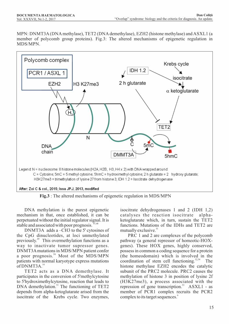
MPN: DNMT3A (DNA methylase), TET2 (DNA demethylase), EZH2 (histone methylase) and ASXL1 (a member of polycomb group proteins). Fig.3: The altered mechanisms of epigenetic regulation in MDS/MPN.
Fig.3 : The altered mechanisms of epigenetic regulation in MDS/MPN
DNA methylation is the purest epigenetic mechanism in that, once established, it can be perpetuated without the initial regulator signal. It is
47,48stable and associated with poor prognosis. DNMT3A adds a –CH3 to the 5' cytosines of the CpG dinucleotides, at loci unmethylated
49previously. This overmethylation functions as a way to inactivate tumor supressor genes. DNMT3A mutations in MDS/MPN patient confer
50a poor prognosis. Most of the MDS/MPN patients with normal karyotype express mutations
14of DNMT3A. TET2 acts as a DNA demethylase. It participates in the conversion of 5'methylcytosine to 5'hydroximethylcytosine, reaction that leads to
51DNA demethylation. The functioning of TET2 depends from alpha-ketoglutarate arised from the isocitrate of the Krebs cycle. Two enzymes,
isocitrate dehydrogenases 1 and 2 (IDH 1,2) catalyses the reaction isocitrate alpha-ketoglutarate which, in turn, sustain the TET2 functions. Mutations of the IDHs and TET2 are
51mutually exclusive. PRC 1 and 2 are complexes of the polycomb pathway (a general repressor of homeotic-HOX-genes). These HOX genes, highly conserved, possess in common a coding sequence for a protein (the homeodomain) which is involved in the
52,54coordination of stem cell functioning. The histone methylase EZH2 encodes the catalytic subunit of the PRC2 molecule. PRC2 causes the methylation of histone 3 in position of lysine 2f (H3K27me3), a process associated with the
53repression of gene transcription. ASXL1 - as member of PCR1 complex recruits the PCR2
6complex to its target sequences.
“Overlap” syndrome: biology and the criteria for diagnosis. An update.Dan ColiţăDOCUMENTA HAEMATOLOGICA
Vol. XXXVII, Nr.1-2, 2017
16
The mutations of the transcription factors genes (see table 1), have, in general, poor prognosis and the propensity to develop AML. Excepting CMML, their incidence is reduced in MDS/MPN. RUNX1 (formerly AML1, CBFA2) encodes a subunit of CBF (“core binding factor”), essential for the hematopoiesis as activator of growth,
56survival and differentiaion. CEBPA encodes a transcription factor involved in the normal differentiation of
57granulocytes. NPM1 (nucleophosmin 1) gene, encodes a
58phosphoprotein involved in proliferation. It is credited to have a kindly prognosis if it is not associated with a FLT3 mutation.
8 TP53 are uncommon in MDS/MPN , but it is
59strong associated with a poor prognosis. Cohensin is a multimeric protein, highly conserved, involved in sister cromatid separation during the mitosis (RAD21, STAG2, SMC1A,
6 0SMC3). They are mutated in ~ 10% 6
MDS/MPN cases, with an infaust prognosis.The mutations that activate the signalling pathways (table 1) are relative frequent in MDS/MPN diseases. Signalling, means a succession of events which embrace signals and stimuli acting as ligands upon specific receptors (R) that initiate the cell responses through mediators and other propagator pathways untill to the effector genes. A general mechanism of the substrate activation is the phosphorilation performed by the kinases. Some R are kinases (e.g. the tyrosine kinases) which do the phosphorilation directly. Others need to pick up the phosphate from other protein-kinases donors. In this processus is involved the JAK (Janus) kinase and STAT (Signal Transducers and Activators of Transcription) in combination with the RAS superfamily. The propagation of the signals from RAS reaches the final common pathway of MAPK (Mitogen Activated Protein Kinases) that phosphorylates the transcription
61factors and induces genes expression.
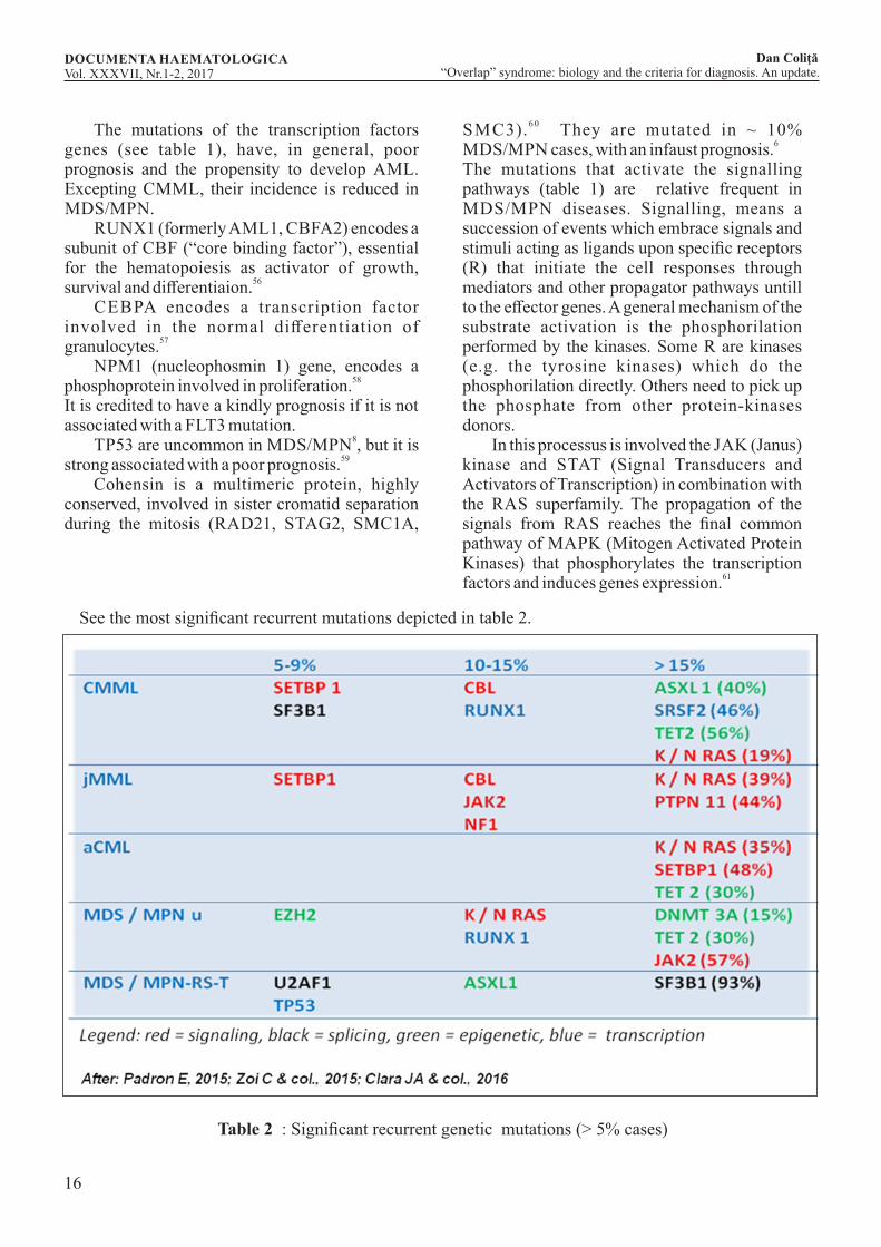
See the most significant recurrent mutations depicted in table 2.
Table 2 : Significant recurrent genetic mutations (> 5% cases)
“Overlap” syndrome: biology and the criteria for diagnosis. An update.Dan ColiţăDOCUMENTA HAEMATOLOGICA
Vol. XXXVII, Nr.1-2, 2017

17
In MDS/MPN there are some reciprocal translocations involving different tyrosine kinase fusion 62
genes. Signaling mutations produce activations of proliferative and anti-apoptotic pathways. These signals affect GF receptors, the downstream cytokine receptor signaling molecules (JAK2, N-, K-Ras) and some
7 V617Fnegative regulations of signaling pathways by CBL, NET. The JAK2 mutations are seen in CMML and in the most cases of MDS/MPN-RS-T. The RAS mutations are in CMML often associated with a MPN phenotype with monocytosis. CSF3R (the receptor of granulocyte colony-stimulating factor 3) is frequent
6,7mutated in aCML (~ 40% of cases).CMMLCMML is the most frequent (1/100 cases/year) and the most severe disease of the group (20% rate of
7, 65survivors at 3 years). The diagnosic criteria of WHO 2016 are presented joined (table 3).
(1, 25)Table 3 : The mandatory diagnostic criteria of CMML
The presence of dysplasia is mandatory for the diagnosis. If it is absent the diagnosis of CMML can be sustained if the monocytosis persists more than 3 mo, other causes of monocytosis are excluded and if exists cytogenetic or molecular clonal genetic anomalies in the hematopoietic
1cells. Also, a previous documented history of 1
M P N s excludes C M M L . Cytogenet ic abnormalities are detected in ~ 30% of cases and influence the overal survival at 5 years: 35% OS if the karyotype is normal or –Y exists; 4% OS if the karyotype anomalies are complex, or exists +8, -7/7q-; 26% OS in the presence of other
6 6abnormal i t i es . The molecular gene t ic abnormalities are not specific but very frequent (90% of cases). In the figure 2 we presented the significant recurrent genetic mutations, the order of their apparitions (TET2 ASXL1) and the fact that they are in majority driver mutations involved in epigenetic and signaling troubles. ASXL1 mutations mark a poor prognosis. RAS mutations contribute to an evolution towards a proliferative
67phenotype. CMML presents under 2 variants: the “MPN – subtype” and the “MDS – subtype”. In the first of them dominates the weight loss, the night sweats, the painful splenomegaly, leukemia cutis, pleural and peritoneal effusions and
cachexia. Patients with the MDS phenotype present cytopenias, mild bleeding, transfusion-dependent anemia and have a better prognosis
1,7,68,69compared with the proliferative variant. The percentage of blasts and promonocytes (the “blast
70equivalent cells”) in the PB and BM have a prognostic impact and divides the CMML cases in 3 groups (after the 2016 WHO revision)(ref.1): CMML-0 (<2% blasts in PB and <5% blasts in BM); CMML-1 (2 – to 4% blasts in PB and 5 – to 9% blasts in BM); CMML-2 (5 – to 19% blasts in PB and 10 – to 19% blasts in BM, and/or Auer rods) . For the correc t ident ificat ion of promonocytes from monocytes (which present occasionally bizzare nuclei and cytoplasmic granules, which rends more difficult their
71diagnosis) , the experts recommend the flow cytometry immunophenotyping and the genetic
1testing. The neoplastic monocytes express the same phenotype as the mature monocytes (CD11b, CD11c, CD14, CD33, CD45, CD64, often with bright expression, plus, sometimes, CD13, HLA-DR also). To distinguish them from the reactive monocytes some aspects as the lack of CD13 and HLA-DR, the aberrant expression of CD16, CD23, CD56, CD117, the presence of abnormal
“Overlap” syndrome: biology and the criteria for diagnosis. An update.Dan ColiţăDOCUMENTA HAEMATOLOGICA
Vol. XXXVII, Nr.1-2, 2017
18
granulocytes and an increased number of blasts can 72
be useful. Recently a more simple method was recommendead since an International Committee admitted the existence of 3 subpopulations of monocytes in the PB, based on the expression of CD14 and CD16 markers: CD14+, CD16- cells,
lowCD14+16+ (the “intermediate” cells) and CD14 CD16+ (the “nonclassical” monocytes). In CMML was described a characteristic increase of CD14+ CD16- monocytes (> 94%) at the expense of the other fractions, that accurately distinguish
73,74CMML from the other confounding situations.
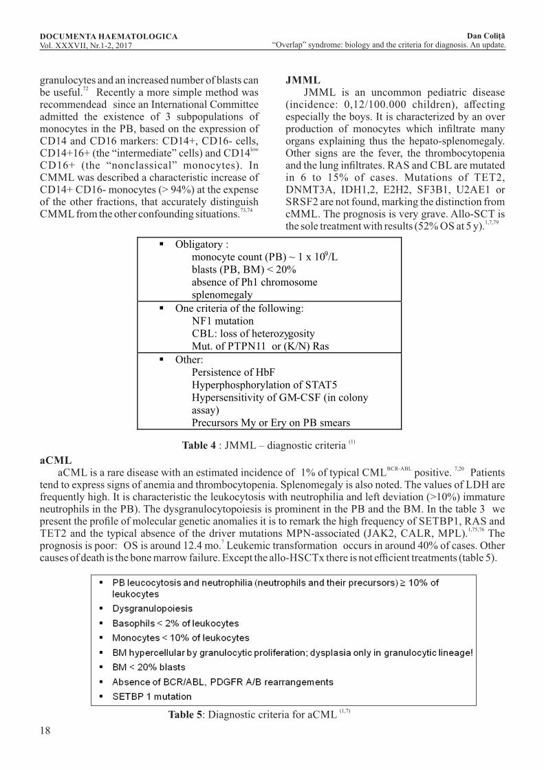
JMML JMML is an uncommon pediatric disease (incidence: 0,12/100.000 children), affecting especially the boys. It is characterized by an over production of monocytes which infiltrate many organs explaining thus the hepato-splenomegaly. Other signs are the fever, the thrombocytopenia and the lung infiltrates. RAS and CBL are mutated in 6 to 15% of cases. Mutations of TET2, DNMT3A, IDH1,2, E2H2, SF3B1, U2AE1 or SRSF2 are not found, marking the distinction from cMML. The prognosis is very grave. Allo-SCT is
1,7,79the sole treatment with results (52% OS at 5 y).
§ Obligatory : monocyte count (PB) ~ 1 x 10⁹/L blasts (PB, BM) < 20% absence of Ph1 chromosome splenomegaly
§ One criteria of the following: NF1 mutation CBL: loss of heterozygosity Mut. of PTPN11 or (K/N) Ras
§ Other: Persistence of HbF Hyperphosphorylation of STAT5 Hypersensitivity of GM-CSF (in colony assay) Precursors My or Ery on PB smears
(1)
Table 4 : JMML – diagnostic criteria
aCMLBCR-ABL 7,20
aCML is a rare disease with an estimated incidence of 1% of typical CML positive. Patients tend to express signs of anemia and thrombocytopenia. Splenomegaly is also noted. The values of LDH are frequently high. It is characteristic the leukocytosis with neutrophilia and left deviation (>10%) immature neutrophils in the PB). The dysgranulocytopoiesis is prominent in the PB and the BM. In the table 3 we present the profile of molecular genetic anomalies it is to remark the high frequency of SETBP1, RAS and
1,75,76TET2 and the typical absence of the driver mutations MPN-associated (JAK2, CALR, MPL). The
7prognosis is poor: OS is around 12.4 mo. Leukemic transformation occurs in around 40% of cases. Other causes of death is the bone marrow failure. Except the allo-HSCTx there is not efficient treatments (table 5).
(1,7)Table 5: Diagnostic criteria for aCML
“Overlap” syndrome: biology and the criteria for diagnosis. An update.Dan ColiţăDOCUMENTA HAEMATOLOGICA
Vol. XXXVII, Nr.1-2, 2017
19
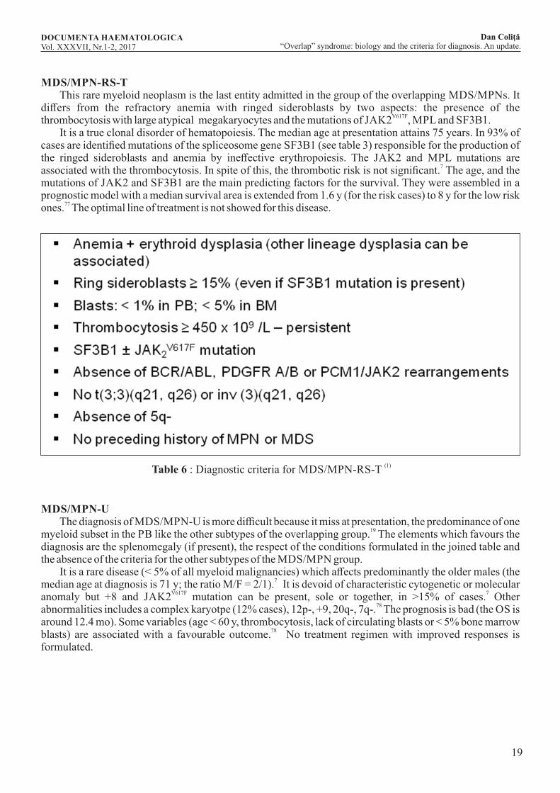
MDS/MPN-RS-T This rare myeloid neoplasm is the last entity admitted in the group of the overlapping MDS/MPNs. It differs from the refractory anemia with ringed sideroblasts by two aspects: the presence of the
V617Fthrombocytosis with large atypical megakaryocytes and the mutations of JAK2 , MPL and SF3B1. It is a true clonal disorder of hematopoiesis. The median age at presentation attains 75 years. In 93% of cases are identified mutations of the spliceosome gene SF3B1 (see table 3) responsible for the production of the ringed sideroblasts and anemia by ineffective erythropoiesis. The JAK2 and MPL mutations are
7associated with the thrombocytosis. In spite of this, the thrombotic risk is not significant. The age, and the mutations of JAK2 and SF3B1 are the main predicting factors for the survival. They were assembled in a prognostic model with a median survival area is extended from 1.6 y (for the risk cases) to 8 y for the low risk
77ones. The optimal line of treatment is not showed for this disease.
(1)Table 6 : Diagnostic criteria for MDS/MPN-RS-T
MDS/MPN-U The diagnosis of MDS/MPN-U is more difficult because it miss at presentation, the predominance of one
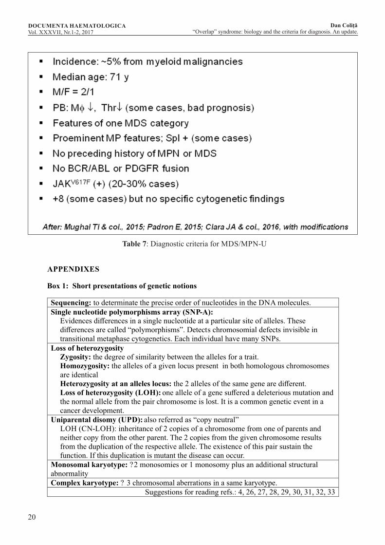
19myeloid subset in the PB like the other subtypes of the overlapping group. The elements which favours the diagnosis are the splenomegaly (if present), the respect of the conditions formulated in the joined table and the absence of the criteria for the other subtypes of the MDS/MPN group. It is a rare disease (< 5% of all myeloid malignancies) which affects predominantly the older males (the
7median age at diagnosis is 71 y; the ratio M/F = 2/1). It is devoid of characteristic cytogenetic or molecular
V617F 7anomaly but +8 and JAK2 mutation can be present, sole or together, in >15% of cases. Other 78
abnormalities includes a complex karyotpe (12% cases), 12p-, +9, 20q-, 7q-. The prognosis is bad (the OS is around 12.4 mo). Some variables (age < 60 y, thrombocytosis, lack of circulating blasts or < 5% bone marrow
78blasts) are associated with a favourable outcome. No treatment regimen with improved responses is formulated.
“Overlap” syndrome: biology and the criteria for diagnosis. An update.Dan ColiţăDOCUMENTA HAEMATOLOGICA
Vol. XXXVII, Nr.1-2, 2017
20
Table 7: Diagnostic criteria for MDS/MPN-U
APPENDIXES
Box 1: Short presentations of genetic notions
Sequencing: to determinate the precise order of nucleotides in the DNA molecules. Single nucleotide polymorphisms array (SNP-A):
Evidences differences in a single nucleotide at a particular site of alleles. These differences are called “polymorphisms”. Detects chromosomial defects invisible in transitional metaphase cytogenetics. Each individual have many SNPs.
Loss of heterozygosity Zygosity: the degree of similarity between the alleles for a trait. Homozygosity: the alleles of a given locus present in both homologous chromosomes are identical Heterozygosity at an alleles locus: the 2 alleles of the same gene are different. Loss of heterozygosity (LOH): one allele of a gene suffered a deleterious mutation and the normal allele from the pair chromosome is lost. It is a common genetic event in a cancer development.
Uniparental disomy (UPD): also referred as “copy neutral” LOH (CN-LOH): inheritance of 2 copies of a chromosome from one of parents and neither copy from the other parent. The 2 copies from the given chromosome results from the duplication of the respective allele. The existence of this pair sustain the function. If this duplication is mutant the disease can occur.
Monosomal karyotype: ?2 monosomies or 1 monosomy plus an additional structural abnormality Complex karyotype: ? 3 chromosomal aberrations in a same karyotype.
Suggestions for reading refs.: 4, 26, 27, 28, 29, 30, 31, 32, 33
“Overlap” syndrome: biology and the criteria for diagnosis. An update.Dan ColiţăDOCUMENTA HAEMATOLOGICA
Vol. XXXVII, Nr.1-2, 2017

21
Box 2 : MDS/MPN : functional classes of the genes
Transcription-factors genes:
The deciphering the genetic code (groups of 3 nucleotides = a message) and translate ian it in corresponding amino acids chains begins with the transcription of the genic DNA into the messenger RNA (mRNA). mRNA expulses its introns (the splicing). So, it becomes able to migrate out of the nucleus, and reaches the cytoplasmic ribosomes, where is accomplishes the protein synthesis with the support of other 2 species of RNA: rRNA (the ribosomal RNA – the manager of the lecture of the message) and tRNA (the transfer RNA), which achieve the translation of the message in a chain of amino acids). This whole process is assured by specific functional genes.
The splicing:
To becomes functional, the primitive mRNA must get rid of its in trons and fusions the exons head to head (the splicing). This process is mediated in specific proteic structures (the spliceosomes). After the splicing can begins the translation. The genes of splicing factors are the most frequently mutated in MDS and MDS /MDP. These mutations are mutually exclusive. Some mutations are very frequent associated with definite diseases (SRSF2 with CMML; SFR3B1 with MDS/MPN -RS, for example).
The epigenetic regulators:
Epigenetics refers to changes in the genes expressions but without changes in their structures. The epigenetic changes are stable and heritable and operate mainly by histone modifications and DNA methylation. Histones are the main proteic component of the chromatin. The histone proteins form together with DNA the nucleosomes, by wrapping around them, the DNA chains. Two genes (E2H2 and ASXL1), implied in the histone modifications are frequent mutated in MDS/MPN. The first encodes the catalytic subunit of a repressor of the homeotic genes (the policomb complex) which participate at the modulation of the chromatin structure. DNA methylation acts on cytosine grouped in CG doublets (cytosine/guanine) in specific areas, where regulates some histone modifications (e.g.: DNMT3A or DNA methyltransferase) which add a methyl
to CG dinucleotides and have a poor prognostic.
Signaling mutations
cause aberrant activations of proliferation and antiapoptotic pathways governed by the growth factors.
Some mutations were described also at tyrosine-kynases (TK) gene-fusions. In MDS/MPN only the fusions which are not the result of BCR/ABL translocation have diagnostic value. Other mutations involve growth factors receptors and the downstream signaling pathways starting from them (e.g.: JAK, RAS). Signaling mutations are frequent in MDS/MPN.
Suggestions for reading refs.: 6, 7, 8, 15, 34, 35, 36, 37, 38, 39
“Overlap” syndrome: biology and the criteria for diagnosis. An update.Dan ColiţăDOCUMENTA HAEMATOLOGICA
Vol. XXXVII, Nr.1-2, 2017
22
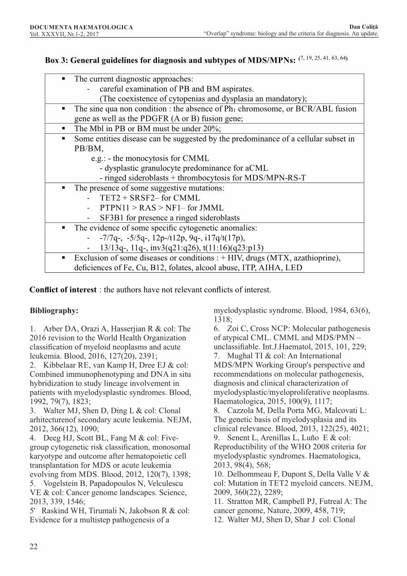
Box 3: General guidelines for diagnosis and subtypes of MDS/MPNs: (7, 19, 25, 41, 63, 64)
§ The current diagnostic approaches:
- careful examination of PB and BM aspirates.
(The coexistence of cytopenias and dysplasia an mandatory);
§ The sine qua non condition : the absence of Ph1 chromosome, or BCR/ABL fusion gene as well as the PDGFR (A or B) fusion gene;
§ The Mbl in PB or BM must be under 20%;
§ Some entities disease can be suggested by the predominance of a cellular subset in PB/BM,
e.g.: - the monocytosis for CMML
- dysplastic granulocyte predominance for aCML
- ringed sideroblasts + thrombocytosis for MDS/MPN-RS-T
§
The presence of some suggestive mutations:
-
TET2 + SRSF2 –
for CMML
-
PTPN11 > RAS > NF1 –
for JMML
-
SF3B1 for presence a ringed sideroblasts
§
The evidence of some specific cytogenetic anomalies:
-
-7/7q-, -5/5q-, 12p-/t12p, 9q-, i17q/t(17p),
-
13/13q-, 11q-, inv3(q21:q26), t(11:16)(q23:p13)
§
Exclusion of some diseases or conditions : + HIV, drugs (MTX, azathioprine), deficiences of Fe, Cu, B12, folates, alcool abuse, ITP, AIHA, LED
Conflict of interest
: the authors have not relevant conflicts of interest.
Bibliography:
1. Arber DA, Orazi A, Hasserjian R & col: The 2016 revision to the World Health Organization classification of myeloid neoplasms and acute leukemia. Blood, 2016, 127(20), 2391;2. Kibbelaar RE, van Kamp H, Dree EJ & col: Combined immunophenotyping and DNA in situ hybridization to study lineage involvement in patients with myelodysplastic syndromes. Blood, 1992, 79(7), 1823;3. Walter MJ, Shen D, Ding L & col: Clonal arhitecturenof secondary acute leukemia. NEJM, 2012, 366(12), 1090;4. Deeg HJ, Scott BL, Fang M & col: Five-group cytogenetic risk classification, monosomal karyotype and outcome after hematopoietic cell transplantation for MDS or acute leukemia evolving from MDS. Blood, 2012, 120(7), 1398;5. Vogelstein B, Papadopoulos N, Velculescu VE & col: Cancer genome landscapes. Science, 2013, 339, 1546;5' Raskind WH, Tirumali N, Jakobson R & col: Evidence for a multistep pathogenesis of a
myelodysplastic syndrome. Blood, 1984, 63(6), 1318;6. Zoi C, Cross NCP: Molecular pathogenesis of atypical CML. CMML and MDS/PMN – unclassifiable. Int.J.Haematol, 2015, 101, 229;7. Mughal TI & col: An International MDS/MPN Working Group's perspective and recommendations on molecular pathogenesis, diagnosis and clinical characterization of myelodysplastic/myeloproliferative neoplasms. Haematologica, 2015, 100(9), 1117;8. Cazzola M, Della Porta MG, Malcovati L: The genetic basis of myelodysplasia and its clinical relevance. Blood, 2013, 122(25), 4021;9. Senent L, Arenillas L, Luño E & col: Reproductibility of the WHO 2008 criteria for myelodysplastic syndromes. Haematologica, 2013, 98(4), 568;10. Delhommeau F, Dupont S, Della Valle V & col: Mutation in TET2 myeloid cancers. NEJM, 2009, 360(22), 2289;11. Stratton MR, Campbell PJ, Futreal A: The cancer genome, Nature, 2009, 458, 719;12. Walter MJ, Shen D, Shar J col: Clonal
“Overlap” syndrome: biology and the criteria for diagnosis. An update.Dan ColiţăDOCUMENTA HAEMATOLOGICA
Vol. XXXVII, Nr.1-2, 2017

23
diversity of recurrently mutated genes in myelodysplastic syndromes. Leukemia, 2013, 27(6), 1275;13. Welch JS, Ley TJ, Link DC & col: The origin and evolution of mutations in acute leukemia. Cell, 2012, 150(2), 264;14. Papaemmanuil E, Gerstung M, Malcovati L & col: Clinical and biological implications of driver mutations in myelodysplastic syndromes. Bloos, 2013, 122(22), 3616;15. Murati A, Brecqueville M, Devillier R & col: Myeloid malignancies: mutations, models and management. BMC Cancer, 2012, 12, 304;16. Cazzola M – Prognostic biomarkers in myelodysplastic syndromes. EHA, Edu.Book, 2014, 8(1), 237;17. Malcovati L, Papaemmanuil E, Ambaglio M & col: Driver somatic mutations identify distinct disease entities within myeloid neoplasms with myelodysplasia. Blood, 2014, 124(9), 1513;18. Vardiman JW, Thiele J, Arber DA & col: The 2008 revision of the WHO classification of myeloid neoplasms ans acute leukemia. Rationale and important changes. Blood, 2009, 114(5), 937;19. Padron E : Surveing the landscape of MDS/MPN research: overlap among the overlap syndromes. Hematology, 2015 (Amer.Soc.Hem.Edu.Book) p349;20. Clara JA & col.: Clinical management of myelodysplastic syndrome/myeloproliferative neoplasms overlap syndrome. Cancer Biol Med, 2016.doi:10.20892/j.issn.2095-3941.2016.0043;21. Bain JB & col: Bone marrow pathology. Blackwell Sci.Ltd.UK, 1996, 108;22. Ursea C : Tratatul de Medicina Interna (Hematologie partea a II-a) capit.68.2, p1040, Editura Medicala, Bucuresti;23. Steensma DP, Bejar R, Jaiswal S & col: Clonal hematopoiesis of indeterminate potential and its distinction from myelodysplastic syndromes. Blood, 2015, 126(1), 9;24. Kwok B, Hall JM, Witte JS & col: MDS – associated somatic mutations and clonal hematopoiesis are common in idiopathic cytopenias of undeterminated significance. Blood, 2016, 126(21), 2355;25. Cazzola M, Malcovati L, Invermizzi R: Myelodysplastic/myeloproliferative neoplasms. ASH Edu.book.Hemat. 2011, p264;26. Orosco JJ, Appelbaum FR: Unfavourable complex and monosomale karyotypes, the most challenging forms of acute myeloid leukemia.
Oncology J.Hemato. Malignancies, Leukemia & Lymphoma, Myelodysplastic syndromes, 2013, august 03;27. O'Keefe C, Mc Devitt MA, Maciejewski P: Copy neutral loss of heterozygosity: a novel chromosomial lesion in malignancies. Blood, 2010, 115(14), 2731;28. Pettersson E, Lundeberg J, Ahmadian A: Generations of sequencing technologies. Genomics, 2009, 93(2), 105;29. Maciejewski P, Tiu RV, O'Keefe C: Application of array-band whole genomic scanning technologie as a cytogenetic tool in haematological malignancies. BJ Haematol, 2009, 146(5), 479;30. Sheils S, Finn S, O'Leary J: Nucleic acid microarrays. An overview. Cur.Diagnostic Pathol.,2003, 9(3), 155;31. O'Malley DP, Orazi A, Dunphy CH & col: Loss of heterozygosity identifies genetic changes in chronic myeloid disorders, myelodysplastic syndromes and chronic myelomonocytic leukemia. Modern Pathology, 2007, 20, 1166;32. Gondek LP, Tiu R, O'Keefe C & col: Chromosomial lesions and uniparental disomy detected by SNP arrays in MDS, MDS/MPD and MDS – derived AML. Blood, 2008, 112(3), 1534;33. O'Keefe C, Mc Devitt MA, Maciejewski P: Copy neutral loss of heterozygosity: a novel chromosomial lesion in malignancies. Blood, 2010, 115(14), 2731;34. Colita D : Medicina Interna – vol.Hematologie I, p492, Editura Medicala, Bucuresti, 1997;35. Passarge E : Color atlas of genetics, 3rd edition. Thieme (Stuttgart, N.York), 2007;36. Esteller M : Epigenetics in cancer. NEJM, 2008, 358(11), 148;37. Rodriguez-Paredes M, Esteller M : Cancer epigenetics reaches mainstream oncology. Nature Medicine, 2011, 17(3), 30;38. Makishima H, Visconte V, Sakaguchi H & col: Mutations in spliceosome machineray, a novel and ubiquitous pathway in leukemogenesis. Blood, 2012, 119(14), 3203;39. Alberts B & col: Molecular biology of the cell. Garland Publ.Inc.,NY, 1983, p388 and next;40. Acquaviva C, Gelsi-Boyer V, Birnbaum D: myelodysplastic syndromes: lost of two states? Leukemia, 2010,24, 105;41. Papaemmanuil E, Cazzola M, Bonttwood J &
“Overlap” syndrome: biology and the criteria for diagnosis. An update.Dan ColiţăDOCUMENTA HAEMATOLOGICA
Vol. XXXVII, Nr.1-2, 2017

24
col: Somatic SF3B1 mutations in myelodysplasia with ring sideroblasts. N.Engl.J.Med., 2011, 365(15), 1384;42. Malcovati L, Papaemmanuil E, Bowen D & col: Clinical significance of SF3B1 mutations in myelodysplastic syndromes and myelodysplastic/myeloproliferative neoplasms. Blood, 2011, 118, 6239;43. Patmaik MM, Lasho T, Fink CM & col: Spliceosome mutations involving SRSF2, SF3B1 and U3AF35 in chronic myelomonocytic leukemia. Prevalence clinical correlates and prognostic relevance. Am.J.Hematol., 2013, 88(3), 201;44. Visconte V, Rogers HJ, Singh J & col: SF3B1 haploinsuficiency leads to formation of ring sideroblasts in myelodysplastic syndromes. Blood, 2012, 120, 3173;45. Yoshida K, Sanada M, Shiraishi Y & col: Frequent pathway mutations of splicing machinery in myelodysplasia. Nature, 2011, 478, 64;46. Alexander R, Beggs JD : Cross-talk in transcription, splicing and chromatin: who makes the first call? Biochem.Soc.Trans., 2010, 38, 1251;47. Bird A : DNA methylation patterns and epigenetic memory. Genes Dev., 2002, 16(1), 6;48. Issa JPN: The myelodysplastic syndrome as a prototypical epigenetic disease. Blood, 2013, 121(9), 3811;49. Krönke J, Ebert BL: Biology of myelodysplastic syndrome. EHA Edu.book 2014, 8(1), 227;50. Walter MJ, Ding L, Shen D & col: Recurrent DNMT3A mutations in patients with myelodysplastic syndomes. Leukemia, 2011, 25(7), 1153;51. Figueroa ME, Abdel_Wahab O, Lu C & col: Leukemic IDH1 and IDH2 mutations result in a hypermethylation phenotype, disrupt TET2 function and impair hematopoietic differentiation. Cancer Cell, 2010, 18, 553;52. Magli MC & col: Coordinate regulation of HOX genes in human hematopoietic cells. Proc.Nath.Acad.Sci. USA, 1991, 88, 6348;53. Morey L, Helin K : Polycomb group protein – mediated repression of transcription. Trends Biochem.Sci, 2010, 35(6), 323;54. Grimaud Ch: From genetics to epigenetics. The tale of Polycomb group and trithorax group genes. Chromosome Research, 2006, 14(4(, 363;
55. Speck NA, Gilliland DG: Core- binding factor in haematopoiesis and leukemia. Nat Rev Cancer, 2002, 2, 502;56. Blyth K, Cameron ER, Neil JC: The RUNX genes: gain or loss of function in cancer. Nat Rev Cancer, 2005, 5, 376;57. Koschmieder S, Halmos B, Levantini E & col: Dysregulation of the C/EBP alpha differentiation pathway in human cancer. J Clin Oncol, 2009, 27, 619;58. Grisendi S, Mecucci C, Falini B & col: Nucleophosmin and cancer. Nat Rev Cancer, 2006, 6, 493;59. Bejar R, Stevenson K, Abdel-Wahab O & col: Clinical effect of point mutations in myelodysplastic syndromes. NEJM, 20111, 364(26), 2496;60. Nasmyth K, Haering CH; Cohesin – its roles and mechanisms. Annu Rev Genet, 2009, 43, 525;61. Israels LG, Israels ED & col: Muchanisms in Hematology: cap 1, Cell cycle, p1. Gnosis Inc., Winnipeg Manitoba, Canada, 2002;62. Delhommeau F, Pisani DF, James C & col: Oncogenic mechanisms in myeloproliferative disorders. Cell Mol Life Sci, 2006, 63(24), 2939;63. Muramatsu H, Makishima H, Maciejewski J: CMML and aCML: novel pathogenetic lesions. Semin Oncol, 2011, 39(1), 67;64. Tatic A, Colita DN, Coriu D: Actualitati in diagnosticul si tratamentul sindroamelor mielodisplazice. Edit Univ C.Davila, Bucuresti, 2015, p56;65. Rollison DE, Howlander N, Smith MT & col: Epidemiology of myelodysplastic syndromes and chronic myeloproliferative disorders in the United States 2001-2004, using data from the NAACCR and SEER programs. Blood, 2008, 112, 45.66. Such E, Cervera J, Costa D & col: Cytogenetic stratification in chronic myelomonocytic leukemia. Haematologica, 2011, 96(3), 375;67. Ricci C, Fermo E, Corti S & col: RAS mutations contribute to evolution of chronic myelomonocytic leukemia to the proliferative variant. Clin Cancer Res, 2010, 16(8), 2246;68. Parikh S, Tefferi A: Chronic myelomonocytic leukemia: 2012 update on diagnosis, rick stratification and management. Amer J Hematol, 2012, 87, 611;69. M.Cîrstea, A.Colita, B.Ionescu & col:
“Overlap” syndrome: biology and the criteria for diagnosis. An update.Dan ColiţăDOCUMENTA HAEMATOLOGICA
Vol. XXXVII, Nr.1-2, 2017

25
Chronic myelomonocytic leukemia “myelodysplastic type” in transformation to acute myeloid leukemia diagnostic and therapeutic options: case report and literature review. Rumanian J Lab Med, 2016, 24(3), 263;70. Schuler E, Schroeder M, Neukirchen J & col: Refined medullary blast and white blood cell count based classification of chronic myelomonocytic leukemias. Leuk Res 2014, 38(2), 1413;71. Itzykson R Saloray E: An evolutionary perspectiveon chronic myelomonocytic leukemias. Leukemia 2013, 27(7), 1441;72. Gorczyka W : Flow cytometry in neoplastic hematologic, Taylor & Francis (Abingdon, UK 2006, p227);73. Ziegler-Heitbrook L, Ancuta P, Crowe S & col: Nomenclature of monocytes and dendritic cells im blood. Blood, 2010, 116(16), 74;74. Selimoglu-Buet D, Wagner-Ballon O, Saada V & col: Characteristic repartition of monocytes subsets as a diagnostic signature of chronic myelomonocytic leukemia. Blood, 2015, 125(23), 3618;75. Feud F, Horn T, Koch I & col: Atypical chronic myeloid leukemia as defined in the WHO classification is a JAK2V617F negative neoplasm. Leuk Res. 2008, 31(12), 1931;76. Piazza R, Valleta S, Winkelmann N & col: Recurrent SETBP1 mutations in atypical chronic myeloid leukemia. Nat Genet, 2013, 45(1), 18;77. Broséus J, Alpermann T, Wulfert S & col: Age, JAK2(V617F) and SF3B1 mutations are the main predicting factors for survival in refractory anaemia with ring sideroblasts and marked thrombocytosis. Leukemia, 2013, 27, 1826;78. Di Mardo CD, Daver M, Jain N & col: Myelodysplastic/Myeloproliferative Neoplasms, unclassifiable (MDS/MPN, U): Natural history and clinical outcome by treatment strategy. Leukemia, 2014 28(4), 958;79. Abu Kar S, Jankowska A, Makishima H & col: Spliceosomal gene mutations are frequent in the diverse mutational spectrum of chronic myelomonocytic leukemia but largely absent in juvenile myelomonocytic leukemia. Haematologica, 2013, 98, 107;
“Overlap” syndrome: biology and the criteria for diagnosis. An update.Dan ColiţăDOCUMENTA HAEMATOLOGICA
Vol. XXXVII, Nr.1-2, 2017

26

27
DOI: 10.1515/dch-2017-0007
The need for transfusion and the risk assessment benefits
1Georgeta Hanganu1 Ploiesti Blood Center, Ploiesti, Romania
Summary
Transfusion therapy is often essential and life-saving therapy for many patients. However, the risks associated with transfusion should not be neglected, which should be evaluated when deciding on a blood product. Risk assessment versus patient benefit, must be the permanent preoccupation of the prescribing physician. Availability in blood products, that covers all blood groups in the ABO and Rh systems, should be a continuing concern of Transfusion Units and Transfusion Centers to avoid unfortunate and irreversible situations for the patient as in certain cases of immunization post-transfusion, as in the following case, extremely exciting. At the X hospital's surgery section, in March 2009, arrives the patient P.E. 28-year-old, with altered general condition, vaginal bleeding, accentuated pain, and abdominal pain. There is a diagnosis of acute abdomen, with suspicion of extra uterine pregnancy. Because there is no gynecology department at X hospital, at the request of the family, the patient is transferred to an others hospital in a larger city where there is a gynecology department. In rescue symptomatology is getting worse. Arrived at Y. hospital, the patient is consulted by the gynecologist who confirms the suspicion of ectopic pregnancy and decides the surgery. Because the intervention was timed and hemorrhage was important and very high intra-operative bleeding, the patient's hemoglobin reached 6.0 g / dl. The patient's blood panel shows Leucocytes 25,000 / mmc, Platelet 576000 / mmc, Hemoglobin 6.0 g / dl, VSH 100 mm / 1h. The doctor first recommends transfusion therapy with 4 units of Concentrated Erythrocyte izo-group, izo-Rh.
Corresponding author: Georgeta Hanganu, Ploiesti Blood Center, Vestului Street, No. 24 A, Ploiesti, Romania, phone 034/802968, e-mail: [email protected]
The pre-transfusion samples determine the blood group of the patient: O, Rh D negative. Because of the degree of urgency and the impossibility of transfusion timing, the patient receives during surgery one red blood cell concentrate negative (the only one in Hospital Transfusion Units) and then 3 units of positive erythrocyte concentrate. It is operated, bleeding is stopped. The next day he gets another 2 red blood cell concentrate positive units. It is surgically cured, externally over 7 days. After 7 years on a blood donation campaign, P.E. presents itself as a potential donor, and in the questionnaire and in the interview with the doctor, regarding the physiological and pathological antecedents, it signals the existence of past transfusions. In this donation action, it is temporarily rejected for hemoglobin 11.5 g / dl at the time of presentation, but taking into account the antecedents and the transfusion history, blood samples are collected, only to investigate the hypothesis of post-transfusion immunization. An Irregular Antibody Research with Bio Vue
Equipment is performed, with a panel of 3 ficinated red cells, the result is positive. Detection of antibodies with Resolve panel with 11 erythrocytes and identification of anti-D antibody is made. Titration of anti-D antibodies is performed by performing binary dilutions: ½, ¼, 1/8, 1/16, 1/32, 1/64, 1/128, 1/256, 1/512, 1/1024, 1/2048, 1/4096, 1/8192, 1/16384, 1/32786, 1/65536, up to dilution 1/131072. Undoubtedly, blood transfusion, especially in vital emergency situations, saves the patient, but in the context of minimal ABO and factor D compatibility, the immunological impact is dramatic. Therefore transfusion prescription physicians must always balance the risks associated with transfusion versus patient's clear benefit, and the Hospital Transfusion Unit should have available enough stock of negative Rh erythrocyte in stock. The question always arises: the need for transfusion is always a reality? How can it be minimized? Is an anemia interpreted in medical context, is diagnosis and
28
early treatment of anemia? Does anemia correlate by iron, folic acid and vitamin B12? Are all possible steps taken to avoid transfusion? Unfortunately, transfusion is often the safest and quickest therapeutic solution. Unfortunately, transfusion is also the cheapest solution because blood components are delivered free of charge to all public hospitals. But everything that is free is devalued, even if the value of blood products is very high! Sometimes blood products get to replace other medicines that load hospital bills! Blood products are replacing a patient's psychological counseling; sometimes a blood product is administered to the patient to gain color, increase morale! They have a mental tonic effect! Often a single product is transfused. Most of the times, transfusion of that single product means that the patient can recover through the forces of his own body, the benefit brought by him.We have to remember. Transfusion therapy is a therapy with a biological product, and should be treated as such.
Any biological product can bring: -HIV infection, HCV, HBV, HGV, HTLV, CMV, Lues, Lyme disease, West Nile Encephalitis, Epstein Barr Virus, Parvovirus B19, Chagas Disease (tripanosomiasis), and any other bacteria or virus . - Immunological rhini t is : erythrocyte immunization, leukocyte immunization, platelet immunization, immune-modulatory effect. -a side effect of a side effect: pyrogenic reactions, non-hemolytic febrile reactions, allergic reactions, anaphylactic reactions, acute hemolytic reaction by inborn blood transfusion in ABO, Rh, or other erythrocyte systems; altered blood transfusion, massive transfusion syndrome; citrate intoxication and hyperkalemia; post-transfusion hemosiderosis; post-transfusion thrombo-cytopenic purpura; post-transfusion "graft versus host" disease. Therefore, when indicating transfusion treatment, the prescribing physician must weigh very well, the risk of transfusion versus the undeniable benefit.
The need for transfusion and the risk assessment benefits
Georgeta HanganuDOCUMENTA HAEMATOLOGICAVol. XXXVII, Nr.1-2, 2017

29
30


