The interaction of Escherichia coli integration host factor with the cohesive end sites of phages Xand 21
Winnie Xin and Michael Feiss
Department of Microbiology, College of Medicine, and Interdepartmental Genetics Ph.D. Program,University of Iowa, Iowa City, IA 52242, USA
Received November 30, 1987; Revised and Accepted January 25, 1988
ABSTRACTThe interaction of E. coli integration host factor (IHF) with the
cohesive end sites (cos's) of phages X and 21 has been studied by theDNAase I footprinting technique. Six potential sites in cos A differ fromthe consensus IHF binding sequence by 1 to 3 base pairs. Of the six, onesite, I1, binds IHF strongly. The I1 segment protected by IHF contains twosequences that closely match the IHF consensus binding sequence. Another site,I2, binds IHF moderately well, and three sites: IO', 13 and I4 bind IHF veryweakly. The IO site does not bind IHF under the conditions used here. Inphage 21 the DNA segment extending to the right from the cohesive ends, whichcontains three potential IHF binding sites, was examined. Two sites bindIHF well; I1, the 21 analogue of one of the X I1 sites, and IO, a site notanalogous to a X site. The third 21 site, I2, binds IHF moderately well, asdoes the analogous I2 site in X. The significance of the results for A DNApackaging is discussed.
INTRODUCTION
A virus depends on its host for many processes, and some cases involve
specific interactions between virus and host elements. An example of a
specific interaction is the interaction of phage X DNA with E. coli integra-
tion host factor (IHF). IHF is a small, heterodimeric, site-specific, DNA
binding protein. The alpha subunit (MW=11,224) is encoded by the himA gene
(1), and the beta subunit (MW=10,581) is the product of the hip gene (2).IHF is required for site-specific integration of A DNA into the host chromo-
some. IHF binds specifically to several sites within A attP (3). More
recent studies indicate IHF acts at attP to assist in the formation of a
nucleosome-like structure involving IHF, A integrase, and attP (4, 5).IHF also acts in the formation of a multiprotein complex at the origin
of replication of pSC101 (6). In addition to acting in the formation of
complex DNA:protein structures, IHF functions in gene regulation. For
example, IHF binds to a site at the beginning of the A cII gene, and enhances
clI exptession at a poettranscriptional level, perhaps by influencing mRNA
dissociation from the DNA (7). Study of a number of IHF binding sites yielded
the following consensus sequence: 5'-PyNPyAANNNNTTGAT(A/T)-3' (8).IHF has been implicated in the DNA packaging of the evolutionarily-
related phages X and 21. The enzyme terminase plays a central role in DNA
packaging in the lambdoid phages (9). The A terminase is a heterooligomer
of two subunits: the small subunit gpNul and the large subunit gpA, the gene
products of the Nul and A genes, respectively. Terminase is a multifunctional
protein that binds A DNA at cos, introduces the staggered nicks to generate
the cohesive ends, and binds the prohead to generate the DNA:terminase:prohead
complex that leads to packaging of the DNA (10). The cohesive end site, cos,
is bipartite, consisting of cosN, the nicking site, and cosB, the site where
terminase initially binds A DNA. The structure of cos is shown in Fig. 1;
the Nul and A genes are just to the right of cos.
Phage 21 produces a terminase that shares about 60% sequence identity
with A terminase (11); the genes 1 and 2 produce the small subunit, gpl, and
the large subunit, gp2, respectively. IHF is essential for an early,
terminase-dependent step in 21 DNA packaging (12). The IHF requirement can
be bypassed, because 21 her mutants can be found that are able to grow in
cells lacking IHF. The her mutations affect the amino-terminal part of gpl,
the part of gpl that is known from other studies to be involved in DNA binding.
These results suggest IHF helps 21 terminase to bind at cos.
IHF plays an accessory role in A DNA packaging. Bear et al. (13)described a mutant, A cosl54, that is dependent on IHF for DNA packaging.
The mutation occurs in a sequence (called R) that is repeated several times
in cos and which was postulated to be the terminase binding site. Because
A cosl54 is IHF-dependent, Bear et al., suggested that A terminase normally
is able to bind to cos well enough for growth, but that when one binding site
is inactive terminase requires IHF to form a functional complex with cos.
With this model in mind, Bear et al. searched cos for the IHF consensus
binding sequence, and found several (Fig. 1). We have identified two
additional sites, I0 and I0' (Fig. 1). In 21, a similar search (11) found
several potential IHF binding sequences (Fig. 2).
We have done DNA footprinting experiments to examine these putative
binding sites as a first step in understanding the role of IHF in viral
DNA packaging.
2016
Nucleic Acids Research
Figure 1: Schematic diagram of presumptive terminase binding sites and IHFbinding sites (I) at X cos. The center structure represents the staggered nicksthat are introduced by terminase at A cosN. Each R site contains a 16 bpsequence postulated to be the terminase binding site. The I sites resemblethe IHF binding consensus sequence. coslS4 is a single base transition muta-tion in one of the R sites. The IO site partially overlaps the leftmost Xgene, Nul.
MATERIALS AND METHODS
Plasmids
Plasmid pLW11O has a A DNA segment extending from the BclI site at
47942 through cosN to an EcoRI site at 194 cloned into pBR322 digested at
the BamHI and EcoRI sites (14). Base pairs of A and 21 are numbered as in
Daniels et al. (15); the base at the 5' end of the left cohesive end is
designated 1 and numbering proceeds rightward. The 5' to 3' strand extending
to the right from position 1 is called the top strand, and the 3' to 5' strand
is the bottom strand. Plasmid pSFl has a A segment extending from the HindIII
site at 44141 through cosN to the BamHI site at 5505 cloned into pBR322 (16).Plasmid pBA2 is a derivative of pACYC184 (17) containing the cos/terminase
segment from X-21 hybrid 51 (18). X-21 hybrid 51 is a recombinant from a A
vs. 21 cross, such that the segment from position 1, at the left cohesive end,
to 490, a site within the 1 gene, is derived from 21 and the remainder of the
chromosome is derived from A. pBA2 was constructed by ligating the X-21 hybrid51 segment extending from the HindIII site at 44141 through cos to the BamHI
Figure 2: Schematic diagram of the phage 21 cos segment to the right of 21cosN. The left cohesive end is shown at the left of the diagram. 'The I sitesare the possible IHF binding sites at phage 21 cos. The leftmost gene on the21 chromosome, gene 1, follows the IO site. The postulated terminase bindingsites (R sequences) are not shown in this diagram. The 21 R sequences are atthe same positions as the A R sites (Fig. 1), but the DNA sequence of the21 repeats is different from the sequence of the A R sites.
2017
Nucleic Acids Research
site at 5505 into HindIII, BamHI-digested pACYC184. pBA4, a shortened
derivative of pBA2, was made by deleting the segment from the A BglI site at
2666 to the BglI site of pACYC184. Plasmids were prepared in large quantity
by the method of Bestwick et al. (19).
3'-labeling
The procedure for DNA-labeling at a 3' end by filling in with DNA poly-merase I (Klenow fragment, Boehringer-Mannheim) and an appropriate a-32P-dNTP
or a-32P-ddNTP (Amersham) was as described in Maniatis et al. (20). Restric-
tion enzymes were obtained from New England Biolabs, Bethesda Research
Laboratories or Boehringer-Mannheim. The labeled fragment was purified from
a 6% polyacrylamide gel by electroelution and ethanol precipitations.
5'-labeling
Plasmid DNA was digested and treated with bacterial alkaline phosphatase
to remove 5' terminal phosphates. Then the digest mix was extracted with
phenol and ethanol precipitated. The sample was then labeled with T4 DNA
polynucleotide kinase (New England Biolabs) and y-32P-ATP (Amersham). A
secondary restriction enzyme digestion was performed after the labeling.The labeled fragment was purified from a 6% polyacrylamide gel by electro-
elution and ethanol precipitations.DNA sequencing
The Maxam and Gilbert DNA sequencing method (21) was used for sequencing
end-labeled DNA fragments.
Footprinting experiments
The method of Galas and Schmitz (22), as modified for IHF by Craig and
Nash (3) was used. Purified end-labeled DNA fragment was incubated with
various amounts of IHF in 100 p1 binding buffer. Binding buffer contained
52 mM Tris-HCl (pH 7.4), 70 mM KCI, 10% (v/v) glycerol, 1.1 mM EDTA, 1.0 mM
P-mercaptoethanol, 7.0 mM MgCl2, 3.0 mM CaCl2, and 200 pg/ml bovine serum
albumin. After 20 min at 25°C, 5.0 p1 of 5 pg/ml pancreatic DNAase I
(Worthington) in the binding reaction buffer was added and incubation con-
tinued for 30 sec. The reactions were then stopped by addition of 100 p1
of stop solution containing 0.6 M NH4Ac, 0.1 M EDTA, and 20 pg/ml sonicated
calf thymus DNA. The sample was ethanol precipitated and washed and loaded
onto an 8% sequencing gel.
Purified IHF is a gift from Dr. Howard Nash from National Institute
of Mental Health. Prior to use, IHF was diluted in 50 mM Tris-HCl (pH 7.4),
10% (v/v) glycerol, 800 mM KCI, and 2 mg/ml bovine serum albumin.
2018
Nucleic Acids Research
IHF binding 1 2 3 4 5 6 7 8 9 1011121314 15consensus 5'-Py N Py A A N N N N T T G A T (A/T)-3'
mis-matches location
cosAIO C t T C C a t t g T T C A T T 3/10 197-183*IO' C a A A A a g c c T C G C T T 3/10 146-132IlA C a T A A c t t a A T G T T T 2/10 72-86IIB C t T A A t g t t T T T A T T 1/10 77-91I2 C g G A A a a t t T T C A T A 2/10 44-30I3 T g T G A t a t g T A G A T G 3/10 48439-48453I4 T c G T A t t a a T T G A T C 3/10 48386-48372
cos421IO G a T A A t g g g T T G T T T 2/10 172-186II T a T A A c t t t T T G T T T 1/10 72-86I2 T g A A A a a t t T T C A T A 2/10 44-30
Figure 3: Consensus sequence of IHF binding sites and the possible bindingsites at X cos and phage 21 cos. The IHF consensus is that of Gardner andNash (8). Base pairs are numbered as according to Daniels et al. (15).
RESULTS
Six sequences that resemble the IHF consensus binding sequence are
located in or near cosAX. Two of these sequences, I3 and I4, are to the
left of cosN and four, IO, IO', II and I2, are to the right of cosN (Fig.1). The II sequence actually contains two overlapping sequences, IlA and
I1B, that closely match the IHF consensus binding sequence. In phage 21,
between cosN and the 1 gene, there are three potential binding sites for
IHF; designated IO, II and I2 (Fig. 2). Figure 3 is a comparison of these
nine sequences with the IHF consensus binding sequence. We have examined
the ability of IHF to protect these potential IHF binding sites againstDNAase I attack.
Protection by IHF at the X Il and I2 sites
Plasmid pLW1lO was cleaved at the BanII site of pBR322 and the EcoRI
site at 194 of X, generating a 850 bp fragment. This fragment was 3'-labeled
by filling in the EcoRI end, using Klenow fragment and a-32P-ddATP, and then
footprinted. Our footprinting data (Figure 4A) shows that IHF stronglyprotects the X II site at all concentrations of IHF studied. At IHF
concentration of 45 nfM in the binding reaction, the same concentration used
to study the A att site (3) a 46-bp segment from 65 to 110 is protected. At
5-fold and 10-fold increased IHF concentrations, the pattern of protection is
very similar to the protection at the 1X IHF concentration except that the
nucleotides C at 72 and A at 94 show enhancement of DNAase I attack.
2019
Nucleic Acids Research
LI
*M.U
I
..
I lw:..:.
S0
_ w. _..
Pxi x*-
_a_l_ -
X,,,,, 6.
_sJst_ _
*.
1 B . }.w , _:_ a;
*'s 1R
':a#:"
__
_g .
._
.i._ ..__ i}iNd3=_F
wE __ ==
=; = _._L
E--v -:a_ 2s:yq
sB':
sgdxMP
_ s
*
W, :c
..::...: ::
:__ :_
:dw". :::}
AAGCC GAr4i
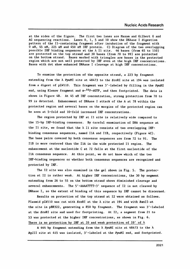
Fiue4: Footprint of IHF at the II site of X. DNAase I digestion patternof A) the top strand, and B) the bottom strand, of the DNA fragment con-taining the A, Ii site. For the top strand (A), an 850 bp fragment extendingfrom the BanII site of pBR322 through cos to the A, EcoRI site at 194 was used.The EcoRI end was 3'-labeled. For the bottom strand (B), a 223 bp fragmentextending from the A PpuMI site at 48473 to the A EcoRI site at 194 was used.The fragment was 3'-labeled at the PpuMI site. Base pair positions are given
2020
a
Nucleic Acids Research
at the sides of the figure. The first two lanes are Maxam and Gilbert G andAG sequencing reactions. Lanes 0, 1, 5 and 10 show the DNAase I digestionpattern of the Il-containing fragment after incubation of the fragment with0 nM, 45 nM, 225 nM and 450 nM IHF protein. C) Diagram of the two overlappingpossible IHF binding sequences at the X I1 site. 46 bases (from 65 to 110)are protected on the top strand and 30 bases (from 70 to 99) are protectedon the bottom strand. Bases marked with triangles are bases in the protectedregion which are not well protected by IHF even at the high IHF concentrations.Bases with dot show enhanced DNAase I cleavage at high IHF concentrations.
To examine the protection of the opposite strand, a 223 bp fragment
extending from the A PpuMI site at 48473 to the EcoRI site at 194 was isolated
from a digest of pLW11O. This fragment was 3'-labeled by filling in the PpuMI
end, using Klenow fragment and a-32P-dGTP, and then footprinted. The data is
shown in Figure 4B. At 45 nM IHF concentration, strong protection from 70 to
99 is detected. Enhancement of DNAase I attack of the A at 78 within the
protected region and several bases on the margins of the protected region can
be seen at 5-fold and 10-fold increased IHF concentrations.
The region protected by IHF at I1 site is relatively wide compared to
the 15-bp IHF-binding consensus. By tareful examination of DNA sequence at
the I1 site, we found that the X I1 site consists of two overlapping IHF-
binding consensus sequences, named IIA and I1B, respectively (Figure 4C).The base pairs covered by both consensus sequences are from 72 to 91. The
IlB is more centered than the IlA in the wide protected I1 region. The
enhancement at the nucleotide C at 72 falls at the first nucleotide of the
IIA consensus sequence. At this point, we do not know which of the two
IHF-binding sequences or whether both consensus sequences are recognized and
protected by IHF.
The I2 site was also examined in the gel shown in Fig. 5. The protec-
tion at I2 is rather weak. At higher IHF concentrations, the 30 bp segment
extending from 26 to 55 on the bottom strand shows diminished cleavage and
several enhancements. The 5'-AAAATTTT-3' sequence of I2 is not cleaved byDNAase I, so the extent of binding of this sequence by IHF cannot be discerned.
Results on protection of the top strand at I2 were obtained as follows.
Plasmid pLWIIO was cut with EcoRI at the A site at 194 and with BanII.at
the site in pBR322, generating a 850 bp fragment. The fragment was 3'-labeledat the EcoRI site and used for footprinting. At I2, a segment from 21 to
53 was protected at the higher IHF concentrations, as shown in Fig. 6.
There is no protection by IHF at I0 and weak protection of I0' of A
A 444 bp fragment extending from the A PpuMI site at 48473 to the A
BglII site at 415 was isolated, 3'-labeled at the PpuMI end, and footprinted.
2021
Nucleic Acids Research
Gc3 0 -
- _ 0
U
G Aef.,O
J: SI
Figure 5:. DNAase I footprints of A II, I2, IO and IO' sites. The G and AGlanes are Maxam and Gilbert sequencing reactions of the DNA fragment containingthe A I1, I2, IO and IO' sites. The 444 bp fragment used extends from the APpuMI site at 48473 to the A BglII site at 415. The fragment was 3'-labeledat the PpuMI end. Base pair positions are given at the sides of the figure.Lanes 0, 1, 5 and 10 are the DNAase I digestion pattern of the same DNAfragment after incubation with 0 nM, 45 nM, 225 nM and 450 nM IHF, respectively.The two overlapping IHF binding sequences are also indicated. Symbols as inFig. 4. Bases with x show reduced DNAase I cleavage at high IHF concentration.
The results, shown in Figure 5, show no significant protection of the bottom
strand at the I0 site. There is weak diminution of cleavage at some base
pairs in I0' at the 10-fold increased concentration of IHF. The top strand
was also examined by using a fragment extending from the A DraI site at 90
2022
11B.oft
-AWw
Nucleic Acids Research
As A0 1 500G G 0 1 5 20
1 41
48380
123
1
1 3;s -
4845080
I..~~~~~~~~~:
Figure 6: DNAase I footprints of the A I2, I3 and I4 sites. The G and AGlanes are Maxam and Gilbert sequencing reactions. The fragment used was the850 bp segment from the pBR322 BanII site to the A EcoRI site at 194. Thefragment was 3'-labeled at the EcoRI end. Base pair positions are given atthe sides of the figure. Lanes 0, 1, 5 and 20 are the DNAase I digestionpattern of the DNA fragment containing the I2, I3 and I4 sites after theincubation of the DNA with 0 nM, 45 nM, 225 nM and 900 nM IHF, respectively.Symbols as in Figures 4 and 5.
to the BglII site at 415. This fragment was 5'-labeled at the DraI end.
Again, there was no significant protection of the Io site and weak protec-tion of some bp at the I0' site was found (data not shown).The I3 and I4 sites of A are weakly protected by IHF at high concentrations
I3 and I4 are present on the footprint of the top strand shown in
Figure 6. The results show that at low IHF concentration (45 nM), neither
I3 nor I4 is protected against DNAase I attack. At 5 and 10-fold increased
2023
Nucleic Acids Research
.S
,.=,;._
.. _
_*
____
__
i_
_
-_,
Drs --
._.,
-s-.f;) ;1
:;4
--
_F__
-
*_E*''VFw,,w,,
.
.-
Lst-jr
0
~~~~~..
.:, G
~~~~~~~I 1
:- 11wo_
'
S....{..._ _
l
Wm _
Figure 7: DNAase I footprints of phage 21 cos fragment containing the 21I0, Ii and I2 sites. The 348 bp fragment extends from the A PpuMI site at48473 to the 21 NcoI site at 319. The DNA was 3'-labeled at the PpuMI end.Base pair positions are given at the sides of the figure. G and AG lanes areMaxam and Gilbert sequencing reactions. Lanes 0, 1, 5, and 20, show theDNAase I digestion patterns of 21 cos fragment after incubation with 0 nM,45 nM, 225 nM and 900 nM IHF, respectively. Symbols as in Fig. 4.
IHF concentrations, both I3 and I4 show some diminution of DNAase cleavage
at some base pairs, suggesting weak binding by IHF. To approach the I3 and
I4 sites more closely, the 693 bp fragment extending from the A XmnI site at
2024
C
'_s
I
ow
i
k.- ': .izrma AMMO :10,
W*
vw....
4*46
"hum,
.::-,X. -s .2
"POWAO,,44-::..
Nucleic Acids Research
39 to the BanII site of pBR322 was 5'-labeled at the XmnI end and footprinted.
Again, weak binding of I3 and I4 by IHF, this time on the bottom strand, was
indicated by diminution of DNAase cleavage at a few base pair positions (data
not shown). It may be noted in Figure 6 that bp outside I3 and I4 show enhanced
cleavage by DNAase I. These enhancements may indicate the formation of a
complex structure at high IHF concentrations. Diminution of cleavage at bp
outside the I sites is not seen, indicating that the diminutions found within
the I sequences represent specific interactions with IHF.
IHF protects three sites at the left end of phage 21 DNA
Lambdoid phage 21 has a cosN that is functionally identical to cosN of
X. In cosB of phage 21 there are also repeat sequences analogous to the R
sequences at A cosB (not shown in Figure 2), but the sequence of the 21
repeats is different from the A R sequence. This divergence of the DNA
sequence in the repeats between the two phages presumably reflects the
different DNA packaging specificities between the two phages. DNA sequencing
at the left end of phage 21 has revealed three potential IHF binding sites
(11), I2, Il and I0 (Figure 2). These sites are at roughly the same locations
as the I2, II and I0 sites of X, respectively, but differ in details. The
21 I2 site differs from the X I2 by only two base pairs. The 21 II site con-
tains only one IHE binding consensus sequence; it is equivalent to X HA.
The 21 I0 site is oriented in the opposite direction and several base pair
upstream from the X I0 site; it includes the putative Shine-Dalgarno sequence
of the 21 1 gene, which encodes the smaller subunit of 21 terminase. Plasmid
pBA4, which contains the 21 DNA sequence from cosN to 490, was used to study
the binding of IHF to 21 cos. The plasmid was digested by PpuMI at 48473 of
the A sequence and NcoI at 319 of the phage 21 sequence. The 348 bp fragment
was isolated and 3'-labeled at the PpuMI end. At 45 nM IHF, positive foot-
printing results were observed at the 21 I1 and I0 sites (Figure 7). The
segments protected on the bottom strand are from 63 to 99 for II and from 163
to 206 for I0. When the IHF concentration was increased S and 20-fold, the
21 I2 site also showed diminished cleavage by DNAase I and the protection at
the 21 II and I0 sites became stronger. The I2 segment protected extends from
26 to 49. There are some sites of enhancement of DNAase I cleavage at the
higher IHF concentrations. The DNAase I cleavage of the A at 42 in the 21
I2 region, the A at 78 in the 21 I1 region and the C at 179 in the 21 I0
region are most enhanced at the high IHF concentration.
A summary of the IHF protection results is shown in Table 1.
2025
Nucleic Acids Research
Table 1. Relative strength of IHf protection
cosX: I4 I3 I2 II IO' IO+/- +/- + +++ -s/- _
cos¢21: I2 II IO+ ++ ++I
+++: strong protection at all the IHF concentrationstested.
++: positive protection at 1X IHF concentration withincreasing protection at higher IHF concentrations
+: no detectable protection at 1X IHF concentrationand weak protection at higher IHF concentrations.
+/-: weak protection of some base pairs at high IHEconcentrations.no detectable protection at all the IHF concentrationstested.
DISCUSSION
IHF binds to specific sites at X cos
Our work shows that IHF binds to specific sites at cosX and cos2l. The
strength of protection by IHF among the sites at X cos varies. None of the
six potential IHF binding sites at A cos has a perfect match to the IHF
binding consensus sequence (Fig. 3). Inspection of the nucleotide sequence
and DNAase I protection patterns of the IHF binding sites at A cos reveals
several common features. First, the more closely a site matches the consensus
sequence, the better the protection. The two binding sequences at the II
site have the best matches to the consensus sequence. IlA has two mismatches
and I1B has only one mismatch and these sites are most strongly protected by
IHF at all concentrations. On the other hand, the IO, IO', I3 and I4 sites
all have three mismatches and they show very poor or no protection by IHF.
Secondly, each of the poor binding sites differs from the consensus
sequence at a position known to be very important for IHF binding. For
example, the A at position 4 of the consensus sequence is important because
an A-to-C transversion mutation in the IHF site in the left end of IS1
abolishes IHF binding (23). IO, I3 and I4 have a base other than A at
position 4. The T at position 11 of the IHF consensus sequence is also
important for the interaction with IHf, as follows. Gardner and Nash (8)
found that changing both T1O and Tll to Gs results in loss of IHF binding.
T1o is apparently not crucial for IHF binding, because pBR322 I site contains
an A at position 10 and is able to bind IHF (24). The above two studies
indicate that T11 is important for IHF binding. The IO' and I3 sites con-
tain a base other than T at position 11. Thus each of the weak or non-binding
2026
Nucleic Acids Research
sequences in cosX is changed for a base that is important for IHF binding.The regions protected by IHF seem very wide compared to the 15-bp IHF
binding consensus. The region protected by IHF at HI of A att covers 24 bps;
the protection of IHF at H2 of X att is 34 bps wide on one strand and 42 bps
on the other (3). At X cos similar results are obtained. In the case of the
A Ii site, the protected region spans about 46-bps on the top strand and 30-bps
on the bottom strand (Figure 4). The A I1 site consists of two possible
binding sequences and the protected region covers both sequences. We do not
know which of the two binding sequences or whether both are recognized by IHF
at this point. A further genetic study is needed to answer this question.
Comparison of the pattern of protection between A II and 21 Il suggests that
only one protein molecule binds to the A II site, as follows. The 21 Il con-
tains only one IHF consensus and it is structurally analogous to the IIA of A,
but the width of the region protected at the 21 Il site is very similar to
that of A II site.
Comparison of IHF binding sites at A cos and phage 21 cos
Lambdoid phage 21 requires IHF as a host factor for terminase action
in DNA packaging while A DNA packaging does not absolutely require IHF as
a host factor in vivo. For A, cleavage of cosN can be carried out in vitro
in a reaction requiring terminase and a host factor, either IHF or THF
(terminase host factor). The latter has been identified biochemically; it
is a basic protein with a molecular weight of 22 kd (25). The gene encoding
THF has not been identified.
Comparing the protection by IHF to the right of the A and 21 cosNs, we
found that certain differences exist between them. The 21 IO site runs in
an opposite direction to the A I0 site and overlaps the Shine-Dalgarno
sequence of the 21 1 gene, which codes for the smaller subunit of 21 terminase.
IHF protects the 21 I0 site but it does not bind to the A I0 site. IHF
has been found to promote the translation initiation of the A cII gene
transcript by binding to the site 3-bp upstream of the clI Shine-Dalgarno
sequence (7). We have not determined whether IHF plays any role at the 21
I0 site in 21 1 gene translation initiation. Also, the DNA sequence divergencebetween the two phages at their Ii sites results in only one possible IHF
binding sequence at the 21 I1 site while there are two possible IHF bindingsequences at the A II. We have not examined the cos fragment to the left of
21 cosN.
From the data we have obtained, we can conclude that A cos and 21 cos
respond differently to IHF. 21 cos contains three clear IHF binding sites
2027
Nucleic Acids Research
to the right of its cosN and two of them are well protected by IHF in foot-
printing experiments. On the other hand, X cos has more sites and each site
seems to have its own characteristics. Also, in vivo and in vitro data have
shown that 21 DNA packaging absolutely requires IHF as a host factor (12)
while A DNA packaging can proceed in cells lacking IHF.
Weak IHF binding sites.
We have found that the IO', I3 and I4 sites of A, which differ from
the consensus sequence at 3 positions, bind IHF weakly. These sites are
unusual in that most of the IHF binding sites have been described to date
are strong sites that match the consensus well. The significance of the
weak sites in cosA is unclear. They may function in vivo, or they may be
sequences that fortuitously match the consensus sequence. The latter is
to be expected in AT-rich segments of DNA such as cosA. On the other hand,
genetic studies indicate that IHF sites that have several mutations retain
residual activity in vivo (8, 26). These studies suggest that IHF binding
sites that bind weakly or not at all in footprint experiments may still be
able to be bound by IHF with sufficient affinity to function in vivo. Further
work on this point is needed.
Possible roles of IHF in K DNA packaging
IHF has a surprisingly varied functional repertoire, acting in gene
expression and in structure formation. IHF acts post-transcriptionally in the
regulation of K cII expression (7). IHF acts positively and negatively at
the transcriptional level in phage Mu (27).
IHF is involved in structure formation in a number of cases. At the
replication origin of plasmid pSC101, IHF assists in the formation of a nucleo-
protein structure that includes the ori DNA, the dnaA protein, and the plasmid
initiator protein. In pSC101, IHF binds at a position that contains a static
bend, and the bending of the DNA is increased by IHF (6). The pSC101 results
indicate that IHF may play a role in structure formation by bending the DNA.
In K site-specific recombination, IHF is involved in formation of complex
nucleoprotein structures at attP and attR (4, 5, 28, 29). Structure formation
involves cooperative interactions between IHF and integrase (3, 5). IHF may
also be involved in bending the DNA at the att sites, because att DNA, like
the pSC101 origin, appears to be bent (30). The role of IHF at cos seems
likely to be analogous to its role at attP, attR and at the pSC101 ori, i.e.,
in structure formation, though there is little direct evidence to date and the
argument is based on the similarity of cos to the att sites and the pSC101 ori.
The structure of cos is complex and is probably an interspersed array of binding
2028
Nucleic Acids Research
sites for IHF and terminase, in a manner analogous to the att and ori sites.
The DNA of cos is also bent (Yeo, personal communication; Kosturko, personal
communication). A third similarity to the att and ori sites is functional.
Echols (31) has argued that complex nucleoprotein structures are formed for
DNA sites that function in processes, like site-specific recombination and
initiation of replication, in which great specificity is required. The intro-
duction of nicks at cosN and the initiation of packaging would also seem to
require great specificity. Therefore, we speculate that the role of IHF at
cos is to assist in structure formation, perhaps by bending the DNA and/or
cooperative interactions with terminase.
ACKNOWLEDGEMENTS
We thank Howard Nash who generously gave us purified IHF for this study.
We appreciate helpful discussions with Linda Kosturko and our coworkers here
at Iowa. We thank Marcia Reeve fc;: preparation of the manuscript. Supported
by NIH grant AI-12581.
REFERENCES1. Miller, H.I., and Friedman, D.I. (1980). Cell 20, 711-718.2. Flamm, E.L., and Weisberg, R.A. (1985). J. Mol. Biol. 183, 117-128.3. Craig, N.L., and Nash, H. A. (1983). Cell 35, 795-803.4. Richet, E., Abcarian, P., and Nash, H.A. (1986). Cell 46, 10i1-1021.5. Thompson, J.F., Moitoso de Vargas, L., Skinner, S.E., and Landy, A.
(1987). J. Mol. Biol. 195, 481-493.6. Stenzel, T.T., Patel, P., and Bastia, D. (1987). Cell 49, 709-717.7. Mahajna, J., Oppenheim, A., Rattray, A., and Gottesman, M. (1986).
J. Bact. 165, 167-174.8. Gardner, J., and Nash, H.A. (1986). J. Mol. Biol. 191, 181-189.9. Feiss, M. (1986). Trends in Genetics 2, 100-104.
10. Becker, A., Marko, M., and Gold, M. (0977). Virology 78, 291-305.11. Miller, G., and Feiss, M. (1985). J. Mol. Biol. 185, 246-249.12. Feiss, M., Frackman, S., and Sippy, J. (1985). J. Mol. Riol.
183, 239-240.13. Bear, S., Court, D., and Friedman, D.I. (1984). J. Virol. 52, 966-972.14. Feiss, M., Widner, W., Miller, G., Johnson, G., and Christiansen, S.
(1983). Gene 24, 207-218.15. Daniels, D., Schroeder, J., Szybalski, W., Sanger, F., Coulson, A., Hong,
G., Hill, D., Peterson, G., and Blattner, F. (1983). In Lambda II,Cold Spring Harbor Laboratory, ed. R.W. Hendrix, J.W. Roberts, F.W.Stahl, and R.A. Weisberg. pp 519-676.
16. Feiss, M., Siegele, D.A., Rudolph, C.A., and Frackman, S. (1982). Gene17, 123-130.
17. Chang, A.C.Y., and Cohen, S.N. (1978). J. Bact. 134, 1141-1156.18. Frackman, S., Siegele, D.A., and Feiss, M. (1985). J. Mol. Biol.
183, 225-238.19. Bestwick et al. (1983). Anal. Biochem. 133, 79-84.820. Maniatis, T., Fritsch, E.F., and Sambrook, J. (1984). Molecular
Cloning, A Laboratory Manual. New York: Cold Spring Harbor Laboratory.
2029
Nucleic Acids Research
21. Maxam, A., and Gilbert, W. (1980). Methods in Enzymology 65, 499-559.22. Galas, D., and Schmitz, A. (1978). Nuc. Acids Res. 5, 3157-3170.23. Prentki, P., Chandler, M., and Galas, D. J. (1987). EMB0 J.
6, 2479-2489.24. Gamas, P., Chandler, M.G., Prentki, P., and Galas, D.J. (1987). J. Mol.
Biol. 195, 261-272.25. Gold, M., and Parris, W. (1986). Nucl. Acids Res. 14, 9797-9809.26. Thompson, J.F., Waechter-Brulla, D., Gumport, R.I., Gardner,
J.F. de Vargas, L.M., and Landy, A. (1986). J. Bact. 168, 1343-1351.27. Krause, H.M., and Higgins, N.P. (1986). J. Biol. Chem. 261, 3744-3752.28. Pollock, T.J., and Nash, H.A. (1983). J. Mol. Biol. 170, 1-18.29. Better, M., Lu, C., Williams, R.C., and Echols, H. (1982). Proc. Natl. Acad
Sci., USA 79, 5837-5841.30. Ross, W., and Landy, A. (1982). J. Mol. Biol. 156, 523-529.31. Echols, H. (1986). Science 233, 1050-1056.