SECONDARY ARTICULATION OF THE AVIAN MANDIBLE WALTER •'. BOCK INTRODUCTION THE avianskull,and especially its jaw apparatus, hasalways been a favorite subject for students of evolutionary and functional anatomy. Any analysis of this ddicateand complicated "machine" requires an extensive knowledge of mechanics as well as of morphology; indeed, the resources of the mostcompetent anatomist are taxed whenever he undertakes a functional study of the avian skull. Omission of even a single featurefrom analysis usually means that the entirestudy must be redone. Such is the problem with functional investigations of the avian mandible. Anatomists have alwaysassumed that the quadrate- articular hinge is the onlysuspension of the mandible, andtheworking hypothesis used in previous studies on the mechanics of the jaw appa- ratus hasbeenthat the forceswhichcounteract the disarticulating forces on the mandible mustbe supplied by the quadrate hinge. The jaw muscles and ligaments attaching to the mandible provide, of course, some support for it, but for the sake of simplicitythesenonbony elements will be omitted from discussion. However, the quadrateis not the only support of the mandible. In many groups of birds, a secondary bony supportof the mandible--the roedial brace of the mandible lies medialto the qnadrate-articular hinge. This braceis formed by the medial process of the mandible abutting against the base of the skull, the hingebetween the two bones beingthe badtemporal articulation of the mandible. The functional consequences of this secondary brace are obvious. For manyanatomists, information about this brace will be comparable to tellingan engineer that thereis a third support in the middleof a bridgeafter he has determined the bridge stresses by assuming that the only supports wereat the ends. The discovery of the roedial brace was quite by accident during a taxonomic review of the plovers. Amongother features of the skull, I had to examinea process on the lateral margin of the basitemporal plate (Bock, 1958: 46). Much to my surprise, this process wascapped by a pad of tissue which was fibrous in nature. At the time, I was preoccupied with the comparisons and did not realizethe significance of this capping pad. Several days later during a lull in the taxonomic investigations, I returned to the problemof why this process of the basitemporal plateshould havea fibrous capping pad. Only thendid I realizethat fibrous padson bony processes mean,in general, only one thing--another bone articulates on this process. But the only bone
Transcript
SECONDARY ARTICULATION OF THE AVIAN MANDIBLE
WALTER •'. BOCK
INTRODUCTION
THE avian skull, and especially its jaw apparatus, has always been a favorite subject for students of evolutionary and functional anatomy. Any analysis of this ddicate and complicated "machine" requires an extensive knowledge of mechanics as well as of morphology; indeed, the resources of the most competent anatomist are taxed whenever he undertakes a functional study of the avian skull. Omission of even a single feature from analysis usually means that the entire study must be redone. Such is the problem with functional investigations of the avian mandible. Anatomists have always assumed that the quadrate- articular hinge is the only suspension of the mandible, and the working hypothesis used in previous studies on the mechanics of the jaw appa- ratus has been that the forces which counteract the disarticulating forces on the mandible must be supplied by the quadrate hinge. The jaw muscles and ligaments attaching to the mandible provide, of course, some support for it, but for the sake of simplicity these nonbony elements will be omitted from discussion. However, the quadrate is not the only support of the mandible. In many groups of birds, a secondary bony support of the mandible--the roedial brace of the mandible lies medial to the qnadrate-articular hinge. This brace is formed by the medial process of the mandible abutting against the base of the skull, the hinge between the two bones being the badtemporal articulation of the mandible. The functional consequences of this secondary brace are obvious. For many anatomists, information about this brace will be comparable to telling an engineer that there is a third support in the middle of a bridge after he has determined the bridge stresses by assuming that the only supports were at the ends.
The discovery of the roedial brace was quite by accident during a taxonomic review of the plovers. Among other features of the skull, I had to examine a process on the lateral margin of the basitemporal plate (Bock, 1958: 46). Much to my surprise, this process was capped by a pad of tissue which was fibrous in nature. At the time, I was preoccupied with the comparisons and did not realize the significance of this capping pad. Several days later during a lull in the taxonomic investigations, I returned to the problem of why this process of the basitemporal plate should have a fibrous capping pad. Only then did I realize that fibrous pads on bony processes mean, in general, only one thing--another bone articulates on this process. But the only bone
20 Boc•:, Secondary Jaw Articulation [ Auk [Vol. 77
which could possibly abut on the lateral basitemporal process is the roedial process of the mandible. If the mandible articulated on the basitemporal plate, the plovers would have two separate suspensions of the lower jaw--a condition unknown in all other recent tetrapods and one that is of extreme importance to studies on the evolution of the mammalian jaw articulation. With this and the functional consequences of a second mandibular brace in mind, the taxonomic comparisons were forgotten, and skulls with the lower jaw in place were examined. The medial brace was discovered in the first specimen. My first thought was that surely someone had described this brace earlier. But a search through the literature and correspondence with other ornithologists and anatomists were fruitless; no one knew of any mention of a secondary brace of the avian mandible in the literature. However, although the medial brace was unknown to recent workers, including myself, it had already been described many years ago by Shufeldt (1890a: 354; 1890b: 71; 1893: 339) in the gulls, terns, and skimmer, even though he did not name it or attach any functional importance to it. For some inexplicable reason, little attention was paid to Shufeldt's discovery by his contemporaries--indeed, I do not know of a single reference to it in the literature. Consequently today the roedial brace is a completely unknown structure. It may seem strange that the medlal brace has escaped the attention of every avian anatomist except Shufeldt. But it is rather obscure in most forms in which it is found, and no one suspected a need for an additional brace of the mandible and hence did not look for one.
Examination of the skull of other avian families revealed that the
roedial brace is present in many diverse groups of birds in numerous degrees of specialization, and this discovery led to an extensive study of this structure. Therefore, I shall present in this paper a description of the roedial brace and the basitemporal articulation, a discussion of its functional significance, and the results of a survey of its occurrence in birds. A discussion of its evolution and of its significance in under- standing the evolution of the mammalian jaw articulation as well as some points of general evolutionary theory has been presented else- where (Bock, 1959). It was also necessary to study in detail two other features of the skullrathe quadrate hinge and the processes of the basicraniummboth of which are closely associated with the development of the roedial brace. These will be discussed along with the survey of the brace.
Jan.] Boc•, Secondary .law Articulation 21 1960J
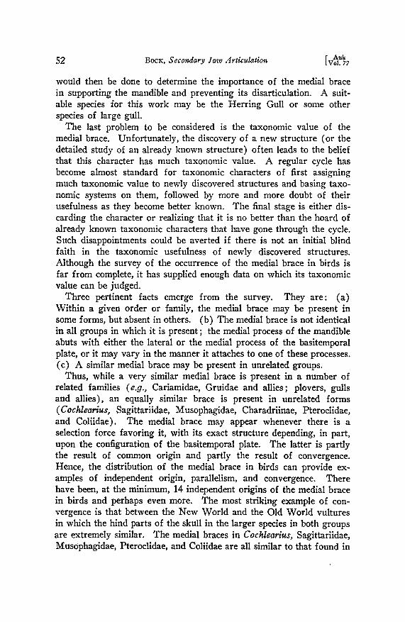
Description of a species lacking the roedial brace. The jaw articula- tion oœ the Boat-tailed Grackle (Cc•sidix mexicanus, Icteridae), a spe- cies having only the quadrate-articular suspension oœ the mandible, is described first to establish a standard against which the roedial brace and the basttemporal articulation can be compared.
The three articular condyles of the quadrate are shown on the right side only in Figure 1. The medial condyle is the largest and provides the greatest amount of support for the lower jaw as is indicated by the well-developed articular surface --a deep cavity--on the mandible for this condyle. The medium-sized lateral and the small posterior condyles are continuous with one another and are separated from the roedial condyle by a shallow groove; the posterior condyle appears merely as a narrow ridgelike extension of the lateral condyle. The corresponding articular surfaces on the mandible for these condyles are continuous with one another and form a shallow groove that faces reedtally. The articular condyles •f the quadrate form a rough horseshoe, which bounds a cavity lying between the lateral and roedial condyles. A knob of bone on the mandible fits into this cavity. This arrangement of the articular surfaces of the mandible embracing the condyles of the quadrate serves to prevent sidewards or backwards displacement of the mandible. The lateral process of the basitemporal plate abuts against the body of the quadrate and serves as a brace to prevent the quadrate from being displaced medially or posterlally. This brace of the quadrate is found in many passerine
I½.
..... ß .. jb.
Ibp' •*•
mpm.'
Figure l. Ventral view of the skull of a grackle. The posterior end of the right ramus of the mandible was removed to expose the condyles of the quadrate. These are ecoss-hatched. On the left side of the skull, the medial process of the mandible approaches the basitemporal plate but does not touch it. The deep-lying bones are shaded for contrast. The key to the abbreviations used in aH figures eau be found on p. 22.
[ Auk 22 Boca, Secondary Yaw ,'trticulation tVol. 77
K•¾ To ABBm•'•ATXONS USE•) xN THE FXGUKES
ac.--articular cavity ap.--articular pad ba.--basitemporal articulation bc.--basicranium
bp.--basitemporal plate bsa.--basisphenoid articulation cf.---collagenous fibers cp.---capping pad on the lateral basitemporal process ep.--exoccipital process jb.--jugal bar lbp.--lateral basitemporal process lc.--lateral condyle of the quadrate lca.--lateral condyle of the articular m.--mandible
mbp.--medial basitemporal process mc.--medial condyle of the quadrate mca.--medial condyle of the articular M.d. m.--M. depressor mandibulae mp.--maxillo-palatine M. pt.--M. pterygoideus mpm.--medial process of the mandible oc.--occipital condyle opq.--orbital process of the quadrate p.--palatine pc.--posterior condyle of the quadrate pca.--posterior condyle of the artlcular pt.--pterygoid q.--quadrate sm.--synovial membrane v.--vomer
birds and in some others, such as the goatsuckers (Caprimulgidae). The only other feature of the mandible of interest to us is its roedial process (best seen in Figure 2), which is relatively narrow in respect to its length. Although the medial process of the mandible approaches the basitemporal plate, there is a distinct gap between the two bones. As the bill opens, the medlal process of the mandible moves away from the basitemporal plate (in the direction of the arrow, Figure 2), thereby increasing the gap between the two bones. Consequently, in the Boat-tailed Grackle, there is never any contact between these bones, and the medial process cannot serve as a brace for the mandible.
In all birds, the function of the roedial process of the mandible is to provide an increased area for muscle attachment; the M. depressor mandibulae inserts along the posterior rim of the medial process, and the M. pterygoideus attaches to its anterior surfaces. The length and shape of the roedial process is then, at least, roughly correlated with the strength of either one or both of these muscles. However, because of its close proximity to the base of the skull,. the medial process is preadapted to provide additional support for the mandible should the need for this support arise. With a slight increase in length and a change in
.Ian.] BocI(, Secondary 7aw Articulation 23 1960J
bp.
qe
Ibp.
q. ib.
mpm.'
Figure 2. Close-up of the left-jaw artleulation of a grackle as seen from the ventro-medial side. The lateral process of the basltemporal plate ap- proaehes the quadrate; in life this process abuts against the quadrate and braces it agains t inward displacement. A distinct gap may be seen between the medlal process of the mandible and the basltemporal plate. •ghen the bill opens, the roedial process of the mandible moves away from the basi- temporal plate in the direetlon of the arrow. lienee there ean never be any contact between the mandible and the base of the skull.
shape, mainly a broadening of the process, the medial process would abut against the basitemporal plate and thereby brace the mandible. Once the two bones are in contact, some type of articulation (a functional requirement whenever two bones rub upon one another) would have to develop•perhaps, at first, some form of simple articulation, which could evolve into a true diarthrosls with increased demand on the newly arisen brace.
The roedial brace in the plovers. The first step in the development of the roedial brace and the basitemporal articulation is the establish- ment of a contact between the medial process of the mandible and the
24 Boca, Secondary .raw Articulation [ Auk tVol. 77
basitemporal plate. The following description of this stage is based on the condition seen in the Ringed Plover (Charadrius hiaticula, Charadriidae).
In contrast to the Boat~tailed Grackle, the largest articular condyle of the quadrate is the lateral, not the roedial, c0ndyle; the roedial condyle is somewhat smaller than the lateral (Figure 3). Furthermore, the posterior condyle is con-
Figure 3. Skull of a Hinged Plover seen from the postero-venlral side. The right ramus of the mandible was removed to expose the condyles of the quadrate. Both the q•adrate condyles and the capping pads on the lateral processes of the basitemporal plate are eross-hatched. The roedial proecss of the mandible articulates a•ainst the capping pad as •hown on the left aide of the skull. Lines "A-A" and "B-B" indieate the plane of sectioning for the histological slides. Again, the deep-lying bones are shaded for eontrast.
tinuous with both the medial and the lateral condyles, not only with the lateral. The qnadrate condyles are 'arranged in the usual horseshoe pattern, but they are more rounded than in the Boat-tailed Grackle with the result that the cavity between the lateral and medial condyles is poorly defined. The articular surfaces on the mandible are relatively flat and lack the knob that fits into the cavity between the lateral and medial condyles of the quadrate. Hence in the Ringed Plover, the articular surfaces of the quadrate-articular hinge do not offer as much protection against possible disarticulation of the mandible when the bill is opened. Again in contrast to the Boat-tailed Grackle, the medial process
Jan.] Boc•c, Secondary law Articulation 25 19601
of the mandible in the plover is broad; in fact, it is almost as wide as it is long. Turning now to the basitemporal plate, we see that its lateral process does not abut against the quadrate. Rather, the outer bony wall of the eustachian tube abuts against the quadrate and braces it against inward displacement. The lateral process of the basitemporal plate is more posterior and roedial than in the grackle. Also, the quadrate slants forward in the plover, while it is almost vertical in the grackle, which accounts for a large part of the gap between it and the lateral process of the basitemporal plate in the plover. More important is a pad of tissue capping the lateral process. This pad is tan in color and appears to be composed of either collagenous fibers or fibrocartilage; it is definitely not bone. The pad is present in all specimens, although it is often lost during cleaning of the skeleton. The distal tip of the roedial process of the mandible abuts against the capping pad on the lateral process of the basitemporal plate. At the point where it touches the capping pad, the roedial process is smooth and rounded, similar to a typical articular surface. Thus, in the Ringed Plover, there is a definite contact or articulation between the roedial process of the mandible and the lateral process of the basitemporal plate.
Dorsal
-• •. .
cf. ha.
Ventral
and eo•enone •be• b• •tJ•l, t maes of eol•enons •e•
[ Auk 26 BocK, Secondary Jaw •lrtlculatlon tVol. 22
The secondary abutment of the mandible on the brain case may be called the roedial brace oi the mandible, and its articulation may be called the basttemporal articulation oi the mandible. The two terms may be shortened to the roedial brace and the basitemporal articulation when discussing the mandible as will be done in the rest of this paper.
Slides were prepared of the two jaw articulations (right and left) of a Ringed Plover. The sections were cut at 10/• and stained with Ehrlich's hematoxylin and eosin. One articulation was sectioned in the plane indicated by line A~A in Figure 3, while the second was cut along line B-B. Because of the difficulties of cutting bone, I was only able to assemble rough serial sections of each series. Neverthe- less, the sections were sufficient to enable me to locate the basitemporal articulation and to determine its structure.
A schematic longitudinal section through the basitemporal articulation is shown in Figure 4. The base of the brain case appears as a thin, wavy line of bone. The roedial process of the mandible, which appears as an oblong ring of bone, is slightly separated from the brain case. Between the brain case and the roedial process is a heavy mass of collagenous fibers continuous with the two bones. This mass of collagenous fibers is the basltemporal articulation. The cavity seen inside
Dotsol
mpm:
M.d
Ventral
Figure 5. Semischematic cross-section through the basitemporal articula- tion of a plover. The articulation was cut in the plane indicated by line "B-B" in Figure 3. The thin band of eollagenous fibers running from the roedial process of the mandible to the basicranium is the anterior edge of the basitemporal articulation.
J'an.'l Bocx•, Secondary law Articulation 27 1960.1
the fibrous mass, although it is probably not an artifact, is not a true articular cavity. Its function may be to insure sufficient flexibility between the two bones.
In the anterior slides of the cross-sectional series (Figure 5), the roedial process of the mandible is present as an oblique ring of bone. Dorsal and lateral to the roedial process is the quadrate with its orbital process extending dorsal and roedial from the main body of the bone. On the roedial edge of the section and slightly dorsal to the roedial process of the mandible is the base of the skull, which appears as a jumbled mass of bone because of disruption and crushing during preparation. Slender bands of collagenous fibers run from the roedial process of the mandible to the base of the skull; these are the beginnings of the basltemporal articulation. In a more posterior slide (Figure 6), most of the structures seen in the last figure are still present, with the addition of a large mass of collagenous fibers just dorsal to the former position of the now-absent roedial process of the mandible. This mass of collagenous fibers is attached to the roedial process (seen in intermediate slides) and extends toward the base of the skull as a band of fibers that finally merges into a bone (= the lateral process of the basitemporal plate ?). From its position and connection to both the mandible and the brain case, I have identified this mass of collagenous fibers as the basftemporal articulation. The thickening seen in the middle of the articulation is probably the capping pad on the lateral process of the basitemporal plate. As the more posterior slides are examined, the roedial process of the mandible drops out first, followed by the
Dorsol
b½.•
cf.- b
M.½
Ventral
Figure 6. Semischematic cross.section through the basitemporal articula- tion of a plover. This section is posterior to the section shown in Figure 5. The roedial process of the mandible has dropped out, but the basitemporal articulation has become more prominent. The thick knob of collagenous fibers is probably the capping pad found on the lateral basitemporal process.
28 Boc•r, Secondary Yaw Articulation [ Auk [Vol. 77
capping pad on the lateral basltemporal process, until all that remains is the now- prominent base of the skull.
These slides show that the basitemporal articulation in the plover possesses neither articular pads of cartilage nor a true articular cavity, both of which are necessary diagnostic features of a diarthrodlal articulation. x Instead, the basi- temporal articulation of the plover belongs to the class of articulations that are only slightly movable---amphiarthroses--and to the subclass--syndesmoses--be- cause the bones are held together by ligaments (Jackson, 1933: 275; Lewis, 1942: 277). This' type of articulation is considered to be intermediate, both morphologically and functionally, between sutures and dlarthroses or true articular joints.
The roedial brace in the Black Skimmer. The most advanced stage in the development of the medial brace in birds may be seen in the Black Skimmer (Rynchops nigra, Rynchopinae). Shufeldt (1890b: 71) described the articulation for the gulls, terns, and skimmer, all members of the Laridae, and stated that it is best developed in the skimmer, which has a true articular joint. He mentioned the very distinct articular surfaces present on the medial process of the mandible and on the lateral basitemporal process in the skimmer, which is doubt- less the basis for his statement that the skimmer has a true articular
joint.
The articular condyles of the quadrate are similar to those seen in the plover, except that the posterior condyle appears as a ridge of the lateral condyle (Fignre 7). In contrast to both the grackle and the plover, the posterior condyle of the skimmer is dorsal, not ventral, to the anterior condyles. Because of the "de- pressed" posterior condyle, the roedial condyle is a sharply defined knob. The articular surfaces of the mandible and of the quadrate form a "peg and socket" joint, which prevents the mandible from slipping sidewards when the bill is opened. Because of the "depressed" posterior condyle, there is no protection against backwards movement of the mandible. The roedial process of the man- dible projects inward and upward toward the base of the skull and bears a smooth articular surface on its distal end. The lateral process of the badtemporal plate is in the same position as its homologue in the plover, but lacks the capping pad. Instead there is an articular surface, which faces the articular surface in the roedial process of the mandible. The quadrate is not braced by any part ot• the brain case.
The roedial brace and the basitemporal articulation of the Black Skimmer are illustrated in Figure 8. This view ot• the skull shows that the articular surfaces
• Articulations are usually classified into three main categories--sutures, amphi- arthroses, and diarthroses--which are convenient, but arbitrary, divisions of a continuous spectrum of structures. Sutures are nonmovable contacts between bones and need not to be considered further since the basitemporal articulation is a movable hinge. The difference between amphiarthroses and diarthroses is that, in the former, the bones are simply held together by bands of collagenous fibers, while, in the latter, an articular cavity, articular surfaces of cartilage, and a synovial membrane must be present. For a general discussion of this subject, see Lubosch (1910), who investigated the histological structure of articulations found in all living classes of vertebrates, or any textbook on histology.
1•] Boca;, Secondary Yaw Articulation 29
mpre. (•••pco.
mco. f• 'lco.
cl.
Figure 7, Ventral view of the skull and dorsal view of the mandible of a skimmer. The articular surfaces of both the quadrate hinge mad the roedial brace are cross-hatched. The deep-lying bones are stippled for
oœ the basitemporal articulation are so oriented that the mediai brace can withstand both backwards and inwards /orees.
Slides of the roedial brace of a Black Skimmer were prepared to determine the histologicai nature of its basitemporal articulation. The entire jaw articulation was removed from a specimen, and the excess bone was trimmed off until all that remained was the medial brace. The tissue was sectioned along the longitudinal axis of the medial brace; however, the direction of sectioning in the radial plane is not known. Sections were cut at 10• and stained with Ehrlich's hematoxylin and eosin or with Mallory's triple stain. They were not mounted serially. The extreme difficulties of embedding and cutting bird skulls prevented sectioning the medial brace of the skimmer while it was in its natural position and assembling serial sections.
Inspection of a section (Figure 9) reveals that the hasitemporal articulation in the Black Skimmer is a true diarthrosis. Pads of articular cartilage on each bone, a typical articular cavity, and articular membrane are all present. The exact size of the cavity and the relationship of the bones to one another cannot be determined from the sections because the bones were moved relative to one
another during preparation. Nevertheless, several features of the articulation (not illustrated) deserve closer attention.
30 Boc•:, Secondary Yaw Articulation [ Auk [Vol. 77
• ,Ibp.
pt. rnprn. bp.
Figure 8. Skull of a skimmer seen from the postero-ventral side. The basltemporal articulation is shown on the left side of the zkuH (rough lines), but it has been removed on the right side to show the processes of the medial brace. The deep-lying bones are stippled for eontrast.
The articular pads are most interesting. Rather than being composed of hyaline cartilage as is usually true for diarthroses, the pads are formed of a dense fibro- cartilage. The collagenous fibers run perpendicularly from the bone toward the surface of the articular cartilage, but before they reach the surface, they turn to the side or back on themselves to form a mass of interwoven fibers near the
surface of the articular pad. These pads are very similar to those shown by Petersen (1930: Figs. 137a and 141) to illustrate articular pads composed of fibrocartilage. The orientation of the fibers in at least part of the articular pad on the roedial process of the mandible is influenced by the insertion of the muscle (probably the M. pterygoideus) seen at the edge of the section. The collagenous fibers associated with the muscle insertion lie parallel to the muscle fibers and thus run across the articular pad and parallel to its surface. Lastly, the surfaces of the articular cartilages are not covered by a squamous epithelium, but are bare of an epithelium and are rough, not smooth as is usual for articular surfaces. This may be, however, an artifact of preparation. Fibrocartilage articular pads may occur when there is much pressure on the articulation. A prime example of fibrocartilage articular pads associated with pressure is the articulations between the thoracic vertebrae in man.
Details of the articular membrane are somewhat obscured because of tearing during preparation. However, the membrane appears to be similar in all respects to typical synovial membranes except that it seems to be looser and more folded than normal, probably to allow the bones to slide further past one another than in most articulations. In some sections, a long tongue of loose connective tissue
•J•] BocIq Secondary Jaw .Zlrtlculatlon 31
Dorsal
M. pt.
pm.
Ventral
Figure 9. Semischematlc longitudinal section through the basitemporal articulation of a skimmer. Note the presence of artlcular pads, an artleular cavity, and synovlal membrane, all of which are necessary features of dlarthro•es. The synovial membrane is shown by rough llnes, while the fibrocartilage artlcular pads are shown by stippling; bone and muscle are the same as in Figure 4.
projects into the articular cavity, and in other sections it may even be continuous across the cavity. I do not know whether this infolding of the synovial membrane is present only at the edges of the articulation or whether it is found throughout the articulation; serial sections are needed to ascertain this point. Membranes or cartilagenous disks have been reported in the cavities of articulations which are subject to pressure. Lastly, it should be noted that the synovial membrane continues without a break into the epimysium surrounding the muscle which inserts on the medlal process of the mandible.
I have also obtained some rather poor sections of the basitemporal articulation of the Herring Gull (Larus argentatu•, Larlnae). In all respects, including the fibrocartilage articular pads and the tongue of connective tissue projecting into the articular cavity, the histological structure of the gull basitemporal articulation
[ Auk 32 Boca, Secondary Jaw Articulation tVol. 77
appears similar to the one just described for the skimmer. The chief difference appears to be the thicker articular pads in the herring gull.
Functional significance of the roedial brace. All of the structural features of the medial brace indicate that it serves to support the mandible and to prevent its disarticulation when the bill is opened. The fibrous capping pad on the lateral basftemporal process in the plover and the fibrocartilage articular pads in the Black Skimmer are strong evidence in favor of this functional conclusion. It must be emphasized, however, that this functional conclusion is based on deductive reasoning from the morphology of the medial brace and the quadrate hinge and must be verified by empirical observations. Assuming, at least for the purposes of this paper, that the medial brace does serve to support the mandible, two additional problems must be solved: whether the medial brace functions as a support of the mandible in all birds in which it is present, and why a second support of the mandible is needed in certain groups of birds.
The roedial process of the mandible may elongate under the action of selection forces for stronger jaw muscles until it becomes so long that it rubs against the base of the skull as the bill opens and closes (see Bock, 1959, for a more detailed discussion). An articulation will develop between the medial process and the basitemporal plate simply because the two bones rub upon one another. These birds now possess most or all of the morphological features of the medial brace--the medial process abutting against the basftemporal plate with an articula- tion between them--but it cannot yet be considered as a support of the mandible unless it is actually functioning as one. •'his could be deter- mined by the presence of certain specializations such as fibrous articular pads and strengthened processes on the basitemporal plate, or better by actual observations or experiments. If the morphological medial brace was not functioning as a support of the mandible, it could be regarded as being in the preadapted stage. Although this problem is important in discussions on the evolution of the media] brace, it shall not be con- sidered further in this paper. The reader is referred to my other paper (Bock, 1959) for a more complete discussion of this point. I shall assume, for the purposes of this study, that if the medial process of the mandible abuts against the basftemporal plate, then that bird has a functional medial brace.
The question of why some birds need a second support of the man- dible may be approached by a consideration of how they catch their food. •'he forelimbs of birds, being modified into wings, have no direct auxiliary function in the ingestion of food. Only a very few groups, such as the hawks and the owls, use their feet to capture their prey.
Jan.] BOChe, Secondary Jaw Articulation $3 19601
Most birds use only their bill in feeding. In addition, birds are very active and frequently capture or break up their food by rapid move- ments of the head. In many species, food is taken in flight. Thus, it can be concluded that the jaw articulation of birds is frequently subject to 'sudden, powerful shocks or to asymmetrical forces, and hence is exposed to the danger of being disarticulated. The problem is, there- fore, whether the quadrate hinge in all birds can withstand these strong and possibly disarticulating forces on the mandible or is additional support of the mandible needed in some groups.
The articular surfaces of the quadrate-articular hinge usually consist of three separate condyles on the quadrate with corresponding articular surfaces on the articular of the mandible. Some birds have only two condyles on the quadrate, while a very few have four. The quadrate condyles are usually arranged in the shape of a rough horseshoe with the base pointing backwards, condyles at the base and the tips of the two arms, and a cavity between the arms of the horseshoe. Either the lateral or the medial condyle is the largest with the posterior condyle usually appearing as a projection of the lateral condyle, but with some connection with the medial condyle. Sometimes the posterior condyle appears to be a projection of the medial condyle. The posterior con- dyle usually projects as far ventrally as the others, but sometimes it is more dorsal, i.e., "depressed," than the other condyles. It is difficult to describe the variation of the quadrate condyles in birds, but it will suffice to say that there is great variation in the size, shape, and position of the condyles between the families and orders of birds. Some indi- cation of this variation may be gained through the survey presented below, but even there a great many details are omitted.
The structure of the corresponding articular surfaces on the articular is even more variable than the condyles of the quadrate. In some birds, the articular surfaces of the mandible are well defined, while in others, they are flat. In general, birds may be divided into two groups: those in which the articular encases the condyles of the quadrate, thus pre- venting backwards and/or sidewards shifting of the mandible, and those in which the articular is flattened and does not clasp the condyles of the quadrate, thus offering limited protection against disarticulation of the mandible. Attention will be focused on the second group. The problem becomes even more complex because the relationships of the articular surfaces shift as the bill opens, with the result that the articu- lar may no longer encase the quadrate condyles. The mandible is depressed by the action of the M. depressor mandibulae, which takes origin from the exoccipital process and inserts on the posterior rim of the mandible including its medial process. Upon contracting, the M.
34 Boc•:, 3¾condary Jaw Articulation [ Auk [Vol. 77
depressor mandibulae draws the posterior end of the mandible upwards, thereby depressing the rest of the mandible, which pivots on the pos- terior articular condyle. Therefore, as the bill opens, the articular surfaces of the anterior quadrate condyles lose contact with the corre- sponding surfaces of the articular. Support of the lower jaw would then rest largely on the posterior condyle. Hence, when the bill is opened while the bird is feeding, the mandible receives the least support from the quadrate at a time when it would need the greatest amount of support; any backwards or sidewards force on the depressed mandible would act mainly on the posterior condyle and the posterior surfaces of the anterior condyles of the quadrate. (In these discussions, all forces on the quadrate hinge within 20 to 30 degrees of the medial plane and in a backwards direction are included within the limits of backwards disarticulation, thereby including most of the possible shifts caused by lateral or twisting forces on the mandible.) If these dis- rupting forces are strong enough and if the structure of the quadrate hinge is such that it offers only limited protection against disarticulation, e.g., the posterior condyle being dorsal, then it would be advantageous for the bird to have some structure that would provide additional support for the mandible. This extra protection can be provided, of course, by the medial brace of the mandible. It should be noted that the need for extra support of the mandible exists only when the bill is opened, hence the roedial brace need be functional only at this time.
If the conclusion that the medial brace functions to support the mandible in compensation for a poorly developed quadrate hinge is correct, then it should be expected that the relative development of the brace is correlated directly with the strength of the force on the de- pressed mandible and inversely with the ability of the quadrate hinge to withstand these forces. The medial brace of the Boat-tailed Grackle, the Ringed Plover, and the Black Skimmer may be compared to test this latter conclusion.
Grackles feed mainly on insects, seeds, and other small morsels in which the main action of the bill is a forceful bite; hence backwards and probably sidewards forces on the mandible would be small or lack- ing. The peg and socket arrangement of the quadrate hinge is probably able to withstand all of the forces that may act on the mandible. Thus, the probability of the quadrate hinge of the grackle disarticulating is very small or perhaps even nonexistent, which is •n agreement with the absence of the medial brace in this species.
The Ringed Plover feeds by picking up small animals from sand and mud beaches, and this should not result in strong forces on the mandible. However, the articular surfaces of the mandible do not
•J•6] BocIq •econdary Jaw •trtlcula•ion 35
clasp the quadrate condyles and thus offer little protection against the possible disruption of the quadrate hinge. The slightly developed medial brace of the plover probably provides the extra support needed to compensate for a poorly developed quadrate hinge.
The mandible of the Black Skimmer is subject to quite severe shocks. This bird catches its prey by flying low over the surface of the water and inserting its knifelike mandible into the water. Each time a fish is caught or an obstacle hits the mandible, the head of the skimmer is snapped violently downwards and backwards. The force at which a skimmer hits an obstacle is sometimes great enough to break off the tip of the horny covering of the mandible. 2 As we have seen, the structure of the quadrate hinge in the skimmer is such that it cannot resist a strong backwards movement of the mandible. Therefore, it can be concluded that each time a skimmer catches a fish or hits an underwater
obstacle with its bill, there is a strong and sudden force on the mandible while the bill is opened and that the quadrate hinge of the skimmer kannot withstand this force; thus the need for a secondary support of the mandible. The medial brace in the skimmer must be well developed as it probably supplies most of the resistance to the backwards forces on the mandible.
A comparison of these three species supports the conclusion that a flattening of the articular surfaces of the quadrate hinge and/or an increase in the forces acting on the mandible are correlated with a more highly developed medial brace. More examples will be given in the review of the medial brace in birds. The degree of development of the medial brace may be estimated roughly by whether the bones are in contact even when the bill is closed and by the structure of the basi- temporal articulation. A disarthrosis with fibrocartilage articular pads indicates a more highly developed medial brace than a syndesmosis.
SURVEY OF THE MEDIAL BRACE AND ASSOCIATED STRUCTURES
The structure and occurrence of the medial brace, the processes of the basicranium, and the quadrate hinge will be described in this section. I shall first outline the general problems associated with each of these structures and then present the details in the synopsis of the families. All material examined is in the skeleton collection of the American
Museum of Natural History. The sequence of families used in the synopsis is that presented by Mayr and Amadon (1951).
• These observations are partly my own, but they were greatly clarified by a discussion with Dr. Richard Zusi, who is studying the functional anatomy of the feeding methods of the skimmer. I am most grateful to Dr; Zusl for supplying me with these unpublished data.
[ Auk 36 BOCK, Secondary .gaze Articulation tVol. 77
The roedial brace. The first question that arose after the medial brace (hereafter abbreviated as mb.) and the basitemporal articulation (hereafter abbreviated as ba.) were found in the Charadriinae and the Laridae was: How widespread are these structures in birds? Are they restricted to a few families of the Laro-Limicolae? Or are they of widespread occurrence in birds ? A survey using dried skulls was undertaken to answer these questions. Although the results of this survey provide much information about the distribution of the medial brace in birds, it must be emphasized that they are preliminary and that much more work is needed before we have an accurate picture of the occurrence and structure of the medial brace and the basitemporal articulation. No attempt was made to examine all available genera of a family, and because dried skulls were used, the observations cannot be considered as conclusive. During preparation of the skeleton, the ligaments between the bones of the brace may tear, the articular pads may dry and fail off the specimen, the quadrate may shift forward, and the mandible may become disarticulated as is the usual case in museum specimens. All these factors contribute toward destroying the basi- temporal articulation and the normal position of the bones that consti- tute the brace, and thus render the specimen unsuitable for observation. Therefore, it is emirely possible that I have overlooked the medial brace in some groups, especially if it is weakly developed, and that I have erroneously reported a medial brace in some groups that do not possess one. For example, early in the study, I examined a dried skull of a gull (Larus) and concluded that it did not possess a medial brace. Later dissection of an alcoholic specimen revealed that gulls have a well-developed roedial brace, but one with thick articular pads. These pads had dried and dropped off the skull, thereby leaving a gap between the bones of the medial brace. Observations on alcoholic specimens are needed to verify my preliminary findings and to ascertain the exact structure of the brace in groups other than the plovers and the skimmer.
During the course of the survey, it became obvious that the medial brace was not only of widespread occurrence in birds, but that its structure varies greatly between the families and orders of birds. The most siguaificant feature of this variation is not the difference in the morphology of the articulation itself, but the fact that the process of the basitemporal plate on which the roedial process of the mandible abuts is not the same in all birds. The medial process of the mandible articulates either with the lateral or the medial process of the basi- temporal plate (Figure 10) or with the main body of the plate itself. Variation exists even within these limits as the mandible may abut against the anterior edge or the ventral tip of the lateral basitemporal
Jan.] BOCa, Secondary .raw Articulation 37 1960J
pt.
p.
Figure 10. Skull of a California condor (C, ymnogyps) seen from the postero-ventral side. The roedial process of the mandible abuts against the enlarged roedial process of the basitemporal plate. The lateral basltemporal proeesz can be seen passing beneath the medlal basltemporal process. The deep-lylng bones are shaded for contrast.
process. The interesting aspect of this variation is that the medial brace is effective as a support of the mandible no matter which process of the basitemporal plate is involved. This problem is discussed at greater length elsewhere (Bock, 1959).
The bony processes of the badcranium. Once it was discovered that the medial process of the mandible (hereafter abbreviated as mpm.) articulated with two different processes of the basftemporal plate, it became necessary to ashertain the correct names for them. A review of the literature not only failed to produce an acceptable set of names, but revealed an almost hopeless confusion in the terminology for the proc- esses of the basicranium. This confusion was the product of the past few decades, because prior to 1900 there was relatively standard terminology for the processes found on the base of the skull. No general review of these structures has been published since the turn of the century. Unfortunately, Barnikol (1952) did not include the base of the skull in his excellent paper on the factors influencing the shape of the skull. The following set of names is a composite one,
[ Auk 38 BOCK, Secondary Jaw Articulation [Vol. •?
picking the ones which are the most descriptive and the least contro- versial. No consideration is given to priority or to homology with the mammalian or the reptilian skull. Thus, such names as the para- occipital, the mastoid, and the opisthotic process will not be used.
(a) The lateral process of the basitemporal plate (hereafter abbrevi- ated as lbp.) is located along the lateral margin of the basitemporal plate just ventral to the posterior opening of the eustachian tube. In some birds, the lateral process appears to be a ridge along the edge of the basitemporal plate and forms the ventral wall of the eustachian tube. Frequently, a ridge of bone continues from the lateral process to the exoccipital process. The base of the lateral process and of its posterior extension is pierced by the foramina for several branches of the carotid artery. When the ventral wall of these foramina does not ossify, there is a break between the lateral basitemporal process and the exoccipital process. The lateral basitemporal process apparently func- tions as a point of attachment for certain cervical muscles.
(b) The medial process of the basitemporal plate (hereafter abbrevi- ated as mbp.) is located near the posterior edge of the basitemporal plate, medial and slightly posterior to the lateral basitemporal process. Sometimes there is a second pair of very small knobs inside the medial processes. The function of the medial process is apparently to provide a point of attachment for some of the cervical muscles (see Boas, 1929). Fisher (1944: 229) erroneously called this process the exoccipital proc- ess, while •Pycraft (1902: 280) referred to it as the mammillary process.
(c) The exoccipital process (hereafter abbreviated as ep.) is located on the ventral end of the exoccipital bone and usually projects down- wards. It is little more than a ventral extension of the exoccipital bone. In some birds, such as the cormorant, the exoccipital process is hori~ zontal and points to the side. As mentioned above, a ridge of bone frequently connects the exoccipital process with the lateral basitemporal process. The }•. depressor mandibulae takes origin from the exoccipi- tal process. This process has been called the paraoccipital, the opis- thotic, and the mastold process as well as the occipital wing.
(d) The auditory bulla can be used for the swollen bulb about the opening of the ear (see Dilger, 1956: 177), although this structure is not homologous with the auditory bulla of the mammals. This swelling is formed by the exoccipital process and the lateral basitemporal process. The squamosal does not take part in its formation, hence it cannot be referred to as "the inflated squamosal area" (Tordoff, 1954: 10). The auditory bulla will not be included in the synopsis.
The quadrate hinge. While examining families for the presence of the medial brace, I became puzzled over the lack of the brace in some
Jan.] Boca, Secondary 7aw Articulation 39 1960J
groups in which I had expected to find one. In some cases, I have probably overlooked it as I had later discovered upon checking my observations; but in other cases, the brace is definitely lacking. A com- parison of the quadrate hinge in a few groups revealed that it varied greatly in its ability to protect the mandible against disarticulation. In some birds, such as the skimmer (Rynchops), it offers very little protection, while in others, such as Balaeniceps, it provides all the necessary protection and support. All possible intermediate steps are found in other birds. It was, thus, considered essential to determine the ability of the quadrate hinge to support the mandible and to correlate this ability with the development of the medial brace. The minute detail of the quadrate suspension is most interesting, but it is of little importance to this study. Consequently, I will restrict the descriptions to the barest necessity, and concentrate attention on the ability of the quadrate hinge to support the mandible. These functional conclusions are based only on observations on the dried skull, and are speculative and inconclusive at best. Observations on the living bird and experi- ments are needed to ascertain the role of the quadrate hinge and other structures, such as ligaments, jaw muscles, and the medial brace, in supporting the mandible when the bill is opened. The lateral, the posterior, and the medial quadrate condyle will be abbreviated as lc., pc., and mc., respectively. For additional details on the morphology of the quadrate-articular hinge, the reader is referred to such general papers as Lebedinsky (1921).
SYNOPSIS OF lq'AI•ILIES
Struthionidae. (a) The mb. appears to be absent in the ostrich, but the mpm. is well developed and approaches the base of the skull when the bill is opened. Perhaps the rob. is present (if so, the artlcular pads or the connecting bands of collagenous fibers of the ha. have dried and fallen off?). (b) Both the lbp. and the mbp. are absent, but the ep. is well developed. (c) The lc. and the pc. are small and dorsal; the pc. is an extension of the lc. The inc. is the largest and the most ventral of the three condyles. It offers excellent protection against sidewards shifting of the mandible, but only poor protection against backwards disarticulation.
Apterygidae. (a) The rob. is present in the kiwis with the mpm. articulating against the mbp. (b) The lbp. is present but poorly developed and is over- shadowed by the well-developed mbp., which is a bony knob well back of the basitemporal plate. The ep. is lateral and well developed. It forms a vertical plate of bone on the back of the skull. (c) The lc. is fused with the pc.; both face laterally. The mc. is the largest and offers good protection against side- wards, but not against backwards, disarticulating forces.
Casuariidae. (a) The mb. is present in the cassowaries (?), with the mpm. articulating with the lbp., or perhaps with the lateral edge of the basitemporal
[ Auk 40 BOCK, Secondary Jaw .4rticulatio• [Vol. 77
plate. (b) The Ibp. forms a slight ledge along the edge of the basitemporal plate. The mbp. is absent or present as a slight bump. The ep. is well developed and forms a vertical plate of bone on the back of the skull. (c) The Ic. is continuous with the pc. to form a flat plate, which faces backwards and outwards. The inc. is the largest condyle but forms a flat plate, which offers little support against either sidewards or backwards disarticulating forces.
Dromaeidae. (a) The rob. is absent in the emus. (b) Both the Ibp. and the mbp. are absent. The ep. is present as a flat plate of bone, which does not flare out to the sides as in the kiwis. (c) The Ic. and the pc. are joined to form a long articular surface. The mc. is the largest with one articular surface facing out- wards. This articular surface has a corresponding abutment on the mandible, which serves to protect the quadrate hinge from disarticulation.
Rheidae. (a) The mb. is absent in the rheas. (b) The ibp. is present (?) as a ridge below the edge of the basitemporal plate, while the mbp. is present as a slight knob. The ep. is well developed. (c) The Ic. is small and the most dorsal of the three condyles. The pc. is connected to the Ic. and is small. The mc. is the largest and the most ventral. It has an outward-facing articular surface with a corresponding face on the mandible, which may serve to protect the mandible from disarticulation.
Tinamidae. (a) The mb. is probably absent in the tinamous, although one skull showed a peculiar set of processes and articular surfaces, which suggests that a mb. is present in this individual. (b) The Ibp. and the mbp. are absent. The ep. is present but not as prominent as in the rheas. (c) The lc. is small and dorsal and is connected to the small, but more ventral, pc. The inc. is large and the most ventral with a lateral articular surface, which could serve to protect the mandible. However, there is not a corresponding articular surface on the mandible. The quadrate hinge of the tinamous does not appear to be able to protect the mandible against disarticulation, but a rob. has not developed.
Spheniscidae. (a) The mb. is present in several genera of penguins, such as .4ptenodytes, with the mpm. articulating against the mbp., but it is absent (?) in others such as Spheniscus. Perhaps the articular pads in these latter genera are thick and have dried out, which would account for the apparent absence of the mb. (b) The lbp. is present as a poorly defined ridge along the margin of the basi- temporal plate. The mbp. is also present, but it is well developed. It is found near the lateral edge of the bone and projects more ventral than does the Ibp. The ep. is present, but it is poorly developed. (c) The lc. is well developed, while the pc. is small and more dorsal than the others. The mc. is the largest and has a concavity on its lateral edge with a corresponding ridge on the mandible. How- ever, the entire articulation is quite flat and offers little protection against disarticu- lation of the mandible; therefore, the seeming absence of a rob. in some genera is a puzzle.
Diomedeidae. (a) The mb. is absent in the albatrosses or may be present (?) with the mpm. articulating with the mbp. If the rob. is present, the articular pads are probably very thick, and have dried and dropped off the skull. (b) The lbp. is absent, but the mbp. is present as a prominent knob of bone. The ep. is well developed and extends ventrally. (c) The Ic. is well developed with the pc. appearing as a posterior and slightly dorsal continuation. The inc. is the largest condyle and has a groove across it at an angle to the longitudinal axis of the skull. A bony ridge on the mandible fits into this groove and apparently prevents dis- articulation of the mandible.
Jan.1 BOCK, Secondary .law Articulation 41 19601
Procellariidae. (a) The mb. is absent in most shearwaters and petrels, but may be present in the largest members of this family, e.g., Macronectes, with the mpm. articulating with the mbp. (b) The lbp. is absent. The mbp. is absent in the storm petrels, bnt present in the shearwaters from weakly developed to well developed in Macronecte& The ep. is present and projects ventrally. (c) The quadrate condyles are similar to those in the albatrosses including the groove on the mc., which prevents the disarticulation of the mandible.
Pelecanoididae. (a) The rob. is absent in the diving petrels. (b) Both the lbp. and the mbp. are absent. The ep. is present, but weak. (c) The lc. is a well- defined knob. The pc. is a ridge extending back from the lc.; it is as ventral as the lc. The me. is a bony ridge at an angle to the other condyles. It has an anterior lip, which apparently serves to protect the mandible against disarticulatlon.
Podlcipitidae. (a) The rob. is present in the grebes with the mpm. abutting against the lbp. (b) The lbp. is present as a poorly developed ridge along the margin of the basitemporal plate, but with some thickening at the point where the mandible articulates with it. The mbp. is present as a slight knob. It is quite posterior and is almost in line with the occipital condyle. The ep. is present and projects ventrally. It extends toward the lbp., but a gap separates the two processes. (c) The lc. is present, but it is quite flat. The pc. is well developed and projects ventrally. It is well separated from the other condyles and appar- ently prevents sidewards slipping of the mandible. The mc. is a strong knob with a lateral groove and aids the pc. in protecting the mandible. However, the quadrate hinge cannot prevent backwards dlsarticulation of the mandible, which agrees with the presence of the mb. in this family.
Gaviidae. (a) The mb. seems to be absent in the loons, but it may be present. The mpm. appears to articulate with the weakly developed mbp., with thick articular pads between the bones. Dissection of alcoholic specimens is needed to verify this point. (b) The lbp. is absent. The mbp. is present as a small knob. The ep. is prominent and projects downward and slightly to the side. (c) The lc. is well developed. The pc. is smaller bnt still well developed. It is more dorsal than the other condyles. The me. is the largest of the three and has a lateral groove as in the albatrosses. However, the entire hinge is very flat and offers little protection against disarticulation of the mandible. When the bill is open, the mandible slips off the pc. with ease. Thus it seems reasonable to suggest that a rob. may be present to protect the mandible.
?ha•thontidae. (a) The mb. is absent in the tropic birds. (b) The Ibp. is absent. The mbp. is present as a well-developed knob. The ep. may be present, but it is not very distinct from the rest of the occipital bone. (c) The lc. is small and rounded, while the pc. is a low ridge; both are quite flat. The mc. is the largest and is quite rounded. The quadrate hinge offers little protection for the mandible. However, the upper jaw is raised when the bill opens, and this could alter the relationships between the articular surfaces of the quadrate hinge, which may then be sufficient to protect the mandible against disarticulation.
Fregatidae. (a) The mb. is absent in the frigate birds. (b) The lbp. is very poorly developed. The mbp. is present as a small knob. The ep. is a slight pro- jection of the occipital plate. (c) The Ic. is a well-developed knob, with the pc. slightly dorsal to it. The mc. is long with an overhanging anterior lip. There is a projection on the mandible, which fits into the lip of the mc. This locking device is not well developed; however, it probably does prevent slipping of the mandible.
42 BOCK, Secondary .taw Articulation [ Auk [Vol. 77
Phalacrocoracidae. (a) The mb. is present in the cormorants and anhingas with the mpm. abutting against the mbp. (b) The lbp. is absent to weakly developed in some cormorants as a long ridge. The mbp. is a well-developed knob of bone. The ep. is high above the base of the skull and extends to the side. There is some question whether the ep. in the Phalacrocoracidae is homologous with that process in other groups of birds. (c) The Ic. is well developed with the pc. being dorsal. The mc. is well developed with an overlapping anterior lip, which fits over a corresponding lip on the mandible. This locking device appears to be sufficient to protect the mandible against sidewards disarticulation; however, there is little protection against backwards slipping, which is the reason for the development of the mb. in this group.
Sulidae. (a) The mb. appears to be absent in the boobies, but it may be present. (b) The Ibp. is absent, but the mbp. is well developed. It projects ventrally and appears to have an articular surface on its lateral surface. The ep. is present but does not project strongly downward. A ridge connects the ep. with the lbp. (c) The lc. is a well-developed knob. The ]pc. is more dorsal than the others. The mc. is oblong in shape and has an anterior lip, which locks with a corre- sponding lip on the mandible. This mechanism appears sufficient to protect the mandible, but the shift in these structures when the bill opens may reduce their effectiveness and may necessitate the development of a mb.
Pelecanidae. (a) The mb. is absent in the pelicans. (b) Both the Ibp. and the mbp. are absent. The ep. is present as a thick projection of the occipital plate. (c) The lc. and the pc. are both flat and equally ventral in position. The me. is better developed and is more ventral with a long, curved, but poorly developed ridge, which fits into a groove on the mandible; this tongue and groove mecha- nism may, however, serve to protect the mandible against disarticulation. The quadrate hinge of the pelicans is really quite poor, but there is probably little stress on it.
Acclpitridae. (a) The mb. is probably absent in most accipitrine hawks, although it is present in some of the larger genera, e.g., Gyps, with the mpm. articulating with the mbp. In a few, the articulation may be with the Ibp. (?). (b) The Ibp. is present as a ridge along the margin of the basitemporal plate. The mbp. is absent in most genera, but it is present in some of the larger forms and is very well developed in the large vultures such as Gyps. In fact, the skull morphology of the large species of the Old World vultures is remarkably similar to that in the New World vultures. The ep. is well developed and connected to the Ibp. by a ridge of bone. (c) The lc. is small and the most dorsal. It is con- tinuous with the pc.; both are relatively flat. The mc. is the best-developed and the most-ventral condyle; however, it does not supply much protection for the mandible. Why the mb. is lacking in most of the true hawks is a mystery, for it is expected because of the structure of the quadrate hinge and the feeding habits of the hawks.
Falconidae. (a) The mb. is present in the falcons with the mpm. abutting against the Ibp. (b) The lbp. is well developed; however, the mbp. is absent. The ep. is well developed and is continuous with the lbp. (c) The lc. and the pc. are connected with one another and are fiat. The mc. is the most ventral and is a rounded knob of bone; however, it does not provide sufficient protection against disarticulation of the mandible.
Pandionidae. (a) The mb. is absent in the osprey. (b) The lbp. is weakly developed as is. the mbp., which is present as a slight knob. The ep. forms a
Jan.] BOC•r, Secondary 7aw Articulation 43 19601
short ventral projection of the occipital bone. (c) The lc. and the pc. are con- tinuous with one another and are relatively flat. The mc. is larger, more ventral, and rounder with a slight anterior overhang, which apparently helps to support the mandible.
Cathartidae. (a) The mb. is present in the New World vultures, with the entire posterior border of the mpm. involved in the articulation. In the smaller forms, such as Cathartes and Cora•7yps, the mpm. abuts against the Ibp., while in the larger genera, such as Gymno•7yps and Vultur, the mpm. articulates with the mbp. Sarcoramphus appears to be intermediate in the structure of its mb. (b) The lbp. is present, but as a low ridge along the margin of the basitemporal plate. The mbp. is present and varies from poorly developed to strongly developed. The ep. is a well-developed ventral extension of the occipital bone and is connected to the lbp. by a ridge of bone. (c) The lc. is small and the most dorsal with the pc. appearing as a posterior extension; both condyles are relatively flat. The mc. is the largest with a slight anterior lip, which fits into a corresponding ridge on the mandible. This mechanism apparently prevents sidewards slipping of the mandible, but offers little protection against backwards slipping.
Sagittariidae. (a) The mb. is present in the secretary bird with the mpm. articulating with the ventral tip of the lbp. (b) The lbp. is present as a prominent ridge along the margin of the basitemporal plate and is continuous with the poorly developed ep. The mbp. is absent. (c) The lc. is the most dorsal and is quite flat. The pc. is a thin ridge running from the lc., and although it is more ventral than the lc., it is still poorly developed. The mc. is the best developed and the most ventral. It is oblong and thin with a slight anterior lip, which hooks into a corresponding lip on the mandible. However, this locking device is not sufficient to prevent disarticulation of the. mandible.
Ardeidae. (a) The mb. appears to be absent in most herons; however, the mpm. may abut against the body of the basltemporal plate when the bill is opened. The mb. is present in the boat-billed heron .(Cochlearius) in which the mpm. articulates with the ventral edge of the Ibp. A similar rob. may be present in a weakly developed state in the night-herons (Nycticorax). (b) The lbp. is present in some herons as a weakly developed ridge along the edge of the basi- temporal plate. It is best developed in Cochlearius. The mbp. is absent. The ep. is present but weakly developed. (c) The lc. is well developed as are the pc. and the inc. A deep hollow separates them. All condyles are equally ventral. The mc. has an anterior lip, which fits into a weakly developed lip on the mandible. This structure is apparently sufficient to protect the mandible from disarticulation except in the wider-billed night-herons and the broad-billed boat-billed herons. In the latter form, there must be a strong force on the mandible when the bird is feeding that has necessitated the development of the mb.
Threskiornithidae. (a) The mb. is absent in the ibises and spoonbills. (b) The Ibp. is only weakly developed as is the mbp., which is a low knob of bone. The ep. is directed laterally but is still quite ventral. (c) The Ic. is flat and dorsal. The pc., which is a continuation of the Ic., is more ventral and rounder. The mc. is the best developed and is oblong. It is divided by a lateral groove as in the storks. Apparently, the quadrate hinge is sufficient to prevent disarticulation of the mandible. The quadrate condyles of the spoonbills are most peculiar in that they are pushed together, have flat surfaces and face forward, but they are appar- ently sufficient to support the mandible.
Ciconildae. (a) The mb. is absent in the storks. (b) The lbp. is absent or
4• BOCK, Secondary Jaw Articulation [ Auk [Vol. 77
very slightly developed as is the mbp. The ep. is a poorly developed ventral pro- jection. (c) The lc. is small and more dorsal than the others. The pc. is an extension of the lc., but it is larger and more rounded than that condyle. The inc. is the largest and is oblong in shape. Its lateral end is separated from the rest of the condyle by a groove. A ridge on the mandible fits into this groove and serves to protect the mandible. In Balaeniceps, the inc. is cut by two longi- tudinal grooves, one dorsal and one ventral, with corresponding ridges on the mandible. This double-locking device is extremely effective and serves to protect the mandible of the shoe-bill against any possible disarticulatlon.
Scopidae. (a) The rob. is absent in the hammerhead. (b) The lbp. is slightly developed, while the mbp. is absent. The ep. is only slightly developed. (c) The lc. is well developed and rounded. The pc. is a ridgelike extension oœ the lc. The mc. is oblong in shape and at right angles to the longitudinal axis of the bill It has incurveal surfaces on its lateral end into which a boss on the mandible fits.
Apparently, this structure is sufficient to protect the mandible. Phoenicopteridae. (a) The rob. is absent in the flamingos. (b) The lbp.
appears to be absent. The mbp. is present as a long ridge that ends in a slight knob. It is difficult to be certain whether this ridge is the lbp. or the mbp. The ep. is present as a downward projection of the occipital plate. (c) The lc. is the most dorsal and is fiat in shape. The pc. is a bit larger and more rounded than the lc. The inc. is the largest and the most ventral. It is oblong and has a lateral concavity. The quadrate hinge apparently provides enough protection for the mandible.
Anatidae. (a) The rob. is absent in the ducks and geese. (b) Both the lbp. and the mbp. are absent. The ep. is present as a prominent ventral projection of the occipital plate. (c) The lc. is the largest and the most ventral. The pc. appears to be absent or may be present as a knob of the lc. that does not take part in the articulation. The inc. is small and dorsal. In the mergansers, a distinct groove separates the lc. and the mc. The quadrate articulation of the ducks is very flat and appears to be insufficient to prevent disarticulation of the mandible, but a mb. is not present. Perhaps the enlarged posterior process of the mandible supports the mandible in some unsuspected way.
Anhimidae. (a) The rob. is absent in the screamers. (b) The lbp. is present. A pair of low bumps on the middle of the basltemporal plate may represent the mbp. The ep. is present and extends into the lbp. without a break. (c) The lc. is the largest and the most ventral condyle. The pc. appears to be absent. The mc. is small and dorsal. As in the ducks, the quadrate hinge supplies little protection for the mandible, although it may be enough to prevent disarticulation of the mandible.
Megapodiidae. (a) The rob. is absent in the megapodes. (b) The lbp. is present as a short, marginal ridge. The mbp. is absent (?), or may be present as a low bump. The ep. is present and fused with the lbp. (c) The lc. is the largest and the most ventral condyle. The pc. is absent, while the mc. is smaller and more dorsal than the lc. The quadrate hinge in the megapodes does not seem sufficient to protect the mandible, yet no mb. has developed.
Cracldae. (a) The rob. is absent in the guans and curassows. (b) The lbp. is present but less prominent than in the megapodes. The mbp. is absent. The ep. is present and fused with the lbp. (c) The quadrate condyles are similar to those of the megapodes except that the inc. extends as far ventral as the lc. Again, the quadrate hinge does not appear sufficient to support the mandible.
•J•{•] Booer, Secondary .raw Articulation 45
Phasianidae. (a) The rob. is absent in the pheasants and grouse. (b) The lbp. is absent except in the Numidinae in which it is a prominent, rounded knob. The mbp. is absent. The ep. is present and fused with the lbp. or the lateral edge of the basitemporal plate. (c) The quadrate condyles are similar to those of the megapodes, but a deep groove separates the lc. and the me. The quadrate hinge provides little protection for the mandible, and it is a puzzle why a mb. does not exist in this family.
Meleagridldae. (a) The rob. is absent in the turkeys. (b) The lbp. is present but small. The mbp. is apparently absent. The ep. is present and fused with the Ibp. (c) The quadrate condyles are similar to those found in the Phasianidae. The quadrate hinge of the turkeys is a poor one and apparently does not supply sufficient support for the mandible against disarticulation.
Opisthocomidae. (a) The rob. may be present in the hoatzin, but the specimen examined was damaged. (b) The lbp. is a prominent projection with what appears to be an articular surface in its lateral face. The mbp. is absent. The ep. is present and continuous with the lbp. (c) The lc. is about equal in size to the inc. The pc. is absent, although the lateral half of the me. may represent the pc. The quadrate hinge of the hoatzin is a poor one and supplies little protection for the mandible.
Musophagidae. (a) The mb. is present in the turacos with the mpm. abutting against the ventral edge of the lbp. (b) The lbp. is well developed. The mbp. is absent. The ep. is present and continuous with the lbp. (c) The Ic. is small and somewhat rounded. The pc. is very small and dorsal. The inc. is the largest, most rotreded, and the most ventral; however, the quadrate hinge is a poor one and does not protect the mandible against disarticulation.
Cuculidae. (a) The rob. is present (?) in the cuckoos with the basal part of the mpm. articulating with the anterior edge of the lbp. (b) The lbp. is large and well developed. The mbp. is absent. The ep. is present and continuous with the lbp. (c) The quadrate condyles are similar to those found in the tufacos except that the inc. has an anterior overhang; however, the quadrate hinge pro- vides little protection for the mandible.
Cariamidae. (a) The rob. is present in the serlamas with the mpm. abutting against the anterior edge of the lbp. (b) The lbp. is a very prominent but thin ridge of bone along the margin of the basitemporal plate. The mbp. is present as a small knob. The ep. is present and continuous with the lbp. (c) The lc. is continuous with the pc.; both are flat and separated from the inc. by a wide groove. The inc. is prominent and well rounded, but it is not like that in the cranes. This quadrate hinge is a poor one and does not provide sufficient protec- tion for the mandible.
Psophildae. (a) The mb. is present in the trumpeters with the mpm. abutting against the anterior edge of the lbp. (b) The processes of the basicranium are similar to those of the Cariamldae. (c) The lc. is continuous with the pc.; both are somewhat rounded. The inc. is oblong and with a concavity on its lateral edge. The quadrate hinge is a poor one and offers little protection for the mandible.
Gruidae. (a) The rob. is present in the cranes with the mpm. articulating with the anterior edge of the lbp. (b) The lbp. is present as a blunt, rounded knob. A small mbp. is present immediately behind the Ibp. The ep. is prominent and continuous with the lbp. (c) The lc. and the pc. are continuous with each other and are flat. The mc. is oblong and with a concavity on its lateral end, much like
[ Auk 46 BOCK, Secondary Jaw Articulation tVol. 77
that seen in the storks. The quadrate hinge is a poor one and apparently offers insufficient support for the mandible.
Aramidae. (a) The mb. is present in the limpkin with the mpm. abutting against the anterior edge of the lbp. (b) The lbp. is a prominent, rounded knob. The mbp. is only slightly developed. The ep. is present and continuous with the lbp. (c) The quadrate condyles are very similar to those found in the Psophiidae and the Gruidae; consequently, the quadrate hinge of the limpkin offers little protection for the mandible.
Eurypygidae. (a) The mb. is present in the sun bitterns with the mpm. abutting against the anterior edge of the lbp., or perhaps with the anterior edge of the basitemporal plate itself. Further study is needed to clarify this point. (b) The Ibp. is present as a very low ridge. The mpb. is absent. The ep. is present and continuous with the lbp. (c) The lc. is large and rounded. The pc. is smaller but still rounded. The me. is oblong but short and with a concavity at its lateral end similar to that seen in the cranes. The quadrate hinge is a poor one and offers little protection for the mandible.
Heliornithidae. No specimens of the sun grebes were available for study. Rhynochetidae. (a) The mb. is present in the kagu with the mpm. abutting
against the anterior edge of the lbp. (b) The lbp. is a heavy, blunt process. The mbp. is present (?) as a small knob close to and almost continuous with the lbp. The ep. is present and continuous with the lbp. (c) The lc. is the largest and the most-rounded condyle. The pc. is small and continuous with the lc. The me. is small and with a slight concave surface on its lateral end. This is .a poor hinge and offers little support for the mandible.
Otididae. (a) The mb. is present in the bustards with the mpm. articulating with the anterior edge of the lbp. (b) The lbp. is prominent, while the mbp. is absent (?) or present as a low knob just behind the lbp. The ep. is present and continuous with the lbp. (c) The lc. and the pc. are continuous with one another; both are flat. The me. is well developed and the most ventral condyle, although it is the smallest condyle. It has a concavity on its lateral end. However, the quadrate hinge is a poor one and does not support the mandible.
Rallidae. (a) The mb. is present in the rails with the mpm. articulating with the anterior edge of the lbp. or with the ventral tip of the lbp. in a few forms such as Gallinula and Fulica. (b) The lbp. is well developed, while the mbp. is absent. The ep. is poorly developed, but it is continuous with the lbp. (c) The le. is flat and slightly dorsal to the others. The pc. is a rounded extension of the lc. The me. is larger and more rounded with a concavity on its lateral end. Yet, the quadrate hinge of the rails is a poor one and offers little support for the mandible.
Mesoenatidae. No specimens of the roatalos were available for study. Turnicidae. (a) The mb. is present in the button quails with the mpm. articu-
lating with the ventral edge of the lbp. (b) The lbp. is present and well de- veloped, while the mbp. is absent. The ep. is present and continuous with the lbp. (e) The Ic. is the largest condyle with the small pc. appearing as a posterior extension. The me. is smaller than the lc., but it is quite prominent and more ventral than the lc. The quadrate hinge is a poor one and apparently offers little support for the mandible.
Jacanidae. (a) The mb. is present in the jaeanas with the mpm. articulating with the ventral edge of the lbp. (b) The lbp. is well developed and apparently separated from the ep. The mbp. is absent. The ep. is present as a downward
Jan.1 BOCK, Secondary 7aw Articulation 47 19601
projection of the occipital plate. (c) The lc. is large and rounded as are the smaller pc. and the subequal mc. However, the quadrate hinge is a poor one and provides little protection for the mandible.
Thinocoridae. (a) The 'mb. is present in the seed snipes with the mpm. articu- lating with the ventral edge of the lbp. (b) The lbp. is well developed, while the mbp. is absent. The ep. is present and continuous into the lbp. (c) The lc. is continuous with the pc.; both are only slightly rounded. The mc. is small, but it is the most ventral and the most rounded. However, the quadrate hinge offers little protection against disarticulation of the mandible.
Chionididae. (a) The mb. may be present in the sheath bills with the mpm. articulating with the ventral edge of the lbp., but further study is needed to verify this point. (b) The lbp. is present and well developed. The mbp. is very slightly developed. The ep. is a very well-developed, ventral projection of the occipital plate. (c) The lc. is large but flat. The pc. is level with the lc. and is somewhat rounder. The mc. is oblong with a slight concavity on its lateral end. The quadrate hinge is a poor one and seems to offer little protection for the mandible, hence a mb. would be expected to exist in this family.
Dromadidae. No specimens of the crab plover were available for study. Burhinidae. (a) The mb. is present in the thick-knees with the mpm. articu-
lating with the ventral edge of the lbp. (b) The lbp. is slightly developed. The mbp. is present but very poorly developed. The ep. is present and separated from the lbp. (c) The lc. is continuous with the pc.; both are flat. The me. is well developed with a small concavity on its lateral end. The quadrate hinge appar- ently does not provide sufficient protection for the mandible.
Haematopodidae. (a) The mb. is present in the oystercatchers with the mpm. articulating with the anterior edge of the blunt lbp. (b) The lbp. is blunt, heavy, and separated from the ep. The mbp. is just behind the lbp. and is almost fused with it. The ep. is present. (c) The lc. is flat and rather dorsal. The pc. is continuous with the lc. but is more rounded and more dorsal. The mc. is oblong with a concavity on its lateral end. The quadrate hinge of the oystercatcher ap- parently cannot protect the mandible from disarticulating.
Charadriidae. (a) The mb. is present in some members of the shorebirds, notably the plovers, with the mpm. articulating with the ventral tip of the lbp. It is absent in the Scolopacinae (except Numenlus ?), the Phalaropinae, the Re- curvirostrinae, and perhaps the Rostratulinae (not seen). (b) The lbp. is well developed only in the plovers. It is absent or poorly developed in the others. The mbp. is absent in all. The ep. is rather well developed but separated from the basitemporal plate in all subfamilies. (c) The lc. is flat and dorsal in all. The mc. is oblong to rounded with a concavity on its lateral end. The pc. is variable. In the plovers, it is more ventral than the lc., but it is still quite flat; hence the quadrate hinge in this subfamily cannot protect the mandible. In the other subfamilies, the pc. is well developed and quite ventral; hence the quadrate hinge is probably able to protect the mandible from disarticulatlng.
Glareolidae. (a) The mb. is present in the pratincoles and the coursers with the mpm. articulating with the ventral edge of the lbp. (b) The lbp. is well developed, while the mbp. is absent. The ep. is present and continuous with the lbp. (c) The lc. is large and rounded. The pc. is smaller and dorsal. In Glareola, it is very dorsal and very similar to that seen in the skimmer. The mc. is ventral and rounded. It is quite prominent. The quadrate hinge is, therefore,
[ Auk 48 Boche, Secondary Jaw Articulation tVol. 77
unable to protect the mandible from disarticnlating in a backwards direction, although it can prevent sidewards slipping.
Laridae. (a) The mb. is present in the gulls, terns, and skimmer with the mpm. articnlating with the ventral edge of the lbp. It is absent in the jaegers (Stercorariinae). (b) The Ibp. is present and well developed except in the jaegers in which it is weakly developed. Th mbp. is absent to slightly developed. The ep. is present and continuous with the Ibp. (c) The Ic. is large and rounded in all snbfamilies as is the mc., which is the most ventral condyle. In the jaegers, the pc. is small and dorsal, but with an anterior concavity. A knob on the mandible fits into this concavity and thus protects the mandible against disarticu- lation. In the others, the posterior condyle is dorsal and without a cavity; the corresponding knob on the mandible is also lacking. In these subfamilies, the quadrate hinge cannot protect the mandible against backwards disarticulation.
Alcidae. (a) The mb. seems to be absent in the auks, but it may be present with the mpm. articulating with the weakly developed mbp. Dissections are needed to clarify this problem. (b) The Ibp. is very poorly developed, while the mbp. is only slightly better developed. The ep. is present and rounded at its distal end. It is separated from the basitemporal plate. (c) The Ic. is the largest condyle, but it is rather flat. The pc. is dorsal, but seems to offer some protection for the mandible. The mc. is small, but roundec• ventral, and with a slight concavity on its lateral end. It is not clear whether the quadrate hinge in the auks can provide sufficient protection for the mandible.
Pteroclidae. (a) The mb. is present in the sand grouse with the base of the mpm. articulating with the ventral tip of the Ibp. (b) The Ibp. is well developed and quite prominent . The mbp. is absent. The ep. is well developed and continu- ous with the lbp. (c) The lc. is the largest and is continuous with the small pc. The mc. is small but the most rounded. However, all condyles are very flat; hence, the quadrate hinge can offer limited protection for the mandible.
Columbidae. (a) The mb. is absent in the pigeons. (b) Both the Ibp. and the mbp. are absent. The ep. is present and continuous with the edge of the basi- temporal plate. (c) The Ic. is small and slightly dorsal. The pc. appears to be absent. The mc. is large, oblong, and well rounded. It is apparently sufficient to protect the mandible from disarticulation.
Raphidae. Published figures of the skull suggest the possibility that the mb. is present in the dodos. The rob. would be with the mpm. articulating with the well-developed Ibp.
Psittacidae. (a) The rob. is absent in the parrots. (b) The Ibp. may be present in the shape of a lateral ridge on the basitemporal plate. The mbp. is absent. The ep. is present and continuous with the edge of the basitemporal plate. (c) The Ic. is small and very dorsal. The pc. is absent. The me. is large and ventral and is probably the sole functional articular condyle of the quadrate hinge. The quadrate hinge does not appear to offer sufficient protection for the mandible, but there may be little if any backwards force on the mandible. Or, perhaps, the large posterior process of the mandible may, in some way, serve to protect the mandible against disarticulation.
Strigidae. (a) The mb. is absent in the owls (?), but there is a slight indica- tion of an articulation between the mpm. and the anterior edge of the Ibp. (b) The lbp. is weakly developed. The mbp. is absent. The ep. is well developed and continuous with the edge of the basitemporal plate. (c) The lc. is continuous with the pc.; both are dorsal and fiat. The mc. is large, rounded, and with an
Jan4 Bocxq Secondary Jaw Articulation 49 19601
anterior lip. There is a corresponding lip on the mandible. This locking device apparently serves to protect the lower jaw from disarticulation.
Aegothelidae. No specimens of the owlet frogmouths were available for study. Podargidae. (a) The mb. is probably absent in the frogmouths. (b) The Ibp.
is present and lies against the quadrate. The mbp. is absent. The ep. is present and fused with the lbp. (c) The lc. is continuous with the pc.; both are dorsal and flat. The inc. is large and ventral. It has an anterior lip, which hooks into a ridge of the mandible. This locking mechanism apparently serves to protect the mandible against disarticulation.
Caprimulgidae. (a) The mb. is present in the goatsuckers with the very short mpm. articulating with the lbp. Because the lbp. abuts against the quadrate, it is probable that the articular cavity of the ha. is continuous with that of the quadrate hinge. Unfortunately, Fourie (1955: 202) stopped his histological investigations of the qnadrate hinge in Caprimulgus just anterior of the ba. In his Figure 15c, some strands of connective tissue can be seen running from the mpm. to the basi- temporal plate (not labeled); these may represent the anterior end of the ha. (b) The lbp. is well developed and abuts against the quadrate. The mbp. is absent. The ep. is present and fused with the lbp. (c) The lc. is small and rounded. The pc. is absent. The mc. is elongated and separated from the lc. by a sharp groove. The quadrate hinge in the goatsuckers is a poor one and seems unable to provide adequate protection for the mandible.
Nyctibiidae. No specimens of potoos were available for study, but it seems likely that they are similar to the Caprimulgidae in their jaw suspension.
Steatornithidae. (a) The mb. is absent in the oilbird. (b) Both the lbp. and the mbp. are absent. The ep. is present. (c) The lc. is small and dorsal. The pc. is larger and more ventral. The mc. is the largest of all and has a small anterior lip. This quadrate hinge appears to be able to protect the mandible from disarticulation.
Trogonidae. (a) The mb. is absent in the trogons. (b) The lbp. is poorly developed, while the mbp. is absent. The ep. is present and continuous with the lbp. (c) The lc. is dorsal, somewhat rounded, and continuous with the pc. The pc. is more rounded and ventral, as is the large mc. These last two condyles enable the quadrate hinge to protect the mandible from disartlculation.
Coraciidae. (a) The rob. may be present in the rollers with the base of the mpm. articulating with the anterior edge of the lbp.; however, further study is needed to verify this point. (b) The lbp. is present and well developed. The mbp. is present as a small knob. The ep. is well developed and continuous with the lbp. (c) The lc. is flat and dorsal in position. The pc. is even flatter and more dorsal. Although the prominent mc. is large and rounded with an anterior lip, it does not seem sufficient to protect the mandible from disarticulation.
Alcedinidae. (a) The mb. is present in the kingfishers, with the mpm. articu- lating with the anterior edge of the lbp. (b) The processes of the basicranium are similar to those in the Coraciidae. (c) The quadrate condyles are similar to those seen in the rollers, except that the pc. and the mc. are better developed and more ventral. However, the quadrate hinge is not sufficient to protect the mandible from disarticulating.
Meropidae. (a) The mb. may be present in the bee-eaters as in the rollers. (b) The processes of the basicranium are similar to those in the Coraciidae. (c) The quadrate condyles are similar to those in the Coraciidae, but the lc. is as
[ Auk 50 BocIc, Secondary Jaw Articulation LVol. 77
far ventral as the mc. Hence the quadrate hinge is not able to provide sufficient protection for the mandible.
Momotidae. (a) The rob. may be present in the motmots as in the rollers. (b) The processes of the basicranium are similar to those in the Coraciidae. (c) The quadrate condyles are similar to those in the Coraciidae; hence the quadrate hinge cannot protect the mandible against disarticulation.
Todidae. No specimens of todies were available for study, but it is suspected that they are similar to the motmots in the structure of their jaw suspension.
Upupidae. (a) The rob. may be present in the true hoopoes as in the rollers. (b) The processes of the basicranium are similar to those found in the Coracildae. (c) The lc. is small and flat. The pc. is elongate and projects back beyond the quadrate hinge. It is flat. The inc. is oblong with a concavity on its lateral edge. The quadrate hinge is a poor one and apparently cannot provide sufficient pro- tection for the mandible.
Bucerotidae. (a) The rob. is absent in the hornbills. (b) The lbp. is small. The mbp. is small but well developed. The ep. is small and rounded but con- tinuous with the lbp. (c) The lc. is small, flat, and dorsal, as is the pc. The inc. is large, rounded, and has some anterior overhang. However, the quadrate hinge is able to provide sufficient protection for the mandible.
Coliidae. (a) The rob. is present in the mousebirds, with the mpm. articulating with the ventral edge of the lbp. This observation has been confirmed by Starck, who dissected an alcoholic specimen of Colius (personal communication). (b) The lbp. is present and well developed. The mbp. is absent. The ep. is present and continuous with the lbp. (c) The lc. is small and rounded. The pc. is small, flat, and dorsal. The inc. is large, oblong, and the most ventral. Yet, the quadrate hinge is apparently not able to provide sufficient protection for the mandible.
Apodidae. (a) The rob. is absent in the swirls. (b) The lbp. is well developed and lies next to the quadrate. The mbp. is present as a small knob. The ep. is present and continuous with the lbp. (c) The lc. is small and dorsal. The pc. is absent. The inc. is large and oblong. It is apparently sufficient to protect the mandible against disarticulation.
Trochilidae. (a) The rob. is absent in the hummingbirds. (b) The processes of the basicranium are similar to those in the swifts except that the ep. is better developed. (c) The quadrate condyles are similar to those in the swifts; hence the quadrate hinge is able to protect the mandible against disartlculation.
Bucconidae. (a) The rob. is absent in the puffbirds. (b) The lbp. is poorly developed. The mbp. is absent. The ep. is present and continuous with the lbp. (c) The lc. is well developed and rounded. The pc. is more dorsal but still rounded. The inc. is large, rounder, and the most ventral. The quadrate hinge is well developed and able to protect the mandible against disartlculation.
Galbulidae. (a) The rob. is absent in the jacamars. (b) The processes of the basicranium are similar to those in the Bucconidae. (c) The lc. is flat and dorsal, as is the pc. The inc. is a well-developed projection. However, the quadrate hinge does not appear to be able to protect the mandible against disartlculation.
Capitonidae. (a) The rob. is absent in the barbets. (b) The processes of the basicranlum are similar to those in the Bucconldae. (c) The quadrate condyles are similar to those in the Bucconidae; hence the quadrate hinge is able to protect the mandible.
Picidae. (a) The rob. is absent in the woodpeckers. (b) The lbp. is present and well developed. The mbp. is absent. The ep. is well developed and continuous
Jan.] BOCK, Secondary Jaw Articulation $1 19601
with the lbp. (c) The lc. is flat, while the pc. is slightly rounded. The mc. is the largest and the most rounded. The quadrate hinge is probably able to protect the mandible from disarticulating.
Ramphastidae. (a) The mb. may be present in the toucans, with the base of the mpm. articulating with the ventral edge of the lbp.; however, further study is needed to clarify this point. (b) The lbp. is well developed, while the mbp. is only poorly developed. The ep. is well developed and continuous with the lbp. (c) All the quadrate condyles are flat and in the same plane. This hinge is a poor one and apparently unable to protect the mandible from dlsartlculating.
Indlcatoridae. No specimens of the honey guides were available for study. Passeres. (a) The mb. is absent in all passerine birds examined, but whether
it is absent in all members of the order is unknown. (b) The lbp. is well devel- oped as a thin ridge along the margin of the basitemporal plate. In many passerine birds, it abuts against the quadrate and supports that bone against inward dis~ placement. The mbp. is absent or present as a very small bump. The ep. is well developed and continuous with the lbp. In fact, these two processes are fused in many forms and in some are swollen to form a distinct auditory bulla. (c) The lc. is flat and dorsal. The pc. is usually more rounded and more ventral than the lc., but sometimes it is more dorsal. The inc. is always the largest, most-ventral, and most-rounded condyle. It may have a slight anterior overhang. A ridge on the mandible fits into this overhang and thus protects the mandible from dis- articulation.
DISCUSSION AND FUTURE PROBLEMS
This section could better be titled just "Future studies," as we know virtually nothing about the medial brace other than the fact that it exists. The purpose of this paper is to record the presence of the medial brace and give a rough idea of its function and its occurrence in birds. We now need detailed studies on its structure and on its functional significance in the many different birds possessing a medial brace. 'Comparative studies of birds possessing and lacking the medial brace are needed so that we can ascertain the significance of the brace in the mechanics of the jaw apparatus. In fact, the rediscovery of the medial brace has one very discouraging note in that most or all of the past studies on the mechanics of the jaw apparatus, and especially of the lower jaw, must be reexamined and reevaluated if not done anew. In this connection, more work must be done on the structure and function of the quadrate hinge in relation to the forces on the mandible. More studies are also needed on the medial process of the mandible and on the jaw muscles and ligaments attaching to the mandible. It is hoped that some experimental investigations can be done on the function of the medial brace. Although I have not attempted it, it seems possible to approach the medial brace from the medial side of the M. depressor mandibulae and sever the basitemporal articulation or the process of the basitemporal plate, thereby destroying the medial brace. Experiments
52 BocK, Secondary Jaw .4rticulation [ Auk [Vol. 77
would then be done to determine the importance of the medial brace in supporting the mandible and preventing its disarticulation. A suit- able species for this work may be the Herring Gull or some other species of large gull.
The last problem to be considered is the taxonomic value of the medial brace. Unfortunately, the discovery of a new structure (or the detailed study of an already known structure) often leads to the belief that this character has much taxonomic value. A regular cycle has become almost standard for taxonomic characters of first assigning much taxonomic value to newly discovered structures and basing taxo- nomic systems on them, followed by more and more doubt of their usefulness as they become better known. The final stage is either dis- carding the character or realizing that it is no better than the hoard of already known taxonomic characters that have gone through the cycle. Such disappointments could be averted if there is not an initial blind faith in the taxonomic usefulness of newly discovered structures. Although the survey of the occurrence of the roedial brace in birds is far from complete, it has supplied enough data on which its taxonomic value can be judged.
Three pertinent facts emerge from the survey. They are: (a) Within a given order or family, the roedial brace may be present in some forms, but absent in others. (b) The medial brace is not identical in all groups in which it is present; the medial process of the mandible abuts with either the lateral or the medial process of the basitemporal plate, or it may vary in the manner it attaches to one of these processes. (c) A similar medial brace may be present in unrelated groups.
Thus, while a very similar medial brace is present in a number of related families (e.g., Cariamidae, Gruidae and allies; plovers, gulls and allies), an equally similar brace is present in unrelated forms (Cochleaflus, Sagittariidae, Musophagidae, Charadriinae, Pteroclidae, and Coliidae). The medial brace may appear whenever there is a selection force favoring it, with its exact structure depending, in part, upon the configuration of the basitemporal plate. The latter is partly the result of common origin and partly the result of convergence. Hence, the distribution of the medial brace in birds can provide ex- amples of independent origin, parallelism, and convergence. There have been, at the minimum, 14 independent origins of the medial brace in birds and perhaps even more. The most striking example of con- vergence is that between the New World and the Old World vultures in which the hind parts of the skull in the larger species in both groups are extremely similar. The medial braces in Cochlearius, Sagittariidae, Musophagidae, Pteroclidae, and Coliidae are all similar to that found in
1Jp•d] BOCK, Secondary Jaw Articulation 53
the Charadriinae, yet all of these groups are unrelated, and in the case of Cochlearius, the medial brace is not present or only very poorly developed in the other herons from which it had evolved.
It can be concluded that the phylogeny of the roedial brace in birds has been very complex and that the task of separating convergence from true affinity is extremely difficult if not impossible at this time. Con- sequently, little taxonomic value should be assigned to the medial brace of the mandible. This conclusion does not mean that studies of the
medial brace will be dull and fruitless. Quite the contrary is true. The structure, function, and evolution of the jaw suspension, including the medial brace, of birds comprise a virtually unknown area of arian anatomy and should provide the student of arian anatomy with many exciting and stimulating problems.
I am indebted to Drs. Ernst Mayr and Ernest Williams who helped in numerous ways throughout the study and who read and criticized the manuscript, to Drs. Gerd yon Wahleft and Carl Gans who read parts of the manuscript, to my wife, Kitty, who prepared the histological slides and checked the manuscript for errors, to Dr. A. B. Dawson for his help and advice in interpreting the histological structure of the articulation, and to Dr. Professor Deitrich Starck who confirmed my conclusion that the medial brace was unknown to recent workers and
who expressed an a•tive interest in the progress of the study. My sincere thanks are extended to all. Dr. Richard Zusi must be thanked
in particular for providing the Shufeldt references and information on the feeding methods of the skimmer. I am most grateful to the officials of the Department of Birds at the American Museum of Natural History for allowing me to examine the skeletal material in their care. This study was done while working under a National Science Founda- tion Predoctoral Fellowship.
In many groups of birds, the medial process of the mandible abuts against the basitemporal plate to form a secondary brace of the mandible. This brace may be called the medial brace of the mandible, while its articulation may be called the basitemporal articulation of the mandible. In the plovers (CharadriuO, the articulation is a syndesmosis. In the skimmer (RynchopO, it is a true diarthrosis, but one with dense fibrocartilage articular pads. The medial brace supports the mandible and prevents its disarticulation in compensation for a poorly developed
quadrate hinge. The development of the brace is correlated directly with the strength of the forces on the depressed mandible and inversely with the ability of the quadrate hinge to withstand these forces. The brace is found in a wide diversity of birds, such as the grebes, cor- morants, boat-billed heron, vultures, cranes and allies, plovers, gulls and allies, goatsuckers, sand grouse, and mousebirds, but its structure varies. The medial process of the mandible articulates with either the lateral or the medial process of the basftemporal plate, and varies in the manner in which it abuts on these processes. Apparently the exact structure of the medial brace is dependent upon which of these processes of the basftemporal is best developed at the time the mandible makes contact with the base of the skull. Because of this variability in the structure of the roedial brace and because of its many independent origins, it is concluded that the roedial brace has little value in showing relationships between families and orders of birds. Nevertheless, future study of its function and evolution should constitute one of the most exciting pages in the history of avian anatomy.
POSTSCRIPT
This paper was already in press before I was able to show it to Professor Starck who has kindly offered the following suggestions on the terminology used for the articulations. First, he suggested that the basitemporal articulation be given the proper anatomical name. i.e., according to the bones involved. Thus, the basitemporal articulation would be the articular-basitemporal articulation (or the articular-basi- papasphenoid articulation if one uses the terminology for the bones of the skull based on their embryological origins). Second, he pointed out that the classification of articulations which I followed (see foot- note, p. 28) is no longer in use. The currently accepted system divides articulations into diarthroses (normal articulations with articular pads, cavity and membrane) and synarthroses (articulations in which there is a continuous intervening substance between the two bones, be it cartilage, fibrous tissue or bone). Synarthroses are divided into syn- chondroses (in which the intervening substance is cartilage), sutures, and syndesmoses (in which the bones are connected by fibrous tissue). Amphiarthrosis refers to a slightly movable articulation, be it a diar- throsis or a synarthrosis; it alludes only to the functional ability of an articulation and not to its structure. Thus, while the basitemporal articulation of the plover is functionally an amphiarthrosis, it is a synarthrosis and more precisely a syndesmosis.
Jan. 1 BOCK, Secondary Jaw Articulation 55 19601
LITERATURE CrrED
BARNIKOL, A. 1952. Korrelafionen in der Ausgestaltung der Sch•idelform bei VSgeln. Morph. ]ahrb., 92: 373--414.
BoAs, J. E. V. 1929. Biologisch-anatomische Studien fiber den Hals der VSgel. Danske Vidensk. Selsk. Skrifter. Naturv. Math., Set. 9, 1-- 102-222.
BoeK, W.J. 1958. A generic review of the plovers (Charadriinae, Aves). Bull. Mus. Comp. Zool., llR'- 27-92.
BocK, W. J. 1959. Preadaptation and multiple evolutionary pathways. Evolu- tion, 15: 194-211.
DILeE•, W.C. 1956. Relationships of the thrush genera Catharu½ and Hylocichla. Syst. Zool., 5; 174-182.
FISHr•, H. I. 1944. The skulls of the oathatrial vultures. Condor, 46'- 2?2-296. b'ouedE, S. 1955. A contribution to the cranial morphology of the Nyctisyrigrnus
pectoralis pectoralis with special reference to the palate and cranial kinesis. Ann. Univ. Stellenbosch, 31 (A): 179--215.
JACKSOn, C. M., ed. 1933. Morris' Human Anatomy., 9th edition, xiv q- 1481 pp. LS, as, m•sK¾, N. G. 1921. Der Unterkiefer der VSgel. Acta Univ. Latviensis,
1: 12-44.
L•;w•s, W. H., ed. 1942. Anatomy of the human body. 24th edition, 1428 pp. Lu•oscH, W. 1910. Bau und Entstehung der Wirbeltiergelenken. Jena, xv q-
350 pp. P•-"•as•, H. 1930. Die Organe des Skeletsystems. In M611endorff, ed. Hand-
buch der Mikroskopischen Anatomie des Menschen. Die C-ewebe, Part 2, pp. 521-580.
P¾c_•T, W. P. 1902. Contributions to the osteology of birds. Part V. Falconl- formes. Proc. Zool. Soc. London. (vol. 1): 277-320.
S•u•,m', R.W. 1890a. Contributions to the comparative osteology of Arctic and sub-Arctic Waterbirds. Part VII. Journ. Anat. and Phys., 24: 343-366.
Sau•I,•'•, R.W. 1890b. (Same title.) Part VIII. Ibid., 25: 60-77. S•u•X,•, R. W. 1893. Comparative osteological notes on the extinct bird
Ichthyornis. Ibid., 27: 336-342. Tom)oss, H. B. 1954. A systematic study of the avlan family Fringillidae based
on the structure of the skull. Misc. Publ., Univ. Mich. Mus. Zool., no. 81, 41 pp.