Final Waterloo Wellington Integrated Wound Care Program: Evidence-Based Care for Diabetic Foot Ulcers May 5 2016 1 Waterloo Wellington Integrated Wound Care Program Evidence- Based Wound Care Interventions Diabetic Foot Ulcers Prevention and Management Recommendations Content: 1. Objectives 2. Background a. Registered Nurses Association of Ontario (RNAO), Best Practices for Assessment, Prevention and Treatment of Diabetic Foot Ulcers b. International Working Group Diabetic Foot (IWGDF), Consensus Guidelines on the management and prevention of the diabetic foot c. Registered Nurses Association of Ontario (RNAO), Clinical Best Practice Guidelines Strategies to Support Self-Management in Chronic Conditions: Collaboration with Clients d. Canadian Association of Wound Care Best Practice Enabler and Quick Reference Guide Wound Bed Preparation Paradigm 3. Address Patient-Centered Concerns a. Assess Psychosocial Needs /Pain and Quality of Life (QOL) b. Socioeconomic Determinates of Health c. Chronic Disease Self-management 4. Identify and Treat the Cause 4.1 Assessment a. Risk Factors, Effects and Etiology of Diabetic Foot Ulcers b. Pathogenesis of Ulceration c. General Inspection, Skin, Musculoskeletal and Neurological Assessments d. Factors and Predictors of Delayed Healing e. Symptoms of Peripheral Neuropathy 4.2 Obtain a Comprehensive Patient History and Perform a Physical Assessment a. Obtain a comprehensive patient history b. Complete a comprehensive physical examination c. Perform a bilateral lower leg assessment d. Assess Wound and Peri-wound e. Wound Measurement
Transcript
Final Waterloo Wellington Integrated Wound Care Program: Evidence-Based Care for Diabetic Foot Ulcers May 5 2016 1
Waterloo Wellington Integrated Wound Care Program Evidence- Based Wound Care Interventions
Diabetic Foot Ulcers Prevention and Management Recommendations
Content:
1. Objectives
2. Background
a. Registered Nurses Association of Ontario (RNAO), Best Practices for Assessment, Prevention and Treatment of Diabetic Foot Ulcers
b. International Working Group Diabetic Foot (IWGDF), Consensus Guidelines on the management
and prevention of the diabetic foot
c. Registered Nurses Association of Ontario (RNAO), Clinical Best Practice Guidelines Strategies to Support Self-Management in Chronic Conditions: Collaboration with Clients
d. Canadian Association of Wound Care Best Practice Enabler and Quick Reference Guide Wound
Bed Preparation Paradigm
3. Address Patient-Centered Concerns
a. Assess Psychosocial Needs /Pain and Quality of Life (QOL)
b. Socioeconomic Determinates of Health
c. Chronic Disease Self-management
4. Identify and Treat the Cause
4.1 Assessment
a. Risk Factors, Effects and Etiology of Diabetic Foot Ulcers
b. Pathogenesis of Ulceration
c. General Inspection, Skin, Musculoskeletal and Neurological Assessments
d. Factors and Predictors of Delayed Healing
e. Symptoms of Peripheral Neuropathy
4.2 Obtain a Comprehensive Patient History and Perform a Physical Assessment
a. Obtain a comprehensive patient history
b. Complete a comprehensive physical examination
c. Perform a bilateral lower leg assessment
d. Assess Wound and Peri-wound
e. Wound Measurement
Final Waterloo Wellington Integrated Wound Care Program: Evidence-Based Care for Diabetic Foot Ulcers May 5 2016 2
f. Vascular Assessment
g. Ankle Brachial Pressure Index (ABPI) / Toe Brachial Pressure Index (TBPI )
h. Nutritional Assessment
i. Assess Infection (NERDS AND STONEES)
j. Relief of Pressure and Protection of Ulcer
k. Determine if the wound is “Healable, Maintenance or Non-Healable”
5. Provide Local Wound Care
a. Intervention Algorithm
b. Signs and Symptoms of Infection
c. Classification Systems
d. Management of Infection
e. Signs and Symptoms of Osteomyelitis
f. Antiseptic Guidelines
g. Antibiotic Guidelines
h. Determine Goals for Treatment
i. Utilize Product Picker from Canadian Association of Wound Care (CAWC)
j. South West Region Wound Care Program: Wound Cleansing Table and Dressing Selection &
Cleansing Enablers
k. Patient Education on Foot Care and Daily Assessment
l. Adjunctive Therapies
6. Provide Organizational Support
a. Multi-disciplinary Referral Criteria
b. Steps to Avoid Amputation
c. Patient/Patient Teaching and Learning Resources
d. Discharge or Transfer Planning and Communications
e. Waterloo Wellington Integrated Wound Care Program Evidence-Based Wound Care Diabetic
Foot Ulcer Clinical Pathway
7. Diabetic Foot Ulcer Toolkit
Final Waterloo Wellington Integrated Wound Care Program: Evidence-Based Care for Diabetic Foot Ulcers May 5 2016 3
RNAO’s
Assessment and Management of Diabetic Foot Ulcers [1]
Levels of Evidence
A Evidence obtained from at least one randomized controlled trial or meta-analysis of randomized controlled trials
B Evidence from well-designed clinical studies but no randomized controlled trials
C Evidence from expert committee reports or opinion and/or clinical experience or respected authorities. Indicates absence
of directly applicable studies of good quality
RNAO’s
Strategies to Support Self-Management in Chronic Conditions: Collaboration with Clients
Levels of Evidence [2]
la Evidence obtained from meta-analysis or systematic review of randomized controlled trial
lb Evidence obtained from at least one randomized controlled trial
lla Evidence obtained from at least one well-designed controlled study without randomization
llb Evidence obtained from at least one other type of well-designed quasi- experimental study, without randomization
lll Evidence obtained from well-designed non-experimental descriptive studies, such as comparative studies, correlation
studies and case studies
lV Evidence obtained from expert committee reports or opinions and/or clinical experiences of respected authorities
Final Waterloo Wellington Integrated Wound Care Program: Evidence-Based Care for Diabetic Foot Ulcers May 5 2016 4
1. Objectives
The objectives of the development and implementation of these resources is to help Health Care Providers to:
a. Find practical, evidence-based resources to use when caring for individuals that have or who are at risk of
developing, diabetic foot ulcers
b. Perform a comprehensive patient assessment including assessing for indicators of diabetic foot ulcers that will not
heal in the inpatient and outpatient care settings (Acute Care, Long Term Care and Community Care Settings)
c. Identify the correct etiology of diabetic foot ulcers
d. Recognize neuropathy and foot deformities specific to diabetic foot ulcers and apply interventions and/or referrals
as appropriate
e. Complete a Lower Leg (LLA) and Diabetic Foot Assessment (DFA) including vascular-ABPIs (toe pressures) in order
to appropriately classify Diabetic Foot Ulcers
f. Complete ongoing monofilament assessment for neuropathy
g. Focus wound care treatment on appropriate offloading, moisture balance, ongoing debridement and infection
prevention and treatment
h. Utilize the PUSH, BWAT or LUMT tool for accurate wound measurement
i. Determine if the wound is “Healable, Maintenance or Non-Healable”
j. Recognize signs & symptoms of infection and identify treatment interventions
k. Assess Infection (PEDIS – IWGDF)
l. Assess for osteomyelitis by identifying bone exposure, probing to bone or stalled wound
m. Perform accurate wound assessment including progress towards healing
n. Increase the use and implementation of evidence-based diabetic foot ulcer treatment plans including pain management using pharmacological and non-pharmacological interventions
o. Identify and implement appropriate best practice wound care
p. Improve the coordination and communication between care providers/care institutions regarding the transfer/discharge plan for patients with diabetic foot ulcer
q. Encourage self-management in treatment and education plans
2. Background
In 2014, the World Health Organization reported that globally the prevalence of diabetes was estimated to be 9% or 347
million people among adults aged 18+ years, creating a global epidemic [3] compelling healthcare organizations around the
world to identify and acknowledge their own challenges regarding the prevention and management of complications
Final Waterloo Wellington Integrated Wound Care Program: Evidence-Based Care for Diabetic Foot Ulcers May 5 2016 5
resulting from diabetes. Since 2014, healthcare organizational globally have been creating, modifying and researching
methods and frameworks to identify and resolve these challenges.
Current trends in diet, population growth, aging, urbanization, a reduction in physical activity and consequent increase of
obesity and other societal changes [4] across the developed world has substantially increased the prevalence of diabetes.
The increase in diabetes has been reported across socioeconomic status, age and geography and growing statistics of
diabetes across Ontario Canada is no exception. The Canadian Diabetes Association (2014) reported that 1.4 million
Ontarians (9.8%) were diabetic at a cost of $5.8 billion dollars in 2014 with estimations into the year 2024 increasing the
prevalence to 2.2 million (13.1%) at a cost $7.6 billion across Ontario [5].
For healthcare organizations with a high population of diabetic patients, prevention and management of complications
associated with diabetes is at the forefront – and for good reason. The effects and consequences of a high population of
diabetic patients across health care organizations are well documented. People with diabetes are “over three times more
likely to be hospitalized with cardiovascular disease, 12 times more likely to be hospitalized with end-stage renal disease
and over 20 times more likely to be hospitalized for a non-traumatic lower limb amputation compared to the general
population,” [6]. The costs of amputations have been found to be 10 to 40 times greater than the cost of effective initiatives
to prevent amputation [7]. The Canadian Institute for Health Information reports alarming statistics regarding foot
ulceration, a common complication for patients on service at CCAC’s: “foot ulceration affects an estimated 15 - 25% of
people with diabetes at some time in their lives. One third of amputations in 2011- 2012 were performed on people
reporting a diabetic foot wound,” in addition statistics across acute care organizations have confirmed that the number one
complication for admission in acute care hospitals in Canada is the diabetic foot ulcer [7].
In addition to the high percentage of diabetic patients in health care organizations; interventions in the prevention and
management of complications associated with the disease must include other areas of concern. In its 2014 report, the
Canadian Diabetes Association estimated that the number of Canadians living with diabetes emphasized that diabetes is the
leading cause of ‘‘blindness, end stage renal disease, foot ulceration and non-traumatic amputation in Canadian adults,” [5].
Capes and Sherifali (2010) state that of the Canadians living with diabetes, an estimated ‘‘325,000 (approximately 16%) will
develop a foot ulcer.’’
Given the high percentage of patients living with diabetes in Ontario, healthcare organizations need to ensure an
enhanced focused care model for patients with diabetes; promotion of healthy behaviours in the prevention of
diabetic complications, reduce health risk for complications associated in the diabetic patient, and a decrease in fiscal
resources used to provide care for diabetic complications [8] [9].
The following document summarizes research, best practice guidelines, literature, expert interprofessional opinions and
ongoing changes to the care received by patients with diabetes across the sectors of care. This work was completed by a
collaborative group of interdisciplinary wound care professionals from Acute Care, Complex Continuing Care/Rehabilitation,
Long-Term Care (public and private), Primary Care, Specialized Ambulatory Clinics and Home Care (including Service
Provider Organizations). This document focuses on the prevention and management of diabetic foot ulcers and should be
utilized by clinicians and policy makers in the endeavor to create enhanced care for these patients.
Best Practices for Assessment, Prevention, and Treatment of Diabetic Foot Ulcers
The Registered Nurses’ Association of Ontario (RNAO) embarked on a multi-year program of nursing best practice guideline
development, pilot implementation, testing, evaluation and dissemination. In 2005, during the fifth cycle of the program,
one of the areas of importance was on the assessment and management of diabetic foot ulcers. This guideline was
developed by a panel of interdisciplinary team members convened by the RNAO [1].
Final Waterloo Wellington Integrated Wound Care Program: Evidence-Based Care for Diabetic Foot Ulcers May 5 2016 6
In 2006, The Canadian Association of Wound Care (CAWC) developed best practice recommendations for the prevention
and treatment of diabetic foot ulcers for clinical practice [10].
Every 5 years, since 1992, the Clinical & Scientific Section (C&SS) of the Canadian Diabetes Association has published
comprehensive, evidence-based recommendations for healthcare professionals to consider in the prevention and
management of diabetes in Canada [11]. They have served as a helpful resource and aid for anyone caring for people with
diabetes and are recognized, not only in Canada but also internationally, as high-quality, evidence-based clinical practice
guidelines. In 2013, the Canadian Diabetes Association (CDA) created a Clinical Practice Guidelines Expert Committee to
review existing literature and best practices. The committee created updated Clinical Practice Guidelines for patients with a
Diabetic Foot Ulcer [12].
All clinicians are expected to use best practices to assess, prevent, and treat diabetic ulcers to improve patient outcomes.
The framework used in this guideline was applied from the Registered Nurses Association of Ontario (RNAO) “Clinical Best
Practice Guidelines of Assessment and Management of Diabetic Foot Ulcers (2005)2 and its supplement (2013) [1]. The
RNAO Clinical Best Practice Guidelines “Strategies to Support Self-Management in Chronic Conditions: Collaboration with
Clients” (2010) [2] was also used for self-management section. A complete list of references used can be found in the
appendices.
The International Working Group on the Diabetic Foot (IWGDF) was founded in 1996. The aim of the IWGDF is to create
awareness of the disease and to improve the management and prevention of the diabetic foot. Since 1999 the IWGDF
produces quadrennial Practical, Specific and Consensus guidelines on the management and prevention of the diabetic foot.
Guidelines designed by the IWGDF (2015) include Prevention and management of foot problems in diabetes [13] ; IWGDF
Guidance on the prevention of foot ulcers in at-risk patients with diabetes; IWGDF Guidance on footwear and offloading
interventions to prevent and heal foot ulcers in patients with diabetes [14]; IWGDF Guidance on the diagnosis, prognosis
and management of peripheral artery disease in patients with foot ulcers in diabetes [15]; IWGDF Guidance on the
diagnosis and management of foot infections in persons with diabetes [16].
2013 brought together another international-interdisciplinary expert group to review and submit best practice guidelines
for wound management in the diabetic foot ulcer. This group formed in recognition of literature gaps in not only
assessment, debridement, infection management but an appropriate dressing selection to optimize healing in patients with
diabetic foot ulcers [17]. These guidelines have been published and referenced in the document.
1. RNAO BPG Assessment and Management
of Foot Ulcers for People with Diabetes
(First Edition)
3. Canadian Journal of Diabetes
5. RNAO BPG Strategies to Support Self-
Management in Chronic conditions:
Collaboration with Clients
4. Assessment and Management of Foot
Ulcers for People with Diabetes
(Second Edition)
6. International Working Group on the
Diabetic Foot: Prevention of Foot Ulcers
7. International Working Group on the
Diabetic Foot: Footwear and Offloading
8. International Working Group on the
Diabetic Foot: PAD Disease & DFU
9. International Working Group on the
Diabetic Foot: Infection and DFU
10. Best Practice Guidelines: Wound
Management in the Diabetic Foot Ulcer
2. Best Practice Recommendations for the
Prevention, Diagnosis and Treatment of
Diabetic Foot Ulcers: Update 2006
Final Waterloo Wellington Integrated Wound Care Program: Evidence-Based Care for Diabetic Foot Ulcers May 5 2016 7
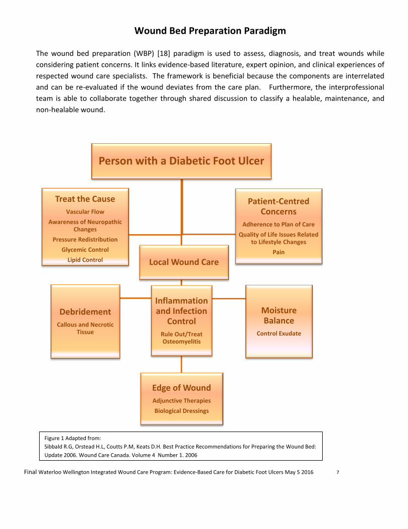
Wound Bed Preparation Paradigm
The wound bed preparation (WBP) [18] paradigm is used to assess, diagnosis, and treat wounds while
considering patient concerns. It links evidence-based literature, expert opinion, and clinical experiences of
respected wound care specialists. The framework is beneficial because the components are interrelated
and can be re-evaluated if the wound deviates from the care plan. Furthermore, the interprofessional
team is able to collaborate together through shared discussion to classify a healable, maintenance, and
non-healable wound.
Person with a Diabetic Foot Ulcer
Debridement
Callous and Necrotic Tissue
Inflammation and Infection
Control
Rule Out/Treat Osteomyelitis
Moisture Balance
Control Exudate
Edge of Wound
Adjunctive Therapies
Biological Dressings
Treat the Cause
Vascular Flow
Awareness of Neuropathic Changes
Pressure Redistribution
Glycemic Control
Lipid Control
Local Wound Care
Patient-Centred Concerns
Adherence to Plan of Care
Quality of Life Issues Related to Lifestyle Changes
Pain
Figure 1 Adapted from:
Sibbald R.G, Orstead H.L, Coutts P.M, Keats D.H. Best Practice Recommendations for Preparing the Wound Bed:
Update 2006. Wound Care Canada. Volume 4 Number 1. 2006
Final Waterloo Wellington Integrated Wound Care Program: Evidence-Based Care for Diabetic Foot Ulcers May 5 2016 8
3. Address Patient-Centered Concerns [2] [1] [19] [20] (see Toolkit Item #6 for worksheet)
(Level B,C: RNAO’s Assessment and Management of Diabetic Foot Ulcers)
(Level la, lb, lll: RNAO’s Strategies to Support Self-Management in Chronic Conditions: Collaboration with Clients)
a Assess Psychosocial Needs /Pain and Quality of Life (QOL)
Communicate with patients, their caregivers and significant others to identify patient-centered goals to
determine realistic expectations for healing or non-healing outcomes.
Although pain is not usually a concern in diabetic foot ulcers, assess pain and in collaboration with patient and
caregivers, create a pain relief plan.6
When pain does occur it may indicate ischemia, infection or Charcot.
Individuals with diabetes need to be informed about the risks and complications, understand the loss of
protective sensation (LOPS), and be taught the problem-solving skills necessary to respond to health-related
problems. 5
Assess quality of life (QOL) (see Toolkit Item #10a and #10b for assessment forms) and screen for mental
health concerns (i.e. depression see Toolkit Item #11 for assessment form)
Encourage and provide ongoing support for smoking cessation if applicable (see Toolkit Item #7a for Smoking,
Chronic Wound Healing, and Implications for Evidence-Based Practice – McDaniel and Browning, Toolkit Item
#7b for Checklist to readiness to quit smoking, see Toolkit Item #7c for Applying 5 A’s to smoking cessation,
see Toolkit Item #7d for WHY test, see Toolkit Item #7e for smoking cessation medication comparison chart
and see Toolkit Item #7f for Strategies to avoid relapse). [20]
b Socioeconomic Determinates of Health (see Toolkit Item #5 for Canadian Nurses Association Social Determinants of
Health and Nursing: A Summary of Issues)
Provide education to patients, caregivers and significant others for care and the management of diabetes
Educate patients, their caregivers and significant others regarding the possible need for long term
compression garments. Assess need for assistance in utilizing garments.
Assess for the presence or absence of social support system for treatment and preventions of diabetic foot
ulcers.
Pressure offloading devices are often expensive and the healthcare professional may need to consult with
appropriate agencies to facilitate access to such treatments for their patients.
Health is a resource for everyday life and is influenced by the determinants of health: income, social status,
support networks, education, employment and working conditions, health services, healthy child development,
physical environment, gender, culture, genetics, and personal health practices. Unemployment, lack of sick
benefits, job insecurity, low income, and homelessness can deter healing and cause more stress. For example,
money is needed to purchase adequate food that is vital for wound healing. Patient may need a referral for a
social worker to assist with finances.
1. RNAO BPG Assessment and Management
of Pain
4. RNAO BPG Integrating Smoking Cessation into
Daily Nursing Practice
3. RNAO BPG Woman Abuse: Screening,
Identification and Initial Response
2. RNAO BPG Assessment and Care of Adults at
Risk of Suicide Ideation and Behaviour
Final Waterloo Wellington Integrated Wound Care Program: Evidence-Based Care for Diabetic Foot Ulcers May 5 2016 9
The following questions could assist in assessing your patient’s financial concerns:
1. Do you have benefits from any other sources to cover cost of insulin/supplies, compression stockings, medical
drugs, parking fees, food allowance. Check for availability for financial compensation (e.g. private insurance,
veterans medical benefits, Ontario Disability Support Program –ODSP/Ontario Works, Non-Insured Health
Benefits -NIHB and Southern Ontario Aboriginal Diabetes Initiative – SOADI for First Nations people and Inuit)
2. Are you the sole bread-winner in your family?
3. How often have you used the food bank or soup kitchen this month?
4. Do you have sick-time benefits or unemployment insurance?
5. Would you like a referral to Meals on Wheels or information on food bank/soup kitchen?
Social Supports
There is evidence to suggest that strong supportive networks improve health and healing [21]. Patients who
have limited social support are more at risk for depression, greater risk for complications, decreased well-being,
poor mental health and physical health. Furthermore, patients who are disabled, migrants from other countries,
ethnic minorities and refugees are vulnerable to racism, discrimination and hostility that may harm their health.
Patients who have stigmatizing conditions such as mental health, addictions (street drug use, methadone
patients and cigarette smokers), and diseases such as HIV/AIDS suffer from higher rates of poverty and limited
supports.
The following questions could assist in assessing your patient’s support system:
1. Do you have someone to help you? Friend, family, neighbor, church member?
2. Does patient seem depressed or suicidal?
3. Do you have transportation to receive medical follow-up and to obtain groceries?
4. Do you have someone to help you with your personal care such as showering?
5. Do you have someone to get your groceries, housekeeping and other necessities?
6. Are you afraid of your partner or family member?
7. Would you like a referral to a social worker or case worker?
c Chronic Disease Self-management
Assess level of patient’s self-management skills
Chronic Disease Self-management
Self-management promotes and strengthens the confidence (self-efficacy) of the patient to be able to care for their
chronic disease [2]. The focus of self-management is to allow the patient to self-identify concerns and to address
these concerns collaboratively with nurses and health professionals. Fostering and promoting independence is
strongly encouraged but the patient and caregiver will need to be assessed by health professional for cognitive and
physical ability.
1. Canadian Nurses Association Social Determinants of
Health and Nursing: A Summary of Issues
Final Waterloo Wellington Integrated Wound Care Program: Evidence-Based Care for Diabetic Foot Ulcers May 5 2016 10
The Self-management Initiative, through the Ontario Ministry of Health and Long-Term Care (MOHLTC), is an
integrated, comprehensive strategy aimed at preventing and improving management of chronic conditions in
Ontario. The goal of this cost-free program is to provide education and skills training workshops to both health care
providers and patients with chronic conditions. For more information, please call 1-866-337-3318 or
www.wwselfmanagement.ca.
2. Self-Management Initiative Link for Health
Care Providers
1. Self-Management Initiative Link for Patients
with Chronic Conditions
Final Waterloo Wellington Integrated Wound Care Program: Evidence-Based Care for Diabetic Foot Ulcers May 5 2016 11
Figure 2: RNAO Clinical Best Practice Guideline: Strategies to Support Self-Management in Chronic Conditions:
Collaboration with Clients [2]
The 5 A’s of Behavioural Change
These activities are not necessarily linear with each step following the other sequentially. The goal of the 5 A’s, in the
context of self-management support, is to develop a personalized, collaborative action plan that includes specific
behavioural goals and a specific plan for overcoming barriers and reaching those goals. The 5 A’s are elements that are
interrelated and are designed to be used in combination to achieve the best results especially when working with patients
in complex health and life situations.
Assess
Beliefs, Behavior and Knowledge
Advise
Provide specific information about health
risks and benefits of change
Agree
Collaboratively set goals based on patient's interest
and confidence in their ability to change the
behaviour
Assist
Identify personal barriers, strategies, problem- solving
techniques and social/environmental
support
Arrange
Specify plan for follow-up (e.g. visits, phone
calls, mailed reminders)
Personal Action Plan
List specific goals in behavioral terms
List barriers and strategies to address
them
Specify follow-up plan
Share plan with practice team and
patient’s social support
Final Waterloo Wellington Integrated Wound Care Program: Evidence-Based Care for Diabetic Foot Ulcers May 5 2016 12
1. ASSESS
Beliefs, Behavior and Knowledge
Establish rapport with patients and families
Screen for depression on initial assessment, at regular intervals and advocate for follow-up treatment of
depression
Establish a written agenda for appointments in collaboration with the patient and family, which may
include:
a) Reviewing clinical data
b) Discussing patient’s experiences with self-management
c) Medication administration
d) Barriers/stressors
e) Creating action plans
f) Patient education including assessing learning style
Consistently assess patient’s readiness for change to help determine strategies to assist patient’s
readiness for change to help determine strategies to assist patient with specific behaviours
Identify patient specific goals
2. ADVISE
Provide specific information about health risks and benefits of change
Combine effective behavioural, psychosocial strategies and self-management education processes as part
of delivering self-management support
Utilize the “ask-tell-ask” (also known as Elicit-Provide-Elicit) communication technique to ensure the
patient receives the information required or requested
Use the communication technique “Closing the Loop” (also known as “ teach back”) to assess a patient’s
understanding of information
Assist patients in using information from self-monitoring techniques (e.g., glucose monitoring, home blood
pressure monitoring) to manage their condition
Encourage patients to use monitoring methods (e.g., diaries, logs, personal health records) to monitor and
track their health condition
Identify community resources for self-management (e.g., support groups)
3. AGREE
Collaboratively set goals based on patient’s interest and confidence in their ability to change the behaviour
Collaborate with patients to:
a) Establish goals
b) Develop action plans that enable achievement of SMART goals (see below)
c) Establish target dates for success of goals and reassessment
d) Monitor progress towards goals
4. ASSIST
Identify personal barriers, strategies, problem-solving techniques and social/environmental support
Use motivational interviewing with patients to allow them to fully participate in identifying their desired
behavioural changes
Final Waterloo Wellington Integrated Wound Care Program: Evidence-Based Care for Diabetic Foot Ulcers May 5 2016 13
Teach and assist patients to use problem-solving techniques
Be aware of community self-management programs in a variety of settings, and link patients to these
programs through the provision of accurate information and relevant resources
5. ARRANGE
Specify plan for follow-up (e.g., visits, phone calls, mailed reminders)
Arrange regular and sustained follow-up for patients based on the patient’s preference and availability
(e.g., telephone, email, regular appointments). Nurses and patients discuss and agree on the
data/information that will be reviewed at each appointment
and share with other interdisciplinary team members involved
Use a variety of innovative, creative and flexible modalities with patients when providing self-management
support such as:
a) Electronic support systems
b) Printed materials
c) Telephone contact
d) Face-to-face interaction
e) New and emerging modalities
Tailor the delivery of self-management support strategies to the patients’ culture, social and economic
context across settings
Facilitate a collaborative practice team approach for effective self-management support
Share with caregiver/family members/circle of care
Final Waterloo Wellington Integrated Wound Care Program: Evidence-Based Care for Diabetic Foot Ulcers May 5 2016 14
Figure 3: College of Nurses Ontario SMART Goals [22]
College of Nurses SMART Goals Link
Time-limited
A time-limited goal has specific timelines and a deadline. This will help motivate you to move toward your goal and to evaluate your progress
Relevant
A relevant goal applies to your current role and is clearly linked to your key role & responsibilities.
Attainable
An attainable goal can be achieved based on your skill, resources and area of practice.
Measurable
A measurable goal is quantifiable, meaning you can see the results.
Specific
A specific goal is detailed, focused and clearly stated. Everyone reading the goal. Everyone should know exactly what you want to learn.
Final Waterloo Wellington Integrated Wound Care Program: Evidence-Based Care for Diabetic Foot Ulcers May 5 2016 15
Stages of Change Model
Table 1: RNAO Clinical Best Practice Guideline: Strategies to Support Self-Management in Chronic Conditions:
Collaboration with Clients [2]
Stage in Transtheoretical Model of
Change
Patient Stage
Pre-contemplation
Not thinking about change; may be resigned Feeling of no control Denial:
does not believe it applies to self Believes consequences are not serious
Contemplation
Weighing benefits and costs of behavior, proposed change
Preparation
Experimenting with small changes
Action
Taking a definitive action to change
Maintenance
Maintaining new behavior over time
Relapse
Experiencing normal part of process of change Usually feels demoralized
Final Waterloo Wellington Integrated Wound Care Program: Evidence-Based Care for Diabetic Foot Ulcers May 5 2016 16
There are 3 self-management strategies that health professionals can use to
promote self-management in patients with Diabetic Foot Ulcers [2]
Final Waterloo Wellington Integrated Wound Care Program: Evidence-Based Care for Diabetic Foot Ulcers May 5 2016 21
PATHOGENESIS OF ULCERATION
The reason for the susceptibility and increase incidence of foot ulcers in the diabetic patient is because of the interaction of several
pathogenic factors. Research supports this reason establishing that “diabetic foot ulcers result from the simultaneous action of
multiple contributing causes,” [26], [45], [46] [47] while the main principal causes are noted to be peripheral neuropathy and
ischemia from peripheral vascular disease, [33] [39] [48] [26] [10] [49]. The four pathogenic factors that trigger the formation of an
ulcer in the diabetic patient includes abnormal foot biomechanics, peripheral arterial disease, neuropathy and poor wound healing
[50] [26] [32] [51] [13] [25] [45] [23] [17].
Abnormal Foot
Biomechanics
Neuropathy
Peripheral Arterial Disease
Poor Wound Healing
SUCCESSFUL DIAGNOSIS AND MANAGEMENT OF PATIENTS WITH
DIABETIC FOOT ULCERS INVOLVES A HOLISTIC-MULTIDISCIPLINARY
APPROACH THAT INCLUDES:
1. Optimal diabetes control
2. Effective local wound care
3. Infection control
4. Pressure relieving strategies
5. Restoring pulsatile blood flow
[17] [31] [34] [12] [78] [57] [40] [6] [11] [1]
Final Waterloo Wellington Integrated Wound Care Program: Evidence-Based Care for Diabetic Foot Ulcers May 5 2016 22
General Inspection
A careful inspection of the feet in a well-lit room should always be carried out after the patient has removed shoes and socks. Because inappropriate footwear and foot deformities are common contributory factors in the development of foot ulceration [1] [13], the shoes should be inspected and the question “Are these shoes appropriate for these feet?” should be asked. Examples of inappropriate shoes include those that are excessively worn or are too small. See page # for more information on offloading.
Skin Assessment
The dermatological assessment should initially include a global inspection, including interdigital, for the presence of
ulceration or areas of abnormal erythema. The presence of callus (particularly with hemorrhage), nail dystrophy, or
paronychia (infection next to nail) should be recorded [54], with any of these findings prompting referral to a specialist or
specialty clinic. Focal or global skin temperature differences between one foot and the other may be predictive of either
vascular disease, infection or ulceration and could also prompt referral for specialty foot care [55] [56] [57] [35].
Musculoskeletal Assessment
The musculoskeletal assessment should include evaluation for any gross deformity [16]. Rigid deformities are defined as
any contractures that cannot easily be manually reduced and are most frequently found in the digits. Common forefoot
deformities that are known to increase plantar pressures and are associated with skin breakdown include metatarsal
phalangeal joint hyperextension with interphalangeal flexion (claw toe) or distal phalangeal extension (hammer toe) [58]
[59] [5] [17].
Neurological Assessment
Peripheral neuropathy is the most common component cause in the pathway to diabetic foot ulceration [60] [31] [44] [61].
The clinical exam recommended, however, is designed to identify loss of protective sensation (LOPS) rather than early
neuropathy. The diagnosis and management of the latter were covered in a 2004 ADA technical review [32]. The clinical
examination to identify LOPS is simple and requires no expensive equipment.
Factors that may affect healing potential:
Local
Presence of necrosis, foreign body and/or infection
Final Waterloo Wellington Integrated Wound Care Program: Evidence-Based Care for Diabetic Foot Ulcers May 5 2016 36
Targets for Glycemic Control for Most People with Type 1 and Type 2 Diabetes
Fasting plasma glucose of 4.0 to 7.0 mmol/L (8.0 for the elderly to prevent chance of hypoglycemia)
A1C ≤ 7.0% to reduce the risk of microvascular and macrovascular complications 2-hour postprandial (after meal) plasma glucose targets of 5.0 to l0.0 mmol/L
(5.0 to 8.0 mmol/L if A1C targets not being met)
If the presence of any of these signs of micronutrient deficiencies is noted, a referral should be made to a Registered Dietitian who can work with the primary care provider for screening of dietary deficiencies and treatment.
The
Nestle Mini-Nutritional Assessment (MNA) ( Toolkit item #11) is a screening and assessment tool that identifies
individuals age 65 and above who are malnourished or at risk of malnutrition, allowing for earlier intervention to
provide adequate nutritional support. It has not been validated for use with younger individuals. The screening
tool consists of 6 questions.
Complete the screen by filling in the boxes with the appropriate numbers.
Total the numbers for the screening score.
The screening score (max 14 points):
12- 14 points = normal nutritional status
8-11 points = at risk of malnutrition
0 -7 points = malnourished
Link to Mini-Nutritional Assessment Form
As recommended by the Canadian Diabetes Association (CDA) Clinical Practice Guidelines
(CDA CPG Expert Committee, 2008)
Final Waterloo Wellington Integrated Wound Care Program: Evidence-Based Care for Diabetic Foot Ulcers May 5 2016 37
i. Assess for Infection
The validated mnemonics “NERDS” and “STONEES” classify the signs and symptoms of localized infection (NERDS) and spreading
infection (STONEES). Increased localized pain is a significant predictor of deep compartment infection.
Presence of Superficial Bacteria
N- Non-healing wound
E- Exudate increased
R- Red friable (fragile tissue that bleeds easily)
D- Debris (presence of necrotic tissue (eschar/slough) in wound
S- Smell
Presence of Spreading Bacteria (< 3 low bacteria count, >3 high bacteria count)
S- Size increasing
T- Temperature increased (> 3 degrees F difference)
O- Os (probes to bone or bone is increased)
N- New areas of breakdown
E- Exudate present
E- Erythema and/or Edema
S- Smell
j. Relief of Pressure and Protection of Ulcer
(Level A: RNAO’s Assessment and Management of Diabetic Foot Ulcers [1])
The International Working Group Diabetic Foot Ulcers outlines the importance of offloading to enhance wound healing:
This is a cornerstone in treating an ulcer associated with increased biomechanical stress
The preferred treatment for a neuropathic plantar ulcer is a non-removable knee-high offloading device, either total
contact cast (TCC) or removable walker rendered irremovable
When a non-removable TCC or walker is contra-indicated or not tolerated, use a removable device
When these devices are contra-indicated, use footwear that best offloads the ulcer
Offloading is for LIFE!
Final Waterloo Wellington Integrated Wound Care Program: Evidence-Based Care for Diabetic Foot Ulcers May 5 2016 38
In non-plantar ulcers, consider offloading with shoe-modifications, temporary footwear, toe-spacers or orthoses
If other forms of biomechanical relief are not available, consider felted foam, in combination with appropriate
footwear
Instruct the patient to limit standing and walking, and to use crutches or wheelchair if necessary
Ongoing Offloading Care (per Waterloo Wellington Pathway for Diabetic Foot Ulcer document)
Ensure appropriate footwear/offloading referrals have been arranged to a qualified offloading specialist (if patient
does not have)
Review weartime of offloading device as per treating practitioner’s directions
Review adherence to using appropriate footwear and/or offloading device(s)
Assess barriers to appropriate offloading
Initial and ongoing callous reduction is part of offloading
Assess for secondary complications of offloading and refer concerns to dispensing practioner
1. look for redmarks, blisters, skin abrasions
2. ask about knee, hip or back issues (including contralateral limb) due to height difference of offloading
device
3. check for unsafe gait (are they stable, using appropriate aids, etc)
Check gait aids such as walker, cane, crutches
Review long term goals of offloading (i.e. transition from cast to shoes, foot orthoses, etc.)
Teach patient to assess for secondary complications
Total Contact Cast or Prefabricated Removable Walking
Casts (rendered irremovable) is GOLD STANDARD of Care
Contraindications for Total Contact Casting (TCC)
Patients with documented lower-extremity arterial disease
Patients with an active wound infection or a sinus tract with deep extension into the foot which requires daily wound
access for topical wound management
Patients with unstable gait
Patients with cast claustrophobia or previously known non-adherence to treatment plan
Patients with fluctuating leg edema or active skin disease
Inadequately trained clinical staff
Restless leg syndrome or conditions which cause leg tremors
Final Waterloo Wellington Integrated Wound Care Program: Evidence-Based Care for Diabetic Foot Ulcers May 5 2016 39
Discuss winter footwear with appropriate offloading specialist
Check for availability for financial compensation (e.g. private insurance, veterans medical benefits, Ontario
Disability Support Program –ODSP, Assisted Devices, Non-Insured Health Benefits -NIHB and Southern Ontario
Aboriginal Diabetes Initiative – SOADI for First Nations people and Inuit)
Final Waterloo Wellington Integrated Wound Care Program: Evidence-Based Care for Diabetic Foot Ulcers May 5 2016 40
k. Determine if the wound is “Healable, Maintenance or Non-Healable
Healable Wounds: Have sufficient vascular supply, underlying cause can be corrected, offloaded & health can be
optimized
Goal: Principles of wound bed preparation and moist wound healing: treat the cause, debridement, bacterial
balance, exudate control, protect peri-wound skin
Maintenance Wounds: have healing potential, but various patient factors are compromising wound healing at
this time
Goal: Principles of wound bed preparation and moist wound healing: treat the cause, debridement, bacterial
balance, exudate control and protect peri-wound skin. Avoid higher cost advanced wound treatments until
factors compromising wound healing are resolved. Focus on quality of life issues, exudate and odour
management
Non-healable/Palliative wounds: has no ability to heal due to untreatable causes such as terminal disease or
end-of-life
Goal: Avoid higher cost advanced wound treatment and focus on exudate and odour management, quality of life
issues. [1].
Final Waterloo Wellington Integrated Wound Care Program: Evidence-Based Care for Diabetic Foot Ulcers May 5 2016 41
Provide Local Wound Care
a. Intervention Algorithm
Final Waterloo Wellington Integrated Wound Care Program: Evidence-Based Care for Diabetic Foot Ulcers May 5 2016 42
b. Signs and Symptoms of Wound Infection
(Level A, B and C: RNAO’s Assessment and Management of Diabetic Foot Ulcers [1])
Diabetic Foot Ulcers, like most chronic wounds, can become infected with superficial or spreading bacteria.
However, the risk for infection in the diabetic foot is especially problematic. Reasons for heightened risk of infection
in the Diabetic Foot include [14]:
• Immune compromised host
• Poor glycemic control
• Poor granulation and prolonged wound healing
• More than 50% of foot infections in diabetics lack elevated WBC and erythrocyte sedimentation rate or fever
• High colonization with staph/fungal
The average cost of healing a single ulcer is $8,000, that of an infected ulcer is $17,000, and that of a major amputation is
$45,000 [70]. Limb-threatening diabetic foot infections are usually polymicrobial. Commonly encountered pathogens
include methicillin-resistant staphylococcus aureus, β-hemolytic streptococci, enterobacteriaceae, pseudomonas
aeruginosa, and enterococci. Anaerobes, such as bacteroides, peptococcus, and peptostreptococcus, are rarely the sole
pathogens but are seen in mixed infections with aerobes. Antibiotics selected to treat severe or limb-threatening infections
should include coverage of gram-positive and gram-negative organisms and provide both aerobic and anaerobic coverage
[70].
Proper debridement is necessary to decrease the risk of infection and reduce peri-wound pressure, which can impede
normal wound contraction and healing [18] [31] [1] [16] [49] [34] [27] [18].
The International Working Group has created the IWGDF Guidance on the diagnosis, and management of infection in
patients with foot ulcers in diabetes [16]. The Guideline outlines assessment procedures, recommendations, treatment and
rationales.
Classification/Diagnosis
1. Diabetic foot infection must be diagnosed clinically, based on the presence of local or systemic signs or
symptoms of inflammation (Strong; Low).
2. Assess the severity of any diabetic foot infection using the Infectious Diseases Society of
America/International Working Group on the Diabetic Foot classification scheme (Strong; Moderate)
Signs and Symptoms Specific to Diabetic Foot Infection
Usual signs and symptoms of infection may be more subtle in patients with diabetes Local Infection: NERDS (non-healing, exudate, red friable tissue, debris, smell) Deep Infection: STONEES (size increasing, temperature increase, os – probes to bone, new areas of breakdown, exudate, erythema, edema, smell)
Elevated blood sugars from patient’s baseline
Increase in pain level (new pain is a red flag in patients with altered sensation)
Generalized malaise/fever
Wound probes to bone (likely osteomyelitis)
Link to IWGDF Guidance on the diagnosis and management of infection
in the Diabetic Foot Ulcer Patient [16]
Final Waterloo Wellington Integrated Wound Care Program: Evidence-Based Care for Diabetic Foot Ulcers May 5 2016 43
c. Classification Systems
Table 8: Classification Systems of Infection and Ischemia - DFU
Classification
system
Key Points Pros/Cons References
Wagner Assesses ulcer depth along with
presence of gangrene and loss of
perfusion using six grades (0-5)
Well established58
Does not fully address infection
and ischemia
Wagner 198159
University of
Texas
(Armstrong)
Assesses ulcer depth, presence of infection and presence of signs of lower-extremity ischemia using a matrix of four grades combined with four stages
Well established58
Describes the presence of
infection and ischemia better
than Wagner and may help in
predicting the outcome of the
DFU
Lavery et al 199660
Armstrong et al
199852
PEDIS Assesses Perfusion, Extent (size),
Depth (tissue loss), Infection and
Sensation (neuropathy) using
four grades (1-4)
Developed by IWGDF
User-friendly (clear definitions,
few categories) for practitioners
with a lower level of experience
with diabetic foot management
Lipsky et al 201246
SINBAD Assesses Site, Ischemia, Neuropathy, Bacterial infection and Depth
Uses a scoring system to help
predict outcomes and enable
comparisons between different
settings and countries
Simplified version of the S(AD)SAD classification system61
Includes ulcer site as data
suggests this might be an
important determinant of
outcome62
Ince et al 200863
Final Waterloo Wellington Integrated Wound Care Program: Evidence-Based Care for Diabetic Foot Ulcers May 5 2016 44
D Infection & Ischemia Infection & Ischemia Infection & Ischemia
Infection & Ischemia
Table 10: Limb-Threatening Infection in Patients with a Diabetic Foot Ulcer [70, 71]
SUPERFICIAL INFECTION
■ Non-healing
■ Bright red granulation tissue
■ Friable and exuberant granulation
■ New areas of breakdown or necrosis
■ Increased exudates
■ Bridging of soft tissue and the
epithelium
■ Foul odour
DEEP WOUND INFECTION
■ Pain
■ Swelling, induration
■ Erythema (> 2 cm)
■ Wound breakdown
■ Increased size or satellite areas
■ Undermining or tunneling
■ Probing to bone
■ Anorexia
■ Flu-like symptoms
■ Erratic glucose control
SYSTEMIC INFECTION
In addition to deep wound infection:
■ Fever
■ Rigour
■ Chills
■ Hypotension
■ Multi-organ failure
Final Waterloo Wellington Integrated Wound Care Program: Evidence-Based Care for Diabetic Foot Ulcers May 5 2016 45
d. Management of Infection
Swabs for C&S not usually helpful if wound is dry; if wet then should be done using LEVINE semi-quantitative method
In addition to recognizing the signs and symptoms of infection in diabetic foot ulcers, it may be helpful to obtain a culture and sensitivity (C&S) using a validated method of sampling to quantify bacteria in wounds
Tissue biopsies are considered the gold standard but unfortunately are not practical in many settings.
A linear relationship between quantitative tissue biopsy and swab for C&S taken using the Levine method of sampling (see below) has been validated and is recommended for assessing any open wound
Swabs for C&S are important in determining the type of bacteria and the appropriate antibiotics, but are not necessary to confirm the presence or absence of infection.
C&S results may not reflect the presence or absence of biofilm.
Levine Method for obtaining C&S laboratory swab [18]
1. Cleanse wound thoroughly
2. Place swab on granulation tissue
3. Apply enough pressure to extract fluid
4. Turn swab 360 degrees on fluid (avoid slough or debris)
5. Place swab in transport medium
IWGDF Guidance on the diagnosis and management of foot infections in persons with diabetes [16]
Diabetic foot infection must be diagnosed clinically, based on the presence of local and systemic signs and symptoms of
inflammation (Strong; moderate). Assess the severity of any diabetic foot infection using the Infectious Diseases Society of
America/International Working Group on the Diabetic Foot classification scheme. [16]
The full PEDIS system (which includes classification of other wound descriptors, such as arterial disease, neuropathy and
wound size) of the IWGDF was originally developed for research purposes, but it can serve as a clinical classification as well
[16]. Classification of DFIs using the full PEDIS system or the infection part of the IWGDF/IDSA DFI scheme has been shown
in several prospective studies to predict the need for hospitalization or lower extremity amputation [16].
Deep foot infections have been identified as the immediate cause of 25 to 51% of amputations in persons
with diabetes
Signs of deep wound and systemic signs of infection are potentially limb and/or life threatening. These
clinical signs and symptoms require urgent medical attention.[73]
Lipinsky (2012) recommends that persons with new diabetic foot infections have plain radiographs to
identify bony abnormalities such as bone deformity or destruction, foreign bodies or soft tissue gas. An
abnormal plain radiograph finding can be helpful in the diagnosis of osteomyelitis [84] [1].
Final Waterloo Wellington Integrated Wound Care Program: Evidence-Based Care for Diabetic Foot Ulcers May 5 2016 46
a. While virtually all clinically infected diabetic foot wounds require antimicrobial therapy do not treat clinically
uninfected wounds with antimicrobial therapy (Strong; Low)
b. Select specific antibiotic agents for treatment based on the likely or proven causative pathogens, their antibiotic
susceptibilities, the clinical severity of the infection, evidence of efficacy of the agent for DFI and costs (Strong;
Moderate)
c. A course of antibiotic therapy of 1-2 weeks is usually adequate for most mild and moderate infections (Strong;
High)
d. Administer parenteral therapy initially for most severe infections and some moderate infections, with a switch to
oral therapy when the infection is responding (Strong; Low)
e. Do not select a specific type of dressing for a diabetic foot infection with the aim of preventing an infection or
improving its outcome (Strong; High)
f. For diabetic foot osteomyelitis we recommend 6 weeks of antibiotic therapy for patients who do not undergo
resection of infected bone and no more than a week of antibiotic treatment if all infected bone is resected (Strong;
Moderate)
g. We suggest not using any adjunctive treatments for diabetic foot infection. (Weak; Low)
h. When treating a diabetic foot infection, assess for use of traditional remedies, previous antibiotic use, and consider
local bacterial pathogens and their susceptibility profile. (Strong; Low)
i. We recommend sending a specimen for culture that is from deep tissue, obtained by biopsy or curettage after the
wound has been cleansed and debrided. We suggest avoiding swab specimens, especially of inadequately debrided
wounds, as they provide less accurate results [72] [73]
j. We recommend that clinicians select an empiric antibiotic regimen on the basis of the severity of the infection and
the likely etiologic agent(s) (strong, low).
k. For mild to moderate infections in patients who have not recently received antibiotic treatment, we suggest that
therapy just targeting aerobic GPC is sufficient (weak, low).
l. For most severe infections, we recommend starting broad-spectrum empiric antibiotic therapy, pending culture
results and antibiotic susceptibility data (strong, low).
m. Empiric therapy directed at Pseudomonas aeruginosa is usually unnecessary except for patients with risk factors
for true infection with this organism (strong, low).
n. Consider providing empiric therapy directed against methicillin-resistant Staphylococcus aureus (MRSA) in a
patient with a prior history of MRSA infection; when the local prevalence of MRSA colonization or infection is high;
or if the infection is clinically severe (weak, low). [73]
Final Waterloo Wellington Integrated Wound Care Program: Evidence-Based Care for Diabetic Foot Ulcers May 5 2016 47
o.
Table 11. Infectious Diseases Society of America and International Working Group on the Diabetic Foot Classifications of
Diabetic Foot Infection
Clinical Manifestation of Infection PEDIS
Grade
IDSA Infection
Severity
No symptoms or signs of infection 1 Uninfected
Infection present, as defined by the presence of at least 2 of the following items:
• Local swelling or induration
• Erythema
• Local tenderness or pain
• Local warmth
• Purulent discharge (thick, opaque to white or sanguineous secretion)
Local infection involving only the skin and the subcutaneous tissue (without
involvement of deeper tissues and without systemic signs as described below). If
erythema, must be >0.5 cm to ≤2 cm around the ulcer.
Exclude other causes of an inflammatory response of the skin (e.g. trauma, gout, acute
Ischemia may increase the severity of any infection, and the presence of critical ischemia often makes the infection severe. Systemic infection may sometimes manifest with other clinical findings, such as hypotension, confusion, vomiting, or evidence of metabolic disturbances, such as acidosis, severe hyperglycemia, and new-onset azotemia
[29, 43, 44]
Final Waterloo Wellington Integrated Wound Care Program: Evidence-Based Care for Diabetic Foot Ulcers May 5 2016 48
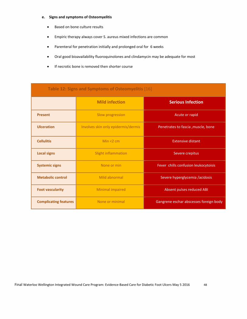
e. Signs and symptoms of Osteomyelitis
Based on bone culture results
Empiric therapy always cover S. aureus mixed infections are common
Parenteral for penetration initially and prolonged oral for 6 weeks
Oral good bioavailability fluoroquinolones and clindamycin may be adequate for most
If necrotic bone is removed then shorter course
Table 12: Signs and Symptoms of Osteomyelitis [16]
Mild infection Serious Infection
Present Slow progression Acute or rapid
Ulceration Involves skin only epidermis/dermis Penetrates to fascia ,muscle, bone
Cellulitis Min <2 cm Extensive distant
Local signs Slight inflammation Severe crepitus
Systemic signs None or min Fever chills confusion leukocytoisis
Metabolic control Mild abnormal Severe hyperglycemia /acidosis
Foot vascularity Minimal impaired Absent pulses reduced ABI
Complicating features None or minimal Gangrene eschar abscesses foreign body
Final Waterloo Wellington Integrated Wound Care Program: Evidence-Based Care for Diabetic Foot Ulcers May 5 2016 49
f. Antiseptics Guidelines
Recommendations for the use of antiseptics and antiseptic dressings
An international consensus panel studied use of silver in healable wounds. This panel recommended that silver be used for a two week period if infections is suspected and then be reassessed. It is the opinion of Dr. David Keast, a leading wound care specialist that these recommendations can be extended to the use of all antiseptics and antiseptic dressings (eg. iodine and PHMB).
Choices for after initial two weeks using antiseptics or antiseptic dressings
Healable wounds
Bacterial burden has been reduced and the wound is progressing to healing
Discontinue use of antiseptics and antiseptic dressings
Bacterial burden has been reduced the wound is progressing but there are patient risk factors that put them at risk of re-infection
Continue to use and monitor Suggest: Low adherent knitted viscose fabric impregnated with a polyethylene glycol (PEG) base containing 10% Povidone Iodine
Bacterial burden is controlled but the location of the wound is such that it is at risk of recontamination e.g. perianal, or exit sites for g-tubes etc
Continue to use as an antimicrobial barrier.
No effect Discontinue and change strategy such as systemic antibiotics or a change of the topical antiseptic or better debridement. As always factors such as adequate plantar pressure redistribution in neuropathic foot ulcers or compression therapy for venous disease must be in place.
Slough/Eschar
No slough or obvious biofilm present Suggest: Iodine gel
Slough is present Topical antiseptic to remove biofilm needed
Suggest: Periodic debridement provided arterial blood supply is adequate
Maintenance
or Non-healable Wounds
Eschar to be kept dry No real limit to use
Use as long as required
Suggest:
Povidone iodine is best as an antiseptic with drying properties. Use it as long as required to keep dry
Table 13: As per Dr. Stephan Landis and Dr. David Keast (Leading Wound Care Specialists) Aug. 2015
Final Waterloo Wellington Integrated Wound Care Program: Evidence-Based Care for Diabetic Foot Ulcers May 5 2016 50
g. Antibiotics Guidelines
Antibiotics should be prescribed using local protocols and, in complex cases, the advice of a clinical microbiologist or
Final Waterloo Wellington Integrated Wound Care Program: Evidence-Based Care for Diabetic Foot Ulcers May 5 2016 51
Agents in bold f ace type are those that have been most commonly used as comparators in clinical trials (see Table 7). The only agents currently specifically FDA approved for
diabetic foot infections are shown in italics.
Narrow-spectrum agents (eg, vancomycin, linezolid, daptomycin) should be combined with other agents (eg, a fluoroquinolone) if a polymicrobial infection (especially
moderate or severe) is suspected.
Use an agent active against MRSA for patients who have a severe infection, evidence of infection or colonization with this organism elsewhere, or epidemiological risk factors
for MRSA infection.
Select definitive regimens after considering the results of culture and susceptibility tests from wound specimens, as well as the clinical response to the empiric regimen.
Similar agents of the same drug class can probably be substituted for suggested agents.
Some of these regimens do not have FDA approval for complicated skin and skin structure infections.
Abbreviations: CPK, creatine phosphokinase; ESBL, extended-spectrum β-lactamase; FDA, US Food and Drug Administration; IV, intravenous; MIC, minimum inhibitory
concentration; MRSA, methicillin-resistant Staphylococcus aureus; MSSA, methicillin-sensitive Staphylococcus aureus; PO, oral; QID, 4 times a day; TID, 3 times a day.
Agents approved for treating skin and skin structure infections on the basis of studies that excluded patients with diabetic foot infections (eg, ceftaroline, telavancin) are not
included. b Agents shown to be effective in clinical trials including patients with diabetic foot infections. Daptomycin or linezolid may be substituted for vancomycin.
excessively broad. High rates of nausea and vomiting and increased mortality warning. Non-equivalentto ertapenem + vancomycin in 1 randomized clinical trial
Levofloxacinb or
ciprofloxacinb with
clindamycinb
Limited evidence supporting clindamycin for
severe S. aureus infections; PO & IV
formulations for both drugs
Imipenem-cilastatinb Very broad-spectrum (but not against MRSA);
use only when this is required. Consider when
ESBLproducing pathogens suspected
MRSA Linezolidb Expensive; increased risk of toxicities when
used >2 wk
Daptomycinb Once-daily dosing. Requires serial monitoring of
CPK
Vancomycinb Vancomycin MICs for MRSA are gradually
increasing
Pseudomonas
aeruginosa Piperacillin-tazobactam
b TID/QID dosing. Useful for broadspectrum coverage. P. aeruginosais an uncommon pathogen indiabetic foot infections except in special circumstances (2)
Link to Infectious Diseases Society of America/International
Working Group on the Diabetic Foot classification scheme
Final Waterloo Wellington Integrated Wound Care Program: Evidence-Based Care for Diabetic Foot Ulcers May 5 2016 52
h. Determining Goals for Local Treatment for Diabetic Foot Ulcers
(Level A, B and C: RNAO’s Assessment and Management of Diabetic Foot Ulcers)
Healable Wounds: Have sufficient vascular supply, underlying cause can be corrected, offloaded & health can be
optimized
Goal: Principles of wound bed preparation and moist wound healing: treat the cause, debridement, bacterial
balance, exudate control, protect peri-wound skin
Maintenance Wounds: have healing potential, but various patient factors are compromising wound healing at this
time
Goal: Principles of wound bed preparation and moist wound healing: treat the cause, debridement, bacterial
balance, exudate control and protect peri-wound skin. Avoid higher cost advanced wound treatments until factors
compromising wound healing are resolved. Focus on quality of life issues, exudate and odour management
Non-healable/Palliative wounds: has no ability to heal due to untreatable causes such as terminal disease or end-
of-life
Goal: Avoid higher cost advanced wound treatment and focus on exudate and odour management, quality of life
issues. [1].
i. Utilize Product Picker from Canadian Association of Wound Care (CAWC)
Product Picker for Classification of Dressing Products
Each organization may use the PDF Fillable CAWC Product Picker to list the products available within their
organization (see Toolkit Item #14)
j. South West Regional Wound Care Program’s Dressing and Wound Cleansing Table:
Healable and Non-Healable/Maintenance Wounds
Link to Product Picker
Canadian Association for Enterostomal Therapy’s ‘Evidence-Based
Recommendations for Conservative Sharp Wound Debridement’
If healing potential is not established, aggressive debridement and moist interactive healing is not recommended. [6] [1] [81] [27] [57]
South West Regional Dressing and Cleansing Enabler
Healable Wounds
South West Regional Dressing and Cleansing Enabler Non-
Healable/Maintenance Wounds
Final Waterloo Wellington Integrated Wound Care Program: Evidence-Based Care for Diabetic Foot Ulcers May 5 2016 53
NOTE: Normal saline and sterile water do NOT contain preservatives and must be discarded 48 hours
after opening
k. Patient Education on Foot Care and Daily Assessment [1]
Ongoing Foot care is a vital element to promote wound healing and prevent recurrence of Diabetic Foot Ulcers
The following information is provided to clients as recommended practices:
• Foot care -- Podiatrist or chiropodist
• Foot wear -- protective shoes and pressure reduction
• Daily foot assessment
• Monofilament testing for neuropathy
• Glycemic control
• Prophylactic surgery
Foot soaks are NOT recommended for patients with a Diabetic Foot Ulcer. There is currently no proven benefit to
soaking diabetic feet, and in fact there is the potential for maceration of tissues and increased risk of infection. Use
of hot water may not be recognized by the patient due to the presence of neuropathy leading to thermal tissue
damage and soaking in antiseptic chemicals such as hydrogen peroxide maybe damaging to healthy granulation
tissue [48] [27] [74] [57] [10] [13] [1].
l. Adjunctive Therapies
Consider Multi-disciplinary referrals for adjunctive therapy.
Adjunctive therapy refers to additional treatment used together with the primary treatment to achieve the
outcome of the primary treatment.
There are many types of adjunctive therapies for wound management. The ones contained in this resource
include only those that have been verified by rigorous research standards and are included in the RNAO/CAWC
best practice guidelines.
Electrical Stimulation Therapy (EST)
(Level B: RNAO’s Assessment and Management of Diabetic Foot Ulcer [1])
Refers to the application of a low level electrical current to the base of a wound or peri-wound using
conductive electrodes to induce cellular activity to facilitate wound healing.
Therapeutic Ultrasound (TU)
(Level A: RNAO’s Assessment and Management of Diabetic Foot Ulcer 4
)
Refers to the therapeutic application of ultrasound waves to the base of a wound or peri-wound to
induce cellular activity to facilitate wound healing.
Final Waterloo Wellington Integrated Wound Care Program: Evidence-Based Care for Diabetic Foot Ulcers May 5 2016 54
6. Provide Organizational Support
a. Multi-disciplinary Team Intervention Referral Criteria Checklist
b. Patient, Caregiver and Healthcare Provider Teaching and Learning Resources
RNAO Learning Package: Assessment and Management of Diabetic Foot Ulcers (see Toolkit Item #18)
Diabetes Passport (Ministry of Ontario)
Regional Resources
c. Discharge or Transfer Planning and Communications
Regardless of the method of providing the information (e.g. Care Connect, photocopy or Discharge Summary), it is
agreed that the following information is critical in providing seamless care when individuals who have diabetic foot
ulcers are being discharged or transferred to a different care setting:
Current blood work results
Vascular study results
Current and past treatment regimes
Any surgical interventions?
Primary Care Physician
Community Nursing
Advanced Wound Specialist
Nurse Practitioner
Infectious Disease Specialist
Vascular Surgeon
Orthopedic Surgeon
Dermatologist
Plastic Surgeon
Internist/Endocrinologist
Nephrologist
Cardiologist
Opthalmologist/Optometrist
Mental Health Specialist
Psychologist/Psychiatrist
Social worker
Registered Dietitian
Pharmacist
Occupational Therapist
Physiotherapist
Physiatrist
Registered Kinesthologist
Chiropodist
Diabetic Education Program Patient self-referral link