45
Wavelets and functional MRI Ed Bullmore Mathematics in Brain Imaging IPAM, UCLA July 21, 2004
| Date post: | 04-Jan-2016 |
| Category: |
Documents |
| Upload: | erasmus-hutchinson |
| View: | 45 times |
| Download: | 0 times |

Wavelets and functional MRI
Ed Bullmore
Mathematics in Brain Imaging
IPAM, UCLA
July 21, 2004

• What’s a wavelet?
• Wavelet, fractal, brain
• Wavelets and functional MRI data analysis
- resampling or “wavestrapping”
- estimating noise and signal parameters
- hypothesis testing
Overview
(See Bullmore et al [2004] NeuroImage, IPAM issue, for more comprehensive literature review)

Wavelets are “little waves”, defined by their scale j = 1,2,3…J and their
location k = 1,2,3…K = N/2j
…a family of orthonormal wavelets tesselates the time-frequency plane
Wavelets are little waves…
ψ φ
1)( dtt 1)( dtt
1)( dtt 0)( dtt

Wavelets provide an orthonormal basis for multiresolution analysis
j
j
jkj
ktt
2
2
2
1)(,
k k
kjkjJj
kJkJ da ,,,, y
The scaling function multiplied by the approximation coefficients, plus the wavelets multiplied by the detail coefficients, recovers the signal losslessly

Wavelets are adaptive to non-stationarity
“If we are interested in transient phenomena – a word pronounced at a particular time, an apple located in the left corner of an image – the
Fourier transform becomes a cumbersome tool.” Mallat (1998)

Wavelets have decorrelating or whitening properties
)(2'
',', '22~,RH
jjkjkj kkdd
Correlation between any two wavelet coefficients will decay exponentially as a exponential function of distance between them depending on…
H = Hurst exponent of time series
R = regularity of wavelet (number of vanishing moments)
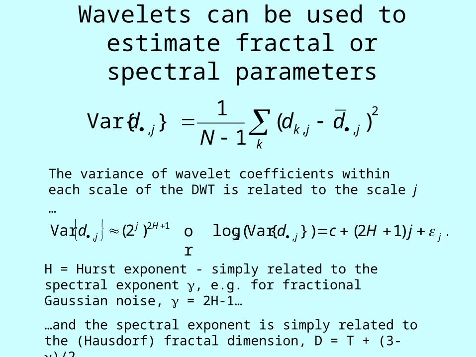
Wavelets can be used to estimate fractal or spectral parameters
2
,,, )(1
1}Var{
kjjkj dd
Nd
The variance of wavelet coefficients within each scale of the
DWT is related to the scale j …
12, )2(Var
Hjjd .)12(}){Var(log ,2 jj jHcd or
H = Hurst exponent - simply related to the spectral exponent , e.g. for fractional Gaussian noise, = 2H-1…
…and the spectral exponent is simply related to the (Hausdorf) fractal dimension, D = T + (3-)/2

Wavelet variance scaling in fMRI
Greater variance at coarser scales of the decomposition
Gradient of straight line fitted to log-linear plot = 2H+1

The discrete wavelet transform is quick to compute
Mallat’s pyramid algorithm
– iterative high and low pass filtering of downsampled coefficients
– O(N) complexity, compared to O(N log(N)) for FFT

• Fractal, scaling or scale-invariant, self-similar or self-affine structure is very common in nature, including biological systems
•A wavelet basis is fractal and so a natural choice of basis for analysis of fractal data
• Brain imaging data often have scaling properties in space and time
• Therefore, wavelets may be more than just another basis for analysis of fMRI data
Wavelet, fractal, brain
Continuous wavelet transform of fractal fMRI time series

Biological fractals…• complex, patterned,
• statistically-self similar,
• scale invariant structures,
• with non-integer dimensions,
• generated by simple iterative rules,
• widespread in real and synthetic natural systems, including the brain.

Fractal cardiologyApparently complex, but simply generated, dendritic anatomy
Statistically self-similar (self-affine) behavior in time
1/f-like spectral properties

Brain images often have fractal properties in space (and time)
Biological
Synthetic

Allometric scaling and self-similar scaling in brain structure
Zhang & Sejnowski (2000) PNAS 97, 5621-5626
Kiselev et al (2004) NeuroImage 20, 1765-1774

Power law scaling of local field
potentials
Leopold et al (2003) Cerebral Cortex

Power law scaling of fMRI time seriesRaw fMRI
Images, Time
MR
sig
na
l
0 20 40 60 80 100 120
10
40
10
60
10
80
11
00
Movement-corrected fMRI
Images, Time
Co
rre
cte
d M
R s
ign
al
0 20 40 60 80 100 120
-20
-10
01
02
0
Brownian head drift
Images,Time
He
ad
mo
vem
en
t, m
m
0 20 40 60 80 100 120
0.0
0.2
0.4
0.6
0.8
Raw fMRI spectrum
Frequency
Sp
ect
ral D
en
sity
10 20 30 40 50 60
05
01
00
15
02
00
25
03
00
Movement-corrected fMRI spectrum
Frequency
Sp
ect
ral D
en
sity
10 20 30 40 50 60
05
01
00
15
02
00
25
03
00
1/f spectral scaling
Log Frequency
Lo
g S
pe
ctra
l De
nsi
ty
1.0 1.5 2.0 2.5 3.0 3.5 4.0
-20
24
6Head movement is often a slow “drift”, which tends to exaggerate low frequencies in fMRI spectra; but, even after movement correction, fMRI time series often demonstrate power law scaling of spectral power -
S{f} ~ f or log(S{f}) ~ log(f)

• Resampling or “wavestrapping”
* Michael Breakspear
• Time series modeling and estimation
* Voichita Maxim
• Hypothesis testing
* Jalal Fadili and Levent Sendur
Wavelets and fMRI data analysis

Time series is autocorrelated or colored
Its wavelet coefficients are decorrelated or white … exchangeable under the null hypothesis
The resampled series is colored
Bullmore et al (2001) Human Brain Mapping 12, 61-78
Resampling or “wavestrapping”

-25
-20
-15
-10
-5
0
5
10
15
20
-25
-20
-15
-10
-5
0
5
10
15
20
25
-15
-10
-5
0
5
10
15
20
-20
-15
-10
-5
0
5
10
15
-10
-5
0
5
10
15
20
-20
-15
-10
-5
0
5
10
15
20
-20
-15
-10
-5
0
5
10
15
20
25
30
35
-1
0
1
2
3
4
5
6
-5
-4
-3
-2
-1
0
1
2
3
d1
d2
d3
d4
d5
d6
s6
DWT
iDWT
Time series resampling in the wavelet domain or 1D “wavestrapping”
Time Wavelets
Observed
Resampled
NB: Boundary correction issues inform choice of Daubechies wavelets – which have most compact support for any number of vanishing moments

1D wavestrapping preserves fMRI autocorrelation function
If the exchangeability assumption was not satisfied, then the wavestrapped interval for the ACF would be unlikely to include the observed ACF…
…the wavestrapped data would have a whiter spectrum and a flatter ACF than the observed data.
• identical resampling at all voxels preserves the spatial correlations in each slice
• permuting blocks of coefficients, or cyclically rotating coefficients within scale, are alternative resampling strategies
• experimental power at frequencies corresponding to a scale of the DWT may be preserved under wavestrapping

Fourier- compared to wavelet-based fMRI time series resampling
Laird et al (2004) MRM
Wavelet resampling is a superior method to Fourier-based alternatives for nonparametric statistical testing of fMRI data

2 or 3D discrete wavelet transform• multiresolution spatial filtering of time series statistic maps
• spatially extended signals are losslessly described by wavelet coefficients at mutually orthogonal scales and orientations
yx
x
xy
y
increased scale j, fewer wavelet coefficients {wj}
2D (i)DWT
spatial map of GLM coefficients {}

2-dimensional wavestrappingBreakspear et al (2004) Human Brain Mapping, in press
2D wavelet coefficients can be reshuffled randomly, in blocks, or rotated cyclically within each scale (left)
Wavestrapping can be restricted to a subset of the image (right)

2D wavestrapping preserves spatial spectrum (autocorrelation structure)
Different versions of 2D wavestrapping algorithm preserve observed spatial spectrum (top left) more or less exactly.
Random shuffling of 2D coefficients (top right), block permutation (bottom left) and cyclic rotation of coefficients (bottom right) show increasingly close convergence with observed spatial spectrum
• relative lack of underpinning theory on decorrelating properties of >1D processes

3 or 4D spatiotemporal “wavestrapping” Breakspear et al (2004) Human Brain Mapping, in press
The only nonparametric method for inference on task-specific functional connectivity statistics?

GLM with white errors; OLS is BLU estimator
y = X +
{} iid ~ N(0,= I2)
Estimating noise and signal parameters
But fMRI errors are generally colored or endogenously autocorrelated
• autoregressive pre-whitening
estimate unsmoothing kernel
• pre-coloring
apply smoothing kernel
tKK
KKXKy
NXKyK
),0(~,11
Autoregressive pre-whitening is more efficient but may be inadequate in the case of long-memory errors…

Autoregressive pre-whitening may not always work well
• FMRI data may frequently have significant residual autocorrelation after attempted pre-whitening by AR(1) and AR(3) models
• A serious side-effect of inadequate pre-whitening is loss of nominal type 1 error control in tests of /SE()

Fractional Gaussian noise as a model for functional MRI errors
For fMRI, we are interested in the GLM with fractional Gaussian noise errors
y = X +
{} iid ~ N(0,)
• Gaussian noise is the increment process of Brownian motion…
…fractional Gaussian noise is the increment process of fractional Brownian motion
• fGn is a zero mean stationary process characterised by two parameters, H and
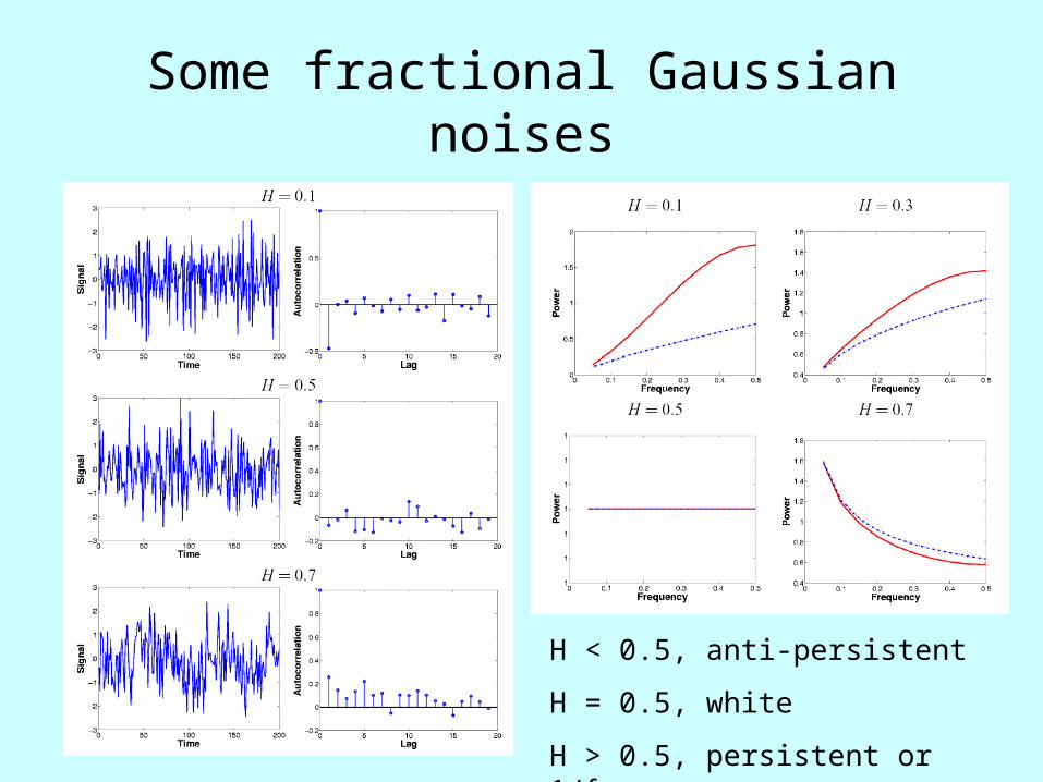
Some fractional Gaussian noises
H < 0.5, anti-persistent
H = 0.5, white
H > 0.5, persistent or 1/f

Estimators of fractional Gaussian noise parameters in time and wavelet domains
Hurst exponent Variance
A maximum likelihood estimator in the wavelet domain (wavelet-ML) gives the best overall performance in terms of bias and efficiency of estimation of H and 2: Fadili & Bullmore (2001); Maxim et al (2004)

Maximum likelihood estimation of fGn parameters in wavelet domain
Likelihood function of G, a fractional Gaussian noise, includes the inverse of its covariance matrix
S is assumed to be diagonalised in the wavelet domain, considerably simplifying its inversion

Wavelet ML estimation of signal and fGn parameters

Resting fMRI data looks like fGn (after head movement correction)
Freq
uenc
y
fGn
fBm
fGnfBm
+16 mm = 0
0
0.12
-1 1-2 0
Fre
quen
cy
Spectral Exponent,
“Raw” fMRI
Movement corrected fMRI
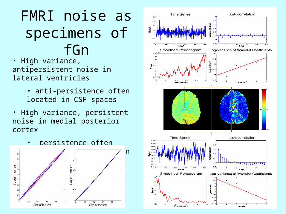
FMRI noise as specimens of fGn
• High variance, antipersistent noise in lateral ventricles
• anti-persistence often located in CSF spaces
• High variance, persistent noise in medial posterior cortex
• persistence often located symmetrically in cortex
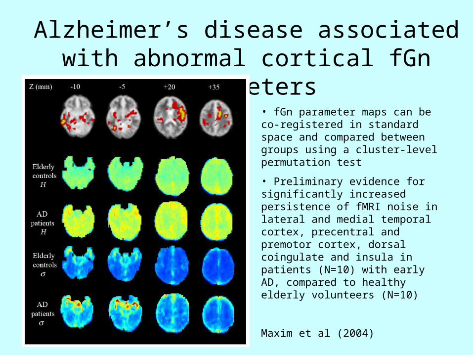
Alzheimer’s disease associated with abnormal cortical fGn parameters
• fGn parameter maps can be co-registered in standard space and compared between groups using a cluster-level permutation test
• Preliminary evidence for significantly increased persistence of fMRI noise in lateral and medial temporal cortex, precentral and premotor cortex, dorsal coingulate and insula in patients (N=10) with early AD, compared to healthy elderly volunteers (N=10)
Maxim et al (2004)

Hypothesis testing
V ~ 20,000 voxels
iV = E(FP), so if
i = 0.05, E(FP) = 1,000
• Bonferroni (i) = 0.05/V
• False discovery rate (FDR)
(i) >= 0.05/V
• GRF (i) < 0.05/V
for small d.f.

Why wavelets for multiple hypothesis testing for spatial statistic maps?
• Compaction (few large, many small coefficients)
•Multiresolution properties obviate difficulties concerning optimal choice of monoresolution Gaussian smoothing kernel (MFT)
• Various strategies, including wavelet shrinkage, available to reduce the search volume prior to hypothesis testing (enhanced FDR)
• Known dependencies of neighboring coefficients under the alternative hypothesis (Bayesian bivariate shrinkage)
• Non-orthogonal wavelets exist and may be advantageous for representing edges and lines (dual tree complex wavelet transform)

Wavelet shrinkage algorithms to control false discovery rate
• Take 2D or 3D wavelet transform of spatial {} maps
• Calculate P-value for each detail coefficient
• Sort the P-values in ascending order and find the order statistic i such that
• Calculate the critical threshold
• Apply hard or soft thresholding to all coefficients and take the inverse wavelet transform to recover the thresholded map in space

Wavelet FDR control is multiresolution• Wavelet FDR = 0.01 retains activation in bilateral motor cortex and ipsilateral cerebellum
• Spatial FDR = 0.01, after monoresolution smoothing with Gaussian kernel, retains activation in contralateral motor cortex with FWHM = 6 mm but loses cerebellar activation with FWHM = 18 mm
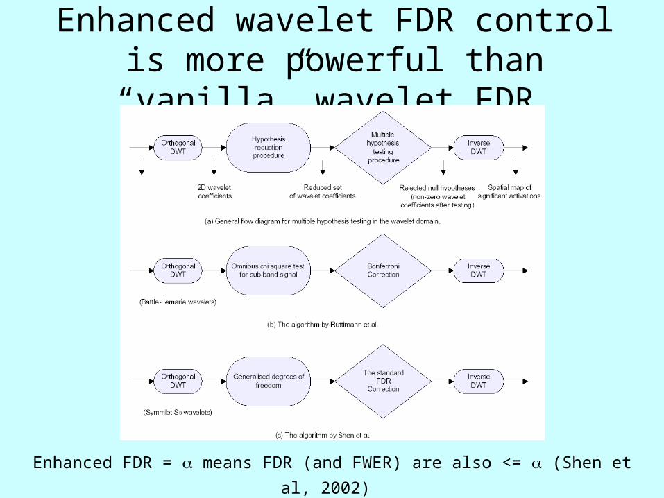
Enhanced wavelet FDR control is more powerful than “vanilla” wavelet FDR
Enhanced FDR = means FDR (and FWER) are also <= (Shen et al, 2002)

Enhanced FDR control in wavelet domain
Shen et al (2002); Sendur et al (2004)
Generalised degrees of freedom is estimated by Monte Carlo integration to reduce the search volume for hypothesis testing
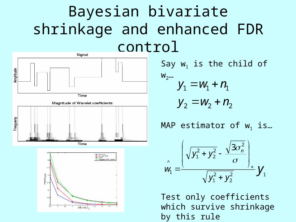
Bayesian bivariate shrinkage and enhanced FDR control
222
111
nwy
nwy
yyy
yy
w
n
122
21
222
21
^
1
3
Say w1 is the child of w2…
MAP estimator of w1 is…
Test only coefficients which survive shrinkage by this rule

Complex (dual tree) wavelet transform may be
advantageous for mapping edges
Complex wavelet coefficients are estimated by dual tree algorithm – their magnitude is shift invariant

Conclusions
• Wavelets are versatile tools, particularly applicable to analysis of scaling, scale-invariant or fractal processes in time (or space, or both)
• Brain images often have scaling or 1/f properties and wavelets can provide estimators of their fractal parameters, e.g. Hurst exponent
• Specific wavelet applications to fMRI data analysis include:
• Resampling (up to 4D)
• Estimation (robust and informative noise models)
• Hypothesis testing (multiresolution and enhanced FDR control)

Thanks
John Suckling
Mick Brammer
Jalal Fadili
Michael Breakspear
http://www-bmu.psychiatry.cam.ac.uk
Levent Sendur
Raymond Salvador
Voichita Maxim
Brandon Whitcher
Human Brain Project, National Institute of Mental Health and National Institute of Biomedical Imaging & Bioengineering
Wellcome Trust
GlaxoSmithKline plc


