15


Contents lists available at ScienceDirect
Acta Tropica
journal homepage: www.elsevier.com/locate/actatropica
Frequency and risk-factors analysis of Escherichia coli O157:H7 in Bali-cattle
I Wayan Suardanaa,⁎, Dyah Ayu Widiasihb, Widagdo Sri Nugrohob, Michael Haryadi Wibowoc,I Nyoman Suyasad
a Department of Veterinary Public Health, Faculty of Veterinary Medicine, Udayana University, Denpasar. Jl. PB. Sudirman Denpasar-Bali, 80232, Indonesiab Department of Veterinary Public Health, Faculty of Veterinary Medicine, Gadjah Mada University, Jl. Fauna 2, Karang Malang, Yogyakarta 55281, Indonesiac Department of Microbiology, Faculty of Veterinary Medicine, Gadjah Mada University, Jl. Fauna 2, Karang Malang, Yogyakarta 55281, Indonesiad Bali Assesment Institute for Agriculture Technology, Jl. By Pass Ngurah Rai, Pesanggaran, Denpasar - Selatan, 80222, Bali, Indonesia
A R T I C L E I N F O
Keywords:Bali cattleE. coli O157:H7Molecular analysisRisk factors
A B S T R A C T
Cattle are known as the main reservoir of zoonotic agents verocytotoxin-producing Escherichia coli. Thesebacteria are usually isolated from calves with diarrhea and/or mucus and blood. Tolerance of these agents to theenvironmental conditions will strengthen of their transmission among livestock. A total of 238 cattle fecalsamples from four sub-districts in Badung, Bali were used in this study. Epidemiological data observed includecattle age, sex, cattle rearing system, the source of drinking water, weather, altitude, and type of cage floor, thecleanliness of cage floor, the slope of cage floor, and the level of cattle cleanliness. The study was initiated byculturing of samples onto eosin methylene blue agar, then Gram stained, and tested for indole, methyl-red, vogesproskauer, and citrate, Potential E.coli isolates were then cultured onto sorbitol MacConkey agar, and furthertested using O157 latex agglutination test and H7 antisera. Molecular identification was performed by analysis ofthe 16S rRNA gene, and epidemiological data was analyzed using STATA 12.0 software. The results showed, theprevalence of E. coli O157:H7 in cattle at Badung regency was 6.30% (15/238) covering four sub districts i.e.Petang, Abiansemal, Mengwi, and Kuta which their prevalence was 8.62%(5/58), 10%(6/60), 3.33%(2/60), and3.33(2/60)%, respectively. The analysis of 16S rRNA gene confirmed of isolates as an E. coli O157:H7 strain with99% similarities. Furthermore, the risk factors analysis showed that the slope of the cage floor has a highlysignificant effect (P < 0.05) to the distribution of infection. Consequently, implementing this factor must beconcerned in order to decrease of infection.
1. Introduction
Escherichia coli O157:H7 caused multiple food and water-borneoutbreaks worldwide and considered as serious threat to public health.The agent produce verocytotoxin which cause diarrhea and hemor-rhagic colitis which may cause an adverse effects on the central nervoussystem, pancreas, lungs and heart with a case fatality rate ranging from3 to 5% (Mohawk and O'Brien, 2011). Infection by these bacteria isoften followed by life-threatening hemolytic uremic syndrome (HUS)and death especially in the elderly and young children (Mendoncaet al., 2012).The transmission of bacteria to human is known usually byconsumption of undercooked ground beef and unpasteurized milk(Rangel et al., 2005).
Verocytotoxin-producing E. coli has been isolated from a variety ofanimals, particularly ruminants, and cattle are regarded as the mainreservoir (Karmali et al., 2010). The proportion of animals infected bythis agent varied. The previous study found 52/257 (20%) were
recovered from cows, 16/71 (23%) from calves (Blanco et al., 1996),16/60 (27%) from cattle faeces, and 7/70(10%) from feedlot pens (Ootet al., 2007). The high prevalence was isolated in dairy cattle 144/198(72.73%) (Ferreira et al., 2014).
Global studies consisting 220,427 cattle were included in the meta-analysis showed the estimated prevalence of E.coli O157 in cattle at theglobal level was 5.68%. The random effects pooled prevalence esti-mates in Africa, Northern America, Oceania, Europe, Asia and LatinAmerica-Caribbean was 31.20%, 7.35%, 6.85%, 5.15%, 4.69%, and1.65%, respectively (Islam et al., 2014).
In Indonesia, the study of E. coli from calves with diarrhea wasinitiated by the researchers at Veterinary Research Institute (Kusmiyatiand Supar, 1998) who discovered alpha hemolytic E. coli isolates whichsome of them were verocytotoxigenic. The study of Drastini identifiedthe prevalence of this agent in dairy cows was 1.3% (Drastini, 2007).Furthermore, other researcher (Suardana et al., 2010) found as many as4/80(5%) of cattle faeces, 2/78(2.6%) of beef, 2/80(2.5%) of chicken
http://dx.doi.org/10.1016/j.actatropica.2017.05.019Received 14 March 2017; Received in revised form 6 May 2017; Accepted 11 May 2017
⁎ Corresponding author.E-mail addresses: [email protected] (I.W. Suardana), [email protected] (D.A. Widiasih), [email protected] (W.S. Nugroho),
[email protected] (M.H. Wibowo), [email protected] (I.N. Suyasa).
Acta Tropica 172 (2017) 223–228
Available online 12 May 20170001-706X/ © 2017 Elsevier B.V. All rights reserved.
MARK
faeces, 2/30(6.7%) of non-clinically human faeces, and 12/76(15.8%)of human suffering kidney failure were positive E. coli O157:H7. Thestudy about the adaptation of this agent to environmental changes alsoconducted by previous researchers who found E. coli O157:H7 survivedat 5 °C for 63–70 days, with the moisture content (74%) of faeces(Wang et al., 1996). These facts indicated that faeces as a potentialvector for the transmission of this organism. In addition, the horizontaltransmission of E. coli O157: H7 may occur during cattle housing. Thetransmission following ingestion of the pathogen at low levels and thatanimal hide as an important source of transmission (McGee et al.,2004).
Previous study showed that the E. coli O157:H7 occurrence in faecesof feedlot cattle depending on the age of the animal (dominant in youngcattle), changes of feed, transportation and hot conditions (Dargatzet al., 1997). Other researchers identified the high infection in cattle iscaused by several factors including feed, stress, livestock density,geography, and season (Kudva et al., 1996). Study of risk-factors onbovine infection with verocytotoxigenic producing E. coli in Ontariofound that calves> 2 weeks of age were at significantly greater risk ofinfection than those under 2 weeks (OR = 2.0) and farm-level calfinfection was negatively associated with herd size, and the maintenanceof a closed herd (Wilson et al., 1993). Based on these facts, identifica-tion of the frequency of local strains of E. coli O157:H7 and analyses ofrisk factors that contributed to the spread of these agents in Bali cattleneed to be revealed as primary step of prevention.
2. Materials and methods
2.1. Samples and epidemiological data
Samples of study were collected aseptically by directly rectalpalpation from communal cattle using sterile arm-length gloves.Approximately 100 g of fecal samples were collected from each cattlein each farm then immediately placed in sample cool box beforetransferred to the laboratory for analysis. In order to representBadung regency, samples were stratified according to sub districk withits characterization i.e. Petang, Abiansemal, Mengwi and Kuta. Samplesize that we used following formula n = 4PQ/L2, where “n” is thesample size, “P” is the assumption of the infection prevalence in thestudy area, “Q” is (1-P), and “L” is the desired error (Martin et al.,1987), wherein the prevalence infection was 3,5% (Suardana et al.,2010) with degree of error was 5%, therefore the number of samples forconfidence level of 95% was minimum 54 samples. The epidemiologicaldata were taken by interviewing the cattle’ owners and direct observa-tion in the location. The epidemiological data including cattle age, sex,cattle rearing system, the source of drinking water, weather, altitude,type of cage floor, the cleanliness of the cage floor, the slope of the cagefloor, and the cleanliness level of cattle.
2.2. Isolation and identification of Escherichia coli
The isolation started by dissolving of 10 g of the fecal samplesderived from a mixture of 100 g of each sample with 90 ml of bufferedpeptone water (BPW). Furthermore, a tenfold series dilution wasperformed using sterile distilled water and aliquots (100 μl) of eachdilution were plated onto eosin methylene blue agar (EMBA), and theplates were incubated at 37 °C for 24 h. A positive result characterizedby distinctive metallic green colony, confirmed using Gram staining,and tested for indole, methyl red, voges-proskauer, and citrate (IMViC)medium to ensure as group of fecal coli (Lodish, 2013). The positiveresults subsequently inoculated onto nutrient agar medium for furtherinvestigation
2.3. Identification of Escherichia coli O157:H7 serotype
Identification of E. coli O157:H7 was carried out by inoculating of
potential E. coli isolates onto sorbitol MacConkey agar (SMAC) mediumand incubated at 37 °C for 24 h. Positive result on SMAC medium wascharacterized by colourless colony. Further test was done by reacting ofall positive isolates on SMAC medium against E. coli O157 latexagglutination test in order to ensure the isolate was E. coli O157 strain.The identification was ended by testing of isolates with H7 serotype testwhich were characterized by the precipitation form on the bottom ofplate (Suardana et al., 2015).
2.4. Analysis of 16S rRNA gene
2.4.1. Extraction of DNA and PCRBacterial DNA was extracted using QIAamp DNA Mini Kits (Qiagen)
according to manufacturer’s instructions with slighty modification(Suardana, 2014). The 16S rRNA gene was amplified using PlatinumPCR Supermix kit (Invitrogen) on Thermocycler Eppendorf Mastercy-cler personal/PTC 100. The PCR program was carried out in 40 μlreaction volumes containing 2μL DNA template (200 ng/μL), 34 μL PCRSupermix 2x, and 2μL (20 pmol/μL) of each primer. The primers used inthis Study i.e. 27F (5′-AGAGTTTGATCCTGGCTCAG-3′) and U1492R(5′-GGTTACCTTGTTACGACTT-3′). The PCR amplification was pro-grammed refered to previously with initial DNA denaturation at 94 °Cfor 5 min, followed by 35 cycles of denaturation at 94 °C for 1 min,annealing at 55 °C for 1 min, and elongation at 72 °C for 1 min. At theend of cycles, it was followed by a final extension at 72 °C for 5 min 5 μLof PCR products were analyzed by electrophoresis (Bio-Rad) in 1%agarose (Gibco BRL) gel, at 90 V for 45 min. The gel was stained with1% solution of ethidium bromide (50 μL/L) and distained with TBE 1 xfor 10 min. Gel was visualized by UV transillumination and recorded bydigital camera FE-270 7.1 megapixels (Suardana, 2014).
2.4.2. Sequencing and phylogenetic analysisThe sequencing of 16S rRNA gene was conducted using genetic
analyzer (ABI Prism 3130 and 3130 xl Genetic Analyzer) at EijkmanInstitute for Molecular Biology, Jakarta. The sequencing used bothprimers: Stx2 (F) and Stx2 (R). The sequences were edited using MEGA5.2 version software. The nucleotide sequence of 16S rRNA gene of E.coli O157:H7 strains that available in the genBank such as E.coli 933W(AE 005174), SM-25(1) (KF768068), KL-48(2) (KF768069) as referenceand one nucleotide sequence E. coli O111:H11 (NZ_AKAX01000438) asan out group were used in this study. The sequences were aligned usingClustal W and the phylogenetic analysis was constructed using neighborjoining algorithm (Saitou and Nei, 1987; Tamura et al., 2007). Criteriafor species identification is 99% sequence similarity or higher forspecies assignment and 95% sequence similarity or higher for genusassignment (Bosshard et al., 2003) or minimum 99% sequence similar-ity and ideally 99.5% sequence similarity or< 1% divergence forspecies assignment (Janda and Abbott, 2007).
2.5. Data analysis
The positive and negative result per sample which was showed bybiochemical test complete with molecular analysis and questioner wasanalyzed descriptively. The association of infection against risk factorswas tested by Chi-Square test (Steel and Torrie, 1996) and Odds Ratiotest to determine the strength of association (Martin et al., 1987). All ofthe data were analyzed by using STATA 12.0 software.
3. Results and discussion
3.1. Descriptive analysis
Fifteen out of 238 samples which were stratified according to foursub districks were positive detected E.coli O157:H7 in survey that wasconducted from Mart to August 2013. All of positive isolates character-ized by a distinctive metallic green sheen on eosin methylene blue agar
I.W. Suardana et al. Acta Tropica 172 (2017) 223–228
224

(EMBA) medium, and showed positive reaction on indole and methylred, but negative reaction on voges proskauer and citrate test.Furthermore, these isolates formed colourless colonies on selectivemedium sorbitol MacConkey (SMAC) agar. They were also demon-strated slighty agglutination on E.coli O157 Latex Test Kit, and fluidmoderately cloudy of supernatant on tested by H7 antisera. Theinfection of E. coli O157:H7 in this study was variously in each subdistrict with the lower prevalence in Mengwi and Kuta but the highercase was found in Petang and Abiansemal sub districts (Table 1).
According to Table 1. the percentage of E. coli O157:H7 positive inBali cattle was 6.30% distributed in four sub districts i.e. Petang,Abiansemal, Mengwi, and Kuta with prevalence of 8.62% (n = 5/58);10.0% (n = 6/60); 3.33% (n = 2/60), and 3.33% (n = 2/60), respec-tively. These results were in accordance with the previous study wherethe occurrence was 5% (Suardana et al., 2010). On the other hands, theprevalence was higher (27.4%) in dairy cattle in Central Java andYogyakarta (Sumiarto, 2004).
3.2. Molecular analysis of 16S rRNA gene
The molecular analysis of 16S rRNA gene of local strain E. coliO157:H7 has been successfully conducted. Spatial sequences (1351 outof 1500 bp) originated from 10 strains as a representation of this studywere alignmed with some database sequences in genBank i.e. E.coli EDL933 W (AE 005174), E.coli SM 25(1) (KF 768068), and E. coli KL-48(2)(KF768069). On the other hand, all strains also aligned against E.coli
O111:H11(NZ_AKAX01000438) as an out group. The result of theanalysis in the form of the phylogenetic tree is showed in Fig. 1, andgenetically distances among isolates were summarized in Table 2.
3.3. Risk factors analysis
As many as ten variables which were subjected in the interview of238 cattle’owners, and direct field investigation of 238 fecal sampleswere analyzed in this study. Cattle’s owners were interviewed using astandardized questionnaire to obtain the information on farm andindividual animal-level characteristics and management practices. Mostof respondents were women and primary school education, and theywere cooperate to share their cattle’status. The analysis of risk factorsshowed the infection level of E. coli O157:H7 was frequently correlatedwith cattle age of> 1 year, male, cattle rearing with housing system,non-tap water as a source of drinking water, and cattle with lower levelcleanliness. The infection also showed frequent occurrence in thehighland at dry weather. The descriptive analysis of each variablewas summarized in Table 3, and the single variable analysis is showedin Table 4.
The study that was performed base on biochemical and molecularanalysis denoted 15 out of 238 (6.30%) samples positive E. coliO157:H7. The E.coli O157:H7 differs from other strains of E. coli inbeing unable to ferment sorbitol. In sorbitol MacConkey agar (SMAC),lactose is replaced by sorbitol, and pathogenic E. coli cannot fermentsorbitol, so this strain uses peptone to grow. This raises the pH of themedium, allowing the pathogenic strain to be differentiated from othernon-pathogenic E.coli strains. Detection of E. coli O157:H7 by usingSMAC medium has a sensitivity of 100%, specificity of 85%, andaccuracy of 86% (March and Ratnam, 1986). Prompt identification wasperformed after all isolates were positive on the O157 latex test whichwas known as a simple, efficient and reliable method in detecting of E.coli O157:H7 with a 100% sensitivity and specificity (March andRatnam, 1989). Moreover, all isolates were also positive to H7 antiseratest as a definitive test for H7 flagella of E.coli O157:H7 (Farmer andDavis, 1985).
Table 1Distribution of E. coli O157:H7 infection in Bali cattle in Petang, Abiansemal, Mengwi,and Kuta sub district at Badung regency, Bali.
Sub districts Numbers of Fecal-samples E. coliO157:H7 positive
Petang 58 5 (8.62%)Abiansemal 60 6 (10.0%)Mengwi 60 2 (3.33%)Kuta 60 2 (3.33%)Total 238 15 (6.30%)
Fig. 1. Phylogenetic tree of local strains of E. coli O157:H7 against some database sequences in genBank. The phylogenetic tree was constructed using Neighbor Joining algorithm of 1351nucleotides sequence of 16S rRNA gene. The number in the branch of phylogram indícated bootstrap valué (%) by 1000 replicatión multiple, and scale indícated one per 1000 substitutionof nucleotide sequences.
I.W. Suardana et al. Acta Tropica 172 (2017) 223–228
225

According to data in Table 1. the occurrence of E. coli O157:H7 ineach sub district showed that Petang and Abiansemal sub districts werehigher than Mengwi and Kuta sub districts. This fact was supported bythe ideal location of both sub districts at northern area of Badungregency as a land field for growing and multiplication of the agent.According to the statistical data of Badung regency, each of the subdistricts has an altitude>350 m above sea levels, and known as anagricultural land. Whereas Mengwi and Kuta sub districts each has analtitude lower than of those, and famously are known as an area oftourism and business. Livelihoods of people in Petang and Abiansemalsub districts are generally as traditional farmers, as well as their activityin the management of livestock (cattle). Furthermore, the geographicalcondition such as the rainfall, humidity and temperature around 24 °Calso support for survival and maintenance of agents.
Molecular analysis of 16S rRNA gene as a new gold standard forspecification of bacteria was also performed as a deep confirmation of15 out of 238 isolates as an E.coli O157:H7 strains (Janda and Abbott,2007). This method has many advantages i.e. by using of 16S rRNAsequences, numerous of bacterial genera and species have beenreclassified and renamed, phylogenetic relationships have been deter-mined, and the discovery and classification of novel bacterial specieshave been facilitated (Bosshard et al., 2003).
The phylogenetic tree of the local strain in Fig. 1 showed that 10 outof 15 strains of E. coli O157: H7 as a representation of this study lies inthe same clade with positive control ATCC 43894, E.coli EDL 933 W (AE005 174), E. coli SM 25 (1) (KF 768 068), and E. coli KL-48 (2)(KF768069), but separated by E.coli O111: H11 (NZ_AKAX01000438)as an out group. Phylogram in Fig. 1 showed highly probability of localstrain genetically linked to the strain of E. coli ATCC 43894, as well asother reference strain.
This conclusion was supported by the data in Table 2 which showedall of local strains only had nucleotide different ranging from 1 to 12nucleotides against ATCC 43894 control, and E. coli nucleotidedatabase that available in the genBank. The conclusion refers to theconcept of similarty or nucleotides differences which was proposedpreviously by some researchers. There were recommended when thesimilarity of 16S rRNA gene was more than 95% or the nucleotidesdifferent less than 1% (15 out of 1500 bp), the query nucleotides shouldbe categorized as the same species (Janda and Abbott, 2007). Thoseresults were well add to the collection of local strain of E. coli O157: H7that have been confirmed molecularly, and have been deposited the 16Ssequences in Genbank, namely E.coli SM 25 (1) (KF 768 068), and E. coliKL-48 (2) (KF768069) derived from cattle and human faeces, respec-tively.
Furthermore, the risk factors of E. coli O157:H7 infection in Balicattle (Table 2) in Badung regency were more associated to variables offemale, age<1 year, and cattle reared in the cages with theirpercentages were 6.55, 6.67, and 7.69%, respectively, although all ofthose factors were not yet significantly affect (p < 0.05) statistically.
The contribution of those factors, especially for the cattle withage< 1 year was resulted by the calves at this age were found kept intheir cages and rarely rearing in the field. The observations also showedthat the calves in this age usually found in less clean condition as aresult of a lot of movement in a narrow space. Moreover, the farmershardly bathe their calves as results of they were raised without straps.These results also supported by previous study which found a strongeffect in young animals (calves within 2–6-month) were as the high-riskage group (8.6% positive) in contrast to older animals (2.4%).However, there was a tendency of non-significant effect in male calvesto have a higher prevalence than heifers within the same age (Nielsenet al., 2002).
On the other hands, several factors like cemented cage-floor, slopeof cage floor and cleanliness of cage floor had a contradictory effect tothe incidence of E. coli O157:H7 infection (Table 3 and 4). These resultswere different with the previous study which found the occurrence of E.coli O157:H7 infection correlated to unclean of cage. The factsTa
ble2
Gen
etic
distan
ceof
16SrR
NA
gene
oflocalstrain
E.coliO15
7:H7ag
ainstseve
ralda
taba
sesequ
encesin
thege
nBan
k.
E.coliED
L93
3**
E.coliATC
C43
894*
FSP7
Caran
gsari
FSP5
Caran
gsari
FSP47
Pelaga
FSA
20Ta
man
FSA
14Bo
ngka
saFS
A30
Ayu
nan
FSM
8Ba
haFS
M58
Lukluk
FSM
57Lu
kluk
FSM
42Werdi
Buan
a
E.coliSM
25(1)**
E.coliKL
48(2)**
E.coli
O11
1:H11
**
E.coliED
L93
3**
E.coliATC
C43
894*
0.00
0FS
P7Caran
gsari
0.00
10.00
1FS
P5Caran
gsari
0.00
90.00
90.00
8FS
P47
Pelaga
0.00
90.00
90.00
80.00
0FS
A20
Taman
0.01
20.01
20.01
10.00
30.00
3FS
A14
Bong
kasa
0.00
90.00
90.00
80.00
00.00
00.00
3FS
A30
Ayu
nan
0.00
00.00
00.00
10.00
90.00
90.01
20.00
9FS
M8Ba
ha0.00
50.00
50.00
60.01
20.01
20.01
50.01
20.00
5FS
M58
Lukluk
0.00
90.00
90.00
80.00
00.00
00.00
30.00
00.00
90.01
2FS
M57
Lukluk
0.00
40.00
40.00
50.01
30.01
30.01
60.01
30.00
40.00
90.01
3FS
M42
Werdi
Buan
a0.00
00.00
00.00
10.00
90.00
90.01
20.00
90.00
00.00
50.00
90.00
4
E.coliSM
25(1)**
0.00
90.00
90.00
80.00
00.00
00.00
30.00
00.00
90.01
20.00
00.01
30.00
9E.
coliKL48
(2)**
0.00
80.00
80.00
70.00
30.00
30.00
60.00
30.00
80.01
10.00
30.01
20.00
80.00
3E.
coliO11
1:H11
**1.21
51.21
51.21
01.21
11.21
11.20
31.21
11.21
51.20
61.21
11.21
01.21
51.21
11.20
4
Note:
*)=
Con
trol
isolates;*
*)Nuc
leotidesequ
ence
from
genB
ankda
taba
se.E
.coliE
DL93
3W
(AE00
5174
),E.coliSM
25(1)(K
F76
8068
),E.
coliKL-48
(2)(K
F768
069),an
dE.coliO11
1:H11
(NZ_AKAX01
0004
38).
I.W. Suardana et al. Acta Tropica 172 (2017) 223–228
226
generated by the cattle’s owners in the area of study only wipe of thecattle faeces from the cage floor, but it was still a pile up around thecage for a long time. So that, although the cage-floor showed cleanli-ness, cemented, and sloping floor, there were not guaranteed to preventthe transmission of agent from manure. Furthermore, the researchersmentioned, environmental adaptations of E. coli O157:H7 play animportant role in the persistence and dissemination of this microorgan-ism on farms and increasing transfer of agent from cattle to others(Maule, 2000).
The long contact of cattle with a conventional managementespecially that reared in the cage resulting in more opportunity of theagent to contact with their host or transmit from infected cattle toothers. There are many evidences in the literatures that support anassociation between cattle movement and the risk of E. coli O157shedding on farms. Cattle movement might contribute significantly tothe observed prevalence of E. coli O157. Cattle movements involvinginfected farms with cattle shedding an exceptional amount of E. coliO157, ‘super-shedders’, also a substantial contribution to the preva-lence of infected farms (Liu et al., 2007). The maintenance of agent inthe environment was also supported by the other researcher (Jianget al., 2002) who found the E. coli O157:H7 cells survived for up to77,> 226, and 231 days in manure-amended autoclaved soil held at 5,15, and 21 °C, respectively.
The infection of cattle by E. coli O157:H7 in this study alsofrequently associated with dry weather, the use of non tap water as adrinking source, breeding of cattle in highland area, and cattle with
poor cleanliness. Each of variables has a percentage i.e. 6.67, 8.73,6.43, and 8.70%. Especially for the use of non tap water as a drinkingsource, the study found odds ratio 2.44 which mean the use of non tapwater would be increasing the E. coli O157:H7 infection as many as 2.44times. These facts might be explained as a result from the cattle in thestudy area usually drink water collected from the small river aroundtheir cage which possible contaminated by some pollutant as well ascattle faeces.
Several previous study showed animals that carrying VTEC O157 donot show clinical signs of illness following infection with this organismand shedding is intermittent and transient (Keen et al., 2006; Wanget al., 1996). Shedding has also been shown to be seasonal, withexcretion rates peaking in the summer and early autumn (Synge, 2000).The distribution of this agent also supported by several factors.Irrigations are known have an important contribution to the occurrenceof E. coli O157:H7. Irrigations such as irrigation water, swimming water(pools, beaches, and lakes), surface water runoff, and municipal watercontaminated with faeces are some of the reservoirs of E. coli O157:H7(Islam et al., 2004a; Islam et al., 2004b).
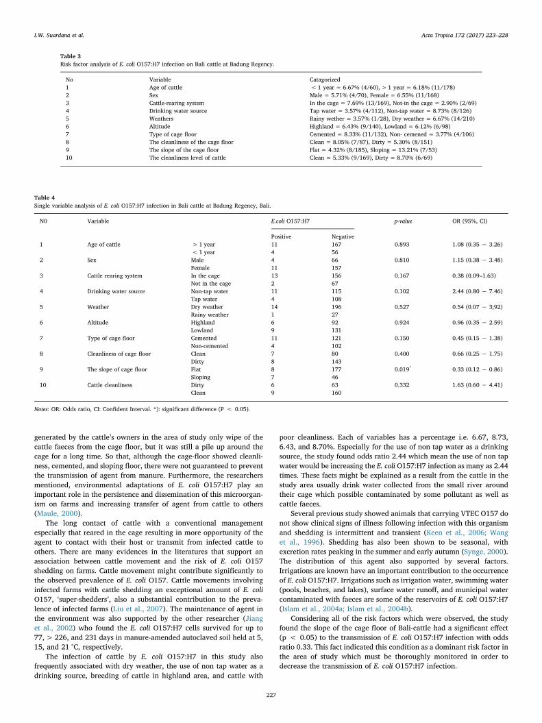
Considering all of the risk factors which were observed, the studyfound the slope of the cage floor of Bali-cattle had a significant effect(p < 0.05) to the transmission of E. coli O157:H7 infection with oddsratio 0.33. This fact indicated this condition as a dominant risk factor inthe area of study which must be thoroughly monitored in order todecrease the transmission of E. coli O157:H7 infection.
Table 3Risk factor analysis of E. coli O157:H7 infection on Bali cattle at Badung Regency.
No Variable Catagorized1 Age of cattle < 1 year = 6.67% (4/60), > 1 year = 6.18% (11/178)2 Sex Male = 5.71% (4/70), Female = 6.55% (11/168)3 Cattle-rearing system In the cage = 7.69% (13/169), Not-in the cage = 2.90% (2/69)4 Drinking water source Tap water = 3.57% (4/112), Non-tap water = 8.73% (8/126)5 Weathers Rainy wether = 3.57% (1/28), Dry weather = 6.67% (14/210)6 Altitude Highland = 6.43% (9/140), Lowland = 6.12% (6/98)7 Type of cage floor Cemented = 8.33% (11/132), Non- cemened = 3.77% (4/106)8 The cleanliness of the cage floor Clean = 8.05% (7/87), Dirty = 5.30% (8/151)9 The slope of the cage floor Flat = 4.32% (8/185), Sloping = 13.21% (7/53)10 The cleanliness level of cattle Clean = 5.33% (9/169), Dirty = 8.70% (6/69)
Table 4Single variable analysis of E. coli O157:H7 infection in Bali cattle at Badung Regency, Bali.
N0 Variable E.coli O157:H7 p-value OR (95%, CI)
Positive Negative1 Age of cattle > 1 year 11 167 0.893 1.08 (0.35 − 3.26)
< 1 year 4 562 Sex Male 4 66 0.810 1.15 (0.38 − 3.48)
Female 11 1573 Cattle rearing system In the cage 13 156 0.167 0.38 (0.09–1.63)
Not in the cage 2 674 Drinking water source Non-tap water 11 115 0.102 2.44 (0.80 − 7.46)
Tap water 4 1085 Weather Dry weather 14 196 0.527 0.54 (0.07 − 3;92)
Rainy weather 1 276 Altitude Highland 6 92 0.924 0.96 (0.35 − 2.59)
Lowland 9 1317 Type of cage floor Cemented 11 121 0.150 0.45 (0.15 − 1.38)
Non-cemented 4 1028 Cleanliness of cage floor Clean 7 80 0.400 0.66 (0.25 − 1.75)
Dirty 8 1439 The slope of cage floor Flat 8 177 0.019* 0.33 (0.12 − 0.86)
Sloping 7 4610 Cattle cleanliness Dirty 6 63 0.332 1.63 (0.60 − 4.41)
Clean 9 160
Notes: OR: Odds ratio, CI: Confident Interval. *): significant difference (P < 0.05).
I.W. Suardana et al. Acta Tropica 172 (2017) 223–228
227
4. Conclusion
This study demonstrates the occurrence of Escherichia coli O157:H7in Bali-cattle at Badung regency was 6.30% which was distributed inthe 4 sub districts i.e. Petang, Abiansemal, Mengwi, and Kuta withprevalence rates of 8.62; 10.0; 3.33; and 3.33%, respectively. Molecularanalysis of 16S rRNA gene showed that local strains have highsimilarity (> 99%) against E.coli ATCC 43894 and several nucleotidesequences as a reference strain. The analysis of risk factors indicatedthat the slope of the cage floor has a significant contribution to theinfection.
Conflict of interest
The authors declare that we have no conflict of interest that mightinappropriately influence the reported work.
Acknowledgements
The authors would like thank Prof. Dr. Supar, MS for his kindness tosupply E. coli ATCC 43894 control isolate, Dr. Aida L.T. Rompis for herEnglish correction, and Minister of Agriculture for their financialsupport in the form of KKP3N grant contract no.795/LB. 620/I.1/2/2013, dated February 25th, 2013
References
Blanco, M., Blanco, J.E., Blanco, J., Gonzalez, E.A., Mora, A., Prado, C., Fernandez, L.,Rio, M., Ramos, J., Alonso, M.P., 1996. Prevalence and characteristics of Escherichiacoli serotype O157:H7 and other verotoxin-producing E-coli in healthy cattle.Epidemiol. Infect. 117, 251–257.
Bosshard, P.P., Abels, S., Zbinden, R., Bottger, E.C., Altwegg, M., 2003. Ribosomal DNAsequencing for identification of aerobic gram-positive rods in the clinical laboratory(an 18-month evaluation). J. Clin. Microbiol. 41, 4134–4140.
Dargatz, D.A., Wells, S.J., Thomas, L.A., Hancock, D.D., Garber, L.P., 1997. Factorsassociated with the presence of Escherichia coli O157 in feces of feedlot cattle. J.Food Prot. 60, 466–470.
Drastini, Y., 2007. Identifcation and characterization based on genotype and phenotype ofVerocytotoxigenic Escherichia coli (VTEC) from livestock in Yogyakarta. Disertation.Gadjah Mada, Yogyakarta.
Farmer, J.J., Davis, B.R., 1985. H7 antiserum-sorbitol fermentation medium: a single tubescreening medium for detecting Escherichia coli O157:H7 associated withhemorrhagic colitis. J. Clin. Microbiol. 22, 620–625.
Ferreira, M.R., Freitas Filho, E.G., Pinto, J.F., Dias, M., Moreira, C.N., 2014. Isolation,prevalence, and risk factors for infection by shiga toxin-producing Escherichia coli(STEC) in dairy cattle. Trop. Anim. Health Prod. 46, 635–639.
Islam, M., Doyle, M.P., Phatak, S.C., Millner, P., Jiang, X., 2004a. Persistence ofenterohemorrhagic Escherichia coli O157:H7 in soil and on leaf lettuce and parsleygrown in fields treated with contaminated manure composts or irrigation water. J.Food Prot. 67, 1365–1370.
Islam, M.A., Chowdhury, R.I., Chakraborty, N., Bari, W., Akhter, H.H., 2004b. Factorsassociated with delivery complications in rural Bangladesh. Eur. J. Contracept.Reprod. Health Care 9, 203–213.
Islam, M.Z., Musekiwa, A., Islam, K., Ahmed, S., Chowdhury, S., Ahad, A., Biswas, P.K.,2014. Regional variation in the prevalence of E: coli O157 in cattle: a meta-analysisand meta-regression. PLoS One 9, e93299.
Janda, J.M., Abbott, S.L., 2007. 16S rRNA gene sequencing for bacterial identification inthe diagnostic laboratory: pluses, perils, and pitfalls. J. Clin. Microbiol. 45,
2761–2764.Jiang, X., Morgan, J., Doyle, M.P., 2002. Fate of Escherichia coli O157:H7 in manure-
amended soil. Appl. Environ. Microbiol. 68, 2605–2609.Karmali, M.A., Gannon, V., Sargeant, J.M., 2010. Verocytotoxin-producing escherichia
coli (VTEC). Vet. Microbiol. 140, 360–370.Keen, J.E., Wittum, T.E., Dunn, J.R., Bono, J.L., Durso, L.M., 2006. Shiga-toxigenic
Escherichia coli O157 in agricultural fair livestock, United States. Emerg. Infect. Dis.12, 780–786.
Kudva, I.T., Hatfield, P.G., Hovde, C.J., 1996. Escherichia coli O157:H7 in microbial floraof sheep. J. Clin. Microbiol. 34, 431–433.
Kusmiyati, Supar, 1998. Verocytotoxigenic E. coli detected from diarhea- dairy calves.Proceedings of the seminar Results in Veterinary Research. Bogor 8–19 Pebruari.
Liu, W.C., Matthews, L., Chase-Topping, M., Savill, N.J., Shaw, D.J., Woolhouse, M.E.,2007. Metapopulation dynamics of Escherichia coli O157 in cattle: an exploratorymodel. J. R. Soc. Interface 4, 917–924.
Lodish, H.F., 2013. Molecular Cell Biology, 7th edition. W.H Freeman and Co., New Yorkxxxiii, 1154, 1158 p. pp.
March, S.B., Ratnam, S., 1986. Sorbitol-MacConkey medium for detection of Escherichiacoli O157:H7 associated with hemorrhagic colitis. J. Clin. Microbiol. 23, 869–872.
March, S.B., Ratnam, S., 1989. Latex agglutination test for detection of Escherichia coliserotype O157. J. Clin. Microbiol. 27, 1675–1677.
Martin, S.W., Meek, A.H., Willeberg, P., 1987. Veterinary Epidemiology. Principle andMethods. Iowa State University Press/Ames, United States of America.
Maule, A., 2000. Survival of verocytotoxigenic Escherichia coli O157 in soil, water and onsurfaces. Symp. Ser. Soc. Appl. Microbiol. 71S–78S.
McGee, P., Scott, L., Sheridan, J.J., Earley, B., Leonard, N., 2004. Horizontal transmissionof Escherichia coli O157: H7 during cattle housing. J. Food Prot. 67, 2651–2656.
Mendonca, R.C.S., Morelli, A.M.F., Pereira, J.A.M., de Carvalho, M.M., de Souza, N.L.,2012. Prediction of Escherichia coli O157:H7 adhesion and potential to form biofilmunder experimental conditions. Food Control 23, 389–396.
Mohawk, K.L., O'Brien, A.D., 2011. Mouse models of Escherichia coli O157:H7 infectionand shiga toxin injection. J. Biomed. Biotechnol. 2011, 258185.
Nielsen, E.M., Tegtmeier, C., Andersen, H.J., Gronbaek, C., Andersen, J.S., 2002.Influence of age, sex and herd characteristics on the occurrence of Verocytotoxin-producing Escherichia coli O157 in Danish dairy farms. Vet. Microbiol. 88, 245–257.
Oot, R.A., Raya, R.R., Callaway, T.R., Edrington, T.S., Kutter, E.M., Brabban, A.D., 2007.Prevalence of Escherichia coli O157 and O157:H7-infecting bacteriophages in feedlotcattle feces. Lett. Appl. Microbiol. 45, 445–453.
Rangel, J.M., Sparling, P.H., Crowe, C., Griffin, P.M., Swerdlow, D.L., 2005. Epidemiologyof escherichia coli O157: H7 outbreaks, United States, 1982–2002. Emerg. Infect. Dis.11, 603–609.
Saitou, N., Nei, M., 1987. The neighbor-joining method: a new method for reconstructingphylogenetic trees. Mol. Biol. Evol. 4, 406–425.
Steel, R.G.D., Torrie, J.H., 1996. Principles and Procedures of Statistics. McGraw-HillCompanies.
Suardana, I.W., Artama, W.T., Asmara, W., Daryono, B.S., 2010. Identification ofEscherichia coli O157:H7 and detection of Shiga like toxin 1 and 2 genes from animals’feces, beef, and human feces. J. Vet. 11, 264–270.
Suardana, I.W., Widiasih, D.A., Mahardika, I.G.N.K., Pinatih, K.J.P., Daryono, B.S., 2015.Evaluation of zoonotic potency of Escherichia coli O157:H7 through arbitrarilyprimed PCR methods. Asian Pac. J. Trop. Biomed. 5, 915–920.
Suardana, I.W., 2014. Analysis of nucleotide sequences of the 16S rRNA gene of novelEscherichia coli strains isolated from feces of human and bali cattle. J Nucleic Acids2014, 475754.
Sumiarto, B., 2004. Infection and contamination of Escherichia coli O157:H7 on lamb atYogyakarta slaughterhouse. J. Vet. 5, 85–90.
Synge, B.A., 2000. Verocytotoxin-producing Escherichia coli: a veterinary view. Symp.Ser. Soc. Appl. Microbiol. 31S–37S.
Tamura, K., Dudley, J., Nei, M., Kumar, S., 2007. MEGA4: molecular evolutionarygenetics analysis (MEGA) software version 4.0. Mol. Biol. Evol. 24, 1596–1599.
Wang, G., Zhao, T., Doyle, M.P., 1996. Fate of enterohemorrhagic Escherichia coliO157:H7 in bovine feces. Appl. Environ. Microbiol. 62, 2567–2570.
Wilson, J.B., Mcewen, S.A., Clarke, R.C., Leslie, K.E., Waltnertoews, D., Gyles, C.L., 1993.Risk-factors for bovine infection with verocytotoxigenic escherichia-Coli in Ontario,Canada. Prev. Vet. Med. 16, 159–170.
I.W. Suardana et al. Acta Tropica 172 (2017) 223–228
228


