The synthesis of novel chromogenic enzyme substrates for detection of bacterial glycosidases and their applications in diagnostic microbiology Michael Burton, a John D. Perry, b Stephen P. Stanforth c and Hayley J. Turner* a a Glycosynth Ltd, 14 Craven Court, Winwick Quay, Warrington, Cheshire, WA2 8QU, UK. b Department of Microbiology, Freeman Hospital, Newcastle upon Tyne, NE7 7DN, UK. c Department of Applied Sciences, Northumbria University, Newcastle upon Tyne, NE1 8ST, UK. Abstract: The preparation and evaluation of chromogenic substrates for detecting bacterial glycosidase enzymes is reported. These substrates are monoglycoside derivatives of the metal chelators catechol, 2,3-dihydroxynaphthalene (DHN) and 6,7-dibromo-2,3- dihydroxynaphthalene (6,7-dibromo-DHN). When hydrolysed by appropriate bacterial enzymes these substrates produced coloured chelates in the presence of ammonium iron(III) citrate, thus enabling bacterial detection. A β-D-riboside of DHN and a β-D- glucuronide derivative of 6,7-dibromo-DHN were particularly effective for the detection of S. aureus and E. coli respectively. Key words: enzyme substrates, chromogenic substrates, glycosidase, pathogenic microorganisms, bacterial detection. Introduction Synthetic enzyme substrates are utilised extensively in diagnostic clinical microbiology for the purpose of detecting and identifying pathogenic microorganisms. 1-3 These substrates are designed to target microbiological species of interest (or groups of species) based upon their enzyme activity. An important sub-class of synthetic enzyme substrates are the chromogenic sugar-based enzyme substrates in which hydrolytic cleavage of the sugar moiety from the aglycone is mediated by an appropriate enzyme resulting in the liberation of a hydroxyaryl derivative, as shown by the representative examples in Scheme 1. The hydroxyaryl derivative can be coloured, thus allowing 1
Transcript
The synthesis of novel chromogenic enzyme substrates for detection of bacterial glycosidases and their applications in diagnostic microbiology
Michael Burton,a John D. Perry,b Stephen P. Stanforthc and Hayley J. Turner*a
a Glycosynth Ltd, 14 Craven Court, Winwick Quay, Warrington, Cheshire, WA2 8QU, UK.b Department of Microbiology, Freeman Hospital, Newcastle upon Tyne, NE7 7DN, UK.c Department of Applied Sciences, Northumbria University, Newcastle upon Tyne, NE1 8ST, UK.
Abstract: The preparation and evaluation of chromogenic substrates for detecting bacterial glycosidase enzymes is reported. These substrates are monoglycoside derivatives of the metal chelators catechol, 2,3-dihydroxynaphthalene (DHN) and 6,7-dibromo-2,3-dihydroxynaphthalene (6,7-dibromo-DHN). When hydrolysed by appropriate bacterial enzymes these substrates produced coloured chelates in the presence of ammonium iron(III) citrate, thus enabling bacterial detection. A β-D-riboside of DHN and a β-D-glucuronide derivative of 6,7-dibromo-DHN were particularly effective for the detection of S. aureus and E. coli respectively.
Synthetic enzyme substrates are utilised extensively in diagnostic clinical microbiology for the purpose of detecting and identifying pathogenic microorganisms.1-3 These substrates are designed to target microbiological species of interest (or groups of species) based upon their enzyme activity. An important sub-class of synthetic enzyme substrates are the chromogenic sugar-based enzyme substrates in which hydrolytic cleavage of the sugar moiety from the aglycone is mediated by an appropriate enzyme resulting in the liberation of a hydroxyaryl derivative, as shown by the representative examples in Scheme 1. The hydroxyaryl derivative can be coloured, thus allowing direct visualisation of the hydrolytic reaction as illustrated by the transformation of the colourless ortho-nitrophenyl β-D-galactopyranoside 1 into the yellow-coloured ortho-nitrophenol (ONP) (Eqn 1).4 Alternatively, if the liberated hydroxyaryl derivative is colourless, a subsequent chemical reaction can be employed to produce a coloured product. Thus, the β-galactoside derivative of 5-bromo-4-chloro-3-hydroxyindole (‘X-gal’) 2 is hydrolysed to produce 5-bromo-4-chloro-3-hydroxyindole which then undergoes an oxidative dimerization in air producing the blue-coloured indigo derivative 3 (Eqn 2).5 The indole-derived substrate, ALDOLTM 455 4,6 similarly generates a reactive 3-hydroxyindole intermediate which participates in a subsequent non-oxidative intramolecular aldol condensation yielding the yellow chromophore 5 (Eqn 3). Sugar-based chromogenic substrates have also been designed around a glycosidated catechol moiety (Figure 1). After enzymatic hydrolysis of the substrate, the resulting catechol aglycone undergoes chelation with metal ions that have been incorporated into the medium, therefore producing coloured metal-chelates. The hydrolysis of esculin 6 by a β-glucosidase enzyme yields D-glucose and esculetin (6,7-dihydroxycoumarin) which, in the presence of iron salts, produced a brown/black complex.7 Cyclohexenoesculetin-β-D-glucoside 78,9 (and also its β-D-galactoside derivative)10 similarly generated a black complex in the presence of iron salts. Alizarin β-D-glucoside 89 (and also its β-D-galactoside derivative)11 yielded a purple-
1
coloured chelate in the presence of iron salts and a pink-coloured chelate with aluminium salts. Hydrolysis of 3’,4’-dihydroxyflavone β-D-ribofuranoside12 gave black colonies in the presence of iron and yellow colonies in the presence of aluminium. Other sugar-based substrates, which after enzymatic hydrolysis produce aglycones capable of chelation with metal ions, have also been prepared from non-catechol cores including glycosides of 8-hydroxyquinoline13 and 3-hydroxyflavone.9
In this paper, we report the synthesis and microbiological evaluation of a series of novel chromogenic sugar-based enzyme substrates based upon catechol, 2,3-dihydroxynaphthalene and 6,7-dibromo-2,3-dihydroxynaphthalene 9 cores (Figure 1).14,15 2,3-Dihydroxynaphthalene is inexpensive and available in large quantities (> 100 g) from several commercial suppliers thus making this an ideal starting material for the synthesis of enzyme substrates. Additionally, halogen atoms can be introduced into this ring-system remote from the hydroxyl-groups, i.e. at the 6,7-positions, whereas the introduction of halogen atoms into known substrates such as compounds 6 and 7 would only be possible adjacent to the hydroxy-groups which may have a detrimental effect on glycosidase activity. We anticipated that the introduction of halogen atoms would be beneficial for reducing diffusion of chelates in solid (agar) media. We envisaged that catechol-derived substrates would have potential applications in liquid media (where the resulting metal chelates would require appreciable aqueous solubility) and that the increased size of the naphthalene-derived substrates 9 would potentially generate a more insoluble end-point better suited for use in solid (agar) media, where diffusion of the chelate must be localised within colonies of microorganisms. The sugar components of structures 9 have been chosen to target a broad range of enzymatic activities across a range of clinically important pathogenic microorganisms. The sugar moieties together with illustrative applications in diagnostic microbiology include: (i) β-D-glucopyranosides (for the detection of enterococci and Listeria monocytogenes), (ii) β-D-galactopyranosides (for the detection of coliforms), β-D-glucuronides (for the detection of Escherichia coli), N-acetylhexosaminides (for the detection of the pathogenic yeast, Candida albicans) and β-D-ribofuranosides (for the detection of Staphylococcus aureus, including MRSA). Catechol β-D-ribofuranoside16 has previously shown efficacy for S. aureus detection in liquid media.17
2
Scheme 1. Hydrolysis of chromogenic enzyme substrates by β-galactosidase giving coloured products.
Figure 1. Chromogenic enzyme substrates possessing a catechol moiety.
Synthesis of substrates
Catechol 2,3,4,6-tetra-O-acetyl-β-D-glucopyranoside 1018-21 was prepared from catechol in low yield using a Michael-type glycosidation procedure (Scheme 2). A Zemplén deprotection of compound 10
3
gave the required β-glucosidase substrate 11. The proton-NMR spectral data of compounds 10 and 11 were consistent with those reported in the literature with large anomeric coupling constants confirming the β-configurations at the anomeric centres.20, 22 The direct reaction of glucose and catechol has been reported to give a 95:5 ratio of α:β anomers in low (11%) overall yield.23 2,3,4,6-Tetra-O-acetyl-β-D-galactopyranoside 12 was prepared using a Michel-type glycosidation reaction and after deprotection, the β-galactosidase substrate 13 was obtained. The coupling constant for the anomeric proton (7.7 Hz, d6-DMSO) and the chemical shift of C-1 (104.0 ppm, d6-DMSO) in the proton and carbon NMR spectra of compound 13 respectively, confirmed the presence of a β-glycoside. The tetraacetyl derivative 12 has been described previously in the literature and was reported as comprising a mixture of both α- and β-anomers.24 Somewhat surprisingly, the substrate 13 appears to be novel although the synthesis of its isomer with the α-configuration has been claimed but no NMR-spectral data was disclosed to support this assignment.24 Additionally, the large negative optical rotation (-33o in DMSO) reported for this proposed α-anomer structure is more aligned to the value expected from a β-D-galactopyranoside. The protected glucuronide derivative 1425, 26 was prepared from catechol following a similar procedure to that reported in the literature.25 Deprotection of compound 14 under basic conditions, followed by acidification using an ion-exchange resin, afforded the required β-glucuronic acid derivative which was conveniently isolated as the cyclohexylamine salt 15. The impure glucuronic acid has been proposed as a catechol metabolite produced from rabbits27 but this compound was not characterised directly. Methylation, per-acylation and finally hydrolysis of the metabolite gave 2-methoxyphenol (guaiacol) which suggested the rabbit metabolite was a mono-glucuronide.
The synthetic routes to the substrates based on the 2,3-dihydroxynaphthalene and 6,7-dibromo-2,3-dihydroxynaphthalene cores are depicted in Scheme 3. A Michael-type glycosylation of 2,3-dihydroxynaphthalene 16a gave the acetylated sugar 17a which was deprotected giving the required β-glucosidase substrate 18a.28-30 A photoluminescence approach for the detection of β-D-glucosidase activity (but not in microorganisms) using compound 18a has recently been reported.31 The physical and spectral data of compounds 17a and 18a were in good accord with published data.28 Similarly prepared were the novel dibrominated analogues 17b and 18b, both of which were associated with the large anomeric proton coupling constants and negative optical rotations confirming the presence of the β-isomers. The novel β-galactosidase substrates 20a and 20b were prepared from compounds 19a and 19b respectively by analogous procedures. The β-glucosaminidase substrate 22a was synthesised from the reaction of 2,3-dihydroxynaphthalene 16a and α-acetochloroglucosamine under basic conditions (giving the intermediate 21a) followed by deprotection. The β-glucuronidase substrate 24a was synthesised using a similar procedure to that shown in Scheme 2 for the preparation of substrate 15. The intermediate 23b was produced from the reaction of naphthalene 16b with a glucuronide trichloroacetimidate32 in the presence of boron trifluoride etherate. Deprotection of compound 23b, followed by treatment with cyclohexylamine then afforded the substrate 24b. The reaction of either α-D-ribofuranosyl trichloroacetimidate33 or 1,2,3,5-tetra-O-acetyl-β-D-ribofuranose with naphthalenes 16a and 16b in the presence of boron trifluoride etherate produced the β-anomers of the acetylated intermediates 25a and 25b. Deprotection of these two compounds afforded the required ribofuranoside substrates 26a and 26b respectively.
5
Scheme 3. Preparation of 2,3-dihydroxynaphthalene and 6,7-dibromo-2,3-dihydroxynaphthalene derived substrates. Reagents and conditions: (i) α-D-acetobromoglucopyranoside or α-D-acetobromogalactopyranoside, acetone, aq. NaOH, rt, overnight; (ii) MeOH, NaOMe, 4 oC, overnight; (iii) α-acetochloroglucosamine, acetone, K2CO3, heat on water bath, 15 min.; (iv) 1,2,3,4-tetra-O-acetyl-α-D-glucuronic acid methyl ester, 4-toluenesulfonic acid (cat.), AcOH, Ac2O, reduced pressure, 120 oC, 1 h (compound 23a) or α-D-glucuronide trichloroacetimidate, BF3•Et2O, CH2Cl2, rt, 30 min. (compound 23b); (v) (a) aq. NaOH, acetone, rt, 2 h, (b) Amberlite IR 120 H+ ion exchange resin, (c) cyclohexylamine; (vi) α-D-ribofuranosyl trichloroacetimidate, BF3•Et2O, CH2Cl2, 5 min., rt (compound 25a); (vii) 1,2,3,5-tetra-O-acetyl-β-D-ribofuranose, BF3•Et2O, 3 Å mol. sieves, CH2Cl2, 15 min., rt (compound 25b).
Evaluation of substrates
In order to simplify the microbiological evaluation of the substrates, each assay comprised a representative panel of 20 clinically important microorganisms which were inoculated simultaneously onto a single Columbia agar plate. A standardised inoculum of approximately 108 colony forming units (CFU)/mL was prepared using a densitometer and 1 µL was delivered onto the agar surface using a multipoint inoculation device (final inoculum: approximately 106 CFU/spot). All assays were conducted at 37oC in air for 18 hours. For each assay, the panel of microorganisms comprised 10 Gram-negative bacteria, 8 Gram-positive bacteria and 2 yeasts. Each substrate was incorporated into the agar medium at a concentration of 300 mgL-1 in the presence of ammonium iron(III) citrate (500 mgL-1). The substrates were added to molten agar at 50°C, after the agar had been sterilised by autoclaving.
Microorganism growth was compared to control plates in which no substrate or metal salt was present. Control plates were also prepared containing metal ions (500 mgL-1) in the absence of substrates. All of the collection of microorganisms exhibited good growth on the substrate-free control plates and no growth inhibition was apparent in the presence of the metal salt.
For illustrative purposes, selected substrates were subjected to additional testing in a broth-based medium. For this purpose, we selected tryptone soya broth (Oxoid) that was supplemented with ammonium iron(III) citrate (500 mgL-1) before sterilization by autoclaving. The broth was then supplemented aseptically with 300 mgL-1 of substrate and inoculated with 106 CFU of test organism before incubation for 18 h at 37°C.
Strong growth was observed for the whole panel of microorganisms in the presence of the catechol-derived substrates 11, 13 and 15 exemplifying the non-inhibitory nature of these substrates (Table 1). Where hydrolysis of the substrates had occurred and catechol was liberated, the brown colour of the resulting iron chelate was extensively dispersed around the border of the colonies and this was attributed to diffusion of the catechol into the surrounding medium. The microbial strains showed mostly expected activity with the glucopyranoside substrate 11 with the exception of an unexpected weak positive reaction with Salmonella typhimurium, which is not known to produce β-glucosidase. The weak reaction observed with E. coli is consistent with the low level of inducible enzyme known to be produced by this species.9, 34 For substrate 13, coloration was only generated by known producers of β-galactosidase and hydrolysis of the glucuronide substrate 15 only occurred with E. coli as expected.
a NCTC: National Collection of Type Cultures; ATCC: American Type Culture Collection; NCPF: National Collection of Pathogenic Fungi.b ++ strong growth, + moderate growth, +/- weak growth.c ++ strong colour, + moderate colour, +/- weak colour, - no noticeable colour. Diffusion of the colour into the medium was noted in all cases.
Table 1. Evaluation of the catechol substrates.
8
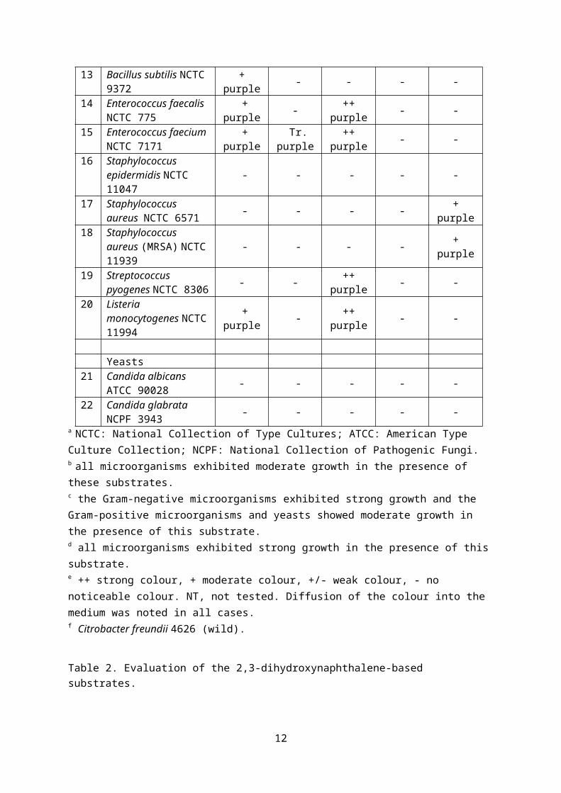
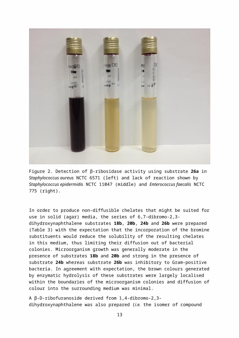
The 2,3-dihydroxynaphthalene-derived substrates 18a, 20a and 22a (Table 2) allowed only moderate growth of all members of the panel of microorganisms suggesting that these substrates were inhibitory to some extent. Strong growth of the Gram-negative microorganisms and moderate growth of the Gram-positive microorganisms and the yeasts were seen with the glucuronide substrate 24a. In contrast, strong growth of all 20 microorganisms was observed in the presence of the ribofuranoside substrate 26a. Purple coloured colonies were produced with this series of substrates when appropriate enzymatic activity was present. As with the catechol-derived substrates, diffusion of colour into the surrounding medium occurred and hence these substrates would be better suited for use in liquid media. For substrate 18a, coloration was generated only by known producers of β-glucosidase including enterococci and Listeria monocytogenes, suggesting this substrate could be exploited for detection of these species. Similarly, for substrate 20a activity was only observed for known producers of β-galactosidase. Substrate 22a is expected to detect β-hexosaminidase activity and several bacteria demonstrated hydrolysis of this substrate including known producers of this enzyme e.g. Serratia marcescens.35 Disappointingly, no coloration was generated by Candida albicans within the 18 h incubation period and this species is known to produce β-hexosaminidase.36 The glucuronide substrate 24a was only hydrolysed by E. coli, as expected, producing strongly coloured colonies. Substrate 26a effectively detected β-ribosidase activity in a number of Gram-negative species and activity was consistent with that previously reported for other β-ribosidase substrates.12, 37 Among Gram-positive bacteria, reactivity was restricted to S. aureus strains and there was clear differentiation from other Gram-positive species, suggesting that this may be a useful substrate for detection of S. aureus. Figure 2 illustrates the differentiation of S. aureus from other Gram-positive microorganisms in liquid media. The usefulness of β-ribosidase detection for the identification of S. aureus has previously been observed.16
a NCTC: National Collection of Type Cultures; ATCC: American Type Culture Collection; NCPF: National Collection of Pathogenic Fungi.b all microorganisms exhibited moderate growth in the presence of these substrates. c the Gram-negative microorganisms exhibited strong growth and the Gram-positive microorganisms and yeasts showed moderate growth in the presence of this substrate.d all microorganisms exhibited strong growth in the presence of this substrate.e ++ strong colour, + moderate colour, +/- weak colour, - no noticeable colour. NT, not tested. Diffusion of the colour into the medium was noted in all cases.f Citrobacter freundii 4626 (wild).
Table 2. Evaluation of the 2,3-dihydroxynaphthalene-based substrates.
10
Figure 2. Detection of β-ribosidase activity using substrate 26a in Staphylococcus aureus NCTC 6571 (left) and lack of reaction shown by Staphylococcus epidermidis NCTC 11047 (middle) and Enterococcus faecalis NCTC 775 (right).
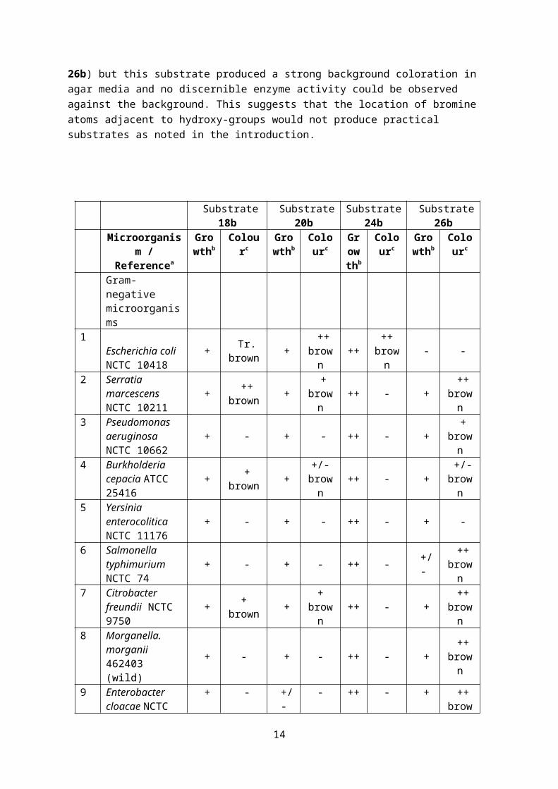
In order to produce non-diffusible chelates that might be suited for use in solid (agar) media, the series of 6,7-dibromo-2,3-dihydroxynaphthalene substrates 18b, 20b, 24b and 26b were prepared (Table 3) with the expectation that the incorporation of the bromine substituents would reduce the solubility of the resulting chelates in this medium, thus limiting their diffusion out of bacterial colonies. Microorganism growth was generally moderate in the presence of substrates 18b and 20b and strong in the presence of substrate 24b whereas substrate 26b was inhibitory to Gram-positive bacteria. In agreement with expectation, the brown colours generated by enzymatic hydrolysis of these substrates were largely localised within the boundaries of the microorganism colonies and diffusion of colour into the surrounding medium was minimal.
A β-D-ribofuranoside derived from 1,4-dibromo-2,3-dihydroxynaphthalene was also prepared ( i.e. the isomer of compound 26b) but this substrate produced a strong background coloration in agar media and no discernible enzyme activity could be observed against the background. This suggests that the location of bromine atoms adjacent to hydroxy-groups would not produce practical substrates as noted in the introduction.
a NCTC: National Collection of Type Cultures; ATCC: American Type Culture Collection; NCPF: National Collection of Pathogenic Fungi.b ++ strong growth, + moderate growth, +/- weak growth, no noticeable growth.c ++ strong colour, + moderate colour, +/- weak colour, Tr. trace of colour, - no noticeable colour.
Table 3. Evaluation of brominated substrates.
Substrate 18b did not show potential as a suitable substrate for detection of β-glucosidase producing pathogens as weak reactions were observed for enterococci and Listeria monocytogenes. Some unexpected (albeit weak) positive reactions were also observed for other species such as S. aureus. β-Galactosidase is most often targeted as a useful marker for coliforms and moderate to strong positive reactions were obtained as expected for E. coli, S. marcescens and C. freundii with substrate 20b. However, E. cloacae (a β-galactosidase producing coliform) did not produce coloration. It is likely that activity of this and other species could be improved by inclusion of an inducer of β-galactosidase such as isopropyl-β-D-thiogalactoside (IPTG). As anticipated, the glucuronide substrate 24b was only hydrolysed by E. coli producing strongly coloured colonies and illustrative agar plates are depicted in Figure 3. In addition to the 20 microorganism plate which clearly shows a positive response for E. coli, four single microorganism plates which included two E. coli strains (expected to give positive responses) and S. marcescens and S. typhimurium (both expected to produce negative responses) were prepared. These four plates also showed a clear difference between positive and negative results and a good contrast between the colour of the colonies and the background. Growth inhibition was even more pronounced with substrate 26b with inhibition observed for E. coli, P. rettgeri and all of the eight Gram-positive bacteria tested. This precluded its potential application for the detection of S. aureus.
14
Figure 3. Agar plates produced using substrate 24b. Left frame: arrangement of 20 microorganisms on a single agar plate (blue spots represent Gram-negative bacteria, red spots represent Gram-positive bacteria and the two yellow spots are the yeast species). Numbers correspond to microorganisms in Table 3. Right frame: left, 20 selected microorganisms on a single agar plate; middle top, E. coli NCTC 10418; top right, E. coli NCTC 12241; middle bottom, S. marcescens NCTC 10211; bottom right, S. typhimurium NCTC 74.
Conclusion
We have reported the synthesis and evaluation of a novel series of chromogenic glycosidase substrates, some of which show potential for the detection of pathogenic bacteria. By introducing two bromine atoms at the 6,7-positions in the naphthalene-based substrates, diffusion of the resulting chromogens into agar media is minimised. The chromogens produced by the non-brominated substrates diffused from the microorganism colonies thus precluding their use in agar media, but these substrates have potential for use in liquid media.
Acknowledgements
We thank the EPSRC UK National Mass Spectrometry Facility at Swansea University, UK, for high resolution mass spectra. We also thank Dr Karen Haggerty for performing the HPLC work.
Experimental
NMR spectra were recorded on a Jeol spectrometer at either 270 or 400 MHz for 1H spectra and either 68 or 100 MHz for 13C spectra. All chemical shifts are quoted in ppm relative to tetramethylsilane. In the assignment of signals the abbreviation DHN is used for 2,3-dihydroxynaphthalene and CHA for cyclohexylamine. Optical rotations were measured on an Optical
15
Activity AA10 polarimeter. High resolution mass spectra (HRMS) were obtained from the EPSRC Mass Spectrometry Facility (Swansea). Flash chromatography was performed using Fluorochem Ltd silica gel (60 Å). Mixed solvents are recorded as volumetric ratios. EtOH is methylated spirit. Water is deionised water. HPLC was performed using an Agilent 1260 modular HPLC instrument fitted with a multiple wavelength detector and a Phenomenex Kinetex 2.6 μ C18, 150 x 2.1 mm column. Data was acquired at 280 nm with a flow rate of 0.4 mL per minute at ambient temperature. Mobile phase A was water containing 0.1% v/v formic acid and mobile phase B was acetonitrile containing 0.1% v/v formic acid. Substrates were prepared in a mixture 95% A/ 5% B. The gradient used was: 0-10 mins 95% A/ 5% B; 10-13.5 mins 5% A/ 95% B; 13.5-18 mins 95% A/ 5% B. The HPLC traces of key substrates are shown in the supplementary information.
6,7-Dibromo-2,3-dihydroxynaphthalene 16b38 was prepared following a literature procedure.
The known acylated sugar derivatives 10,20 1224 and 17a28 were prepared by a Michael-type glycosidation procedure (420-503 mmol scale) in a solution of acetone and aqueous sodium hydroxide. The crude products were purified by column chromatography (eluent toluene: acetone 10:1) followed by trituration with EtOH (compounds 10 and 12) or trituration (MeOH) followed by recrystallization from MeOH (compound 17a). A Zemplén deprotection of compounds 10 and 17a afforded the known glycosides 1118,22,39 and 18a28 respectively. The ester 14 was prepared by a similar method to that described in the literature, m.p. 128-130oC (lit. 136-137 oC).25 1H NMR: (DMSO-d6) 9.28 (1H, s, OH), 6.99 (1H, d, J 7.9 Hz, Ar-H), 6.93-6.83 (2H, m, Ar-H), 6.73 (1H, m, Ar-H), 5.44 (2H, m), 5.09-5.02 (2H, m), 4.62 (1H, d, J4,5 9.9 Hz, H-5), 3.35 (1H, s, OCH3), 2.01 (3H, s, OAc), 2.00 (3H, s, OAc), 1.99 (3H, s, OAc); 13C NMR: (DMSO-d6) 170.1 (Ac), 169.9 (Ac), 169.6 (Ac), 167.8 (C=O), 148.4, 145.0, 124.7, 119.7, 119.3, 117.2 (catechol), 99.4 (C-1), 71.7, 71.5, 71.3, 69.6 (C-2/3/4/5), 53.1 (OCH3), 21.0 (Ac), 20.9 (Ac), 20.8 (Ac).
The synthesis of the β-D-glucuronic acid derivatives and the β-D-ribofuranosides are described below. The preparation of all other compounds is described in the supplementary information.
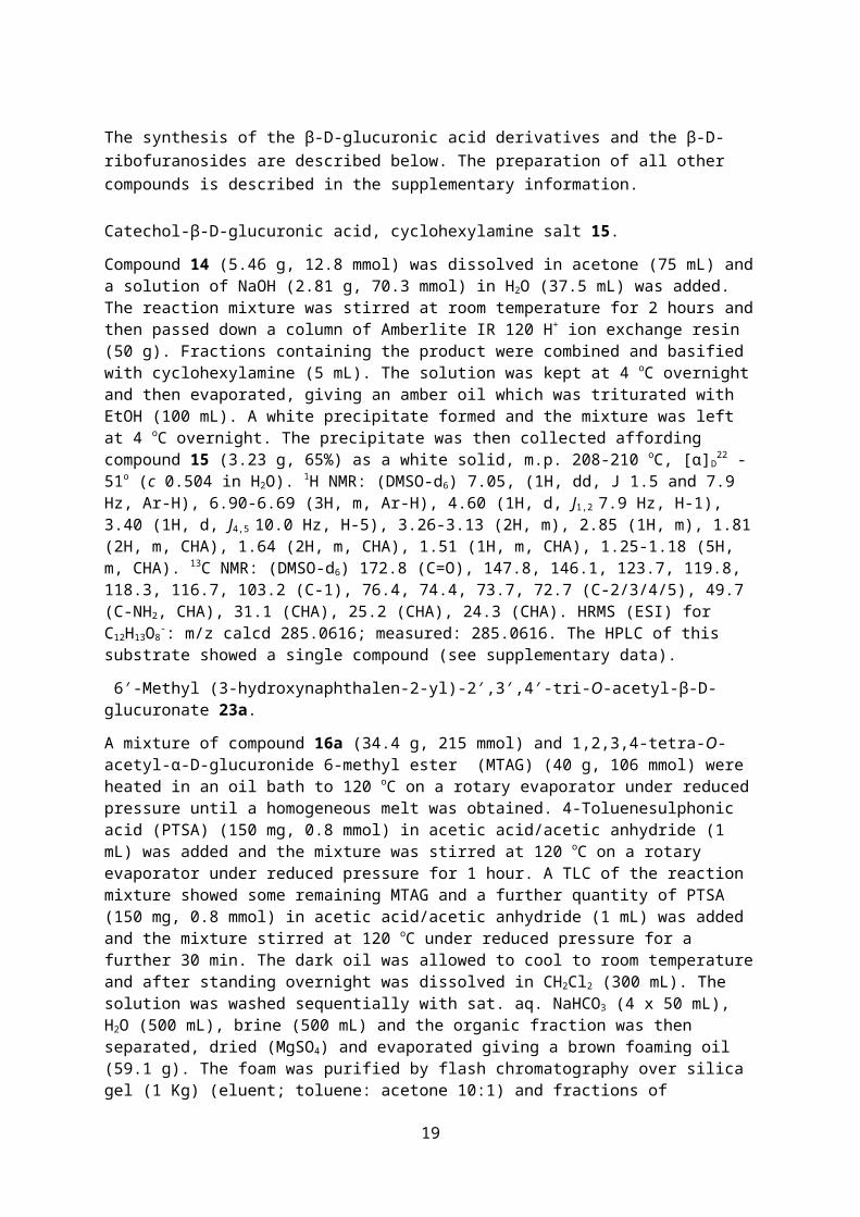
Catechol-β-D-glucuronic acid, cyclohexylamine salt 15.
Compound 14 (5.46 g, 12.8 mmol) was dissolved in acetone (75 mL) and a solution of NaOH (2.81 g, 70.3 mmol) in H2O (37.5 mL) was added. The reaction mixture was stirred at room temperature for 2 hours and then passed down a column of Amberlite IR 120 H+ ion exchange resin (50 g). Fractions containing the product were combined and basified with cyclohexylamine (5 mL). The solution was kept at 4 oC overnight and then evaporated, giving an amber oil which was triturated with EtOH (100 mL). A white precipitate formed and the mixture was left at 4 oC overnight. The precipitate was then collected affording compound 15 (3.23 g, 65%) as a white solid, m.p. 208-210 oC, [α]D
22 -51o (c 0.504 in H2O). 1H NMR: (DMSO-d6) 7.05, (1H, dd, J 1.5 and 7.9 Hz, Ar-H), 6.90-6.69 (3H, m, Ar-H), 4.60 (1H, d, J1,2 7.9 Hz, H-1), 3.40 (1H, d, J4,5 10.0 Hz, H-5), 3.26-3.13 (2H, m), 2.85 (1H, m), 1.81 (2H, m, CHA), 1.64 (2H, m, CHA), 1.51 (1H, m, CHA), 1.25-1.18 (5H, m, CHA). 13C NMR: (DMSO-d6) 172.8 (C=O), 147.8, 146.1, 123.7, 119.8, 118.3, 116.7, 103.2 (C-1), 76.4, 74.4, 73.7, 72.7 (C-2/3/4/5), 49.7 (C-NH2, CHA), 31.1 (CHA), 25.2 (CHA), 24.3 (CHA). HRMS (ESI) for C12H13O8
-: m/z calcd 285.0616; measured: 285.0616. The HPLC of this substrate showed a single compound (see supplementary data).
A mixture of compound 16a (34.4 g, 215 mmol) and 1,2,3,4-tetra-O-acetyl-α-D-glucuronide 6-methyl ester (MTAG) (40 g, 106 mmol) were heated in an oil bath to 120 oC on a rotary evaporator under reduced pressure until a homogeneous melt was obtained. 4-Toluenesulphonic acid (PTSA) (150 mg, 0.8 mmol) in acetic acid/acetic anhydride (1 mL) was added and the mixture was stirred at 120 oC on
16
a rotary evaporator under reduced pressure for 1 hour. A TLC of the reaction mixture showed some remaining MTAG and a further quantity of PTSA (150 mg, 0.8 mmol) in acetic acid/acetic anhydride (1 mL) was added and the mixture stirred at 120 oC under reduced pressure for a further 30 min. The dark oil was allowed to cool to room temperature and after standing overnight was dissolved in CH2Cl2 (300 mL). The solution was washed sequentially with sat. aq. NaHCO3 (4 x 50 mL), H2O (500 mL), brine (500 mL) and the organic fraction was then separated, dried (MgSO4) and evaporated giving a brown foaming oil (59.1 g). The foam was purified by flash chromatography over silica gel (1 Kg) (eluent; toluene: acetone 10:1) and fractions of approximately 200 mL were collected. Fractions 19-26 were combined and evaporated giving a red solid (29.66 g). The red solid was triturated with EtOH (150 mL) and kept at 4 oC overnight. The resulting pale yellow, fluffy solid was collected affording compound 23a (12.6 g, 25%), m.p. 191-192 oC, [α]D
To a stirred solution of α-D-glucuronide trichloroacetimidate (10 g, 21 mmol) and compound 16b (6 g, 18.9 mmol) in CH2Cl2 (100 mL) was added BF3•OEt2 (10 drops, ~ 20 μL). The reaction mixture was stirred at room temperature for 30 min. and then poured into a mixture of CH2Cl2 (500 mL) and sat. aq. NaHCO3 (500 mL). The organic layer was separated and washed with sat. NaHCO3 (4 x 500 mL), H2O (500 mL) and then dried (MgSO4) and evaporated. The residue was triturated with EtOH (100 mL) and the mixture kept at 4 oC overnight. The solid was collected giving compound 23b (2.89 g, 24%) as a white solid, m.p. 176-177 oC, [α]D
(3-Hydroxynaphthalen-2-yl)-β-D-glucuronic acid, cyclohexylamine salt 24a.
Compound 23a (6.1 g, 12.8 mmol) was dissolved in acetone (75 mL) and a solution of NaOH (2.81 g, 70.25 mmol) in H2O (37.5 mL) was added. The mixture was stirred at room temperature for 2 hours and then passed down an Amberlite IR120 H+ ion exchange resin column (50 g). The eluent containing the product was basified using cyclohexylamine (CHA) (5 mL) producing a white precipitate. The mixture was kept at 4 oC overnight and the white fluffy solid was collected, washed with H2O and then acetone affording compound 24a (2.8 g, 53%), m.p. 223-224 oC, [α]D
19 -96o (c 0.5 in H2O). 1H NMR (DMSO-d6): 7.64 (2H, m, Ar-H), 7.46 (1H, s, Ar-H), 7.28 (2H, m, J 6.93 Hz, Ar-H), 7.20 (1H, s, Ar-H), 4.95 (1H, d, J1,2 7 Hz, H-1), 3.58 (1H, d, J3,4=4,5 9 Hz, H-4), 3.35 (2H, m, H-2/3), 3.24 (1H, m, H-5), 2.83 (1H, m, CHA), 1.92-1.48 (5H, m, CHA), 1.23-1.05 (5H, m, CHA); 13C NMR (DMSO-d6): 172.7 (C-6), 147.8, 147.1, 130.7, 128.7, 127.3, 126.3, 124.9, 123.7, 112.1, 110.7 (10 x DHN), 100.0 (C-1), 76.6, 74.8, 73.6, 72.8 (C-2/3/4/5), 49.6 (CHA), 31.0 (CHA), 25.1 (CHA), 24.3 (CHA). HRMS (ESI) for C16H16NO8 [M+H]+: m/z calcd 335.0772; measured: 335.0767. The HPLC of this substrate showed a single compound (see supplementary data).
(6,7-Dibromo-3-hydroxynaphthalen-2-yl)-β-D-glucuronic acid, cyclohexylamine salt 24b.
17
Following the procedure described above for the preparation of compound 24a, compound 23b (2.5 g, 3.9 mmol) gave compound 24b (1.32 g) and a second crop (840 mg) from concentration of the filtrate. Compound 24b (2.16 g, 95%) was obtained as a white solid, m.p. 221-224 oC, [α]D
25 -159o (c 0.25 in H2O). 1H NMR (DMSO-d6): 8.12 (1H, s, Ar-H), 8.09 (1H, s, Ar-H), 7.47 (1H, s, Ar-H), 7.20 (1H, s, Ar-H), 4.96 (1H, d, J1,2 6.9 Hz, H-1), 3.58 (1H, d, J4,5=4,3 9.4 Hz, H-4), 3.39-3.18 (m, H-2/3/5/H2O), 2.86 (1H, m, CHA), 1.85-1.50 (5H, m, CHA), 1.20-1.00 (5H, m, CHA); 13C NMR (DMSO-d6): 172.9 (C-6) 149.3, 148.3, 131.4, 130.6, 130.5, 128.7, 119.2, 117.2, 110.6, 109.7 (10 x DHN), 101.4 (C-1), 76.6, 74.7, 73.5, 72.7 (C-2/3/4/5), 49.7 (CHA), 31.0 (CHA), 25.1 (CHA), 24.3 (CHA). HRMS (ESI) for C16H13Br2O8 [M+H]+: m/z calcd 490.8983; measured: 490.8976. The HPLC of this substrate showed a single compound (see supplementary data).
To a stirred mixture of compound 16a (14.9 g, 93 mmol) in CH2Cl2 (200 mL) was added BF3•OEt2 (3 mL) followed by a solution of α-D-ribofuranosyl trichloroacetimidate (13 g, 30.9 mmol) in CH2Cl2 (100 mL). After approximately 5 min. the reaction mixture was poured into a mixture of sat. aq. NaHCO3 (300 mL) and CH2Cl2 (200 mL). The organic layer was separated and washed with sat. aq. NaHCO3 solution (4 x 500 mL). A TLC showed the organic fraction still contained a significant amount of unreacted compound 16a, therefore it was washed with sat. aq. Na2CO3 solution (2 x 500 mL) (no remaining compound 16a by TLC) and then with H2O (500 mL), dried (MgSO4) and evaporated giving an amber foam. The foam was triturated with MeOH (50 mL) and the resulting white solid collected yielding compound 25a (6.24 g, 54%), m.p. 142-144 oC. [α]D
Compound 16b (3 g, 9.4 mmol), 1,2,3,5-tetra-O-acetyl-β-D-ribofuranose (3.3 g, 10.3 mmol) and 3Å molecular sieves (1 g) were stirred in CH2Cl2 (30 mL) for 10 min. and then BF3•OEt2 (3 mL, 24 mmol) was added. The reaction mixture gradually formed a clear amber solution after approximately 5 min. of stirring at room temperature. After 15 min., a thick white precipitate had formed and the reaction mixture became difficult to stir. The reaction mixture was poured into a mixture of CH2Cl2 (200 mL) and sat. NaHCO3 (200 mL). The organic layer was separated, washed sequentially with sat. aq. NaHCO3 solution (2 x 200 mL), H2O (200 mL) and then dried (MgSO4) and evaporated giving a white solid. The solid was triturated with EtOH (50 mL) and then collected affording compound 25b (4.42 g, 81%), m.p. 185-186 oC, [α]D
Deprotection of compound 25a (1.0 g, 1.7 mmol) by a similar procedure to that described above for the preparation of compound 18b afforded compound 26a (542 mg, 39%) as a white solid, m.p. 181-182 oC, [α]D
22-143o (c 0.55 in acetone/H2O). 1H NMR: (DMSO-d6) 7.63 (2H, m, Ar-H), 7.39 (1H, s, Ar-H), 7.25 (2H, m, Ar-H), 7.14 (1H, s, Ar-H), 5.53 (1H, s, H-1), 4.14 (2H, broad m, H-2/3), 3.92 (1H, m, H-4), 3.59 (1H, dd, J5a,4 4 Hz and J5a,5b 11 Hz, H-5a), 3.41 (1H, m, H-5b and H2O); 13C NMR: (DMSO-d6) 148.2,
18
146.5, 130.5, 128.7, 127.1, 126.1, 124.8, 123.7, 112.4, 110.5 (10 x DHN), 106.9 (C-1), 85.1, 75.1, 70.9, 63.0 (C-2/3/4/5). HRMS (ESI) for C15H16NaO6 [M+Na]+: m/z calcd 315.0839; measured: 315.0844. The HPLC of this substrate showed a single compound (see supplementary data).
Deprotection of compound 25b (1.0 g, 1.7 mmol) by a similar procedure to that described above for the preparation of compound 18b afforded compound 26b (697 mg, 89%) as a white solid, m.p. >240 oC (decomp.), [α]D
23 -90o (c 0.5 in MeOH). 1H NMR (DMSO-d6): 7.73 (1H, s, Ar-H), 7.53 (1H, s, Ar-H), 7.04 (1H, s, Ar-H), 6.30 (1H, s, Ar-H), 5.25 (1H, d, J1,2 2.0 Hz, H-1), 4.32 (1H, t, J3,4=2,3 5.0 Hz, H-3), 4.05 (1H, dd, J1,2 2.0 Hz and J2,3 5 Hz H-2), 3.79 (1H, m, H-4), 3.52 (1H, dd, J4,5a 3.0 Hz and J5a,5b 12 Hz, H-5a), 3.41 (1H, H-5b and H2O); 13C NMR (DMSO-d6): 163.3, 153.4, 134.3, 130.7, 127.1, 124.3, 117.7, 111.8, 110.6, 108.6 (10 x DHN), 108.4 (C-1), 85.5, 75.0, 70.2, 61.7 (C-2/3/4/5). HRMS (ESI) for C15H18Br2NO6 [M+H]+: m/z calcd 465.9495; measured: 465.9495. The HPLC of this substrate showed a single compound (see supplementary data).
References
1. Perry JD. A decade of development of chromogenic culture media for clinical microbiology in an era of molecular diagnostics. Clin. Microbiol. Rev. 2017;30:449-479. 2. Orenga S, James AL, Manfi M, Perry JD, Pincus DH. Enzymatic substrates in microbiology. J. Microbiol. Methods 2009;79:139-155. 3. Váradi L, Lin Luo J, Hibbs DE, Perry JD, Anderson RJ, Orenga S, Groundwater PW. Methods for the detection and identification of pathogenic bacteria: past, present, and future. Chem. Soc. Rev. 2017;46:4818-4832. 4. Lederberg J. The-β-D-galactosidase of Escherichia coli strain K-12. J. Bacteriol. 1950;60:381-392. 5. Rambach A. Appl. Environ. Microbiol. New plate medium for facilitated differentiation of Salmonella spp. from Proteus spp. and other enteric bacteria. 1990;56:301-303. 6. Spitz U, Wick L, Bayer A, Weymuth C, Schabert G. EP 2427431, 2009. 7. Swan A. The use of bile-aesculin medium and of Maxted’s technique of Lancefield grouping in the identification of enterococci (group D streptococci). J. Clin. Pathol. 1954;7:160-163. 8. James AL, Perry JD, Ford M, Armstrong L, Gould FK. Cyclohexenoescletin-β-D-glucoside: a new substrate for the detection of bacterial β-D-glucosidase. J. Appl. Microbiol. 1997;82:532-536. 9. Perry JD, Morris KA, James AL, Oliver M, Gould FK. Evaluation of novel chromogenic substrates for the detection of bacterial β-glucosidase. J. Appl. Microbiol. 2007;102:410-415. 10. James AL, Perry JD, Ford M, Armstrong L, Gould FK. Evaluation of cyclohexenoesculetin-β-D-galactoside and 8-hydroxyquinoline-β-D-galactoside as substrates for the detection of β-galactosidase. Appl. Environ. Microbiol. 1996;62:3868-3870.11. James AL, Perry JD, Chilvers K, Robson IS, Armstrong L, Orr KE. Alizarin-β-D-galactoside: a new substrate for the detection of bacterial β-galactosidase. Lett. Appl. Microbiol. 2000;30:336-340. 12. Butterworth LA, Perry JD, Davies G, Burton M, Reed RH, Gould FK. Evaluation of novel β-ribosidase substrates for the differentiation of Gram-negative bacteria. J. Appl. Microbiol. 2004;96:170-176. 13. Rambach A. EP 0025467, 1981.14. Turner HJ, Burton M. EP 3066107, 2017.15. Turner HJ, Burton M. EP 3066209, 2017.16. Burton M. EP 1438423, 2004.17. Halimi D, Orenga S, Perry JD. US 0297692, 2010. 18. Helferich B, Lang O, Schmitz-Hillebrecht E. Glucosidische Azofarbstoffe. J. Prakt. Chem. 1933;138:275-280.
19
19. Bretschneider H, Beran K. Überführung ein- und zweiwertiger Phenole in acetylierteβ-d-Glucoside mitβ-Pentaacetyl-d-glucose und Borfluorid. Monatsh. Chem. 1949;80:262-270. 20. Praly J-P, Descotes G, Grenier-Loustalot M-F, Metras F. Nouvelle synthèse de spiro-orthoesters D-glucosidiques par photocyclisation stéréoselective d’hydroxyalkyl-D-glucosides. Carb. Res. 1984;128:21-35. 21. Luo S-Y, Jang Y-J, Liu J-Y, Chu C-S, Liao C-C, Hung S-C. Carbohydrate-templated asymmetric Diels-Alder reactions of masked ortho-benzoquinones for the synthesis of chiral bicyclo[2.2.2]oct-5-en-2-ones. Angew. Chem. Int. Ed. 2008;47:8082-8085. 22. Bjerre J, Nielsen EH, Bols M. Hydrolysis of toxic natural glucosides catalyzed by cyclodextrin dicyanohydrins. Eur. J. Org. Chem. 2008;4:745-752.23. Onodera J-I, Takano M, Kishi Y, Yokoyama N, Ishida R. Direct condensation of polyhydric phenols with glucose. Chem. Lett. 1983;1487-1488.24. Peng S-Q, Pu LL, Yi A, Tie-Ming C, Meng-Shen C. Studies on glycosides IV. Synthesis of Arbutin analogs. Acta Chimica Sinica. 1989;47:512-515. 25. Bollenback GN, Long JW, Benjamin DG, Lindquist JA. The synthesis of aryl-D-glucopyranosiduronic acids. J. Am. Chem. Soc. 1955;77:3310-3315. 26. Almeida AF, Santos CN, Ventura MR. Synthesis of new sulfated and glucuronated metabolites of dietary phenolic compounds identified in human biological samples. J. Agric. Food Chem. 2017;65:6460-6466. 27. Garton GA, Williams RT. Studies in detoxification. 17. The fate of catechol in the rabbit and the characterization of catechol monoglucuronide. Biochem. J. 1948;43:206-211. 28. Sakuma YK, Yokoe I. JP 224763, 2004. 29. Bhowmik S, Maitra U. A novel “pro-sensitizer” based sensing of enzymes using Tb(III) luminescence in a hydrogel matrix. Chem. Commun. 2012;48:4624-4626. 30. Xie K, Zhang Y, Chen R, Chen D, Yang L, Liu X, Dai J. Enzymatic glucosylation of unnatural naphthols by a promiscuous glycosyltransferase from Aloe arborescens. Tetrahedron Lett. 2017;58:2118-2121. 31. Goria T, Maitra U. Supramolecular Approach to Enzyme Sensing on Paper Discs UsingLanthanide Photoluminescence. ACS Sensors, 2016;1:934-936. 32. Fischer B, Nudelman A, Ruse M, Herzig J, Gottlieb HE. A novel method for stereoselective glucuronidation. J. Org. Chem. 1984;49:4988-4993. 33. Chiu-Machado I, Castro-Palomino JC, Madrazo-Alonso O, Lopetegui-Palacios C, Verez-Bencomo V. Synthesis of ribofuranosides by catalysis with Lewis acids. Glycosidation versus transacetylation. J. Carb. Chem. 1995;14:551-561. 34. Edberg SC, Pittman S, Singer JM. Esculin hydrolysis by Enterobacteriaceae. J. Clin. Microbiol. 1977;6:111-116.35. Godsey JH, Matteo MR, Shen D, Tolman G, Gohlke JR. Rapid identification of Enterobacteriaceae with microbial enzyme activity profiles. J. Clin. Microbiol. 1981;13:483-490.36. Perry JL, Miller GR. Umbelliferyl-labeled galactosaminide as an aid in identification of Candida albicans. J. Clin. Microbiol. 1987;25:2424-2425.37. Burton M. EP 1438424, 2004.38. Cammidge AN, Chambrier I, Cook MJ, Garland AD, Heeney MJ, Welford K. Octaalkyl- and octaalkoxy-2,3-naphthalocyanines. J. Porphyrins and Phthalocyanines, 1997;1:77-86. 39. Bjerre J, Nielsen EH, Bols M. Hydrolysis of natural glucosides catalyzed by cyclodextrin dicyanohydrins. Eur. J. Org. Chem. 2008;4:745-752.