34
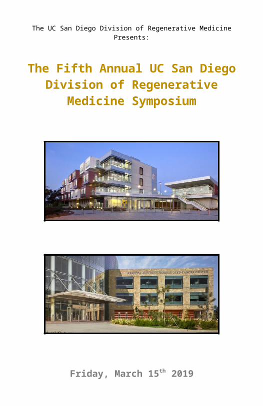
The UC San Diego Division of Regenerative Medicine Presents: The Fifth Annual UC San Diego Division of Regenerative Medicine Symposium Friday, March 15 th 2019
The UC San Diego Division of Regenerative MedicinePresents:
The Fifth Annual UC San Diego Division of Regenerative Medicine
Symposium
Friday, March 15th 20197:00am – 7:00pm

Schedule
Registration and Breakfast (7:00am – 8:00am)Introductions
8:00am – 8:10am Catriona Jamieson, MD, PhD
Session 1: Regenerative MedicineModerator: John Hood, PhD
8:10am – 8:50am Stem Cells in the Adult Human Retinal Pigment Epithelium and their Therapeutic PotentialSally Temple, PhD
8:50am – 9:20am Stem cell niches: the healthy, the old, and the artificialPaul Frenette, MD
9:20am – 9:50am From Human Pluripotent Stem cells to Engineered T cellsGay Crooks, MD
Coffee Break (9:50am – 10:05am)Session 2: Stem Cell Bioengineering
Moderator: Karen Christman, PhD
10:05am – 10:35am Emergence of brain oscillatory waves from engineered human brain organoidsAlysson Muotri, PhD
10:35am – 11:05am Engineered Tissues and Organs: Intelligent DesignLaura Niklason, MD, PhD
11:05am – 11:35am Distinct strategies for epithelial renewal versus regenerationOphir Klein, MD, PhD
Lunch Break (11:35am – 12:35pm)Session 3: Round Table Discussion
Moderator: Bob Klein, JD
12:35pm – 12:45pm Michael Choi, MD to introduce patient12:45pm – 1:35pm Catriona Jamieson, MD, PhD12:45pm – 1:35pm Mark Walters, MD12:45pm – 1:35pm Mehrdad Abedi, MD12:45pm – 1:35pm Maria Millan, MD12:45pm – 1:35pm Jeremy Pettus, MD12:45pm – 1:35pm Joseph Ciacci, MD12:45pm – 1:35pm Sheila Chari, PhD

Schedule
Trainees in Stem Cell ResearchIntroductions by Leslie Crews, PhD
1:35pm – 1:45pm Ryan C. Gimple, Award of Excellence 1:45pm – 1:50pm Rodrigo Chaves, PhD1:50pm – 1:55pm Joyce Chen1:55pm – 2:00pm Antonella Pinto, PhD
Session 4: Cancer Stem CellsModerator: Scott Lippman, MD
2:00pm – 2:30pm Regulation of lung progenitors and their microenvironmentCarla Bender Kim, PhD
2:30pm – 3:00pm Therapeutic targeting of CD47: from cancer stem cells to immuno-oncologyMark Chao, MD, PhD
3:00pm – 3:30pm The Origin of New Cells in the LiverRoel Nusse, PhD
Coffee Break (3:30pm – 3:45pm)Session 4: Neural Stem Cells
Moderator: Jeremy Rich, MD, MHS, MBA
3:45pm – 4:15pm Stem cell-based therapy for Parkinson’s diseaseJun Takahashi, MD, PhD
4:15pm – 4:45pm Human iPSC-derived microglial models of neurodegenerative diseaseStuart Lipton, MD, PhD
4:45pm – 5:15pm Modeling ALS using induced pluripotent stem cells combined with organ-on-chip technologyClive Svendsen, PhD
5:15pm – 5:55pm Using stem cells to probe the secrets of Alzheimer’s diseaseLawrence S. B. Goldstein, PhD
Closing Remarks5:55pm – 6:00pm Catriona Jamieson, MD, PhDCocktail Hour and Poster Presentations (6:00pm – 7:00pm)

Speakers
Joseph Ciacci, MDProfessor, UC San Diego Division of NeurosurgeryJoseph Ciacci, MD, is a board-certified neurosurgeon with extensive experience in neuro-oncology of the spine and brain. Dr. Ciacci’s primary interests are tumors of the spine and brain, complex spinal reconstruction, and stereotactic radiosurgery, a form of radiation therapy that focuses high-power energy on a small area of the body. Dr. Ciacci instructs medical students, residents and fellows at UC San Diego School of Medicine. He also directs the Neurosurgery Residency Program. Additionally, Dr. Ciacci is a trial expert for the CIRM Alpha Stem Cell Clinic at UC San Diego Health as well as the principal investigator for the clinical
trial on spinal cord injury.
Mark Chao, MD, PhDCo-founder, Vice President of Clinical Development, Forty Seven, Inc.Visiting Scholar, Stanford UniversityMark Chao is a physician-scientist and entrepreneur specializing in translational research in hematology/oncology and stem cell biology. Mark conducted seminal research on the role of CD47 in cancer immune evasion and is a founder of Forty Seven, Inc., developing a first-in-class CD47 targeting antibody in cancer. Mark received his medical degree, Ph.D. in cancer biology, and completed his hematology fellowship training at Stanford
University.
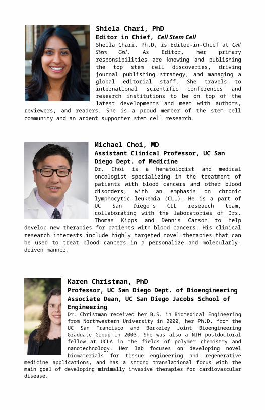
Shiela Chari, PhDEditor in Chief, Cell Stem CellSheila Chari, Ph.D, is Editor-in-Chief at Cell Stem Cell. As Editor, her primary responsibilities are knowing and publishing the top stem cell discoveries, driving journal publishing strategy, and managing a global editorial staff. She travels to international scientific conferences and research institutions to be on top of the latest developments and meet with authors, reviewers, and readers. She is a proud member of the stem cell community and an ardent supporter stem cell research.
Michael Choi, MDAssistant Clinical Professor, UC San Diego Dept. of MedicineDr. Choi is a hematologist and medical oncologist specializing in the treatment of patients with blood cancers and other blood disorders, with an emphasis on chronic lymphocytic leukemia (CLL). He is a part of UC San Diego's CLL research team, collaborating with the laboratories of Drs. Thomas Kipps and Dennis Carson to help develop new therapies for patients with blood cancers. His clinical research interests include highly

targeted novel therapies that can be used to treat blood cancers in a personalize and molecularly-driven manner.
Speakers
Karen Christman, PhDProfessor, UC San Diego Dept. of BioengineeringAssociate Dean, UC San Diego Jacobs School of EngineeringDr. Christman received her B.S. in Biomedical Engineering from Northwestern University in 2000, her Ph.D. from the UC San Francisco and Berkeley Joint Bioengineering Graduate Group in 2003. She was also a NIH postdoctoral fellow at UCLA in the fields of polymer chemistry and nanotechnology. Her lab focuses on developing novel biomaterials for tissue engineering and regenerative medicine applications, and has a strong translational focus with the main goal of developing minimally invasive therapies for cardiovascular disease.
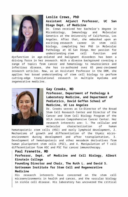
Leslie Crews, PhDAssistant Adjunct Professor, UC San Diego Dept. of MedicineDr. Crews received her bachelor’s degree in Microbiology, Immunology and Molecular Genetics at the University of California, Los Angeles. After that, she embarked upon an exciting research career in stem cell biology, completing her PhD in Molecular Pathology at UC San Diego. Her passion for understanding stem cell function and dysfunction in age-related and malignant disorders has been a driving force in her research. With a diverse background covering a range of topics from cancer and hematology to neuroscience and infectious
disease, she has co-authored over 65 peer-reviewed research articles. Now, as an Assistant Professor in the DRM, she applies her broad understanding of stem cell biology to perform cutting-edge translational research in multiple myeloma and regenerative medicine.
Gay Crooks, MDProfessor, Department of Pathology & Laboratory Medicine, and Department of Pediatrics, David Geffen School of Medicine, UC Los AngelesDr. Crooks serves as Co-Director of the Broad Stem Cell Research Center and Director of the Cancer and Stem Cell Biology Program of the UCLA Jonsson Comprehensive Cancer Center. Her research interests are: 1. The cellular and molecular characterization of human hematopoietic stem cells (HSC) and early lymphoid development, 2. Mechanisms of growth and differentiation of the thymic micro-environment during development and after transplantation, 3. Development of hematopoiesis and other
mesoderm derivatives from human pluripotent stem cells (PSC), and 4. Manipulation of T cell differentiation from HSC and PSC for cancer immunotherapy.Paul Frenette, MDProfessor, Dept. of Medicine and Cell Biology, Albert Einstein CollegeFounding Director and Chair, The Ruth L. and David S. Gottesman Institute for Stem Cell and Regenerative MedicineHis research interests have concerned on the stem cell microenvironments in health and cancer, and the vascular biology in sickle cell disease. His laboratory has uncovered the critical role of the sympathetic nervous system in the regulation of hematopoietic stem cell (HSC) egress from
their niches, in the pathogenesis of acute myeloid leukemia and prostate cancer. He has elucidated circadian rhythmicity in stem cell release from the bone marrow, and identified the major cellular constituents forming niches in the bone marrow.
Lawrence S. B. Goldstein, PhD, Keynote SpeakerDistinguished Professor, UC San Diego Department of Cellular and Molecular Medicine, Department of NeuroscienceScientific Director, Sanford Consortium for Regenerative MedicineDr. Goldstein has held several roles at UCSD. He served as Director of Sanford Clinical Stem Cell Center 2013-2019, Director of the UCSD Stem Cell Program from 2006 – 2015. His awards include a Senior Scholar Award from the Ellison Medical Foundation, an American Cancer Society Faculty Research Award, appointment as The Loeb Chair in Natural Sciences at Harvard University, election to the American Academy of Arts and Sciences, 2009 Public Service Award from the American
Society for Cell Biology and the ARCS Scientist of the Year 2015 and ASCB Lifetime Achievement Fellow 2016.
John Hood, PhDCo-Founder/Founder: Impact Biomedicines, Endeavor Biomedicines, Neuronomix, and SamumedCEO, ActavalonBoard Chairman, Endeavor BiomedicineJohn Hood is the founder and/or co-founder of Impact Biomedicines, Endeavor Biomedicines, Neuronomix and Samumed. Currently, John is the acting Chief Executive Officer at Actavalon, a San Diego biotechnology company developing therapies for autoimmune disease and oncology, Board Chairman of Endeavor Biomedicines and Founder of Neuronomix. Most recently he was the Founder and Chief Executive Officer of Impact
Biomedicines, a company that was acquired for up to $7 billion by Celgene in 2018. He is an inventor on 100+ patents and author on 50+ scientific articles. Dr. Hood obtained a Ph.D. in medical physiology and B.S. in biochemistry from Texas A&M University.
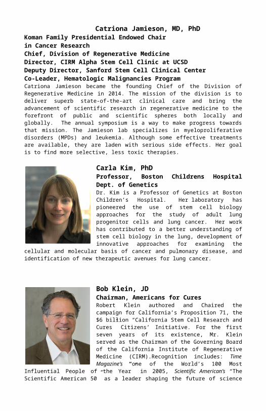
Catriona Jamieson, MD, PhDKoman Family Presidential Endowed Chair in Cancer ResearchChief, Division of Regenerative MedicineDirector, CIRM Alpha Stem Cell Clinic at UCSDDeputy Director, Sanford Stem Cell Clinical CenterCo-Leader, Hematologic Malignancies ProgramCatriona Jamieson became the founding Chief of the Division of Regenerative Medicine in 2014. The mission of the division is to deliver superb state-of-the-art clinical care and bring the advancement of scientific research in regenerative medicine to the forefront of public and scientific spheres both locally and globally. The annual symposium is a way to make progress towards that mission. The Jamieson lab specializes in myeloproliferative disorders (MPDs) and leukemia. Although some effective treatments are available, they are laden with serious side effects. Her goal is to find more selective, less toxic therapies.
Carla Kim, PhDProfessor, Boston Childrens Hospital Dept. of GeneticsDr. Kim is a Professor of Genetics at Boston Children’s Hospital. Her laboratory has pioneered the use of stem cell biology approaches for the study of adult lung progenitor cells and lung cancer. Her work has contributed to a better understanding of stem cell biology in the lung, development of innovative approaches for examining the cellular and molecular basis of cancer and pulmonary disease, and identification of new therapeutic avenues for lung cancer.
Bob Klein, JDChairman, Americans for CuresRobert Klein authored and Chaired the campaign for California’s Proposition 71, the $6 billion “California Stem Cell Research and Cures” Citizens’ Initiative. For the first seven years of its existence, Mr. Klein served as the Chairman of the Governing Board of the California Institute of Regenerative Medicine (CIRM).Recognition includes: Time Magazine’s “one of the World’s 100 Most Influential People of the Year” in 2005, Scientific American’s “The Scientific American 50” as a leader shaping the future of science in November 2005, the Biotechnology Industry Organization’s “Biotech Humanitarian Award” in 2010, the International Society
for Stem Cell Research’s (ISSCR) “ISSCR Public Service Award” in 2011, and Research! America’s Gordon & Llura Gund Leadership Award in 2010. He is working with a coalition of California leaders to explore the possibility of returning to the ballot in 2020 to continue state funding for the research.Ophir Klein, MD, PhDLarry L. Hillblom Distinguished Professor in Craniofacial AnomaliesCharles J. Epstein Professor of Human GeneticsProfessor of Orofacial Sciences and PediatricsChief, Divisions of Genetics and Craniofacial AnomaliesUniversity of California, San FranciscoOphir Klein is the Hillblom Distinguished Professor in Craniofacial Anomalies and the Epstein Professor of Human Genetics at UCSF. He serves as Chief of the Divisions of Medical Genetics and Craniofacial Anomalies. Dr. Klein attended UC Berkeley as an undergraduate and received a Ph.D. in Genetics and an M.D. from Yale University. He completed residencies at Yale in Pediatrics and at UCSF in Clinical Genetics. Dr. Klein’s research focuses on understanding how organs form in the embryo and how they regenerate in the adult, with a particular emphasis on understanding how stem cells in the intestinal epithelium enable renewal and regeneration.
Scott M. Lippman, MDProfessor, UC San Diego Dept. of MedicineSenior Associate Dean and Associate Vice Chancellor for Cancer Research and CareChugai Pharmaceutical Chair in CancerDr. Lippman is the Chugai Professor of Medicine and Associate Vice Chancellor for Cancer Research and Care; and Director of the UC San Diego NCI-designated Comprehensive Moores Cancer Center. His research interest is focused on leveraging premalignant biology for immune interception of oral and pancreatic neoplasia, currently funded by the NCI and a SU2C dream team. His work is providing insight into the biology of genomic instability and immune evasion in premalignancy, and adaptive T-cell memory induction for durable prevention.

Stuart A. Lipton, MD, PhDHannah and Eugene Step Chair, Professor and Director, Neuroscience Translational Center, Scripps Research InstituteStuart A. Lipton, MD, PhD is a Professor at Scripps Research and a clinical neurologist at UCSD. He is best known for developing the FDA-approved Alzheimer’s drug memantine (Namenda®), co-discovering the ubiquitous posttranslational modification known as protein S-nitrosylation, cloning the GluN3 family of NMDA receptor subunits from the human brain, and discovering a gene called Mef2c whose haploinsufficiency results in a form of Autism Spectrum Disorder (ASD). Here, Lipton will discuss his group’s new
work on human iPSC-derived microglial inflammatory responses to misfolded proteins in neurodegenerative disorders.
Maria T. Millan, MDPresident and CEO of California Institute For Regenerative Medicine (CIRM) Maria T, Millan, MD, is the President and CEO of CIRM, an agency created by Californians in 2004 to accelerate stem cell treatments to patients with unmet medical needs. With $3B in funding, CIRM has invested in over 1000 programs, set up critical infrastructure and a world-class ecosystem of top scientists and has recently funded its 50th clinical trial. Dr. Millan began her career as a transplant surgeon and physician scientist after completing her surgical training and research postdoctoral training at Harvard Medical School—Beth Israel Deaconess Medical Center and her
transplant surgery fellowship at Stanford University School of Medicine. At Stanford, she served as an associate professor and head of the pediatric organ transplant program, led a basic research lab and served on the medical school faculty senate and hospital leadership committees. She pursued her interest in stem cell biology and regenerative medicine into the biotechnology sector before joining CIRM in 2012 where she was appointed as president and chief executive officer in 2017.
Alysson R. Muotri, PhDProfessor, UC San Diego Dept. of Pediatrics/Cellular Molecular MedicineCo-Director of the UCSD Stem Cell ProgramDr. Muotri earned a Ph.D. in Genetics in 2001 from University of Sao Paulo, in Brazil. He moved to the Salk Institute as Pew Latin America Fellow in 2002 for a postdoctoral training in the fields of neuroscience and stem cell biology. He has been a Professor at the School of Medicine, University of California in San Diego since late 2008. His research focuses on modeling neurological diseases, such as Autism Spectrum Disorders, using human pluripotent stem cells.
Laura E. Niklason, MD, PhDNicholas M. Greene Professor of Anesthesiology & Biomedical Engineering, Yale UniversityDr. Niklason has been faculty at Yale University since 2006. Her research focuses primarily on regenerative strategies for cardiovascular and lung tissues. She was inducted into the National Academy of Inventors in 2014, and was elected to the National Academy of Medicine in 2015. She was also named (along with Bill Gates and Joe Biden) as one of 34 leaders who are changing healthcare by Fortune Magazine in 2017. Niklason
received her PhD from the University of Chicago in Biophysics in 1988, and received her MD from the University of Michigan in 1991. Dr. Niklason completed her medical training in anesthesiology and critical care medicine at the Massachusetts General Hospital in 1996.
Roel Nusse, PhDProfessor and Chair, Department of Developmental BiologyStanford University, School of MedicineInvestigator, Howard Hughes Medical InstituteDr. Nusse received his PhD from the University of Amsterdam in 1980. He completed postdoctoral studies at the University of California, San Francisco in 1982 working with Dr. Harold Varmus. Dr. Nusse joined the Stanford faculty in 1990. He has been a Howard Hughes Medical Institute Investigator since 1990. In 1999 he was appointed as chair of the department of Developmental Biology at Stanford. Currently, he is the Virginia and Daniel K. Ludwig Professor of Cancer Research. In 2010, he was elected as a member of the National Academy of Sciences. Roel Nusse is also a fellow of the American Academy of Arts and Sciences and a member of the Royal Dutch Academy of Sciences. In 2016, he received the Breakthrough Prize in Life Sciences. His pioneering research has elucidated the mechanism and role of Wnt signaling, one of the paradigms for the fundamental connections between normal development and cancer.
Jeremy Pettus, MDAssistant Professor, UC San Diego Dept. of MedicineAfter being diagnosed with type 1 diabetes at the age of 15, Dr. Pettus has dedicated his career toward treating and educating others with the disease. He now specializes in type 1 diabetes-related clinical research with studies involving immunotherapeutics, islet cell encapsulation, and adjunctive therapies beyond insulin. Dr. Pettus also has a background in immunology research with a focus on biomarkers for type 1 diabetes disease activity. Interests in patient empowerment through education and works with the not-for-profit organization Taking Control of Your Diabetes (TCOYD). With this organization,
Dr. Pettus speaks at the organization’s patient-centered conferences around the U.S., heading the "Type 1 Diabetes" track at these events.
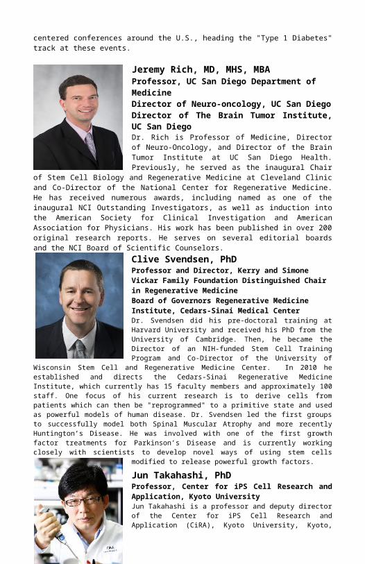
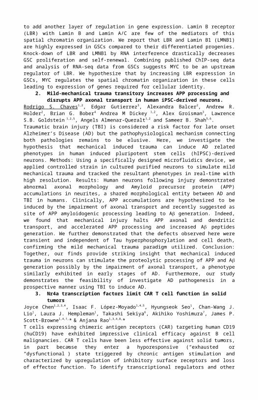
Jeremy Rich, MD, MHS, MBAProfessor, UC San Diego Department of MedicineDirector of Neuro-oncology, UC San DiegoDirector of The Brain Tumor Institute, UC San DiegoDr. Rich is Professor of Medicine, Director of Neuro-Oncology, and Director of the Brain Tumor Institute at UC San Diego Health. Previously, he served as the inaugural Chair of Stem Cell Biology and Regenerative Medicine at Cleveland Clinic and Co-Director of the National Center for Regenerative Medicine. He has received numerous awards, including named as one of the inaugural NCI Outstanding Investigators, as well as induction into the American Society for Clinical Investigation and American Association for Physicians. His work has been published in over 200 original research reports. He serves on several editorial boards and the NCI Board of Scientific Counselors.Clive Svendsen, PhDProfessor and Director, Kerry and Simone Vickar Family Foundation Distinguished Chair in Regenerative MedicineBoard of Governors Regenerative Medicine Institute, Cedars-Sinai Medical Center

Dr. Svendsen did his pre-doctoral training at Harvard University and received his PhD from the University of Cambridge. Then, he became the Director of an NIH-funded Stem Cell Training Program and Co-Director of the University of Wisconsin Stem Cell and Regenerative Medicine Center. In 2010 he established and directs the Cedars-Sinai Regenerative Medicine Institute, which currently has 15 faculty members and approximately 100 staff. One focus of his current research is to derive cells from patients which can then be "reprogrammed" to a primitive state and used as powerful models of human disease. Dr. Svendsen led the first groups to successfully model both Spinal Muscular Atrophy and more recently Huntington’s Disease. He was involved with one of the first growth factor treatments for Parkinson’s Disease and is currently working closely with scientists to develop novel ways of using stem cells modified to release powerful growth factors.
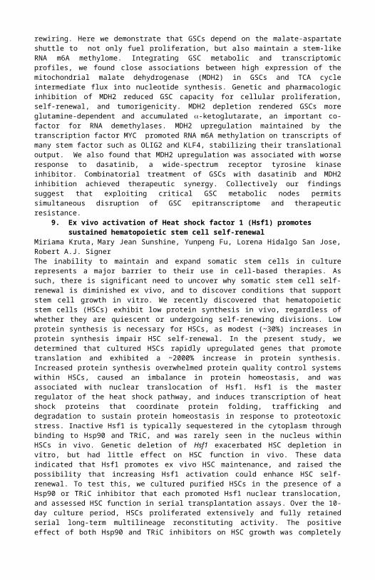
Jun Takahashi, PhDProfessor, Center for iPS Cell Research and Application, Kyoto UniversityJun Takahashi is a professor and deputy director of the Center for iPS Cell Research and Application (CiRA), Kyoto University, Kyoto, Japan. He graduated from the Kyoto University Faculty of Medicine in 1986 and thereafter started his career as a neurosurgeon at Kyoto University Hospital. After he earned his Ph.D. from the Kyoto University Graduate School of Medicine, he worked as a postdoctoral research fellow at the Salk Institute (Dr. Fred Gage), CA, USA., where he started research work on neural stem cells. In 2012, he became a full professor at CiRA, pursuing stem cell-based therapies using iPS cells, and started a clinical trial for Parkinson’s disease in 2018.
Sally Temple, PhD, Keynote SpeakerScientific Director, Neural Stem Cell InstituteSally Temple, PhD, is the Co-Founder and Scientific Director of the Neural Stem Cell Institute located in Rensselaer NY, USA. Dr. Temple’s group is focused on studies of neural stem cells (NSCs) and using this knowledge to develop therapies for central nervous system disorders.
Mark C. Walters, MDProfessor of Pediatrics, Interim Director of Research, UC San Francisco Benioff Children’s HospitalMark is the Jordan Family Director of the Blood and Marrow Transplantation Program at UCSF Benioff Children’s Hospital, Oakland and Program Director of the California Institute of Regenerative Medicine alpha stem cell clinic at UCSF. Dr. Walters received his A.B. with honors in Genetics from the University of California, Berkeley and his MD from the University of California, San Diego. He has been active in cooperative clinical transplantation trials and has led several NIH-supported investigations of hematopoietic cell transplantation for sickle cell anemia and thalassemia. Currently, research interests are focused on genomic editing and gene addition therapies as a strategy
to extend curative therapy in all patients who inherit a clinically significant hemoglobinopathy.
Abstracts1. Spatial chromatin organization maintains cellular identity in glioblastoma
Shruti Bhargava,4 Briana Prager,3,4,5 Sisi Lai,1,2 Xiuxing Wang,4 and Jeremy Rich4
Glioblastomas are heterogeneous tumors and they contain a population of self-renewing cells called glioma stem cells (GSCs). Chromatin landscape of GSCs is being delineated and is demonstrated to play an important role in maintaining cellular identity. Along with chromatin modifications, 3D arrangement of

chromatin in the nucleus has been shown to add another layer of regulation in gene expression. Lamin B receptor (LBR) with Lamin B and Lamin A/C are few of the mediators of this spatial chromatin organization. We report that LBR and Lamin B1 (LMNB1) are highly expressed in GSCs compared to their differentiated progenies. Knock-down of LBR and LMNB1 by RNA interference drastically decreases GSC proliferation and self-renewal. Combining published ChIP-seq data and analysis of RNA-seq data from GSCs suggests MYC to be an upstream regulator of LBR. We hypothesize that by increasing LBR expression in GSCs, MYC regulates the spatial chromatin organization in these cells leading to expression of genes required for cellular identity.
2. Mild-mechanical trauma transitory increases APP processing and disrupts APP axonal transport in human iPSC-derived neurons.
Rodrigo S. Chaves1,2, Edgar Gutierrez3, Alexandra Balcer2, Andrew R. Holder2, Brian G. Bober4 Andrea M Dickey 1,2, Alex Groisman3, Lawrence S.B. Goldstein 1,2,5, Angels Almenar-Queralt1,2 and Sameer B. Shah5,6.Traumatic brain injury (TBI) is considered a risk factor for late onset Alzheimer’s Disease (AD) but the pathophysiological mechanism connecting both pathologies remains to be elusive. Here, we investigate the hypothesis that mechanical induced trauma can induce AD related phenotypes in human induced pluripotent stem cells (hIPSC)-derived neurons. Methods: Using a specifically designed microfluidics device, we applied controlled strain in cultured purified neurons to simulate mild mechanical trauma and tracked the resultant phenotypes in real-time with high resolution. Results: Human neurons following injury demonstrated abnormal axonal morphology and Amyloid precursor protein (APP) accumulations in neurites, a shared morphological entity between AD and TBI in humans. Clinically, APP accumulations are hypothesized to be induced by the impairment of axonal transport and recently suggested as site of APP amyloidogenic processing leading to A generation. Indeed, we found that mechanical injury halts APP axonal and dendritic transport, and accelerated APP processing and increased A peptides generation. We further demonstrated that the defects observed here were transient and independent of Tau hyperphosphorylation and cell death, confirming the mild mechanical trauma paradigm utilized. Conclusion: Together, our finds provide striking insight that mechanical induced trauma in neurons can stimulate the proteolytic processing of APP and A generation possibly by the impairment of axonal transport, a phenotype similarly exhibited in early stages of AD. Furthermore, our study demonstrates the feasibility of investigate AD pathogenesis in a prospective manner using TBI to induce AD.
3. Nr4a transcription factors limit CAR T cell function in solid tumorsJoyce Chen1,2,3,4, Isaac F. López-Moyado1,4,5, Hyungseok Seo1, Chan-Wang J. Lio1, Laura J. Hempleman1, Takashi Sekiya6, Akihiko Yoshimura7, James P. Scott-Browne1,4,†,* & Anjana Rao1,3,4,8,*T cells expressing chimeric antigen receptors (CAR) targeting human CD19 (huCD19) have exhibited impressive clinical efficacy against B cell malignancies. CAR T cells have been less effective against solid tumors, in part because they enter a hyporesponsive (“exhausted” or “dysfunctional”) state triggered by chronic antigen stimulation and characterized by upregulation of inhibitory surface receptors and loss of effector function. To identify transcriptional regulators and other candidates contributing to the diminished CAR T cell function in solid tumors, we developed a CAR T cell model in which recipient mice bearing murine melanoma tumors expressing human CD19 were adoptively transferred with CD19-targeted CAR T cells. We demonstrate that in both CD8+ CAR+ tumor-infiltrating lymphocytes and CD8+
endogenous tumor-infiltrating lymphocytes expressing high levels of PD-1 and TIM3, the Nr4a family proteins Nr4a1 (Nur77), Nr4a2 (Nurr1), and Nr4a3 (Nor1) are prominent effectors of the transcriptional program downstream of NFAT: they promote the expression of inhibitory receptors and genes associated with early hyporesponsive state, and limit effector function. CD8+ T cells from humans with cancer or chronic viral infections also expressed high levels of NR4A transcription factors and displayed enrichment of NR4A binding motifs in accessible chromatin regions. Most importantly, treatment of tumor-bearing mice with CAR T cells lacking all three Nr4a transcription factors (Nr4a TKO) resulted in tumor regression and prolonged survival. Nr4a TKO CAR tumor-infiltrating lymphocytes displayed a transcriptomic profile characteristic of effector function, including upregulation of granzymes and cytokines, and many of these gene expression changes were associated with altered regulatory element accessibility near effector genes. Chromatin regions uniquely accessible in Nr4a TKO CAR tumor-infiltrating lymphocytes compared to wild type were enriched for binding motifs for NFB and AP-1, transcription factors involved in T cell activation. Our data identify Nr4a transcription factors as major players in the cell-intrinsic program of T cell hyporesponsiveness and point to Nr4a inhibition as a promising strategy for cancer immunotherapy.
4. Targeting glioma stem cell miRNA networksDeobrat Dixit, Briana Prager, Lisa C. Wallace, Christine Lee, Tyler E. Miller, Jeremy N. RichGlioma stem cells (GSCs) are self-renewing, highly tumorigenic cells that often display therapeutic resistance, supporting a need for discovery of novel molecular targeting strategies. Using a whole genome miRNA screen, we identified novel miRNAs that regulate GSC stemness and survival. Leveraging three

patient-derived GSC models, we performed a primary miRNA screen using 2051 miRNA-mimics. The top 5% miRNAs (18 miRNAs) that increased or decreased cell viability were selected for further validation, with 10 passing validation in a secondary screen in six patient-derived GSCs. In vitro limiting dilution assays confirmed that these miRNAs significantly modulate stemness of GSCs. IPA analysis predicted these miRNAs as regulators in key biological processes important for cancer, cell cycle, cell death and survival. Ongoing experiments are dissecting the molecular mechanisms regulated by selected miRNAs and optimizing in vivo targeting. Collectively, this novel miRNA screening may inform novel glioblastoma therapies and provide global gene regulatory networks integral to GSC biology.
5. Glioblastoma Stem Cells Display Reprogrammed Circadian Regulation Amenable to Novel Therapeutic Targeting
Zhen Dong1,*, Guoxin Zhang1,*, Meng Qu3,*, Ryan C. Gimple1,2, Qiulian Wu1, Briana C. Prager1,2, Xiuxing Wang1, Leo J. Y. Kim1,2, Deobrat Dixit1, Wenchao Zhou4, Haidong Huang4, Bin Li5, Zhe Zhu1, Zhixin Qiu1, Shideng Bao4, Stephen C. Mack6, Steve A. Kay3,†, Jeremy N. Rich1,†
Glioblastomas are highly lethal cancers, containing self-renewing glioblastoma stem cells (GSCs). Here, we show that GSCs, differentiated glioblastoma cells (DGCs), and neural stem cells (NSCs) displayed robust circadian rhythms, yet GSCs alone displayed exquisite dependence on core clock regulators, BMAL1 and CLOCK, with targeting inducing GSC cell cycle arrest, apoptosis and reduced in vivo tumor growth. To explain selective dependency, chromatin immunoprecipitation revealed BMAL1 preferentially bound at metabolic network genes in GSCs, associated with differences in active chromatin regions compared to NSCs. Targeting BMAL1 or CLOCK attenuated mitochondrial metabolic function and reduced expression of the tricarboxylic acid (TCA) cycle enzymes. Pharmacologic agonists of negative circadian regulators, the Cryptochromes and REV-ERBs, downregulated stem cell regulators and reduced GSC growth as monotherapies with combined pharmacologic targeting inducing synergistic anti-tumor efficacy. Collectively, GSCs coopt circadian molecular regulators beyond canonical circadian circuitry to promote cancer stem cell maintenance and metabolism, offering novel therapeutic paradigms.
6. Super-enhancer screen identifies lipid metabolism as an epigenetically regulated cancer stem cell dependency in glioblastoma
Ryan C. Gimple 1,2 , Jeremy N. Rich2*. Glioblastoma is the most prevalent and aggressive primary intrinsic brain tumor and is among the most lethal of all human cancers with poor responses to all therapeutic modalities. Functionally defined glioblastoma stem cells (GSCs) contribute to this poor prognosis by driving therapeutic resistance and maintenance of cellular heterogeneity. To understand the molecular processes essential for GSC maintenance and tumorigenicity, we interrogated the super-enhancer landscapes of primary glioblastoma specimens and in vitro GSC models. Super-enhancers function as master epigenetic regulators that define cell state, orchestrate cell-type dependent transcriptional networks, and drive key oncogenes in a variety of cancers. This epigenomic landscaping approach revealed polyunsaturated fatty acid synthesis as an epigenetically regulated GSC dependency pathway. Patient derived GSCs containing the identified super-enhancer exhibited high expression of a fatty acid metabolism gene, whose expression was sensitive to BRD4 inhibition. Analysis of the upstream transcription factor control circuits that underlie this super-enhancer reveal that two stem and neuro-developmental factors, OLIG2 and SOX2, may drive the persistence of this super-enhancer. Functionally, knockdown of this gene in patient-derived GSCs resulted in decreased cell proliferation, diminished self-renewal, and induction of apoptosis in vitro, as well as impaired tumor formation in vivo. Global lipidomics profiling revealed that loss of this enzyme altered cellular membrane phospholipid composition, disrupted membrane structural properties, and diminished EGFR signaling. In support of the translational potential of these findings, dual targeting of polyunsaturated fatty acid synthesis and EGFR signaling had a combinatorial cytotoxic effect on GSCs. This discovery implicates aberrant lipid metabolism networks in glioma pathogenesis and suggests that targeting of fatty acid synthesis may provide a specific way to more effectively treat patients with glioblastoma.
7. Hematopoietic stem cells require elevated protein homeostasis capacityLorena Hidalgo San Jose (post-doc)1, Christopher H. Dillingham1, Mary Jean Sunshine1, Miriama Kruta1, Yuning Hong2, Danny M. Hatters3, Robert A.J. Signer1
Background and aims: Stem cells in adult tissues can persist throughout life to regenerate mature cells lost to turnover and injury. Protein homeostasis (proteostasis) is a key regulator of longevity that is maintained by a network of physiological processes that control protein synthesis, folding, repair and degradation. The aim of this study is to compare protein quality control function within long-lived haematopoietic stem cells (HSCs) and short-lived restricted progenitors. Methods: To test if protein synthesis rates influence proteostasis, we examined the abundance of defective proteins in Pten-deficient HSCs. We conditionally deleted Pten from adult hematopoietic cells in Mx1-Cre; Ptenfl/fl mice, which leads
to hyperactivation of the PI3k signaling pathway and increases protein synthesis in HSCs. Additionally, to test if the maintenance of proteostasis is essential for stem cell function, we examined Aarssti/sti mice that harbor a mutation in the alanyl-tRNA synthetase, which causes a tRNA editing defect that reduces translational fidelity and can lead to an accumulation of misfolded proteins. Results: We found that HSCs exhibited high protein quality, as they contained less ubiquitylated and unfolded protein as compared to restricted progenitors in vivo. Modest increases in protein synthesis reduced HSC protein quality in vitro and in vivo, indicating that HSCs depend upon low protein synthesis to maintain the integrity of their proteome. Accumulation of unfolded proteins within HSCs resulted in impairment of the ubiquitin proteasome system, stabilization of the proteasome target c-Myc, and increased proliferation. To investigate how an impaired proteostasis maintenance affects stem cell function, we examined Aarssti/sti
mice. Aarssti/sti mice exhibited modestly reduced proteostasis that specifically impaired HSC maintenance and serial reconstituting activity in vivo but did not impair restricted progenitors. Conclusions: The results presented here demonstrate that HSCs have a lower protein synthesis rate compared to more committed progenitors and that HSCs are particularly dependent on the maintenance of proteostasis to regulate their self-renewal activity in vivo. Low protein synthesis is a broadly conserved feature of somatic stem cells. Our findings raise the possibility that the maintenance of proteostasis is similarly required by stem cells in diverse tissues. Interestingly, interventions that reduce protein synthesis and enhance proteostasis extend organismal lifespan in an evolutionarily conserved manner. Our current findings suggest that mechanisms that promote organismal longevity are conserved at the cellular level to maintain long-lived stem cells in vivo.
8. Reprogrammed TCA cycle promotes stem-like epitranscriptome and therapeutic resistance in glioblastoma stem cells.
Leo J.Y. Kim1,2,3,4, Esther Lim5, Qiulian Wu3,4, Xiuxing Wang3,4 , Qi Xie3,4, Kailin Yang3,4, Christian M. Metallo5, Jeremy N. Rich3,4*
Glioblastomas are lethal cancers harboring diverse cancer stem cell (CSC) hierarchies. Glioma stem cells (GSCs) modulate their differentiation potentials through dynamic epigenetic and metabolic rewiring. Here we demonstrate that GSCs depend on the malate-aspartate shuttle to not only fuel proliferation, but also maintain a stem-like RNA m6A methylome. Integrating GSC metabolic and transcriptomic profiles, we found close associations between high expression of the mitochondrial malate dehydrogenase (MDH2) in GSCs and TCA cycle intermediate flux into nucleotide synthesis. Genetic and pharmacologic inhibition of MDH2 reduced GSC capacity for cellular proliferation, self-renewal, and tumorigenicity. MDH2 depletion rendered GSCs more glutamine-dependent and accumulated -ketoglutarate, an important co-factor for RNA demethylases. MDH2 upregulation maintained by the transcription factor MYC promoted RNA m6A methylation on transcripts of many stem factor such as OLIG2 and KLF4, stabilizing their translational output. We also found that MDH2 upregulation was associated with worse response to dasatinib, a wide-spectrum receptor tyrosine kinase inhibitor. Combinatorial treatment of GSCs with dasatinib and MDH2 inhibition achieved therapeutic synergy. Collectively our findings suggest that exploiting critical GSC metabolic nodes permits simultaneous disruption of GSC epitranscriptome and therapeutic resistance.
9. Ex vivo activation of Heat shock factor 1 (Hsf1) promotes sustained hematopoietic stem cell self-renewal
Miriama Kruta, Mary Jean Sunshine, Yunpeng Fu, Lorena Hidalgo San Jose, Robert A.J. SignerThe inability to maintain and expand somatic stem cells in culture represents a major barrier to their use in cell-based therapies. As such, there is significant need to uncover why somatic stem cell self-renewal is diminished ex vivo, and to discover conditions that support stem cell growth in vitro. We recently discovered that hematopoietic stem cells (HSCs) exhibit low protein synthesis in vivo, regardless of whether they are quiescent or undergoing self-renewing divisions. Low protein synthesis is necessary for HSCs, as modest (~30%) increases in protein synthesis impair HSC self-renewal. In the present study, we determined that cultured HSCs rapidly upregulated genes that promote translation and exhibited a ~2000% increase in protein synthesis. Increased protein synthesis overwhelmed protein quality control systems within HSCs, caused an imbalance in protein homeostasis, and was associated with nuclear translocation of Hsf1. Hsf1 is the master regulator of the heat shock pathway, and induces transcription of heat shock proteins that coordinate protein folding, trafficking and degradation to sustain protein homeostasis in response to proteotoxic stress. Inactive Hsf1 is typically sequestered in the cytoplasm through binding to Hsp90 and TRiC, and was rarely seen in the nucleus within HSCs in vivo. Genetic deletion of Hsf1 exacerbated HSC depletion in vitro, but had little effect on HSC function in vivo. These data indicated that Hsf1 promotes ex vivo HSC maintenance, and raised the possibility that increasing Hsf1 activation could enhance HSC self-renewal. To test this, we cultured purified HSCs in the presence of a Hsp90 or TRiC inhibitor that each promoted Hsf1 nuclear translocation, and assessed HSC function in serial transplantation assays. Over the 10-day culture period, HSCs proliferated extensively and fully

retained serial long-term multilineage reconstituting activity. The positive effect of both Hsp90 and TRiC inhibitors on HSC growth was completely ablated in the absence of Hsf1. Furthermore, Hsf1 activation reduced the unfolded protein load and partially rebalanced protein homeostasis. These findings indicate that maintaining protein homeostasis is a key factor in promoting ex vivo stem cell self-renewal, and reveal new strategies that enable sustained HSC growth in culture.
10. Modeling the Effects of Cholesterol on Alzheimer’s Disease Pathogenesis in iPSC Derived Neurons
Vanessa F. Langness1 Rik van der Kant1,2 Louie Wang1Cheryl M. Herrera1 Daniel A. Williams1Lauren K. Fong1 Yves Leestemaker3 Evelyne Steenvoorden4 Kevin D. Rynearson5Jos F. Brouwers6 J. Bernd Helms6
Huib Ovaa3 Martin Giera4 Steven L. Wagner5,7 Anne G. Bang8Lawrence S.B. Goldstein1,9,12
Alzheimer’s disease (AD) is a progressive neurodegenerative disease that results in loss of neurons and synapses. Brains from human patients with AD exhibit two defining pathological changes: Accumulation of extracellular amyloid plaques which are composed of amyloid beta (Aß) and accumulation of intracellular neurofibrillary tangles which are made up of hyperphosphorylated tau (pTau) protein. These changes are toxic and contribute to the devastating neurodegeneration that occurs in AD. Recent developments in disease modeling using human induced pluripotent stem cells (hiPSCs) have allowed for modeling of Alzheimer’s disease in human neurons in a dish. We can measure Aß and pTau levels in these hiPSC derived neurons allowing us to study how the levels of these proteins become dysregulated in AD. We performed a phenotypic screen for drugs that reduce pTau accumulation in AD-patient iPSC-derived neurons and identified cholesterol lowering drugs as lead compounds. We find that these cholesterol lowering drugs reduce both Aß and pTau levels in iPSC derived neurons. Genetic, biochemical, pharmacological, and epidemiological data support a role for cholesterol in AD pathogenesis, however, the molecular pathways that link cholesterol to AD phenotypes are only partially understood. Recent studies have shown that amyloid precursor protein (APP), the precursor to Aß contains a cholesterol binding site. We use cholesterol lowering drugs and CRISPR/CAS9 genome editing to study how cholesterol levels and mutations that abolish APP-Cholesterol binding influence Aß and pTau burden. Our data indicate that cholesterol lowering drugs can reduce levels of these toxic proteins and thus could lead to new therapeutic avenues for AD treatment.
11. In vivo lineage conversion of vertebrate muscle into early endoderm-like cellsJoseph J. Lancman1*, Clyde Campbell1,2*, Raquel Espin Palazone3, Jonatan Matalonga1, Jiaye He4,5, Alyssa Graves4, Xin-Xin I. Zeng1, Jan Huisken4,5, David Traver3, and P. Duc Si Dong1,2
The extent to which differentiated cells, while remaining in vivo, can be reprogrammed to assume a different identity will reveal fundamental insight into cellular plasticity and will impact regenerative medicine. To investigate in vivo cell lineage potential, we utilized the zebrafish as a practical vertebrate platform to determine factors and mechanisms necessary to induce differentiated cells of one germ layer to adopt the lineage of another. We found that ectopic co-expression of zebrafish Sox32 with mouse Oct4 in several non-endoderm lineages, including skeletal muscle, is able to specifically and cell-autonomously trigger the early endoderm genetic program. Gene expression, live imaging, and functional studies reveal that the endoderm induced muscle cells can proceed to lose muscle gene expression and morphology, while specifically gaining endoderm organogenesis markers, such as the pancreatic specification genes, hhex and ptf1a, via a mechanism resembling normal development. Endoderm induction with a pluripotent defective form of Oct4, a lack of cell division, and a lack of mesoderm, ectoderm, and pluripotency gene activation indicates that reprograming is endoderm specific and occurs via direct lineage conversion. Our work demonstrates that within a vertebrate animal, stably differentiated cells can be induced to directly adopt the identity of a completely unrelated cell lineage without passing through a pluripotent intermediate. This discovery that the lineage potential of differentiated cells in vivo can be manipulated, challenges our understanding of cell lineage restriction and may pave the way towards an in vivo supply of replacement cells for aging or degenerative related diseases such as diabetes
12. TAPBPL specifically induces T cell immune tolerance in glioma stem cellsXiqing LiBackgroundTABPPL acts as an oncogene in Gliomas,rather than TABPPL acts to inhibit tumors growth in many tumors. Methods: Knockout of the TABPPL gene in glioma stem cells and differentiated cells, co-culture of differently treated glioma cells with specially processed T cells. Results: TAPBPL has a specially immunosuppressive effects in glioma stem cells, Knockout of the TABPPL gene in glioma stem cells will increase the sensitivity of T cell killing,TAPBPL is a key checkpoint for T cell recognition. When TABPPL is on the cell membrane, it directly leads to the activation of HLA-ABC, which increases the Antigen presentation ability. It was found by tissue microarrays that in glioma tissues, expression of TABPPL can result in a decrease in the number of total T cells and activated T cells. Conclusion: TAPBPL specifically
induces T cell immune tolerance in glioma stem cells, which is a special immunosuppression gene in the Glioma stem cell.
13. Paradoxical association of TET loss of function with genome-wide hypomethylationIsaac F. López-Moyado1,2,5,*, Ageliki Tsagaratou1, Romain O. Georges1, Benjamin Delatte1, Sven Heinz3, Christopher Benner3, and Anjana Rao1,4,5
Cancer genomes are characterized by focal increases in DNA methylation, co-occurring with widespread hypomethylation. Here we show that TET loss-of-function results in a similar genomic footprint, with expected localized increases in DNA methylation as well as unexpected widespread losses of DNA methylation. We show that 5hmC in wildtype genomes, and DNA hypermethylation in TET-deficient genomes, are largely confined to the active euchromatic compartment. This finding is consistent with the known functions of TET proteins in DNA demethylation and the known distribution of 5hmC at active enhancers and in the gene bodies of highly transcribed genes. In contrast, we show that the unexpected DNA hypomethylation noted in TET-deficient genomes is primarily present in the heterochromatic genomic compartment. In a mouse model of T cell lymphoma driven by TET deficiency (Tet2/3 DKO T cells), genomic analysis of malignant T cells revealed reactivation of repeat elements and enrichment for single nucleotide alterations, again primarily in heterochromatic regions of the genome. Additionally, the expanded Tet2/3 DKO T cells displayed recurrent aneuploidies, as also observed in DNMT-deficient cells, concomitantly with increased accumulation of DNA double-strand breaks. TET loss-of-function may underlie the characteristic pattern of global hypomethylation coupled to regional hypermethylation observed in cancer genomes, and explain the unexpected synergy between DNMT3A and TET2 mutations in myeloid and lymphoid malignancies. Together our data point to a functional interaction between TET proteins and DNMTs, and highlight the potential contribution of heterochromatic dysfunction to oncogenesis
14. Induced in vivo lineage conversion of muscle cells requires loss of myodJonatan Matalonga-Borrel, Joseph Lancman, Clyde Campbell, Duc DongDirect in vivo lineage reprogramming has emerged as a potential alternative approach to replenish loss cells in degenerative diseases, such as beta-cells in diabetes. Recent works have successfully generated beta-cells in vivo in mouse from developmentally related cells restricted to the foregut endoderm. We aim to increase the pool of cells amenable for in vivo lineage conversion, particularly to reprogram cell identity among distinct germ layers, such as muscle, skin, fat, and vasculature cells. These cells are abundant, regenerative, and non-vital, and therefore would be ideal sources of cells to be converted into beta-cells. However these cells are unrelated in lineage and microenvironment, leading us to investigate the mechanisms required to directly reprogram cells in vivo, across germ layers, and in distinct tissues. Leveraging the zebrafish as a vertebrate platform for in vivo lineage conversion, we have identified two transcription factors, oct4 and sox32, that can reprogram several differentiated cell types such as muscle cells and neurons (mesoderm and ectoderm) into endoderm lineages. In muscle cells, we observed that oct4 and sox32 downregulates the expression of the muscle master regulator, myod. Forcing myod to remain expressed prevents induction of the endoderm program by Oct4 and sox32 in muscle cells but not in skin and neuron cells, suggesting that loss myod is specifically required for in vivo reprogramming of muscle cells. Conversely, inhibition of myod expression can increase muscle lineage conversion efficiency. These findings lead us to suggest that repressing the endogenous lineage genetic program is critical for and may enhance induced in vivo lineage conversion of muscle cells and potentially other cell types. Fundamentally, our study supports an active model for maintaining lineage identity and has implications for cell lineage homeostasis and neoplasia. This work is supported by the Larry L. Hillblom Foundation and the Keck Foundation.
15. CHROMATIN LANDSCAPES REVEAL DEVELOPMENTALLY ENCODED TRANSCRIPTIONAL STATES THAT DEFINE GLIOBLASTOMA
Stephen C. Mack1,*,#, Irtisha Singh2,*, Xiuxing Wang3,*, Rachel Hirsch2, Quilian Wu3, Jean A.Bernatchez4,5, Zhe Zhu3, Ryan C. Gimple3,6, Leo J.Y. Kim3,6, Andrew Morton7, Sisi Lai7, Zhixin Qiu3, Rosie Villagomez2, Briana C. Prager3,8,9, Kelsey C. Bertrand1, Clarence Mah10, Wenchao Zhou8, Christine Lee8,11, Gene H. Barnett12, Michael A. Vogelbaum12, Andrew E. Sloan13, Lukas Chavez10, Shideng Bao8, Peter C. Scacheri7, Jair L. Siqueira-Neto4,5, Charles Y. Lin2,14,#, Jeremy N. Rich3Glioblastoma is an incurable brain cancer characterized by high genetic and pathological heterogeneity. Here, we mapped active chromatin landscapes with gene expression, wholeexomes, copy number profiles, and DNA methylomes across 44 glioblastoma stem cell (GSCs) models, 50 primary tumors, and 10 neural stem cells (NSCs) to identify essential super enhancer (SE)-associated genes and the core transcription factors that establish SEs and maintain GSC identity. GSCs segregate into two groups dominated by distinct enhancer profiles and unique developmental core transcription factor regulatory programs. Group specific transcription factors enforce GSC identity — they exhibit higher activity in

glioblastomas versus normal neural stem cells, are associated with poor clinical outcomes, and are required for glioblastoma growth in vivo. Although transcription factors are commonly considered undruggable, we show that group specific enhancer regulation of the MAPK/ERK pathway predicts sensitivity to MEK inhibition. These data demonstrate that transcriptional identity can be leveraged to identify novel dependencies and therapeutic approaches.
16. Impaired axonal regeneration in ALS motor neurons is rescued by restoration of the neuronal microtubule-associated protein stathmin-2
Ze’ev Melamed 1,2 , Jone Erauskin-Lopez 1,2, Michael W Baughn1,2, Ouyang Zhang2, Kevin Drenner1,2, Fernande Freyermuth4,5, Ying Sun1,2, Moira A McMahon6, Melinda Beccari1,2, Jon Artates1,2, Takuya Ohkubo3, Maria Rodriguez3, Frank Bennett6, Frank Rigo6, Sandrine Da Cruz1, John Ravits3, Clotilde Lagier-Tourenne4,5,* and Don W Cleveland1-3,*
Amyotrophic lateral sclerosis (ALS) and frontotemporal dementia (FTD) are associated with loss of nuclear RNA-binding protein TDP-43. Given the central role of TDP-43 in RNA metabolism, it is anticipated that nuclear clearance of TDP-43 in affected neurons of ALS and FTD patients drives processing alterations of multiple RNAs. However, a direct link to increased neuronal vulnerability from reduction or mutation in TDP-43 remains unclear. Here, we have identified a critical role for TDP-43 in regulating expression of stathmin-2, a microtubule-associated protein and neuronal growth factor essential for axon stability and regeneration. Mechanistically, reduction of TDP-43 suppresses stathmin-2 levels by uncovering a premature polyadenylation site in stathmin-2 pre-mRNA, producing a short non-functional mRNA. Suppression of stathmin-2 encoding mRNA is found in neurons trans-differentiated from patient fibroblasts expressing an ALS-causing TDP-43 mutation, in iPSC-derived motor neurons depleted of TDP-43, and in motor cortex and spinal motor neurons isolated from familial ALS patients carrying the most frequent genetic cause of ALS (repeat expansion in C9orf72). Remarkably, aberrant processing of stathmin-2 pre-mRNA is consistently found in sporadic ALS. Finally, we show that lowering TDP-43 or stathmin-2 impairs the ability of iPSC-derived motor neurons to regenerate. Importantly, restoration of stathmin-2 levels rescues axonal regeneration ability in the absence of TDP-43, evidence supporting rescue of stathmin-2 levels as a potential therapeutic approach in neurodegenerative diseases—especially ALS and FTD—affected by TDP-43 proteinopathy. These findings establish that premature polyadenylation-mediated reduction in stathmin-2 is a hallmark of ALS that functionally links TDP-43 proteinopathy to enhanced neuronal vulnerability.
17. Targeting Endocytosis and Intracellular Trafficking of APP and BACE-1 to Reduce TBI-Induced Amyloidogenesis
My Tran 1,2 , Edgar Gutierrez3, Lawrence S.B. Goldstein2,4,5, Angels Almenar-Queralt2,4, Sameer B. Shah5,6, and Rodrigo S. Chaves2,4
Traumatic brain injury (TBI) is a debilitating neurological disorder that can result in short- and long-term disabilities, and while it shares several pathophysiological features with Alzheimer’s disease (AD), their complex relationship is still poorly understood. Excessive accumulation of amyloid beta (Aβ), derived from aberrant proteolytic cleavages of amyloid precursor protein (APP) by β-secretase (BACE-1), has been implicated in the etiology of both TBI and AD. Using human induced pluripotent stem cell (hiPSC)-derived neurons, we have found that mechanical stretch enhances APP-BACE-1 interaction and elevates the generation of Aβ peptides, suggesting that mechanical injury is an upstream regulator of APP proteolytic processing via BACE-1. To reduce APP cleavage by BACE-1, here we modified the co-residence of APP and BACE-1 by treating hiPSC neurons with pharmacological agents that disrupt internalization and intracellular trafficking of APP and BACE-1 proteins. Endocytosis of APP is regulated by clathrin-mediated pathway, which is highly dependent upon the activities of GTPase molecule dynamin. However, the internalization of BACE-1 is reported to be independent of clathrin-mediated endocytosis, and instead, the endosomal sorting of BACE-1 is regulated by ADP ribosylation factor 6 (ARF6). In this experiment, we treated hiPSC-derived neurons either with dynamin inhibitor (dynasore) or ARF6 inhibitor (NAV-2729), and following 16 hours of drug treatment, we detected a significant lower level of extracellular Aβ in neurons treated with either inhibitors. These results indicate that alterations of APP and BACE-1 intracellular trafficking may shelter APP from cleavage by attenuating the physical approximation between APP and BACE-1 through two possible mechanisms: diminishing the amount of endocytosed APP or re-routing BACE-1 to other subcellular compartments. In future experiments, we will establish whether inhibition of dynamin and ARF6 would decrease amyloidogenesis in hiPSC-derived neurons following mechanical trauma. Understanding the trafficking pathways of APP and BACE-1 and how these proteins interact in specific subcellular compartments in neurons will provide remarkable insights that could aid in the development of therapeutic strategies for TBI and AD.
18. Derivation of folliculogenic derma papilla cells from human iPSC.

Antonella Pinto1 (Post-doc), Jasmin Hamidi1, Elina Chermnykh2, Ekaterina Kalabusheva2, Ekaterina Vorotelyak2, Wolfgang Steiger3, Aleksandr Ovsianikov3, Richard Chaffoo4 and Alexey V. Terskikh1
Human induced Pluripotent Stem Cells (iPSCs) have been directed to various cell fates, however, the derivation of Dermal Papilla (DP) cells from iPSCs was challenging. DP plays a dominant role during hair follicle morphogenesis and is critical in defining hair thickness, length, and hair cycle. We report, for the first time, the derivation of functional human DP cells from iPSCs using a Neural Crest (NC) cell intermediate. We investigated the effect of growth factors such as Wnt, FGF, BMP, and R-spondin on the differentiation of iPSC-derived NC cells into DP cells. We observed that the activation of the Wnt pathway enhances the differentiation process, as demonstrated by the expression of classical DP cell markers (Versican and Alkaline Phosphatase) and the up-regulation of relevant DP genes (Nexin, Corin, LEF1, SDC1, HEY1, EGR3). To confirm the identity of iPSC-DP cells in vitro, we compared, at a single cell level, the gene expression profile of iPSC-DPs with that of freshly isolated human adult DP cells including the 3D environment-induced modulation of gene expression. Transplantation of human iPSC-derived DP cells aggregated with mouse E18.5 epithelial cells inside the microscaffolds into the skin of Nude mice resulted in prolonged and robust hair growth. The ability to regenerate hair follicles from cultured iPSC-induced human cells will transform the management of hair loss disorders, which represent an unmet medical need. In addition, modeling of hair growth using organoids will advance the field of regenerative medicine and patient-derived organoids could be used to interrogate drug response for the purpose of personalized medicine.
19. Development of Macrophages from Human Induced Pluripotent Stem Cells: Novel Application for Cell-Based Therapy of Cancers
Somayeh Pouyanfard1, Luis Cruz1, Dan S. Kaufman1*The tumor microenvironment is a complex ecology of cells that provides support to tumor cells during transition to malignancy. Among the innate and adaptive immune cells recruited to the tumor site, macrophages are particularly important and are present at all stages of tumor progression. Distinct populations of macrophages show significant contribution to tumor microenvironment. Macrophages specially show plasticity and diversity which allow their classification along a M1-M2 polarization axis. While M2 phenotype associates with immunosuppression and pro-tumoral activity, classically activated macrophages (M1) contribute to pro-inflammatory and anti-tumor response. Our studies provide a method to routinely generate a large number of macrophages from human induced pluripotent stem cells (hiPSCs). Moreover, these iPSC-derived macrophages can be polarized to inflammatory (M1) phenotype. Briefly, hematopoietic progenitor cells (HPCs) are derived from undifferentiated human iPSCs using a defined serum-free and stromal-free protocol. These HPCs then cultured in stage II conditions in serum-free media supplemented with M-CSF and IL-3. Macrophage progenitor cells are continuously produced after 7-10 days. These cells can be collected and further differentiated in stage III conditions to mature macrophages (hiPSCs-Mφ) in presence of M-CSF. HiPSCs-Mφ exhibited morphology, surface marker expression (CD14, CD11b, CD36, SIRP-α) similar to peripheral blood-derived macrophages (PB-Mφ). These cells are also able to phagocytose beads and Raji tumor cells in presence of anti-CD20 antibody. We can further polarize these hiPSCs-Mφ toward M1 pro-inflammatory macrophages (hiPSCs-M1) by culture with LPS and IFN-γ for 48 hours. Notably, both hiPSCs-M1 and PB-M1 expressed high level of HLA-DR upon stimulation in pro-inflammatory conditions and exhibited similar expression profile of pro-inflammatory cytokines TNF-α, IL-6 and IL-12. Collectively, these studies provide an efficient system for rapid generation of M1 macrophage, facilitating their use as a novel source for cell-based therapy as part of combination therapy in cancer treatment.
20. The enhancer landscape drives subgroup-specific dependencies in meningiomaPrager, BC1-3,*, Vasudevan, HN2, Lee, D2, Xie, Q2, Mack, SC3, Raleigh, DR2, Rich, JN2
Meningiomas are the most common primary brain tumor, and account for 20% of all intracranial neoplasms. Despite this high prevalence, there are no effective medical therapies, and a subset of tumors are not effectively treated by the current therapeutic modality of radiation and resection. Transcriptional and genetic studies have yielded few actionable results with respect to identification of new therapies in grade II and grade III meningiomas. The majority of these high grade tumors are characterized by NF2 mutation, but further attempts to identify dependencies or clinically relevant signatures have been broadly unsuccessful. To address the lack of therapeutic targets in meningioma, we performed H3K27ac ChIP-sequencing to characterize the enhancer landscape of a panel of 33 primary meningioma tissues across all three histologic grades compared with 3 normal arachnoid granulation cell lines. Enhancer profiles cleanly segregated the cohort into three highly prognostic subgroups. These groups exhibit distinct enhancer profiles and an integrated, multi-omics network analysis strategy reveals unique transcriptional programs active in each group. We further identified progesterone receptor as a regulatory hub maintaining the super enhancer network in 2 out of 3 groups. This finding provides a

potential mechanistic basis for the previously unexplained increased meningioma risk among women, and a putative explanation for the equivocal results of previous clinical studies investigating progesterone antagonists as a therapeutic strategy in meningiomas. We further demonstrate that super enhancer-associated genes represent pan-meningioma dependencies, distinguishing tumor tissue from normal cells. Knock-down or inhibition of these genes significantly impairs meningioma cell viability in vitro with a 10-fold greater potency than in normal cells. Thus, we have identified novel and prognostically significant subgroups in meningiomas and defined new, druggable dependencies in both aggressive and more indolent subsets of tumors. Furthermore, enhancer profiling has identified a previously unknown mechanistic basis for the strong epidemiologic risk factors in meningiomas, implicating hormonal programs in maintaining the super enhancer network. Ongoing studies are investigating the efficacy of subgroup-specific druggable strategies and further defining the biologic basis for distinct hormonal dependencies in meningioma.
21. Epigenetic Regulator Landscape in Glioblastoma HeterogeneityZhixin Qiu (Post-doc), Xiuxing Wang, Linjie Zhao, Qiulian Wu, Ryan Gimple, Briana Prager, Guoxin Zhang, Jeremy RichGlioblastoma (GBM) is the most lethal primary brain cancer characterized by chemotherapy and radiotherapy resistance, which is largely driven by glioma stem cells (GSC). GBM is highly heterogeneous with three major transcriptomic subtypes and the presence of GSCs. Understanding the epigenetic reprogramming in subtypes and stemness of GBM is in urgent need to explore therapeutic opportunities. By interrogation of RNA-Seq data in 42 patient-derived GSCs and large-scale ChIP-Seq profiles, we identified epigenetic regulators that control transcriptome reprogramming in different GBM subtypes. Among these subtype-specific regulators is Ying Yang 1 (YY1), a transcription and chromatin regulator involved in both activating and repressing gene expression. Consistent with in silico analysis, YY1 is experimentally validated to be essential in certain subtypes of GBM. RNA-Seq profiles showed that YY1 regulated multiple pathways, including transcription, translation, cell cycle and DNA repair in GSCs. GSCs with YY1 knockdown also showed decreased proneural signature and slightly increased mesenchymal signature, which is in line with its role in proneural subtype and interactions with other regulators. Further, YY1 HiChIP and ChIP-Seq analysis will inform the role of YY1 and chromatin interactions, and ultimately identify therapeutic strategies for GBM. Collectively, these results suggest that global chromatin regulation acts to define cell fates connected to tumor malignancy linking cellular differential and therapeutic resistance.
22. Pharmacological activation of REV-ERBs is lethal in cancer and oncogene-induced senescence
Gabriele Sulli1, Amy Rommel2, Xiaojie Wang3, Matthew J. Kolar4, Francesca Puca5, Alan Saghatelian4, Maksim V. Plikus3, Inder M. Verma2 & Satchidananda Panda1
The circadian clock imposes daily rhythms in cell proliferation, metabolism, inflammation and DNA damage response. Perturbations of these processes are hallmarks of cancer and chronic circadian rhythm disruption predisposes individuals to tumour development. This raises the hypothesis that pharmacological modulation of the circadian machinery may be an effective therapeutic strategy for combating cancer. REV-ERBs, the nuclear hormone receptors REV-ERBα (also known as NR1D1) and REV-ERBβ (also known as NR1D2), are essential components of the circadian clock. Here we show that two agonists of REV-ERBs —SR9009 and SR9011— are specifically lethal to cancer cells and oncogene-induced senescent cells, including melanocytic naevi, and have no effect on the viability of normal cells or tissues. The anticancer activity of SR9009 and SR9011 affects a number of oncogenic drivers (such as HRAS, BRAF, PIK3CA and others) and persists in the absence of p53 and under hypoxic conditions. The regulation of autophagy and de novo lipogenesis by SR9009 and SR9011 has a critical role in evoking an apoptotic response in malignant cells. Notably, the selective anticancer properties of these REV-ERB agonists impair glioblastoma growth in vivo and improve survival without causing overt toxicity in mice. These results indicate that pharmacological modulation of circadian regulators is an effective antitumour strategy, identifying a class of anticancer agents with a wide therapeutic window. We propose that REV-ERBagonists are inhibitors of autophagy and de novo lipogenesis, with selective activity towards malignant and benign neoplasms.
23. Targeting Pyrimidine Synthesis Accentuates Molecular Therapy Response in Glioblastoma Stem Cells
Xiuxing Wang,1,* Kailin Yang,2,3,* Qiulian Wu,1 Leo J.Y. Kim,1,4 Andrew R. Morton,5 Ryan C. Gimple,1,4 Briana C. Prager,1,3,4 Yu Shi,6 Wenchao Zhou,7 Shruti Bhargava,1 Zhe Zhu,1 Li Jiang,1 Weiwei Tao,7 Zhixin Qiu,1 Linjie Zhao, 1 Guoxing Zhang, 1 Xiqing Li,1 Sameer Agnihotri,8 Paul S. Mischel,9 Stephen C. Mack,10 Shideng Bao,7
Jeremy N. Rich1,†
Glioblastoma stem cells (GSCs) reprogram glucose metabolism by hijacking high-affinity glucose uptake to survive in a nutritionally dynamic microenvironment. Here, we trace the metabolic aberration in GSCs to a node between glioblastoma driving mutations and de novo pyrimidine synthesis. Targeting the pyrimidine synthetic rate-limiting step enzyme CAD (carbamoyl-phosphate synthetase 2, aspartate transcarbamyolase, dihydroorotase) or the critical downstream enzyme, DHODH (dihydroorotate dehydrogenase) inhibited the survival, self-renewal, and in vivo tumor initiation of GSCs through the depletion of the pyrimidine nucleotide supply. Core glioblastoma driver mutations in EGFR or PTEN generated distinct CAD phosphorylation patterns to activate carbon influx through pyrimidine synthesis. Simultaneous abrogation of tumor-specific driver mutations and DHODH activity with clinically approved inhibitors demonstrated sustained inhibition of metabolic activity of pyrimidine synthesis and GSC tumorigenic capacity. Higher expression of pyrimidine synthesis genes portended a poor prognosis of glioblastoma patients. Collectively, our results demonstrate a novel therapeutic approach of precision medicine through targeting the nexus between driver mutations and metabolic reprogramming in stem-like glioma cells.
24. KIR expression on in vitro-derived natural killer cells does not regulate killing of allogeneic targets
YunZu M. Wang, Huang Zhu, Eivind H. Ask, Hanna J. Hoel, Alessa Ruiz-Cisneros, Naveen S. Heragu, Kalle-Johan Malmberg, Dan S. KaufmanNatural killer (NK) cells play an essential role in early innate killing of virally-infected and tumor targets. NK cell-mediated activity is regulated by a repertoire of activating and inhibitory receptors that recognize ligands on diseased, stressed, or tumor targets. Killer cell immunoglobulin-like receptors (KIRs) are a family of polymorphic receptors that can be inhibitory or activating based on their intracellular signaling motifs. Expression of certain KIR haplotypes plays a key role in survival and relapse prevention for patients with acute myelogenous leukemia (AML) who receive allogeneic hematopoietic cell transplantation. Therefore, KIR haplotypes are an important consideration in selecting allogeneic donors for patients with AML. However, it is unclear if KIRs play a role in adoptive transfer of NK cells that are becoming more routinely utilized to treat refractory AML and other malignancies. To better address this question we used umbilical cord blood to isolate both CD34+ hematopoietic stem cells and CD45+CD56+ NK cells (UCB-56-NK) from the same umbilical cord blood unit. The CD34+ hematopoietic stem cells were then differentiated in vitro into CD56+ NK cells (UCB-34-NK). Despite originating from the same donor and sharing the same genetic background, as well as comparable expression of Fas ligand, TRAIL, NKp46, NKp44, NKG2A, and NKG2D, the UCB-34-NK cells have characteristically low KIR expression, whereas the UCB-56-NK cells have high KIR expression. This phenotype was further confirmed by mass cytometric (CyTOF) analysis of UCB-56-NK cells and UCB-34-NK cells with a panel of 36 phenotypic and functional NK cell markers. This unique system allows us to study the role of KIR expression independent of any other variations in donor or cell characteristics. The cytotoxicity and NK cell activation of UCB-34-NK cells and UCB-56-NK cells are compared to control NK cells isolated from peripheral blood (PBNK) with standard in vitro cytotoxicity assays against neuroblastoma lines with varying HLA genotypes and a control K562 leukemic target. Our data demonstrates that there is no statistical difference in NK cytotoxicity and activation of UCB-34-NK and UCB-56-NK across a spectrum of target cell HLA types, despite the differences in KIR expression. For example, at effector to target (E:T) ratios of 1:5 and 1:20 against neuroblastoma line IMR32, UCB-34-NK cells demonstrated 68.5% and 84.1% maximal Caspase 3/7 activation, compared to 81.3% and 89.6% by UCB-56-NK cells. Additionally, we have used human induced pluripotent stem cells to derive NK cells (iPSC-NKs) that vary in KIR expression levels. These iPSC-NKs are differentiated from the same well-established iPSC line in the laboratory and therefore again share the same genetic background, and they have similar NK cell surface receptor expression, but differ in levels of KIR expression. Again in vitro cytotoxicity against K562 cells do not demonstrate a significant difference in killing, despite these KIR differences. For example, in targeting erythroleukemia K562 cells, iPSC-NK cells with high levels of KIR expression at E:T ratios of 1:2.5, 1:5, and 1: 10 have Caspase 3/7 activation of 21.1%, 28.2%, and 41.0%, compared to 20.1%, 22.0%, and 31.2% by iPSC-NK cells with low KIR expression. Together, these studies demonstrate that in vitro-derived NK cells do not require KIR expression to become licensed for anti-tumor activity and these cells are able to kill tumor targets whether or not they express KIRs. These studies better enable use of these allogeneic NK cell populations for off-the-shelf NK cell-based therapies without the need to optimize for KIR profiles for patients of differing HLA haplotypes.
25. Switchable chimeric antigen receptor-engineered iPSC-derived natural killer cellsXiao-Hua Li 1, Eduardo Laborda 2, Eric Hampton 2, Diana Gumber 1, Myan Do 1, Robert Blum 1, Huang Zhu 1, Karl Willert 1, Travis Young 2 and Dan S. KaufmanSwitchable chimeric antigen receptors (sCAR) provide an important strategy to preciselyregulate CAR-mediated anti-tumor activity. The sCAR system combines a CAR with an injectable “switch” molecule

(derived from an antibody Fab) for tumor antigen recognition. Previous studies have demonstrated that this sCAR system expressed in T cells provides tight control over T cell-mediated anti-tumor activity. Here, we translated this approach to engineer natural killer (NK) cells with the sCAR to provide a universal, targeted cell-therapy approach that does not have to be derived in a patient-specific manner. First, we expressed the sCAR scFV combined with our previously described NK cell-optimized CAR4 signaling motifs consisting of the NKG2D transmembrane domain, 2B4 co-stimulatory domain and the CD3ζ chain into NK92 cells. We then used both an anti-CD19 switch to target B cell malignancies and an anti-Frizzled7 (FZD7) switch to target solid tumors. Both systems demonstrated a switch-specific dose response in killing either CD19 Raji B cell lymphoma cells or FZD7MA-148 ovarian cancer cells. Next, we engineered human induced pluripotent stem cells (iPSCs) with the sCAR and derived sCAR-expressing NK cells. Again, these sCAR-iPSC-NK cells killed the target tumor cells in a switch-dependent manner. Additionally, specificity of this system is demonstrated by only a basal level of tumor killing when a control switch lacking the sCAR recognition was used, despite of the increased dose of the switch. Similarly, there is no switch mediated killing when Raji-CD19KO or MA148-FZD7KO cells lacking the specific antigen were used. Current studies are testing the iPSC-sCAR system in vivo. Together, this iPSC-sCAR strategy enables close control over CAR mediated activity. Additionally, this system provides flexibility to target multiple antigens on tumor cells to prevent antigen-loss escape variants that can lead to relapsed disease.
26. N6-methyladenine DNA Modification in GlioblastomaQi Xie1,*, Tao P. Wu2,3*, Ryan C. Gimple1,4*, Zheng Li2, Briana C. Prager1,4,5, Qiulian Wu1, Yang Yu6, Pengcheng Wang6, Yinsheng Wang6, David U. Gorkin7, Cheng Zhang7, Alexis V. Dowiak7, Kaixuan Lin2, Chun Zeng8, Yinghui Sui8, Leo J. Y. Kim1,4, Tyler E. Miller4, Li Jiang1, Christine H. Lee9, Zhi Huang10, Xiaoguang Fang10, Kui Zhai10, Stephen C. Mack11, Maike Sander8, Shideng Bao10, Amber E. Kerstetter-Fogle12,13, Andrew E. Sloan12,13, Andrew Z. Xiao2,#, Jeremy N. Rich1,14,15,#
Genetic drivers of cancer can be dysregulated through epigenetic modifications of DNA. While the critical role of DNA 5-methylcytosine (5mC) in the regulation of transcription is recognized, the functions of other non-canonical DNA modifications remain obsure. Here, we report the identification of novel N(6)-methyladenine (N6-mA) DNA modifications in human tissues and implicate this epigenetic mark in human disease, i.e., the highly malignant brain cancer, glioblastoma. Glioblastoma greatly upregulates N6-mA levels, which co-localize with heterochromatic histone modifications, specifically H3K9me3. N6-mA levels are dynamically regulated by the DNA demethylase, ALKBH1, depletion of which leads to transcriptional silencing of oncogenic pathways through decreasing chromatin accessibility. Targeting the N6-mA regulator, ALKBH1, in patient-derived human glioblastoma models inhibited tumor cell proliferation and extended survival of tumor-bearing mice, supporting this novel DNA modification as a potential therapeutic target for glioblastoma. Collectively, our results uncover a novel epigenetic node in cancer through the DNA modification, N6-mA.
27. Modeling APOE4-HDL Mediated Pathogenesis of Alzheimer’s Disease Using Human Pluripotent Stem Cells
Chao-Shun Yang Ph.D.1, Michael Pollante2, 3, Kayla Nguyen3, Mariam Eickn1, Hinal Parikh1, Lawrence S.B. Goldstein Ph.D.1.Alzheimer’s disease (AD) is a non-curable and devastating dementia among the elderly population. However, the genetic and environmental causes of sporadic AD are unknown. Among three genomic variants (2, 3, 4), apolipoprotein E 4 allele (APOE4), instead of most common 3, is the greatest risk factor associated with AD progression. In the adult brain, the APOE protein is the core component forming high-density lipoprotein (HDL) complexes, which are synthesized, secreted, and lipidated by astrocytes and are the major cholesterol/phospholipid supply to mature neurons. Recently, APOE4 has been shown to promote AD progression by exacerbating neurodegeneration caused by the Tau P301S mutation, increasing APP transcript and A secretion, or inducing neuron death when ectopically expressed in neurons. However, the regulation of APOE-containing HDL (APOE-HDL) cellular trafficking and how APOE modulates phosphorylation of Tau (pTau) in human neurons remains unknown. To tackle this question, we have developed a new system to study APOE-HDL cellular trafficking and its potential pTau regulation in human brains by using human astrocytes and human induced pluripotent stem cell derived neurons. First, we established a system to mimic APOE-HDL biogenesis in human brains. This system recapitulates astrocyte-mediated APOE-HDL biogenesis and lipidated APOE-HDLs will provide a well-controlled tool for further study. Second, we discovered that APOE4-HDL has a significantly stronger effect on increasing pTau/total Tau ratio, compared with APOE2 and APOE3. Additionally, APOE4-HDL increases hyperphosphorylation (multiple phosphorylation sites) and conformational changes of Tau. We found that APOE-HDLs are internalized and majorly distributed in perinuclear regions in human neurons. Furthermore, Lipidation of APOE complexes is critical to its pTau regulation. Finally, APOE-HDL may promote AD pathogenesis in part through activating GSK3 in human neurons. Our findings may shed

light on the initial stages of AD progression and lay the foundation to advance therapeutic development for the early stage of AD.
28. Deletion of CISH in human stem cell derived natural killer cells promotes in vivo persistence and enhances anti-tumor activity
Huang Zhu1, Robert H Blum1, Eivind Heggernes Ask3 , Zhengming Wu2, Hanna Julie Hoel3, , Kun-Liang Guan2, Karl-Johan Malmberg3, Dan S. Kaufman1
in natural killer (NK) cells and is encoded by CISH gene in human. Recent studies in a murine model demonstrate that CIS is a potent inhibitory checkpoint in NK cell-mediated tumor immunity. However, it is unclear if CIS similarly regulates human NK cell-mediated anti-tumor activity. Unlike CTLA-4 and PD1 that are expressed at the cell surface and can be blocked by antibody-mediated therapy, CIS is expressed intracellularly. Attempts of deleting CISH in primary NK cells yield controversial results, likely due to low and variable gene editing efficiency in primary NK cells. Therefore, we deleted CISH gene in human induced pluripotent stem cells (iPSCS) using CRISPR/Cas9 technology and characterized the CISH-knockout iPSC lines. We then derived NK cells from these gene-modified iPSCs. Interestingly, deletion of CISH in iPSCs delayed in vitro NK cell differentiation. Specifically, whereas NK cell differentiation was typically fully complete with >90% NK cells after 4 weeks using WT iPSCs, the CISH-/- iPSC- cells only produced ~10% CD45+CD56+ NK cells at 4 weeks, though by 5 weeks these cultures were >80% NK cells. The CISH-/- iPSC-derived NK cells demonstrated typical NK surface maker expression. Analysis of CISH-/- NK cells via mass cytometry (CyTOF) and RNA sequencing demonstrated deletion of CISH substantially altered receptor expression profile and transcriptome. Genes involved in positive regulation of lymphocyte activation were significantly enriched in CISH-/- NK cells. Surprisingly, when challenged with low concentrations of either IL15 or IL2, the CISH-/- NK cells maintained better expansion and exhibited improved cytotoxic function. CISH-/- NK cells demonstrate increased cytotoxic activity, degranulation and IFN-γ production against leukemia cell lines K562 and MOLM-13 cells in vitro. Moreover, single-cell cytokine response analysis of CISH-/- NK cells showed >10 fold enrichment of polyfunctional cell subsets with effector cytokine production (Granzyme B, IFN-γ, MIP-1α, Perforin, TNF-α) compared with WT NK cells or NK cells from peripheral blood. In a MOLM-13 xenograft model, CISH-/- NK cells displayed significantly increased in vivo persistence in peripheral blood in comparison with WT NK cells More importantly, CISH-/- NK cells demonstrate significantly better control of tumor progression in the MOLM-13 xenograft model compared with WT NK cells. Together, these studies demonstrate CISH plays a key role to regulate human NK cell in vivo persistence and anti-tumor activates. Our data justify further clinical validation regulating this intracellular checkpoint to enhance NK cell mediated anti-tumor activities.
Notes

Thank you!
The UC San Diego Division of Regenerative Medicine thanks you for attending the Fifth Annual Symposium. We thank our sponsors, guest speakers, moderators, and all those who made this event possible.

We invite you to join us next year here in San Diego for the 6th Annual UC San Diego Division of Regenerative Medicine Symposium. For more information, please contact Michelle Ghani at: [email protected]
Check out our website for news and events such as our seminar series: http://regenmed.ucsd.edu/
Follow us on Twitter @UCSandiegoDRM
________________________________________________________________________
A Special Thanks to our Sponsors:


