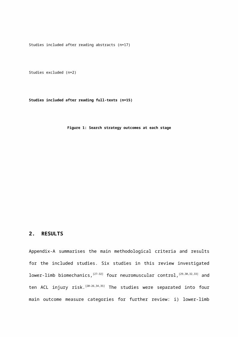
Effects of the Menstrual Cycle on Lower-limb Biomechanics,
Neuromuscular Control, and ACL Injury Risk: A Systematic Review
Vivek Balachandar1,2
1University of Sheffield, School of Medicine and Biomedical Sciences
2Centre for Sports and Exercise Medicine, Queen Mary University of London, United Kingdom
Correspondence to:
Vivek Balachandar
5th year Medical Student, University of Sheffield
07745678360
Word count: 1489 (exc. Title page, references, and appendix)
INTRODUCTION
The incidence of anterior cruciate ligament (ACL) rupture is 3-6 times higher in female
athletes than males during the same cutting and landing sports.[1-3] In the military setting,
relative risks in females have been reported as high as 9.74 compared with men. [4] The
evidence suggests that ACL injury has severe negative implications on mobility, athletic
performance, academic performance, and personal finances.[5,6] Return to previous level of
play is reported between 62%(7) and 67%(8) upto 5-years, with reduced knee function and
fear of re-injury the most common reasons for failure.[7,8] Furthermore, return to previous level
of play is significantly lower in females than males. [8] Long-term consequences are well
investigated, and a recent systematic review reported upto 13% and 48% greater risk of
knee osteoarthritis in patients with a previous isolated ACL rupture or combined ACL and
meniscal injury.[9]
The cause of ACL injury is multifactorial, with both extrinsic and intrinsic factors thought to
contribute.[10,11] Extrinsic factors include physical and visual perturbations,[10-13] and shoe-
surface interactions.[10,11,14,15] Proposed intrinsic factors include female gender,[1-3,10,11] previous
musculoskeletal injury (including prior ACL rupture),[10,11,16] and abnormal lower-limb anatomy,
[10,11] biomechanics,[10,11,17] and neuromuscular control.[10,11,18] Hormonal mechanisms have also
been proposed as a risk factor in female athletes. Hewett et al [19] published a high-quality
systematic review investigating the effects of the menstrual cycle on ACL injury risk until
2005. The review concluded that female athletes may be more predisposed to anterior
cruciate ligament injuries during the pre-ovulatory phase of the menstrual cycle. However,
only seven studies were reviewed,[20-26] and the effects of the menstrual cycle on lower-limb
biomechanics and neuromuscular control were not evaluated. Considering the high number
of recent publications, a systematic review evaluating the effects of the menstrual cycle on
lower-limb biomechanics, neuromuscular control, and ACL injury risk is warranted.
• METHODS
1.1 Search Strategy
MEDLINE, CINAHL, SPORTSDiscus (SD), Web of Science (WoS), and Google Scholar
databases were searched from inception until September 2011 (table 1).
1.2 Inclusion and Exclusion Criteria
Studies evaluating the effects of the menstrual cycle on lower-limb kinematics, kinetics, and
neuromuscular control during weight-bearing functional activities, and non-contact ACL
injury risk in females (18-45) were included. Unpublished studies, case-reports, non-peer
reviewed publications, studies not involving humans, reviews, letters, and opinion articles
were excluded. Studies including participants with previous oral contraceptive pill (OCP) use
were excluded unless non-OCP participant groups were separated.
1.3 Review Process and Data Extraction
All retrieved studies were downloaded to Endnote version X4 (Thomson Reuters
Philadelphia, PA). Results were cross-referenced and duplicated studies were deleted.
Relevant titles were highlighted, with abstracts and full texts reviewed independently for
inclusion (figure 1). Data was extracted from each paper to assist with interpretation of
findings (appendix A)
Keywords MEDLINE
#1 Anterior AND cruciate AND ligament 11194
#2 ACL 7373
#3 Combine #1 OR #2 13539
#4 Biomechanics OR biomechanical OR kinetic OR kinematic OR neuromuscular
7975
#5 Combine #3 OR #4 71009
#6 Menstrual OR menstruation 10665
Combine #3 AND #4 518
Table 1: Search strategy
Potentially relevant studies identified and screened for retrieval (n=518)
Studies excluded (n=490)
Studies included after reading titles (n=28)
Studies excluded (n=11)
Studies included after reading abstracts (n=17)
Studies excluded (n=2)
Studies included after reading full-texts (n=15)
Figure 1: Search strategy outcomes at each stage
2. RESULTS
Appendix-A summarises the main methodological criteria and results for the included
studies. Six studies in this review investigated lower-limb biomechanics,[27-32] four
neuromuscular control,[29,30,32,33] and ten ACL injury risk.[20-26,34,35] The studies were separated
into four main outcome measure categories for further review: i) lower-limb kinematics, ii)
lower-limb kinetics, iii) neuromuscular control, and iv) ACL injury risk.
3. DISCUSSION
The aims of the review were to summarise the effects of the menstrual cycle on lower-limb
kinematics, lower-limb kinetics, neuromuscular control, and ACL injury risk. Greater knee
valgus and lower knee adduction moments, lower hip external rotation moments, and lower
knee joint position sense in the follicular phase may be risk factors for ACL injury in females.
Evidence from this review suggests that females are at significantly greater risk of non-
contact ACL injury during the pre-ovulatory phase of the menstrual cycle, with greatest risk
during the follicular phase.[21,22,24,36-38] Cyclic fluctuations of gonadal hormones have been
reported as a possible cause of ACL injury. As a result, many studies have investigated
changes in lower-limb biomechanics and neuromuscular control during the menstrual cycle,
and a number of mechanisms have been proposed.
• Possible Mechanisms
Levels of oestrogen fluctuate throughout the menstrual cycle, with the greatest surges in the
late follicular phase (days 8-14 in a 28-day cycle).[39,40] Previous studies have reported that
tensile properties of ligaments are influenced by levels of oestrogen, and that oestrogen
receptors are present within fibroblasts of human anterior cruciate ligaments. [41,42] A number
of studies have focused on changes in passive structures during the menstrual cycle.
Studies investigating changes in ACL laxity have reported an increase in knee joint laxity
during the ovulatory and luteal phases compared with follicular phase. [28,31,33,34] One
hypothesis suggests that reduced knee joint laxity or greater stiffness during the follicular
phase may lead to increased risk of ACL injury in females.[19] This represents a possible
mechanism of ACL injury, however, prospective research investigating menses and knee-
joint laxity prior to injury is required to identify inconsistencies between study designs, and
whether retrospective data is adequate.
The effects of the menstrual cycle on neuromuscular control has been investigated as a
possible mechanism behind ACL injury in females. Evidence from this review suggests no
significant changes in gluteus medius onset timing[29] or hamstring and quadriceps
strength[29,32] during functional movements between phases of the menstrual cycle. However,
studies have also reported that oestrogen increases quadriceps contractile strength and
slows relaxation during the ovulatory phase,[43] and decreases knee joint kinesthesia during
the pre-menstrual phase.[35] One hypothesis suggests that fluctuations in serum oestrogen
concentrations during the menstrual cycle affects muscle function and represent a
mechanism of ACL injury in females.[43] A second hypothesis suggests the menstrual cycle
has a significant influence on knee joint kinesthesia and ACL injury risk in females.[35]
Considering the conflicting evidence, and the different variables measured, further high-
quality evidence is required to identify changes in neuromuscular control during the
menstrual cycle and how this contributes to ACL injury risk in females.
Evidence from this review suggests an increase in knee valgus, and decrease in knee
adduction moments and hip external rotation moments during functional movements in the
follicular phase of the menstrual cycle.[28,29] A combination of hip adduction and internal
rotation, knee abduction, tibial external rotation, and ankle eversion has been described as a
dynamic lower-extremity valgus.[19] One hypothesis suggests that increased dynamic valgus
increases the risk of frontal plane valgus collapse and ACL injury in females.[16,44] A number
of cadaveric, in vivo, and computer modeling studies have found that knee valgus can
increase ACL strain high enough to cause rupture.[45-47] Increases in dynamic valgus between
phases of the menstrual cycle may increase risk of ACL injury in females. While the
evidence suggests a proximal risk factor for ACL injury in females, further investigation into
distal mechanisms such as tibial internal/rotation and ankle eversion/inversion are required
to identify the significance of these factors.
• Limitations and future research
A number of movements were used during biomechanical assessment: cutting, single-leg
drop landing, two-leg drop landing, horizontal jump, vertical jump, and hop tests. The
variability of movements between studies makes direct comparisons difficult to make.
Furthermore, whilst these are reported as functional, the literature suggests that
investigations of the ACL should focus on dynamic movements such as rapid deceleration
and change of direction.[10] It is possible that negative findings from studies using movements
such as two-leg drop landing, horizontal jump, and vertical jump may be a result of these
less functional movements. Future studies should focus on high-risk movements that mimic
ACL injuries and allow direct comparisons to be made.
Anterior cruciate ligament injury has significant consequences in athletes at all levels.
Interventions aimed at altering menstrual cycle pattern such as oral contraception are
commonly used by athletes.[48] The evidence suggests that oral contraception may help to
stabilise lower-limb biomechanics and neuromuscular control, and reduce ACL injury risk,
[49,50] however no direct relationship has been established. Further high-quality, RCTs
investigating the effects of oral contraception on lower-limb biomechanics, neuromuscular
control, and ACL injury risk are warranted to understand effects of the menstrual cycle on
knee joint stability in female athletes.
• CONCLUSION
The evidence suggests that females are at significantly greater risk of non-contact ACL
injury during the pre-ovulatory phase of the menstrual cycle, with greatest risk during the
follicular phase. [21,22,24,36-38] Geater dynamic lower-extremity valgus, reduced knee joint laxity,
and reduced knee joint kinesthesia represent possible mechanisms behind ACL injury risk in
the follicular phase. Further high-quality research is required to identify the effects of
neuromuscular control on ACL injury risk in the menstrual cycle. Further high-quality, RCTs
investigating high-risk movements that mimic ACL injuries, and the effects of oral
contraception on lower-limb biomechanics, neuromuscular control, and ACL injury risk are
warranted.
REFERENCES
• Arendt E, Dick R. Knee injury patterns among men and women in collegiate basketball
and soccer: NCAA data and review of literature. Am J Sports Med. 1995;23:694-701.
• Agel J, Arendt EA, Bershadsky B. Anterior cruciate ligament injury in national collegiate
athletic association basketball and soccer: A 13-year review. Am J Sports Med.
2005;33:524–530.
• Griffin LY, Agel J, Albohm MJ, et al. Noncontact anterior-cruciate ligament injuries: risk
factors and prevention strategies. J Am Acad Orthop Surg. 2000;8:141-150.
• Gwinn DE , Wilckens JH, McDevitt ER, et al. The relative incidence of anterior cruciate
ligament injury in men and women at the United States Naval Academy. Am J Sports
Med. 2000;28(1):98-102.
• Freedman KB, Glasgow MT, Glasgow SG, et al. Anterior cruciatate ligament injury and
reconstruction among university students. Clin Orthop Relat Res. 1998;356:208-212.
• Ruiz AL, Kelly M, Nutton RW. Arthroscopic ACL reconstruction: a 5-9 year follow-up.
Knee. 2002;9:197-200.
• Lee DYH, Karim SA, Chang HC. Return to Sports After Anterior Cruciate Ligament
Reconstruction – A Review of Patients with Minimum 5-year Follow-up. Ann Acad Med
Singapore 2008;37:273-8
• Ardern CL, Webster KE, Taylor NF, et al. Return to the Preinjury Level of Competitive
Sport After Anterior Cruciate Ligament Reconstruction Surgery : Two-thirds of Patients
Have Not Returned by 12 Months After Surgery. Am J Sports Med 2011 39: 538
• Oiestad BE, Engebretsen L, Storheim K, et al. Knee osteoarthritis after anterior cruciate
ligament injury: a systematic review. Am J Sports Med. 2009;37:1434–1443
• Hewett TE, Myer GD, Ford KR. Anterior Cruciate Ligament Injuries in Female Athletes :
Part 1, Mechanisms and Risk Factors.. Am J Sports Med 2006 34: 299
• Alentorn GE , Myer GD, Silvers HJ, et al Prevention of non-contact anterior cruciate
ligament injuries in soccer players. Part 1: Mechanisms of injury and underlying risk
factors. Knee Surg Sports Traumatol Arthrosc. 2009 Jul;17(7):705-29.
• McLean SG, Lipfert SW, van den Bogert AJ. Effect of gender and defensive opponent on
the biomechanics of sidestep cutting. Med Sci Sports Exerc. 2004;36:1008-1016.
• Olsen OE, Myklebust G, Engebretsen L, et al. Injury mechanisms for anterior cruciate
ligament injuries in team handball: a systematic video analysis. Am J Sports Med.
2004;32:1002-1012.
• Orchard JW, Powell JW. Risk of knee and ankle sprains under various weather
conditions in American football. Med Sci Sports Exerc. 2003; 35:1118-1123.
• Scranton PE, Whitesel JP, Powell JW, et al. A review of selected noncontact anterior
cruciate ligament injuries in the National Football League. Foot Ankle Int. 1997;18:772-
776.
• Hewett TE, Myer GD, Ford KR, et al. Biomechanical measures of neuromuscular control
and valgus loading of the knee predict anterior cruciate ligament injury risk in female
athletes: a prospective study. Am J Sports Med. 2005;33:492-501.
• Boden BP , Sheehan FT, Torg JS, et al. Noncontact anterior cruciate ligament injuries:
mechanisms and risk factors. J Am Acad Orthop Surg. 2010 Sep;18(9):520-7.
• Hewett TE. Neuromuscular and hormonal factors associated with knee injuries in female
athletes: strategies for intervention. Sports Med. 2000;29:313-327.
• Hewett TE, Zazulak BE, Myer GD. Effects of the Menstrual Cycle on Anterior Cruciate
Ligament Injury Risk : A Systematic Review. Am J Sports Med 2007 35: 659
• Arendt EA, Agel J, Dick R. Anterior cruciate ligament injury patterns among collegiate
men and women. J Athl Train. 1999;34:86-92.
• Arendt EA, Bershadsky B, Agel J. Periodicity of noncontact anterior cruciate ligament
injuries during the menstrual cycle. J Gend Specif Med. 2002;5:19-26.
• Myklebust G, Engebretsen L, Braekken IH, et al. Prevention of anterior cruciate ligament
injuries in female team handball players: a prospective intervention study over three
seasons. Clin J Sport Med. 2003;13:71-78.
• Myklebust G, Maehlum S, Holm I, et al. A prospective cohort study of anterior cruciate
ligament injuries in elite Norwegian team handball. Scand J Med Sci Sports. 1998;8:149-
153.
• Slauterbeck JR, Fuzie SF, Smith MP, et al. The menstrual cycle, sex hormones, and
anterior cruciate ligament injury. J Athl Train. 2002;37: 275-278.
• Wojtys EM, Huston LJ, Boynton MD, et al. The effect of the menstrual cycle on anterior
cruciate ligament injuries in women as determined by hormone levels. Am J Sports Med.
2002;30: 182-188.
• Wojtys EM, Huston LJ, Lindenfeld TN, et al. Association between the menstrual cycle
and anterior cruciate ligament injuries in female athletes. Am J Sports Med.
1998;26:614-619.
• Chaudhari AMW, Lindenfeld TN, Andriacchi TP. Knee and Hip Loading Patterns at
Different Phases in the Menstrual Cycle: Implications for the Gender Difference in
Anterior Cruciate Ligament Injury Rates. Am J Sports Med 2007;35:793
• Park SK, Stefanyshyn DJ, Ramage B. Alterations in Knee Joint Laxity During the
Menstrual Cycle in Healthy Women Leads to Increases in Joint Loads During Selected
Athletic Movements. Am J Sports Med 2009;37:1169
• Cesar BM, Pereira VS, Santiago PRP. Variations in dynamic knee valgus and gluteus
medius onset timing in non-athletic females related to hormonal changes during the
menstrual cycle. The Knee. 2011;18(4):224-230
• Abt JP, Sell TC, Laudner KG, et al. Neuromuscular and biomechanical characteristics do
not vary across the menstrual cycle. Knee Surg Sports Traumatol Arthrosc.
2007;15(7):901-7
• Shultz SJ , Schmitz RJ, Beynnon BD. Variations in varus/valgus and internal/external
rotational knee laxity and stiffness across the menstrual cycle. J Orthop Res.
2011;29(3):318-25.
• Hertel J , Williams NI, Olmsted-Kramer LC, et al. Neuromuscular performance and knee
laxity do not change across the menstrual cycle in female athletes. Knee Surg Sports
Traumatol Arthrosc. 2006;14(9):817-22.
• Shultz SJ , Schmitz RJ, Nguyen AD, et al. Knee joint laxity and its cyclic variation
influence tibiofemoral motion during weight acceptance. Med Sci Sports Exerc. 2011
Feb;43(2):287-95.
• Heitz NA, Eisenman PA, Beck CL, et al. Hormonal Changes Throughout the Menstrual
Cycle and Increased Anterior Cruciate Ligament Laxity in Females. J Athl Train. 1999;
34(2): 144–149.
• Fridén C , Hirschberg AL, Saartok T, Renström P. Knee joint kinaesthesia and
neuromuscular coordination during three phases of the menstrual cycle in moderately
active women. Knee Surg Sports Traumatol Arthrosc. 2006;14(4):383-9.
• Beynnon BD, Robert J. Johnson, Stuart Braun The Relationship Between Menstrual
Cycle Phase and Anterior Cruciate Ligament Injury : A Case-Control Study of
Recreational Alpine Skiers. Am J Sports Med 2006;34:757
• Ruedl G, Ploner P, Linortner I. Are oral contraceptive use and menstrual cycle phase
related to anterior cruciate ligament injury risk in female recreational skiers?
2009;17(9):1065-1069
• Ruedl G, Ploner P, Linortner I. Interaction of potential intrinsic and extrinsic risk factors
in ACL injured recreational female skiers. Int J Sports Med. 2011 Aug;32(8):618-22
• Stricker R , Eberhart R, Chevailler MC, et al. Establishment of detailed reference values
for luteinizing hormone, follicle stimulating hormone, estradiol, and progesterone during
different phases of the menstrual cycle on the Abbott ARCHITECT analyzer. Clin Chem
Lab Med. 2006;44(7):883-7.
• Pauerstein CJ , Eddy CA, Croxatto HD, et al. Temporal relationships of estrogen,
progesterone, and luteinizing hormone levels to ovulation in women and infrahuman
primates. Am J Obstet Gynecol. 1978;15;130(8):876-86.
• Booth FW, Tipton CM. Ligamentous strength measurements in prepubescent and
pubescent rats. Growth. 1970;34:177-185.
• Liu SH, Al-Shaikh RA, Panossian V, et al. Primary immunolocalization of Estrogen and
progesterone target cell in the human anterior cruciate ligament. J Orthop Res.
1996;14:526-533.
• Sarwar R, Beltran NB, Rutherford OM:. Changes in muscle strength, relaxation rate and
fatiguability during the human menstrual cycle. J Physiol. 1996;493:267-272.
• Quatman CE, Hewett TE. The anterior cruciate ligament injury controversy: is ‘‘valgus
collapse’’ a sex-specific mechanism? Br J Sports Med. 2009;43:328–335
• Fukuda Y, Woo SL, Loh JC, et al. A quantitative analysis of valgus torque on the ACL: a
human cadaveric study. J Orthop Res. 2003;21:1107-1112.
• Lloyd DG, Buchanan TS. Strategies of muscular support of varus and valgus isometric
loads at the human knee. J Biomech. 2001;34:1257-1267.
• Markolf KL, Burchfield DM, Shapiro MM, Shepard et al. Combined knee loading states
that generate high anterior cruciate ligament forces. J Orthop Res. 1995;13:930-935.
• Agel J, Bershadsky B, Arendt E. Hormonal therapy: ACL and ankle injury. Med Sci
Sports Exerc. 2006;38:7-12.
• Moller-Nielson J, Hammar M. Sports injuries and oral contraceptive use. Is there a
relationship? Sports Med. 1991;12:152-160.
• Moller-Nielsen J, Hammar M. Women’s soccer injuries in relation to the menstrual cycle
and oral contraceptive use. Med Sci Sports Exerc. 1989;21:126-129.
Appendix A: Summary of studies included in systematic reviewAuthor/year
Participant details Protocol Results
Beynnon BD (2011)
45 females athletes with ACL injury and 45 healthy femalesGroups matched for age, height, weight
Serum sample and self-reported menstrual history data immediately after injury. Both serum concentrations of progesterone andmenstrual history were used to group subjects
Serum concentrations of progesterone revealed that alpine skiers in the preovulatory phase of the menstrual cycle were significantly more likely to tear their ACL than skiers in the postovulatory phase Analysis of menstrual history found similar results, but the difference was not statistically significant
Cesar MG (2011)
23 female non-athletes
Single leg drop landing maneuver while 3-D knee kinematics and gluteus medius muscle onset timing were assessed throughout three distinct phases of the menstrual cycle (confirmed by blood hormone analysis)
Knee valgus angles were significantly less in the luteal phase compared to both follicular phases, while differences were not observed for gluteus medius onset timing.
Ruedl G (2011)
93 female athletes with ACL injury and 93 healthy females
Self-reported questionnaire relating to intrinsic risk and extrinsic risk factors
Preovulatory phase of menstrual cycle (odds ratio, 2.59) was a independent ACL injury risk factor for female skiers
Shultz SJ (2011a) 64 healthy females
Cyclic variations in genu recurvatum (GR), general joint laxity (GJL), varus-valgus (VV), and internal-external (IER) rotational laxities and stiffnesses were examined
Cyclic increases in AKL (9.5%), GR (37.5%), and GJL (13.6%) were observedCyclic increases in VV and IER laxity were negligible. Females had lower VV stiffness at T2 vs T1, but no difference across time points for IER stiffness.Across both time points, females had consistently greater VV and IER laxity and less VV and IER incremental stiffness
Park SK (2009) 26 female athletes
Knee joint biomechanics were measured. Each subject was designated with low, medium, or high knee joint laxity.Knee joint mechanics were compared between low, medium, and high laxity
No significant differences in knee joint mechanics. An increase in KJL was associated with higher knee joint loads during movement. A 1.3-mm increase in KJL resulted in an increase of approximately 30% in adduction impulse in a cutting maneuver, an increase of approximately 20% in knee adduction moment, and a 20% to 45% increase in external rotation loads during a jumping and stopping task
Ruedl G (2009)93 female athletes with ACL injury
Menstrual history, athletic activity, and injury history were collected from the athletes
Analysis revealed that recreational skiers in the preovulatory phase were significantly more likely to sustain ACL injury than skiers in postovulatory phase
Adachi N (2008)
18 female athletes with ACL injury
Menstrual history, athletic activity, and injury history were collected from the athletes
72% of subjects had premenstrual symptoms, 83% had menstrual symptoms.Significant association between the phase of the menstrual cycle and ACL injuries. There were more injuries in the ovulatory phase than expected
Abt JP (2007)
10 healthy femalesMean age-21.4Mean height-1.67cmMean mass-59.9kg
Single-leg postural stability, fine motor coordination, knee strength, knee biomechanics, and serum estradiol and progesterone were assessed at the menses, post-ovulatory, and mid-luteal phases
No significant difference between phases of the menstrual cycle for: i) fine motor coordination, ii) postural stability, iii) hamstring - quadriceps strength ratio at 60 degrees or 180 degrees, iv) knee flexion excursion, v) knee valgus excursion, vi) peak proximal tibial anterior shear force, vii) flexion moment at peak proximal tibial anterior shear force, vii) valgus moment at peak proximal tibial anterior shear force
Chaudhari AMW (2007)
12 female athletes
Horizontal and vertical jump, and drop from a 30-cm box on the left leg. Lower limb kinematics and peak externally applied moments were calculatedWomen were tested for each phase of the menstrual cycle as determined from serum analysis
No significant differences in moments or knee angle were observed between phases in female group
Friden C (2006)32 healthy female athletes
Knee joint kinaesthesia and neuromuscular coordination was measured with the square hop test in the menstrual phase, ovulation phase and premenstrual phase determined by hormone analyses in three consecutive menstrual cycles.
Impaired knee joint kinaesthesia was detected in the premenstrual phase and the performance of square hop test was significantly improved in the ovulation phase compared to the other two phases
Hertel J (2006)14 healthy female athletes
Measures of knee neuromuscular performance and laxity once during the mid-follicular, ovulatory, and mid-luteal stages of menstrual cycle.
No significant differences in the measures of strength, joint position sense, postural control, or laxity across the three testing sessions.No significant correlations were found between changes in E3G or PdG levels and changes in the performance and laxity measures between sessions
Arendt EA (2002)
58 female athletes with ACL injury
Menstrual history, athletic activity, and injury history were collected from the athletes
A significant 28-day periodicity of injuries was present in the entire population as well as in the two subgroups. High- and low-risk time intervals were associated primarily with follicular and luteal phases
Wojtys EM (2002)69 female athletes with ACL injury
Mechanism of injury, menstrual cycle details, use of oral contraceptives, and history of previous injury were recorded. Urine samples validated menstrual cycle phase at the time of the ACL tear.
Results from the hormone assays indicate that the women had a significantly greater than expected percentage of ACL injuries during midcycle (ovulatory phase) and a less than expected percentage of those injuries during the luteal phase of the menstrual cycle.
Myklebust G (1998) 23 female athletes
Menstrual history, athletic activity, and injury history were collected from the athletes
Five of the injuries occurred in the menstrual phase, 2 in the follicular phase, 1 in the early luteal phase and 9 in the late luteal phase
Wojtys EM (1998) 28 female athletes with ACL injury
Mechanism of injury, menstrual cycle details, use of oral contraceptives, and history of previous injury were recorded.
A significant statistical association was found between the stage of themenstrual cycle and the likelihood for an ACL injury. There were more injuries than expected in the ovulatory phase of