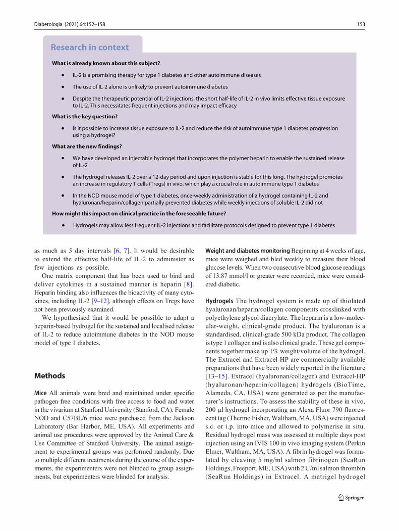
SHORT COMMUNICATION Weekly injection of IL-2 using an injectable hydrogel reduces autoimmune diabetes incidence in NOD mice Nadine Nagy 1 & Gernot Kaber 1 & Michael J. Kratochvil 1,2 & Hedwich F. Kuipers 1 & Shannon M. Ruppert 1 & Koshika Yadava 1 & Jason Yang 1 & Sarah C. Heilshorn 2 & S. Alice Long 3 & Alberto Pugliese 4 & Paul L. Bollyky 1 Received: 15 May 2020 /Accepted: 25 August 2020 # Springer-Verlag GmbH Germany, part of Springer Nature 2020 Abstract Aims/hypothesis IL-2 injections are a promising therapy for autoimmune type 1 diabetes but the short half-life of this cytokine in vivo limits effective tissue exposure and necessitates frequent injections. Here we have investigated whether an injectable hydrogel could be used to promote prolonged IL-2 release in vivo. Methods Capitalising on the IL-2-binding capabilities of heparin, an injectable hydrogel incorporating clinical-grade heparin, collagen and hyaluronan polymers was used to deliver IL-2. The IL-2-release kinetics and in vivo stability of this material were examined. The ability of soluble IL-2 vs hydrogel-mediated IL-2 injections to prevent autoimmune diabetes in the NOD mouse model of type 1 diabetes were compared. Results We observed in vitro that the hydrogel released IL-2 over a 12-day time frame and that injected hydrogel likewise persisted 12 days in vivo. Notably, heparin binding potentiates the activity of IL-2 and enhances IL-2- and TGFβ-mediated expansion of forkhead box P3-positive regulatory T cells (FOXP3 + Tregs). Finally, weekly administration of IL-2-containing hydrogel partially prevented autoimmune diabetes while injections of soluble IL-2 did not. Conclusions/interpretation Hydrogel delivery may reduce the number of injections required in IL-2 treatment protocols for autoimmune diabetes. Keywords Autoimmune . Controlled release . Diabetes . Heparin . Hyaluronan . Hydrogels . IL-2 . Treg Abbreviations FOXP3 Forkhead box P3 HA Hyaluronan HI Heparin LN Lymph node Tconv Conventional T cell Treg FOXP3 + regulatory T cell Introduction Type 1 diabetes is characterised by the progressive immune cell-mediated destruction of pancreatic beta cells and the fail- ure of regulatory mechanisms that normally prevent this, including regulatory T cells (Tregs) [1]. One critical factor that governs Treg function is the cytokine IL-2 [2]. The potential role of low-dose IL-2 in type 1 diabetes is a subject of active exploration and a frontier in the treatment and prevention of autoimmu- nity [3]. Low-dose IL-2 affects two critical cell popula- tions of the immune system: T cells and natural killer (NK) cells. However, IL-2 has a short half-life, depend- ing on the route of administration, from 7 min for i.v. to several hours for i.p. and s.c. administration [4, 5]. In addition, the dosing protocols vary depending on the disease. Cancer treatment usually requires daily dosing [6], whereas autoimmune disease protocols are variable, with some calling for administration of doses spaced at * Nadine Nagy [email protected]1 Division of Infectious Diseases and Geographic Medicine, Department of Medicine, Stanford University School of Medicine, Stanford, CA, USA 2 Department of Materials Science and Engineering, Stanford University, Stanford, CA, USA 3 Benaroya Research Institute, Seattle, WA, USA 4 Diabetes Research Institute, Leonard M. Miller School of Medicine, University of Miami, Miami, FL, USA https://doi.org/10.1007/s00125-020-05314-1 / Published online: 30 October 2020 Diabetologia (2021) 64:152–158
Transcript
SHORT COMMUNICATION
Weekly injection of IL-2 using an injectable hydrogel reducesautoimmune diabetes incidence in NOD mice
Nadine Nagy1 & Gernot Kaber1 & Michael J. Kratochvil1,2 & Hedwich F. Kuipers1 & Shannon M. Ruppert1 &
Koshika Yadava1 & Jason Yang1& Sarah C. Heilshorn2
& S. Alice Long3& Alberto Pugliese4
&
Paul L. Bollyky1
Received: 15 May 2020 /Accepted: 25 August 2020# Springer-Verlag GmbH Germany, part of Springer Nature 2020
AbstractAims/hypothesis IL-2 injections are a promising therapy for autoimmune type 1 diabetes but the short half-life of this cytokinein vivo limits effective tissue exposure and necessitates frequent injections. Here we have investigated whether an injectablehydrogel could be used to promote prolonged IL-2 release in vivo.Methods Capitalising on the IL-2-binding capabilities of heparin, an injectable hydrogel incorporating clinical-grade heparin,collagen and hyaluronan polymers was used to deliver IL-2. The IL-2-release kinetics and in vivo stability of this material wereexamined. The ability of soluble IL-2 vs hydrogel-mediated IL-2 injections to prevent autoimmune diabetes in the NOD mousemodel of type 1 diabetes were compared.Results We observed in vitro that the hydrogel released IL-2 over a 12-day time frame and that injected hydrogel likewisepersisted 12 days in vivo. Notably, heparin binding potentiates the activity of IL-2 and enhances IL-2- and TGFβ-mediatedexpansion of forkhead box P3-positive regulatory T cells (FOXP3+ Tregs). Finally, weekly administration of IL-2-containinghydrogel partially prevented autoimmune diabetes while injections of soluble IL-2 did not.Conclusions/interpretation Hydrogel delivery may reduce the number of injections required in IL-2 treatment protocols forautoimmune diabetes.
AbbreviationsFOXP3 Forkhead box P3HA HyaluronanHI HeparinLN Lymph nodeTconv Conventional T cellTreg FOXP3+ regulatory T cell
Introduction
Type 1 diabetes is characterised by the progressive immunecell-mediated destruction of pancreatic beta cells and the fail-ure of regulatory mechanisms that normally prevent this,including regulatory T cells (Tregs) [1].
One critical factor that governs Treg function is thecytokine IL-2 [2]. The potential role of low-dose IL-2in type 1 diabetes is a subject of active exploration anda frontier in the treatment and prevention of autoimmu-nity [3]. Low-dose IL-2 affects two critical cell popula-tions of the immune system: T cells and natural killer(NK) cells. However, IL-2 has a short half-life, depend-ing on the route of administration, from 7 min for i.v.to several hours for i.p. and s.c. administration [4, 5]. Inaddition, the dosing protocols vary depending on thedisease. Cancer treatment usually requires daily dosing[6], whereas autoimmune disease protocols are variable,with some calling for administration of doses spaced at
as much as 5 day intervals [6, 7]. It would be desirableto extend the effective half-life of IL-2 to administer asfew injections as possible.
One matrix component that has been used to bind anddeliver cytokines in a sustained manner is heparin [8].Heparin binding also influences the bioactivity of many cyto-kines, including IL-2 [9–12], although effects on Tregs havenot been previously examined.
We hypothesised that it would be possible to adapt aheparin-based hydrogel for the sustained and localised releaseof IL-2 to reduce autoimmune diabetes in the NOD mousemodel of type 1 diabetes.
Methods
Mice All animals were bred and maintained under specificpathogen-free conditions with free access to food and waterin the vivarium at Stanford University (Stanford, CA). FemaleNOD and C57BL/6 mice were purchased from the JacksonLaboratory (Bar Harbor, ME, USA). All experiments andanimal use procedures were approved by the Animal Care &Use Committee of Stanford University. The animal assign-ment to experimental groups was performed randomly. Dueto multiple different treatments during the course of the exper-iments, the experimenters were not blinded to group assign-ments, but experimenters were blinded for analysis.
Weight and diabetes monitoringBeginning at 4 weeks of age,mice were weighed and bled weekly to measure their bloodglucose levels. When two consecutive blood glucose readingsof 13.87 mmol/l or greater were recorded, mice were consid-ered diabetic.
Hydrogels The hydrogel system is made up of thiolatedhyaluronan/heparin/collagen components crosslinked withpolyethylene glycol diacrylate. The heparin is a low-molec-ular-weight, clinical-grade product. The hyaluronan is astandardised, clinical-grade 500 kDa product. The collagenis type 1 collagen and is also clinical grade. These gel compo-nents together make up 1% weight/volume of the hydrogel.The Extracel and Extracel-HP are commercially availablepreparations that have been widely reported in the literature[13–15]. Extracel (hyaluronan/collagen) and Extracel-HP(hyaluronan/heparin/collagen) hydrogels (BioTime,Alameda, CA, USA) were generated as per the manufac-turer’s instructions. To assess the stability of these in vivo,200 μl hydrogel incorporating an Alexa Fluor 790 fluores-cent tag (Thermo Fisher,Waltham,MA,USA)were injecteds.c. or i.p. into mice and allowed to polymerise in situ.Residual hydrogel mass was assessed at multiple days postinjection using an IVIS 100 in vivo imaging system (PerkinElmer, Waltham, MA, USA). A fibrin hydrogel was formu-lated by cleaving 5 mg/ml salmon fibrinogen (SeaRunHoldings, Freeport,ME,USA)with 2U/ml salmon thrombin(SeaRun Holdings) in Extracel. A matrigel hydrogel
153Diabetologia (2021) 64:152–158
(Corning, Corning, NY, USA) was prepared according tomanufacturer’s instruction.
IL-2 The IL-2 used in this study was Proleukin (aldesleukin;Clinigen, PA, USA). It is a human recombinant IL-2 product,a highly purified protein with a molecular weight of approxi-mately 15,300 Da. This is a clinical-grade product that hasbeen used widely for over 20 years in numerous animalmodels and human clinical trials [6, 16].
IL-2 in vivo studies For one set of experiments, 6-week-oldNOD mice were treated with either 25,000 IU IL-2 onceweekly or the same amount of IL-2 delivered in the contextof a single hydrogel injection, or PBS injections as a negativecontrol. After 1 month, mice were killed, mesenteric lymphnodes (LNs) were collected, and populations of lymphocyteswere assessed by flow cytometry. For another set of experi-ments the above-mentioned treatment was administered tomice from 6 to 21 weeks of age. The mice were subsequentlymonitored for diabetes onset until 33 weeks of age.
Measurement of IL-2 release from hydrogels Hydrogels werecast into a 96 well plate, 100 μl per well. After the hydrogelswere formed, 200 μl of 1600 IU IL-2/ml PBS was added perwell and incubated overnight. The loading solution was collect-ed for later analysis. Release was conducted by incubating thesamples in 200 μl of PBS, which was collected and replaced atthe specified time points. IL-2 concentration was measuredusing an IL-2 ELISA (BioLegend, San Diego, CA, USA).
IL-2 proliferation assay CTLL2 cells (ATCC, Manassas, VA,USA), were cultured for 48 h in the context of increasingconcentrations of IL-2 with or without heparin. Proliferationwas measured using a resazurin-based Reliablue CellViability Reagent (ATCC, Manassas, VA, USA).
Isolation and analysis of leucocyte populations Total leuko-cytes were isolated from lymph nodes from 10-week-old NODmice, as previously described [17]. CD4+ T cells were isolatedfrom the pooled cell suspensions using an EasySep MouseCD4+ T Cell Isolation Kit (Stemcell, Vancouver, BC, Canada),following the manufacturer’s instructions. For T cell activationand forkhead box P3-positive (FOXP3+) induction studies, cellculture plates (96-well) were coated overnight with 5μg/ml anti-CD3 antibody (catalogue no. 145-2C11; BD Biosciences, SanJose, CA, USA) and 2.5 μg/ml anti-CD28 antibody (catalogueno. 37.51; BD Biosciences). Subsequently, 1× 10⁵ cells werecultured with or without soluble 100 IU/ml IL-2 (Proleukin[aldesleukin]; Clinigen) and 50 ng/ml TGFβ (catalogue no.21C11; BioLegend), as indicated using previously publishedprotocols [18]. Cells were stained and flow cytometry wasperformed as previously described [17]. Analysis was done onan LSR II instrument (BD Biosciences) in the Stanford Shared
FACS Facility (Stanford, CA, USA). Analysis was carried outon Flowjo v10 (Ashland, OR, USA).
Statistical analysis Data are expressed as means ± SEM of nindependent measurements. A p value of <0.05 was consid-ered significant. Significance of the difference between themeans of two or three groups of data was evaluated using atwo-tailed t test or one-way ANOVA with Šidák’s multiplecomparisons post-test.
Results
Heparin potentiates IL-2 Treg induction While heparin isreported to enhance the activity of IL-2, we investigatedwhether heparin-bound IL-2 likewise potentiates the effectsof the cytokine on Tregs. In vitro we observed that prolifera-tion with heparin complexed with IL-2 was more than twofoldthat with IL-2 alone (Fig. 1a). We then sought to determinewhether heparin complexed with IL-2 likewise enhances theimpact of IL-2 on Treg induction from CD4+GFP/FOXP3−
conventional T cells (Tconv) precursors activated with anti-CD3 and anti-CD28 in the presence of TGFβ and IL-2 (Fig.1b) [19]. The presence of heparin and IL-2 greatly increasedTreg induction in this assay (Fig. 1c).
Heparin- and IL-2 containing hydrogels potentiates Treginduction We next studied whether heparin in the context ofa hydrogel also potentiates the effects of IL-2 on Treg induc-tion. To this end, we coated tissue culture plates with acommercially available hyaluronan/heparin/collagen hydro-gel preparation (Fig. 1d) and used the same induction protocol(Fig. 1e). In the presence of IL-2, the hyaluronan/heparin/collagen hydrogel significantly increased Treg induction vsthe non-hydrogel-containing condition (Fig. 1f). Notably,repeating the experiment with a hyaluronan/collagen hydrogellacking the heparin component, a fibrin hydrogel or a matrigelhydrogel did not potentiate Treg induction (data not shown).
Together, these data indicate that heparin potentiates theeffects of IL-2 on Treg expansion, and that this stimulus canbe delivered as a hydrogel.
Hydrogels release IL-2 in a sustained mannerWe next soughtto quantify the capacity of hydrogels to bind and slowlyrelease IL-2 over time. The hyaluronan/heparin/collagenhydrogel eluted IL-2 through day 12, while the hyaluronan/collagen hydrogel eluted IL-2 through day 7 (Fig. 1g,h).These data indicate that hydrogels retain IL-2, releasing it overtime, and that heparin enhances this.
Hydrogel persists in vivo To determine whether thehyaluronan/heparin/collagen hydrogel is stable when injectedin vivo, the hydrogel was delivered via s.c. and i.p. injection
154 Diabetologia (2021) 64:152–158
and allowed to polymerise in situ. Residual hydrogel massin the mice was then assessed using the IVIS in vivo imag-ing system at 1, 5 and 12 days post hydrogel injection (Fig.1i-k). Injection of the hydrogel s.c. resulted in longerpersistence, which was still clearly seen at day 12 postinjection (Fig. 1k). In general, hydrogel introduced viai.p. injection did not show a strong signal using the IVISin vivo imaging (Fig. 1i-k). These data demonstrate that ahyaluronan/heparin/collagen hydrogel polymerises in vivoand is stable for up to 12 days.
Hydrogel-mediated IL-2 delivery is associated with an in vivoTreg increase To investigate whether hydrogel-mediated IL-2delivery could be used to promote Treg expansion in vivo,NOD mice were treated by once weekly i.p. injection of25,000 IU IL-2 either in its soluble form or in a hyaluronan/heparin/collagen hydrogel. After 1 month of treatment, themice were killed and T cell populations in the mesentericLNs were then assessed via flow cytometry.
We observed that the percentage of CD3+CD4+ T cells andCD3+CD8+ T cells in the LNs was significantly decreased in
a cb d
g he f
i j k
Polyethylene glycol
diacrylate + IL-2
Thiol-modified heparin
Thiol-modified denatured
collagen
Thiol-modified hyaluronan
Day 1
s.c. s.c. i.p. i.p.
Day 5 Day 12
s.c. s.c. i.p. i.p. s.c. s.c. i.p. i.p.
Epi-fluo-
rescence
200
1000
800
400
600
Epi-fluo-
rescence
200
1000
800
400
600
Epi-fluo-
rescence
200
1000
800
400
600
IL-2
- H
I
IL-2
+HI
FO
XP
3+C
D4
+ T
cells (
%)
100 200 300
0
2000
4000
6000
8000
10000
12000
IL-2
HI/IL-2
HI
IU IL-2IL
-2 - H
I
IL-2
+HI
FO
XP
3+C
D4
+ T
cells (%
)
IL-2 (
pg/m
l)HA/H
I/COL
IL-2
alone
HA/C
OL +
IL-2
HA/H
I/COL +
IL-2
0
10
20
30
40
50
*
*
AU
Fig. 1 Hydrogels release IL-2 in a sustained manner and potentiate Treginduction in the presence of heparin (HI). (a) CTLL2 cells were culturedin the presence of increasing concentrations of IL-2 with or without HI.Proliferation was measured by resazurin incorporation and expressed asarbitrary units (AU). Data are shown for triplicate wells per condition. (b)Schematic of CD4+GFP/FOXP3− T cells isolated from healthy (non-diabetic) mice, cultured in the setting of anti-CD3 and anti-CD28,TGFβ and IL-2 in the absence or presence of HI. (c) Percentage ofCD4+ T cells that were FOXP3+ from the experiment described in (b).Quantification of n = 3 identical experiments. Data represent mean ±SEM; *p < 0.05 as determined by a two-sided t test. (d) Schematic ofhydrogel composition. (e) Schematic of CD4+GFP/FOXP3− T cells,isolated from healthy (non-diabetic) mice cultured in the setting of anti-CD3/-CD28, TGFβ and IL-2 in the absence or presence of HI and
hydrogel. COL, collagen; HA, hyaluronan. (f) Percentage of CD4+ T cellsthat were FOXP3+ from experiment described in (e). Quantification ofn = 3 identical experiments. Data represent mean ± SEM; *p < 0.05 asdetermined by a two-sided t test. (g) Hydrogel IL-2 release curve fordifferent hydrogel compositions (HA, HI, COL and/or IL-2). (h) IL-2release from hydrogels measured by IL-2 ELISA at day 7. Data representmean ± SEM; *p < 0.05 as determined by ANOVA followed by Šidák’smultiple comparisons test. Data in (g) and (h) are shown for the mean oftriplicate wells per condition. (i–k) Hydrogels of 50 μl volume incorpo-rating an Alexa Fluor 790 fluorescent tag were injected into mice s.c. andi.p. and allowed to polymerise in situ. Residual hydrogel mass was thenassessed at 1 (i), 5 (j) and 12 days (k) post injection using an IVIS in vivoimaging system. Data are representative of three independent experiments
155Diabetologia (2021) 64:152–158
the IL-2 hydrogel treatment group (Fig. 2a,b). The percentageof CD3+CD4+FOXP3+ Tregs was significantly increased inthe setting of the IL-2 hydrogel group (Fig. 2c), while theFOXP3 mean fluorescence intensity (MFI) was unchangedby either treatment (Fig. 2d). Together, these data indicate thathydrogel-mediated IL-2 delivery increases Tregs in vivo.
IL-2 delivery via hydrogel reduces the incidence of diabetesonset in NODmice In parallel to studying the effects of the IL-
2 hydrogel on Treg expansion, we also investigated whetherthis treatment regimen could prevent autoimmune diabetes inNOD mice.
NOD mice were i.p. injected once weekly with 25,000 IUIL-2 either in its soluble form or in a hyaluronan/heparin/collagen hydrogel. This treatment was administered to 6-week-old mice for 15 weeks, a Kaplan–Meier curve showsthe percentage of non-diabetic mice in the different treatmentgroups over time (Fig. 2e). Three months later, at 33 weeks of
f g
e
a b c
0 5 10 15 20 25 30 35
0
20
40
60
80
100
Treatment
start
Treatment
end
No
n-d
iab
etic (
%) Control
IL-2
IL-2 hydrogel
Weeks of age
*
Control IL-2 IL-2 hydrogel
0
2
4
6
8
10
Diabetic
Non-diabetic
*
d
PBS
IL-2
25,0
00
IL-2
hydro
gel
0
5
10
15
20
25 *
PBS
IL-2
25,0
00
IL-2
hydro
gel
LN
FO
XP
3 (
MF
I)
Fig. 2 In vivo heparin (HI) hydrogels are associated with an increase inFoxP3+ Tregs and reduce diabetes onset in NOD mice. (a–d) Six-week-old NODmice were treated with either 25,000 IU IL-2 once weekly or thesame amount of IL-2 delivered in the context of a single hydrogel injec-tion, or PBS injections as a negative control. After 1 month, mice werekilled, mesenteric LNs were collected, and populations of lymphocyteswere assessed by flow cytometry. In particular, the percentage ofCD3+CD4+ T cells (a), the percentage of CD3+CD8+ T cells (b),CD3+CD4+/FOXP3+CD25+ Tregs (c) and FOXP3 mean fluorescenceintensity (MFI) (d) were assessed. Data are representative of two inde-pendent experiments; n = 5 mice per group. Data represent mean ± SEM;
*p < 0.05 as determined by ANOVA followed by Šidák’s multiplecomparisons test. (e) Starting at 6 weeks of age, NOD mice receivedeither 25,000 IU IL-2 once weekly, or the same amount of IL-2 deliveredin the context of a once a week hydrogel injection. PBS injections servedas negative controls.Mice were thenmonitoredweekly for diabetes onset.This treatment was administered to mice from 6 to 21 weeks of age andmice were subsequently monitored until 33 weeks of age. (f, g) Numberof diabetic mice per group (f) and blood glucose (g) at 33 weeks of age,3 months after treatment ended. Data represent mean ± SEM, n = 10miceper group. *p < 0.05 as determined by ANOVA followed by Šidák’smultiple comparisons test
156 Diabetologia (2021) 64:152–158
age, all mice in the control group, eight out of ten in the IL-2group, and five out of ten in the IL-2 hydrogel group werediabetic (Fig. 2f). At 33 weeks of age, the blood glucosevalues of the control mice were >33.33 mmol/l ,~27.75 mmol/l for the IL-2 treatment group and~20.53 mmol/l for the IL-2 hydrogel group (Fig. 2g). Thesedata suggest that once weekly hydrogel delivery of IL-2 couldpartially prevent autoimmunity in this animal model.
Discussion
We report that once weekly injections of a hydrogelcontaining heparin and IL-2 promote Treg expansion andpartially prevent autoimmunity in the NOD mouse modelof type 1 diabetes. In contrast, the same IL-2 regimeninjected without hydrogel did not. Other groups have usedprotocols in which IL-2 was administered daily or severaltimes a week [20–23]; these different treatment regimensmake it difficult to compare data between studies. Our datasuggest that it may be possible to reduce the likelihood ofonset of autoimmune diabetes with less frequent injectionsusing IL-2-releasing hydrogel preparations. Currently, IL-2 use alone cannot be used in humans to prevent autoim-mune diabetes; we used an exploratory approach in ourheparin- and IL-2-containing hydrogel study.
We have demonstrated that heparin delivered in diverseways, alone or in the form of a hydrogel, amplifies the impactof IL-2 on multiple cell types. We report that a hydrogelcontaining heparin can release IL-2 over time using an assayperformed in the absence of cells that does not thereforeinvolve cellular responses to IL-2. Thus, we conclude thatheparin has effects both on the potency of IL-2 as well as itssustained release from a hydrogel over time. We believe thatenhanced IL-2 activity, as well as the extended release of IL-2through very slow diffusion and degradation of the hydrogelitself, contribute to the effects reported here.
This hydrogel may have potential in treating other autoim-mune disorders that are responsive to IL-2 [24], perhapsparticularly autoimmune diseases of the skin. However, thesafety and tolerability of hydrogel materials in the context ofIL-2 will need to be examined [25, 26]. Moreover, it would beimportant to evaluate the performance of these hydrogel mate-rials over a range of IL-2 concentrations and formulations.Based on our findings, we conclude that hydrogel-mediatedIL-2 delivery may be a useful approach for delivering IL-2therapeutically.
Acknowledgements We thank N. L. Haddock (Department of Medicine,Stanford University, Stanford, CA, USA) for her assistance with draftingthe revised manuscript and for generating the graphical abstract.
Data availability The data are available from the corresponding author.
Funding This work was supported in part by the National Institutes ofHealth (NIH) grants R01 DK096087-01, R01 HL113294-01A1, R01DK116782-01A1 and U01 AI101984 to PLB. KY was supported bythe Swiss National Science Foundation early postdoctoral mobility grantand the Stanford Child Health Research Institute and the Stanford NIH-NCATS-CTSA (grant no. UL1 TR001085). This work was also support-ed by grants from the JDRF 3-PDF-2014-224-A-N to NN and 1-SRA-2018-518-S-B Innovation Award to PLB and by grants from theHarrington Institute, Stanford SPARK, the Stanford Child HealthResearch Institute, all to PLB.
Authors’ relationships and activities The authors declare that there areno relationships or activities that might bias, or be perceived to bias, theirwork.
Contribution statement NN, GK and PLB designed the study. NN, GK,MJK, HFK, SMR, KY and JY analysed the data. All authors interpretedthe results. NN and PLB drafted the first version of the manuscript. Allauthors critically revised earlier versions of the manuscript. All authorsread and approved the final version of the manuscript to be published.PLB is the guarantor of this work.
References
1. Bollyky PL, Falk BA, Long SA et al (2009) CD44 costimulationpromotes FoxP3+ regulatory T cell persistence and function viaproduction of IL-2, IL-10, and TGF-beta. J Immunol 183(4):2232–2241. https://doi.org/10.4049/jimmunol.0900191
2. Bayer AL, Yu A, Malek TR (2007) Function of the IL-2R forthymic and peripheral CD4+CD25+ Foxp3+ T regulatory cells. JImmunol 178(7):4062–4071. https://doi.org/10.4049/jimmunol.178.7.4062
3. Rosenzwajg M, Churlaud G, Mallone R et al (2015) Low-doseinterleukin-2 fosters a dose-dependent regulatory T cell tunedmilieu in T1D patients. J Autoimmun 58:48–58. https://doi.org/10.1016/j.jaut.2015.01.001
4. Lotze MT, Frana LW, Sharrow SO, Robb RJ, Rosenberg SA(1985) In vivo administration of purified human interleukin 2. I.Half-life and immunologic effects of the Jurkat cell line-derivedinterleukin 2. J Immunol 134(1):157–166
5. Cheever MA, Thompson JA, Kern DE, Greenberg PD (1985)Interleukin 2 (IL 2) administered in vivo: Influence of IL 2 routeand timing on T cell growth. J Immunol 134:3895–3900 https://www.jimmunol.org/content/134/6/3895
6. Jeal W, Goa KL (1997) Aldesleukin (recombinant interleukin-2): areview of its pharmacological properties, clinical efficacy and toler-ability in patients with renal cell carcinoma. BioDrugs 7(4):285–317. https://doi.org/10.2165/00063030-199707040-00005
7. Tahvildari M, Dana R (2019) Low-dose IL-2 therapy in transplan-tation, autoimmunity, and inflammatory diseases. J Immunol203(11):2749–2755. https://doi.org/10.4049/jimmunol.1900733
8. Xu D, Esko JD (2014) Demystifying heparan sulfate-protein inter-actions. Annu Rev Biochem 83(1):129–157. https://doi.org/10.1146/annurev-biochem-060713-035314
9. Yayon A, Klagsbrun M, Esko JD, Leder P, Ornitz DM (1991) Cellsurface, heparin-like molecules are required for binding of basicfibroblast growth factor to its high affinity receptor. Cell 22 64(4):841–848. https://doi.org/10.1016/0092-8674(91)90512-w
10. Wrenshall LE, Carlson A, Cerra FB, Platt JL (1994) Modulation ofcytolytic T cell responses by heparan sulfate. Transplantation 57(7):1087–1094. https://doi.org/10.1097/00007890-199404000-00018
11. Najjam S, Mulloy B, Theze J, Gordon M, Gibbs R, Rider CC(1998) Further characterization of the binding of human recombi-nant interleukin 2 to heparin and identification of putative bindingsites. Glycobiology 8(5):509–516. https://doi.org/10.1093/glycob/8.5.509
12. Ramsden L, Rider CC (1992) Selective and differential binding ofinterleukin (IL)-1a, IL-1b, IL-2 and IL-6 to glycosaminoglycans.Eur J Immunol 22:3027–3031. https://doi.org/10.1002/eji.1830221139
13. Bollyky PL, Wu RP, Falk BA et al (2011) ECM components guideIL-10 producing regulatory T-cell (TR1) introduction from effectormemory T-cell precursors. PNAS 108(19):7938–7943. https://doi.org/10.1073/pnas.1017360108
14. Elia R, Fuegy PW, VanDelden A, FirpoMA, Prestwich GD, PeattieRA (2010) Stimulation of in vivo angiogenesis by in situcrosslinked, dual growth factor-loaded, glycosaminoglycanhydrogels. Biomaterials 31(17):4630–4638. https://doi.org/10.1016/j.biomaterials.2010.02.043
15. Advanced BioMatrix (2020) HyStem: Thiol-Modified HyaluronanHydrogel Kit. Available from https://advancedbiomatrix.com/hystem.html. Accessed 20 July 2020
17. Ruppert SM, Falk BA, Long SA, Bollyky PL (2015) Regulatory Tcells resist cyclosporine-induced cell death via CD44-mediatedsignaling pathways. Int J Cell Biol 2015(4):614297–614210.https://doi.org/10.1155/2015/614297
18. NagyN, Kaber G, Johnson PY et al (2015) Inhibition of hyaluronansynthesis restores immune tolerance during autoimmune insulitis. JClin Invest 125(10):3928–3940. https://doi.org/10.1172/JCI79271
19. Walker MR, Kasprowicz DJ, Gersuk VH et al (2003) Induction ofFoxP3 and acquisition of T regulatory activity by stimulated human
CD4+CD25- T cells. J Clin Invest 112(9):1437–1443. https://doi.org/10.1172/JCI19441
21. Rabinovitch A, Suarez-Pinzon WL, Shapiro AMJ, Rajotte RV,Power R (2002) Combination therapy with sirolimus andinterleukin-2 prevents spontaneous and recurrent autoimmunediabetes in NOD mice. Diabetes 51(3):638–645. https://doi.org/10.2337/diabetes.51.3.638
22. Tang Q, Adams JY, Penaranda C et al (2008) Central role of defec-tive interleukin-2 production in the triggering of islet autoimmunedestruction. Immunity 28(5):687–697. https://doi.org/10.1016/j.immuni.2008.03.016
23. Grinberg-Bleyer Y, Baeyens A, You S et al (2010) IL-2 reversesestablished type 1 diabetes inNODmice by a local effect on pancre-atic regulatory T cells. J Exp Med 207(9):1871–1878. https://doi.org/10.1084/jem.20100209
24. Castela E, Le Duff F, Butori C et al (2014) Effects of low-doserecombinant interleukin 2 to promote T-regulatory cells in alopeciaareata. JAMA Dermatol 150(7):748–751. https://doi.org/10.1001/jamadermatol.2014.504
25. Long SA, Buckner JH, GreenbaumCJ (2013) IL-2 therapy in type 1diabetes: “Trials” and tribulations. Clin Immunol 149(3):324–331.https://doi.org/10.1016/j.clim.2013.02.005
26. Long SA, Rieck M, Sanda S et al (2012) Rapamycin/IL-2 combi-nation therapy in patients with type 1 diabetes augments Tregs yettransiently impairs β-cell function. Diabetes 61(9):2340–2348.https://doi.org/10.2337/db12-0049
Publisher’s note Springer Nature remains neutral with regard to jurisdic-tional claims in published maps and institutional affiliations.