IntroductionCommon atrium is a rare congenital heart defect compris-
ing < 0.5–1% of all congenital heart diseases.1)2) Common atrium is characterized by complete absence of the interatrial septum, and is commonly accompanied by malformation of the atrioventricular valve.3) Most patients with common atri-um experience symptoms during childhood. However, we de-scribe a patient with common atrium who experienced his first obvious symptom at 48 years of age.
CaseA 48-year-old man presented to the outpatient clinic with a
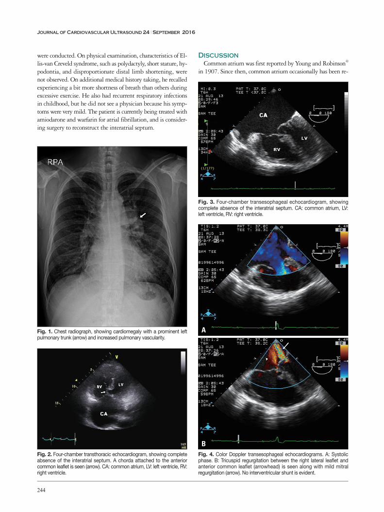
1-week history of intermittent palpitations. He had no signifi-cant medical history. On physical examination, wide splitting of S2 during auscultation was noted, but no other cardiac murmur was found. He had mild digital clubbing, but labial cyanosis at rest was not observed. Electrocardiography showed atrial fibrillation with rapid ventricular response. Laboratory data revealed an elevated hemoglobin level of 17.9 g/dL. Oxy-gen saturation obtained from pulse oximetry was 88%. Chest radiography showed mild cardiomegaly with a prominent left hilar shadow and increased pulmonary vascularity (Fig. 1).
In order to evaluate the patient’s atrial fibrillation, transtho-racic echocardiography was conducted, which showed com-plete absence of the interatrial septum. At this point, we looked for an atrioventricular valve malformation, which fre-quently accompanies common atrium. The anterior common
CASE REPORT J Cardiovasc Ultrasound 2016;24(3):243-246
•Received: February 10, 2016 •Revised: June 8, 2016 •Accepted: July 26, 2016•Address for Correspondence: Jae Beom Lee, Division of Cardiology, Department of Internal Medicine, Anyang SAM Hospital, 9 Samdeok-ro, Manan-gu, Anyang
14030, Korea Tel: +82-31-467-9106, Fax: +82-31-467-9198, E-mail: [email protected]•This is an Open Access article distributed under the terms of the Creative Commons Attribution Non-Commercial License (http://creativecommons.org/licenses/by-nc/3.0) which permits unrestricted non-commercial use, distribution, and reproduction in any medium, provided the original work is properly cited.
leaflet appeared to be attached to the crest of the ventricular septum by a chorda (Fig. 2), and a definite mitral and tricus-pid valve cleft was not noticed. The right ventricle was mark-edly enlarged with mild pulmonary hypertension (estimated right ventricular pressure, 39 mm Hg).
Because transthoracic echocardiography was slightly subop-timal, transesophageal echocardiography was conducted, which also showed complete absence of the interatrial septum. The anterior common leaflet appeared to be attached to the crest of the ventricular septum by a chorda (Fig. 3), as seen on transthoracic echocardiography. However, no interventricular shunt was found. In addition to right ventricular dilation with borderline hypertrophy, mild mitral and tricuspid regurgitation was observed (Fig. 4). These findings corresponded to com-mon atrium with atrioventricular valve malformation, which is an atrioventricular septal defect with separate atrioventricu-lar valves and an intra-atrial shunt only.
In order to check for anomalies of the systemic and pulmo-nary veins as well as accompanied cardiac anomaly, cardiac computed tomography was performed. The pulmonary veins drained to the left side of the common atrium, while the sys-temic veins drained to the right side. Visceroatrial, atrioven-tricular, and ventriculoarterial concordance were noted. There were no other accompanied anomalies (Fig. 5).
In order to evaluate the patient for genetic diseases, such as Ellis-van Creveld syndrome, which is related to common atrium, additional medical history taking and physical examination
Well-Tolerated and Undiscovered Common Atrium until Late Adulthood
Kyungjoong Kim, MD, Jiwook Choi, MD, Youngjae Doo, MD, Yeong Seop Yun, MD, Jongwook Kim, MD, and Jae Beom Lee, MDDivision of Cardiology, Department of Internal Medicine, Anyang SAM Hospital, Anyang, Korea
Common atrium is a rare congenital heart disease characterized by complete absence of the interatrial septum, and is commonly accompanied by malformation of the atrioventricular valve. Most patients with common atrium experience symptoms during childhood. Here, we describe a patient with common atrium who experienced his first obvious symptom at 48 years of age.
Journal of Cardiovascular Ultrasound 24 | September 2016
244
were conducted. On physical examination, characteristics of El-lis-van Creveld syndrome, such as polydactyly, short stature, hy-podontia, and disproportionate distal limb shortening, were not observed. On additional medical history taking, he recalled experiencing a bit more shortness of breath than others during excessive exercise. He also had recurrent respiratory infections in childhood, but he did not see a physician because his symp-toms were very mild. The patient is currently being treated with amiodarone and warfarin for atrial fibrillation, and is consider-ing surgery to reconstruct the interatrial septum.
DiscussionCommon atrium was first reported by Young and Robinson4)
in 1907. Since then, common atrium occasionally has been re-
Fig. 1. Chest radiograph, showing cardiomegaly with a prominent left pulmonary trunk (arrow) and increased pulmonary vascularity.
Fig. 2. Four-chamber transthoracic echocardiogram, showing complete absence of the interatrial septum. A chorda attached to the anterior common leaflet is seen (arrow). CA: common atrium, LV: left ventricle, RV: right ventricle.
Fig. 3. Four-chamber transesophageal echocardiogram, showing complete absence of the interatrial septum. CA: common atrium, LV: left ventricle, RV: right ventricle.
Fig. 4. Color Doppler transesophageal echocardiograms. A: Systolic phase. B: Tricuspid regurgitation between the right lateral leaflet and anterior common leaflet (arrowhead) is seen along with mild mitral regurgitation (arrow). No interventricular shunt is evident.
A
B
Late-Identified Common Atrium | Kyungjoong Kim, et al.
245
ported as a cardiac component in patients with Ellis-van Crev-eld syndrome, trisomy 21, or heterotaxy syndrome with asple-nia, whereas it is rare in nonsyndromic patients.3)5)6) The hemodynamic features of shunting in common atrium are very similar to those of a large atrial septal defect. However, in pa-tients with common atrium, pulmonary and systemic venous blood tend to mix more easily, and systemic arterial oxygen sat-uration tends to be lower than in those with a large atrial sep-tal defect.7) Consequently, most patients with common atrium experience such symptoms as exercise intolerance, exertional dyspnea, palpitations, frequent upper respiratory tract infections, cyanosis, and physical underdevelopment during childhood. Common atrium is frequently accompanied by anomalies of systemic venous drainage and atrioventricular regurgitation, which may increase mixing of blood and result in decreased systemic arterial oxygen saturation.2)7) Considering that increased mixing of arterial and venous blood can generate more severe symptoms at an earlier time, our patient is thought to have experienced relatively mild symptoms because he had no mi-tral and tricuspid valve cleft nor anomalies of systemic venous drainage, which are expected to increase regurgitation.
Because most patients with congenital heart disease show symptoms and receive a diagnosis before reaching adulthood, it can be difficult for physicians to consider the possibility of congenital heart disease in patients in whom symptoms are absent or well tolerated until late adulthood, as in the present case. Therefore, clinical suspicion is vital. If congenital heart disease, such as common atrium, is suspected, diagnosis can be made easily by using echocardiography. Our patient had been experiencing dyspnea during excessive exercise, and had such objective findings as digital clubbing and polycythemia, which increase the possibility of chronic hypoxemia. He also showed a prominent left hilar shadow and increased pulmonary vascu-larity, which can occur in patients with a left-to-right shunt,2)3) as well as cardiomegaly. Although he did not possess any anom-aly related to Ellis-van Creveld syndrome, trisomy 21, or het-
erotaxy syndrome with asplenia, investigating the clinical char-acteristics of such diseases also may be useful for clinical suspicion.
The natural course of common atrium is poor. Later identi-fication of common atrium increases the possibility of progres-sion to arrhythmia, pulmonary vascular disease, or cardiac dys-function.5)6) Moreover, surgical correction can considerably slow progression of the disease.3) Therefore, early diagnosis is im-portant even in well-tolerated cases.
Asuman Kaftan et al.8) reported a similar case of undiscov-ered common atrium in a 23-year-old patient. In addition, Demirelli et al.9) reported a case of incidentally diagnosed as-ymptomatic common atrium in a 23-year-old woman, while Hasanin and Kinsara10) reported a case of late-discovered com-mon atrium in a patient with persistent left superior vena cava. However, to our knowledge, our case is the oldest patient with common atrium to have remained asymptomatic. More-over, the present case shows that clinical characteristics found on physical examination, imaging studies, and laboratory tests can be useful for clinical suspicion of common atrium as long as physicians consider that patients with congenital heart dis-ease can remain asymptomatic until late adulthood.
References1. Campbell M. Incidence of cardiac malformations at birth and later, and
Single atrium. Embryologic, anatomic, electrocardiographic and other diag-nostic features. Am J Cardiol 1968;21:639-52.
3. Jiang H, Wang H, Wang Z, Zhu H, Zhang R. Surgical correction of common atrium without noncardiac congenital anomalies. J Card Surg 2013; 28:580-6.
4. Young AH, Robinson A. Some malformations of the human heart. Med Chron 1907-1908;47:96-106.
5. Digilio MC, Marino B, Giannotti A, Dallapiccola B. Single atrium, atrioventricular canal/postaxial hexodactyly indicating Ellis-van Creveld syndrome. Hum Genet 1995;96:251-3.
6. Ferdman DJ, Brady D, Rosenzweig EB. Common atrium and pulmo-nary vascular disease. Pediatr Cardiol 2011;32:595-8.
Fig. 5. Cardiac computed tomographic images. A: The right and left pulmonary veins drain to the left side of the common atrium. B and C: The superior vena cava and inferior vena cava normally drain to the right side of the common atrium. CA: common atrium, RV: right ventricle, RPV: right pulmonary vein, LPV: left pulmonary vein, SVC: superior vena cava, IVC: inferior vena cava.
A B C
Journal of Cardiovascular Ultrasound 24 | September 2016
246
7. Rastelli GC, Rahimtoola SH, Ongley PA, McGoon DC. Common atrium: anatomy, hemodynamics, and surgery. J Thorac Cardiovasc Surg 1968;55:834-41.
8. Asuman Kaftan H, Tanriverdi H, Kuru O, Bir LS. An asymptomatic case with single atrium. Echocardiography 2006;23:701-3.
9. Demirelli S, Fırtına S, Ermiş E, İnci S. Common atrium: a rare congeni-tal heart anomaly. Turk Kardiyol Dern Ars 2015;43:579.
10. Hasanin AM, Kinsara AJ. Single atrium associated with persistent left superior vena cava in asymptomatic adult: case report and review of literature. Congenit Heart Dis 2008;3:368-71.