Wettability and osteoblast cell response modulation through UV laser processing of nylon 6,6 D.G. Waugh and J. Lawrence School of Engineering, University of Lincoln, Brayford Pool, Lincoln, Lincolnshire, LN6 7TU, UK. Corresponding Author: D.G. Waugh School of Engineering University of Lincoln Brayford Pool Lincoln Lincolnshire LN6 7TS, UK Email: [email protected]Tel: +44 (0) 1522 668891
Transcript
Wettability and osteoblast cell response modulation through
UV laser processing of nylon 6,6
D.G. Waugh and J. Lawrence
School of Engineering, University of Lincoln, Brayford Pool, Lincoln, Lincolnshire, LN6 7TU, UK.
Corresponding Author:
D.G. Waugh School of Engineering University of Lincoln Brayford Pool Lincoln Lincolnshire LN6 7TS, UK Email: [email protected]: +44 (0) 1522 668891
It has been realized worldwide within the scientific community and various industries that lasers
offer major advantages over alternative techniques for materials processing [1-4]. Some of the
main advantages of using a laser for materials processing are:
• Relative cleanliness.
• Accurate processing.
o Allows much control over the Heat Affected Zone (HAZ) due to the ability of
relative precise control over the thermal profile and thermal
penetration/absorption.
• Precise placement of the beam onto the target material allowing user specified areas of
the target material to be processed.
• Post-processing techniques required are usually minimal.
• Non-contact processing.
• Automation (repeatability) of the various processing techniques using a laser is relatively
easy to implement.
With a large number of different lasers now commercially available it is possible for one to deduce
that almost all materials can be processed using a laser due to the wide range of laser parameters
that can be utilized. With the many benefits of using lasers for materials processing it has been
found that the interest in laser-induced surface treatment has grown, especially within the
biomedical industry. This is due to the fact that lasers offer the user a highly selective, rapid
technique to induce surface modification in both organic and inorganic materials [5].
In order to modify the surface properties of polymers for use in biological environments and
enhance the cell response by improving upon the adhesion characteristics, a vast number of
techniques and methods have been developed. These techniques range from surface topography
modification [6-8] to surface chemistry modification [9-11] and have given rise to the increased
interest in using polymers as biomaterials [12-19]. Nylon 6,6, the strongest and most abrasive
resistant unreinforced nylon, has been used for such biological applications as sutures, tracheal
tubes and gastrointestinal segments [20]. With regards to orthopedic applications it can be seen
that nylon is not commonly employed due to the hygroscopic nature having a large affect on the
mechanical properties over long periods of time [21]. Having said that, the use of nylon 6,6 within
this work gives high value experimentally insofar as to ascertain generic factors for polymeric
materials which could be used to predict the osteoblast cell response. Also, by modifying the
surface of polymeric materials it may be possible to identify other biological applications as this
may enhance the osteoblast cell response and biocompatibile properties. On account of nylon 6,6
being a relatively inexpensive polymer when compared to other polymer types, by identifying
other applications for this material the biological industry would benefit by being able to
implement cheaper, more economic bio-implant materials.
With numerous advances in medicine it has been seen that the population is ageing both within the
U.K. and within other major countries around the world. On account of people living longer, a
number of institutions have developed a focus on bioengineering to meet the ever growing
demands on medical facilities [22]. Furthermore, it is possible for one to realize that with an
ageing population there is an ever increasing demand for biological implants. As a result, these
needs of the ageing population need to be met more economically and efficiently so that costs and
the need for unnecessary surgery are considerably reduced. Therefore, it is imperative for the
biomedical industry to devise a way to manufacture cheap implants which can be used in
confidence to ensure a dramatic reduction in failure rates. This can be met by the laser surface
treatment of polymeric materials to enhance their biomimetic properties.
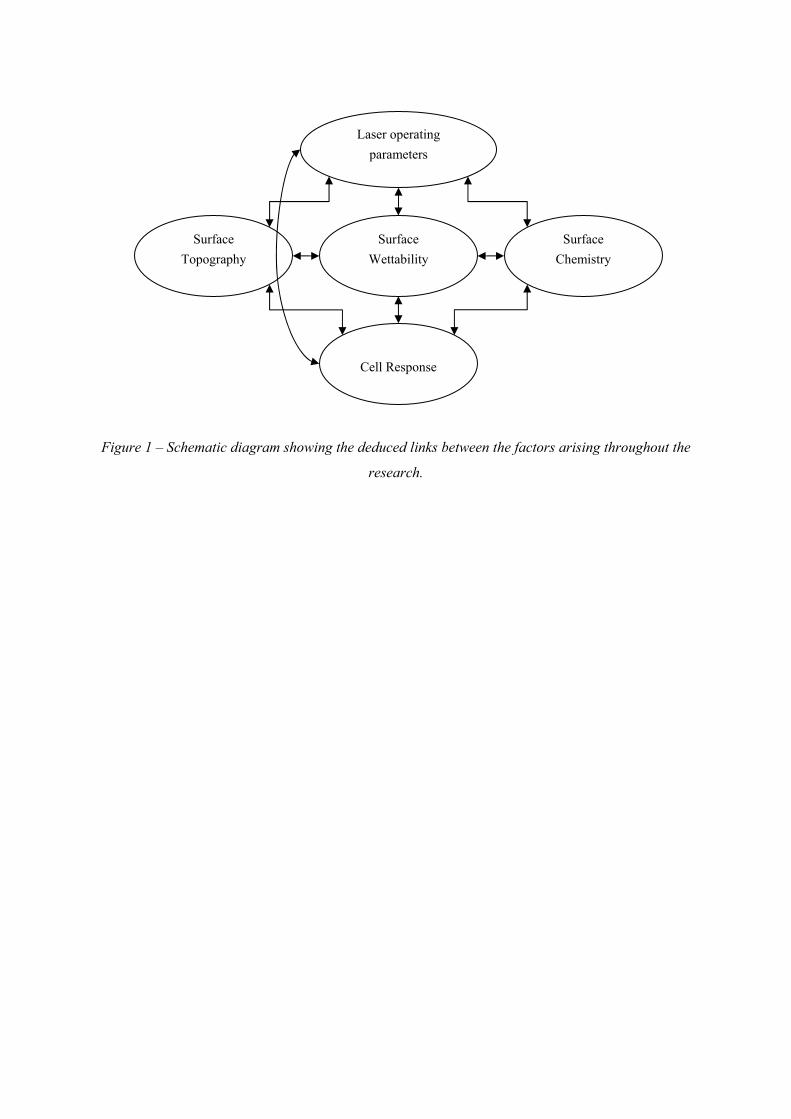
On account of the many benefits offered by laser materials processing to life sciences this paper is
a contribution to the endeavour of enhancing polymer surface properties to determine generic
parameters which give rise to the level of biofunctionality. That is, this work undertakes an
approach by which the laser modified surface parameters, wettability characteristics and osteoblast
cell response are discussed in order to ascertain the variables which link the factors together as
shown in Figure 1.
3.0 – Experimental Technique
3.1 – Nylon 6,6 Material
The nylon 6,6 (Tm: 255°, ρ: 1.3 gcm-3) was sourced in 100 mm2 sheets with a thickness of 5 mm
(Goodfellow Cambridge, Ltd). To obtain a conveniently sized sample for experimentation the as-
received nylon sheet was cut into 20 mm diameter discs using a 1 kW continuous wave (cw) CO2
laser (Everlase S48; Coherent, Ltd). No discernible heat affected zone (HAZ) was observed under
optical microscopic examination.
3.2 – KrF Excimer Laser-Induced Patterning
For the patterned experiments the repetition rate of the KrF excimer laser (LPX 200i; Lambda
Physik, Inc.) was kept constant at 25 Hz, with a number of 10 pulses per site and used Aerotech
CNC programming to induce the required pattern. A constant laser energy of 80±7 mJ was used
with the attenuator set to 0.3 (30%) giving a measured energy at the target sample of
23.67±2.5 mJ, resulting in a fluence of 858±91 mJ/cm2. In order to induce the intended pattern a
projection imaging system was implemented with a focusing lens of x10 demagnification. The
patterns induced using this technique were 50 μm trench (ET50), 100 μm trench (ET100), 50 μm
hatch (EH50 ) and 100 μm (EH100). Two non-contact masks were used for both dimensioned
patterns which included a brass mask with six 1 mm diameter holes spaced by 2 mm, centre to
centre, for the 100 μm dimensions and a SS316 foil (Laser Micromachining Ltd., UK) with five
0.5 mm diameter holes spaced by 1.5 mm, ‘centre to centre’, for the 50 μm dimensions. To keep
the constant 10 pulses per site it should also be noted here that scanning velocities of 0.125 mms-1
and 0.25 mms-1 were used for the 50 µm and 100 µm dimensioned patterns, respectively.
No processing gases were used throughout the experimentation and all laser processing was
carried out in an enclosure in which the ambient gas was air. Also, for all laser processing no
homogenizer was implemented meaning that the raw beam was used which would have given rise
to energy spikes pulse to pulse, having some possible affect on the incident laser fluence and laser
material processing.
3.3 –KrF Excimer Laser Whole Area Irradiative Processing
For the whole area processing with the KrF excimer laser (LPX 200i; Lambda Physik, Inc.) the
raw 23 × 12 mm2 beam was used to irradiate a large section of each sample at a time. In order to
hold the sample normal to the beam a bracket on the optical train was used. For the large area
processing experiments 6 samples where studied; these being 100 pulses at 100 mJ (EWA100),
100 pulses at 150 mJ (EWA150), 100 pulses at 200 mJ (EWA200), 100 pulses at 250 mJ
(EWA250), 500 pulses at 250 mJ (EWA250_500) and 1000 pulses at 250 mJ (EWA250_1000).
This gave fluences of 36±3, 54±5, 72±8 and 91±10 mJ/cm2, respectively for the different energies
used. Throughout the whole area excimer experiments the repetition rate was kept constant at
25 Hz and Aerotech CNC programming ensured that the correct number of pulses was applied to
each sample.
3.4 – Topography, Wettability Characteristics and Surface Chemistry Analysis
After laser irradiation the nylon 6,6 samples were analysed using a number of techniques. The
surface profiles were determined using a white light interferometer (WLI) (NewView 500; Zygo,
Ltd) with MetroPro and TalyMap Gold Software. The WLI was set-up using a ×10 Mirau lens
with a zoom of ×0.5 and working distance of 7.6 mm. This system also allowed Sa and Ra
roughness parameters to be determined for each sample. Where Ra can be defined as the arithmetic
average of the absolute values along a single specified direction and Sa the arithmetic average of
the absolute values over the whole of the laser surface treated area.
In accordance with the procedure detailed by Rance [23] the samples were ultrasonically cleaned
in isoproponal (Fisher Scientific Ltd.) for 3 minutes at room temperature before using a sessile
drop device to determine various wettability characteristics. This was to allow for a relatively clean
surface prior to any contact angle, θ, measurements being taken. To ensure that the sample
surfaces were dry a specimen dryer (Metaserv, Ltd.) was employed to blow ambient air across the
samples. A sessile drop device (OCA20; Dataphysics Instruments, GmbH) was used with relevant
software (SCA20; Dataphysics Intrsuments, GmbH) to allow the recent advancing and receding θ
for triply distilled water and the recent advancing angle for diiodomethane to be determined for
each sample. By starting with a droplet with a volume of 8 µl then the advancing θ were achieved
by adding and removing 0.5 µl, respectively, for each measurement. Thereafter the advancing θ for
the two liquids were used by the software to draw an OWRK plot to determine the surface energy
of the samples. For the two reference liquids the SCA20 software used the Ström et al technique
(triply distilled water – SFT(total:72.80), SFT(D:21.80), SFT(P:51.00); diiodomethane –
SFT(total:50.80), SFT(D:50.80), SFT(P:0.00)) to calculate the surface energy of the material. It
should be noted here that ten θ, using two droplets in each instance, were recorded to achieve a
mean θ for each liquid and surface.
All samples were analysed using x-ray photoelectron spectroscopy (XPS). This allowed any
surface modifications in terms of surface oxygen content due to the laser irradiation to be revealed.
Further details on XPS measurements of surface oxygen content have been stated previously [24].
3.5 – In Vitro Experimentation
Prior to any biological testing being carried out the samples were autoclaved (D-Series Bench-Top
Autoclave; Systec, GmbH) to ensure that all samples were sterilized. For all biological work
undertaken, unless stated, a biological safety cabinet (BSC) (Microflow Class II ABS Cabinet;
BioQuell UK, Ltd) was used to create a safe working environment and to provide a clean, sterile
environment to manipulate the cells used.
Normal human osteoblast cells (Clonetics CC-2538; Lonza, Inc.) were initially cultured in a T75
(75 ml) flask by suspending the cells in 19 ml culture medium comprising of 90% eagle minimum
essential medium (Sigma-Aldrich, Ltd., UK) and 10% foetal bovine serum (FBS) (Sigma-Aldrich
Ltd., UK). The flask was then placed in an incubator and left for 24 hrs. After 24 hrs the cells were
assessed and the spent media was aspirated before dispensing 15 ml of fresh media and returning
the flask to the incubator for three days.
The period of three days allowed the cells to become confluent in the flask providing enough cells
for seeding onto the samples. The cells were detached from the flask using 5 ml Trypsin-EDTA
(Sigma-Aldrich Ltd., UK) whilst placed in the incubator for seven minutes. Once all cells had
become detached 10 ml culture medium was added to neutralize the Trypsin. In order to aspirate
the supernatant the cell culture was centrifuged (U-320R; Boeco, GmbH) for five minutes at 200 g.
To ensure the cells were ready for seeding they were resuspended in 10 ml of culture medium and
dispensed between the samples in the 6-well plates. This equated to 0.55 ml (2×10⁴ cells/ml) for
each sample. The well plates were then placed in the incubator for a set time. One plate was
removed after 24 hrs and two other plates after four days. A well plate after 24 hrs and 4 days was
prepared for the SEM as will be discussed later and the other plate, removed after 4 days, was
prepared for counting using an improved neubauer hemacytometer (Fisher Scientific Ltd., UK) by
mixing 10 μl of each cell suspension with 10 μl of trypan-blue (Sigma-Aldrich Ltd., UK). In order
to harvest the cells for counting the cells were detached from the samples using 2 ml Trypsin-
EDTA (Sigma-Aldrich Ltd., UK) whilst placed in the incubator for seven minutes. Once all cells
had become detached 4 ml culture medium was added to neutralize the Trypsin. In order to
aspirate the supernatant the cell culture was centrifuged (U-320R; Boeco, GmbH) for five minutes
at 200 g. To ensure the cells were ready for counting they were resuspended in 2 ml of culture
medium and 2 ml of the trypan-blue was added.
3.6 – Scanning Electron Microscopy and Cell Cover Density of In Vitro Samples
In order to view the attached cells using SEM it was necessary to undertake a procedure to produce
a sample that was dehydrated ready for Au coating. The samples were initially rinsed with
phosphate-buffered saline (PBS) (Sigma-Aldrich, UK) to remove any unattached cells and then
adherent cells were fixed using 1.2% glutaraldehyde in water (Sigma-Aldrich, UK) at room
temperature for one hour within the BSC. After an hour the glutaraldehyde solution was removed
and the fixed cells were washed with PBS prior to carrying out a graded series of ethanol/distilled
water mixtures of 50/50, 80/20, 90/10, 95/5, 98/2 and 100/0. Each sample was left in these
mixtures for 10 min to ensure dehydration. Once this procedure was carried out, the samples were
mounted and sputter coated with Au so that SEM micrographs could be obtained. In order to
produce the best images possible each image was manipulated in terms of brightness, contrast and
gamma by using ImagePro Version 5.0.0.39 for Windows XP/Professional software (Media
Cybernetics Inc., USA).
In addition to the cell count described in Section 3.5, the cell cover density was determined
following both 24 hrs and 4 day incubation. This was done by analysing the cell coverage on each
sample using SEM and optical micrographs with the ImagePro software. The optical micrographs
were obtained using an up-right optical microscope (Flash 200 Smartscope; OGP, Ltd) with
magnifications varying between ×20 and ×100.
4.0 – Results and Discussion
4.1 - Topography
4.1.1 – KrF Excimer Laser-Induced Patterning
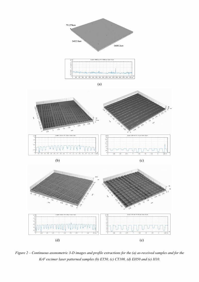
It can be seen in Figure 2 that the topography of the nylon 6,6 had been modified significantly through
excimer laser patterning when compared to the as-received sample (see Figure 2(a)). For the 50 µm
dimensioned patterns (see Figure 2(b) and Figure 2(d)) trench depths of around 1.5 µm was obtained
from the employed laser processing parameters. These constant laser processing parameters also gave
rise to trench depths of roughly 4 to 5 µm for the 100 µm dimensioned patterned samples (see Figure
2(c) and Figure 2(e)). What is more, on account of the accuracy and precision of the excimer laser
system implemented, the trenches produced in the nylon 6,6 materials which can be seen in Figures
2(b) to (e) were highly periodic. This periodicity was also confirmed upon obtaining the profile
extractions as shown in Figures 2(b) to (e).
Table 1 gives the surface roughness values for each of the KrF excimer laser-induced patterned
samples and shows that the surface roughness had dramatically increased for the excimer laser
patterned samples. For instance, the largest increases were seen with the 100 µm dimensioned patterns
which had an Sa of approximately 1.5 µm. This can be seen to be on account of the fact that the
100 µm dimensioned patterned samples gave rise to deeper trenches in comparison to the 50 µm
dimensioned samples.
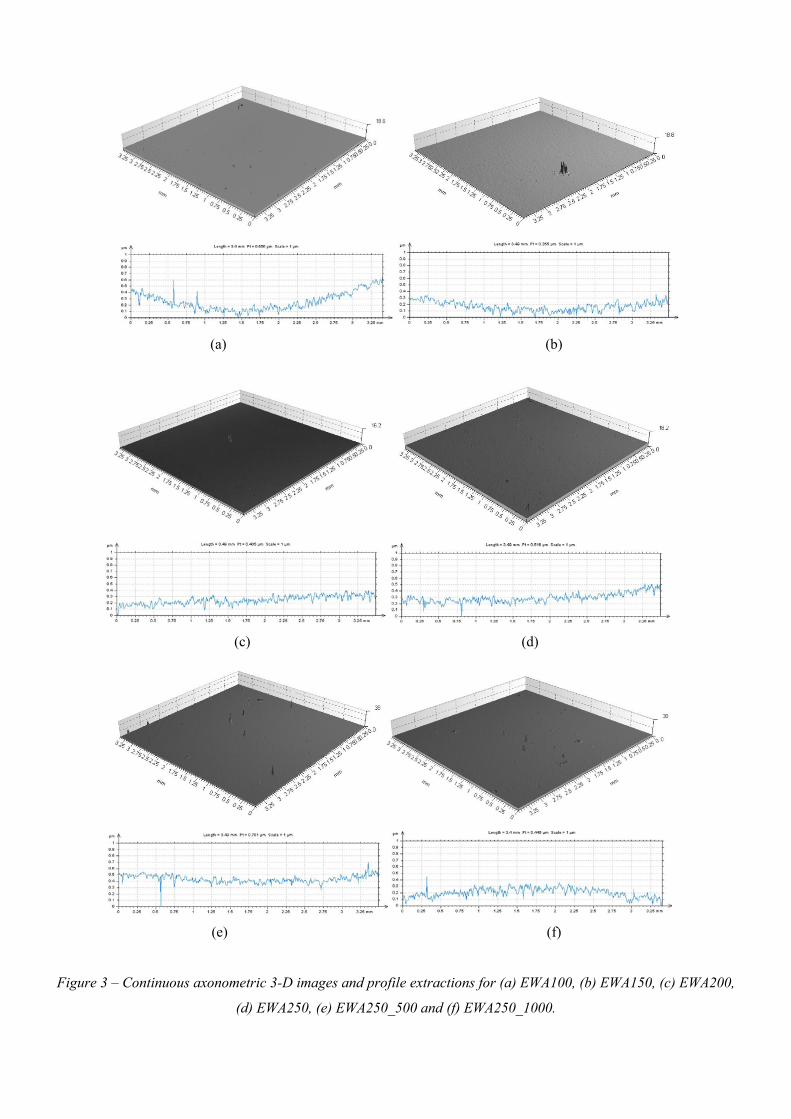
4.1.2 – KrF Excimer Laser Whole Area Irradiative Processing
Through WLI it was found that the KrF excimer laser whole area irradiative processed samples (see
Figure 3) appeared to have a similar topography to that of the as-received sample (see Figure 2(a)).
This can be attributed to the fact that the whole sample was irradiated meaning that any ablation
taking place would remove a somewhat uniform layer from the surface of the nylon 6,6. On the other
hand, especially for the samples with low incident fluences, it may be possible that the material did
not ablate on account of the threshold fluence not being achieved.
With regards to surface roughness the whole area irradiated samples had roughnesses equivalent to
that of the as-received sample (AR) which had an Sa of 0.126 µm and an Ra of 0.029 µm (see Table
1). This can be accounted for by the observation through the WLI which showed a negligible effect on
the surface topography for these samples. This further confirms that the fluences implemented for
these samples was not sufficient to elicit an ablative response from the nylon 6,6 samples. Having said
that, it may still have been possible for surface chemistry changes to occur due to oxidation. This is on
account of the fact that directly after the irradiation of the samples using the KrF excimer laser, the
samples were warm to the touch, with the warmest sample being WA250_1000 which had
1000 pulses and the highest fluence of 91 mJcm-2.
4.2 – Wettability and Surface Oxygen Content
4.2.1 – KrF Excimer Laser-Induced Patterning
Table 1 gives a summary of the surface parameters and wettability characteristics for each of the
samples studied. On account of the KrF excimer laser-induced patterning of the nylon 6,6 samples θ
increased by up to 24° in comparison to the as-received sample (AR) which had a θ of 56.4±1.2°. This
did not appear to corroborate with current theory as an increase in surface roughness for a hydrophilic
material should bring about a reduction in θ [5]. This increase in θ arising from the excimer patterned
samples can be accounted for by the reduction in apparent polar component, γP, and apparent total
surface free energy, γT, which is modified by the liquids equilibrium state. That is, the wetting regime
had changed on account of the surface topography with the likelihood being that the transition was to
a mixed-state wetting regime in which both Wenzel and Cassie-Baxter regimes formed along the
liquid-surface interface [25-31]. It should also be noted here that Table 1 confirms that as a result of
the KrF excimer laser-material oxidation of the surface was observed, allowing the surface oxygen
content to increase by at most 1.6 %at. The largest increase in surface oxygen content was found to be
from the hatch patterned samples (EH50 and EH100) owed to the fact that more material was ablated
inducing more surface oxidation.
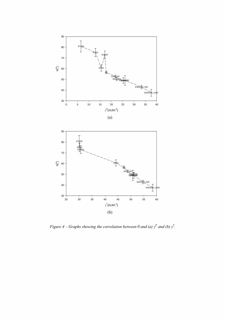
From Figure 4 it can be seen that θ was an inverse function of γP (see Figure 4(a)) and γT (see Figure
4(b) which does agree with current theory for a hydrophilic material such as nylon 6,6. This suggests
that even though there is a likely-hood of a mixed-state wetting regime is arising on the KrF excimer
laser patterned samples γP and γT still play a distinct role in terms of the wettability of the nylon 6,6. It
is also possible to ascertain from Figure 4(b) that similar values of γT give rise to equivalent values of
θ. That is, with a γT of approximately 30 mJm-2, a θ of between 75 and 80° could be expected.
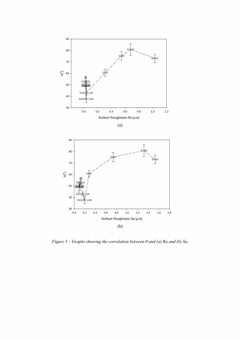
Figure 5 shows the correlation arising between θ and the surface roughness parameters Ra and Sa. For
the KrF excimer laser-induced patterned samples it can be seen that θ is an increasing function of both
Ra and Sa with the exception of the last data point which was for sample EH100 which had an Ra of
1.032 µm, an Sa of 1.530 µm and a resulting θ of 73±3.7°. This could suggest that an increase in the
surface roughness gives rise to an increase in θ until a certain point at which θ remains constant or
begins to decrease by which time would then start to correspond with current theory insofar as an
increase in surface roughness gives rise to a more hydrophilic response [5]. Furthermore, this could
suggest that over the different rough periodic surfaces a transition in wetting regime could have taken
place and would explain the decrease in θ for the roughest sample (sample EH100).
4.2.2 – KrF Excimer Laser Whole Area Irradiative Processing
From Table 1 it can be seen that the KrF excimer laser whole area irradiated samples gave rise to a
decrease in θ of up to 20°. The largest decrease in θ was observed for samples WA250_500 and
WA250_1000 which had the largest fluence of 91 mJm-2 and more pulses compared to the other
samples. The reduction observed in θ can be attributed to the increase in γP and γT. In terms of surface
oxygen content it was seen that there was an increase of up to 5 %at. which could have also given rise
to the reduction in θ. Another factor which can be taken from Table 1 is that the surface roughness
variation was negligible compared to the as-received sample (AR) which had an Ra of 0.029 µm and
Sa of 0.12 µm. Therefore, it stands to reason that γP and γT or the surface oxygen content in this
instance had to be the most dominant in determining the wettability of nylon 6,6 following KrF
excimer laser whole area irradiative processing.
Figure 4 shows that θ over all KrF excimer laser whole area processed samples was an inverse
function of γP (see Figure 4(a)) and γT (see Figure 4(b) with very good correlation. What is more it can
be seen from Figure 4 that those samples which had equivalent γP or γT values gave rise to similar
observed θ. For instance, for γP values of around 22 and 25 mJm-2, θ was approximately 51 and 48°
(see Figure 4(a)). Also, from Figure 4(b) it can be seen that samples with similar γT of around
51 mJm-2 gave rise to θ in the region of 50°.
Figure 5 shows a reasonable inverse correlation between the surface roughness and θ for all KrF
excimer laser whole area irradiative processed samples. However, on account of the relatively small
variations in Ra and Sa for these samples, a conclusive relationship between surface roughness and θ
could not be determined.
4.2.3 – Comparison Between KrF Excimer Laser-Induced Patterning and KrF Excimer Laser
Whole Area Irradiative Processing
Table 1 allows one to identify that there was some significant differences between the as-received
(AR), KrF excimer laser-induced patterned and KrF excimer laser whole area processed nylon 6,6
samples in terms of surface parameters and wettability characteristics. It was observed that for the
KrF laser-induced patterned samples θ increased even though there was a significant increase in
surface roughness. This increase in θ can be attributed to the reduction in γP and γT and the likelihood
of a transition in wetting regime. On the other hand, it was found that on account of an increase in γP
and γT, θ decreased for the KrF excimer laser whole area irradiative processed nylon 6,6 samples. It
was observed that for all KrF excimer laser processed samples the surface oxygen content increased
by up to 5 %at. when compared to the as-received sample (AR). This increase in surface oxygen
content indicates that this may have given rise to the observed reduction in θ for the laser whole area
processed samples. Still, this does not seem to be the case as a reduction in θ was not observed for the
KrF excimer laser-induced patterned samples. This suggests that a significant variation in surface
topography of the nylon 6,6 may bring about the change in wettability regime; whereas when there is
no significant variation in topography it is possible that a variation in wettability is brought about by
other parameters such as γP and γT. In addition, even though there was a change in wetting regime for
the KrF excimer laser-induced patterned samples to account for the increase of Sa and Ra, it was still
found that θ remained strongly linked to variations in γP and γT.
In terms of collating the γP and γT results for the entire KrF excimer laser processed samples and
the effects thereof on θ, Figure 4 shows that there was a strong inverse function correlation
between θ and the surface energy parameters regardless of the processing technique used. This
indicates that γP and γT could be the main driving parameters determining the wettability of the
nylon 6,6 samples in this instance. As a result it may be possible to use these parameters as
indicators of how nylon 6,6 will wet due to similar values of γP and γT giving rise to equivalent θ,
as discussed in Section 4.2.1 and Section 4.2.2.
4.3 – Osteoblast Cells: 24 hours Incubation
4.3.1 – KrF Excimer Laser-Induced Patterning
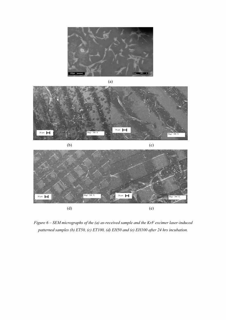
After 24 hrs incubation time for the osteoblast cell seeded samples it can be seen from the SEM
micrographs shown in Figure 6 that the cells had begun to adhere and proliferate across each of the
KrF excimer laser-induced patterned nylon 6,6 samples. Similar to the as-received sample (see Figure
6(a)) the KrF excimer laser-induced patterned samples gave rise to osteoblast cells with a bipolar cell
morpohology. In addition to this, one major difference between the cell growth on the as-received
sample (see Figure 6(a)) and the KrF excimer laser-induced patterned samples is that the excimer
patterned nylon 6,6 samples gave rise to some form of directionality; that is, the cells appeared to be
preferentially growing along the grooves formed by the excimer laser. This could have been induced
by the surface roughness and surface oxygen content being higher (see Table 1) in the etched grooves
allowing for preferential cell growth.
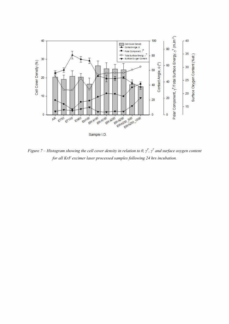
Figure 7 allows one to see that in terms of cell cover density, following 24 hrs of incubation, the KrF
excimer laser-induced patterned nylon 6,6 samples gave rise to a cell cover density equivalent to that
observed with the as-received sample (AR). That is, all of the KrF excimer laser-induced patterned
samples gave a cell cover density of around 20% suggesting that the nylon 6,6 surfaces had not given
rise to a more enhanced osteoblast cell response following KrF excimer laser patterning.
4.3.2 – KrF Excimer Laser Whole Area Irradiative Processing
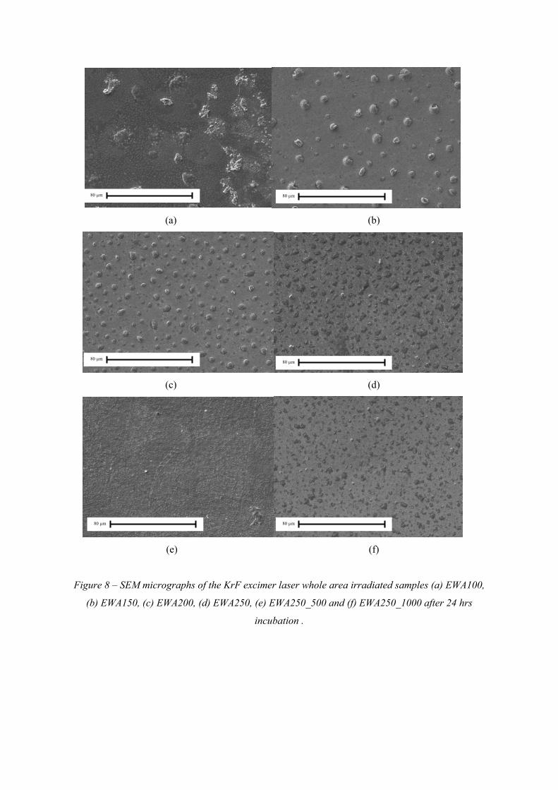
Figure 8 shows the SEM micrographs for the KrF excimer laser whole area irradiative processed
nylon 6,6 samples after 24 hrs incubation. From this it can be seen that the KrF excimer whole area
irradiative processed samples gave rise to variations in cell morphology in comparison to the as-
received sample (see Figure 6(a)) in that, the cells shown in Figure 8 appeared to be more clumped
radial. This can be attributed to the fact that the combination of differing wettability characteristics
and surface oxygen content is likely to have contributed to cell differentiation. As a result, one can
deduce that by modulating cell differentiation through varying wettability characteristics and surface
oxygen content there is a large potential for using laser surface treated polymeric materials within
regenerative medicine.
It is apparent from Figure 7 that all of the KrF excimer laser whole area irradiative processed samples
gave rise to larger cell cover densities when compared to the as-received sample (AR) with the
exception of sample EWA250_500 and sample EWA250_1000. The largest cover density of 25% was
obtained with samples EWA100, EWA150, EWA250 and EWA250 which was 5% more than what
was observed for the as-received sample (AR). Sample EWA250_500 and sample EWA250_1000
gave rise to a reduction in osteoblast cell response (see Figure 7) and could be attributed to the
samples becoming too hydrophilic (see Table 1) in which the modified nylon 6,6 surfaces were not
sufficient to promote an enhanced osteoblast cell response. That is, materials that are too hydrophilic
are well known for their cell-repellant properties and hinder the initial protein adsorption needed for a
positive cell response [18,32].
4.3.3 – Wettability Characteristics and Surface Parameters
By taking into account the effects of θ on the osteoblast cell response following 24 hrs incubation, it
can be seen that the data plotted in Figure 7 suggested a possible threshold window for θ between 47
and 53° which gave rise to an enhanced osteoblast cell response in terms of cell cover density. On the
other hand, this also indicates that above or below this θ threshold window the osteoblast cell
response would inherently be hindered slowing the cell proliferation and reducing the cell cover
density. Also, for the KrF excimer laser-induced patterned samples which gave rise to the largest θ of
approximately 70 to 80° it can be seen from Figure 7 that the cell cover density reduced to around
18%. This is highly significant when compared to the other samples with θ between 47 and 53°
because these samples gave rise to the largest cell cover densities of 25% or more allowing one to
extrapolate that a θ threshold window is likely.
The likelihood of a threshold window is further confirmed when accounting for γP and γT. Such that
Figure 7 shows there was a γP threshold window of 17 to 25 mJm-2 and a window of 47 to 53 mJm-2
for γT which gave rise to an enhanced osteoblast cell response in terms of cell cover density. It should
also be noted here that the relationship between the cell cover density, θ and γP are mostly the inverse
to one another and can be attributed to the relationship between γP and θ. From this the inverse
relationship for the two parameters with the cell cover density further attests to the relationship
between θ and γP.
It should noted here that Figure 7 also suggests that even though a threshold window could be
possible, the KrF excimer laser-induced patterned nylon 6,6 samples did not conclusively follow the
trend. This is due to those samples with γT of around 30 mJm-2 giving rise to a variation of cell cover
densities ranging between 16 and 21%. This can be accounted for by the likelihood of a mixed-state
wetting regime arising on these samples which would have had a large impact upon how the
osteoblast cells reacted to the patterned nylon 6,6 surfaces.
In addition, Figure 7 allows one to see that the cell cover density for the KrF laser processed samples
after 24 hrs of incubation was a decreasing function of the surface oxygen content such that the lowest
cell cover densities of around 17% were achieved when the surface oxygen content had increased up
to 18 %at. Having said that, it is well known that an increase in surface oxygen content should bring
about an enhanced cell response [5]. An explanation for this observed phenomena can be attributed to
a potential increase in surface toxicity whilst oxidation took place during the laser surface treatment.
This is on account of the nylon 6,6 potentially becoming excessively toxic through degradation of the
nylon 6,6 surface giving rise to toxic substances which would hinder cell growth. Even though these
substances may be present it is likely that surface oxidation would have taken place and would
explain the increase in surface oxygen content and reduction in cell count for the KrF excimer laser
processed samples. This can be attributed to the fact that at the higher fluences further melting could
cause the surface of the nylon 6,6 to become more toxic which would hinder cell growth compared to
the other samples. That is, upon considerable degradation of the nylon 6,6 it is known that toxic
substances such as CO and HCN can evolve [33]. These evolved toxic substances could have been
present on the surface on account of the rapid heating and cooling, trapping these substances until
they leached out into the cell media. This would have had a considerable deleterious effect on the
osteoblast cell growth as it has been shown that CO and HCN affect cell growth and proliferation
[34].
Following 24 hrs of incubation it has been observed that θ, γP, γT and indirectly the surface oxygen
content all had an influence on modulating the osteoblast cell response to the laser surface treated
samples. From the results obtained it was also found that there was no specific trend identified
between the surface roughness and the osteoblast cell response following 24 hr incubation. This
allows one to see that the surface roughness parameters were not a dominant parameter in determining
the osteoblast cell response. Also, on account of some erroneous results from the laser-induced
patterned samples the laser whole area irradiative processed samples seemed to give rise to the highest
potential for use in regenerative medicine. This is because of the likely mixed-state wetting regime
arising on the laser-induced patterned samples having a large impact upon the osteoblast cell
response.
4.4 – Osteoblast Cells: 4 Days Incubation
4.4.1 – KrF Laser-Induced Patterning
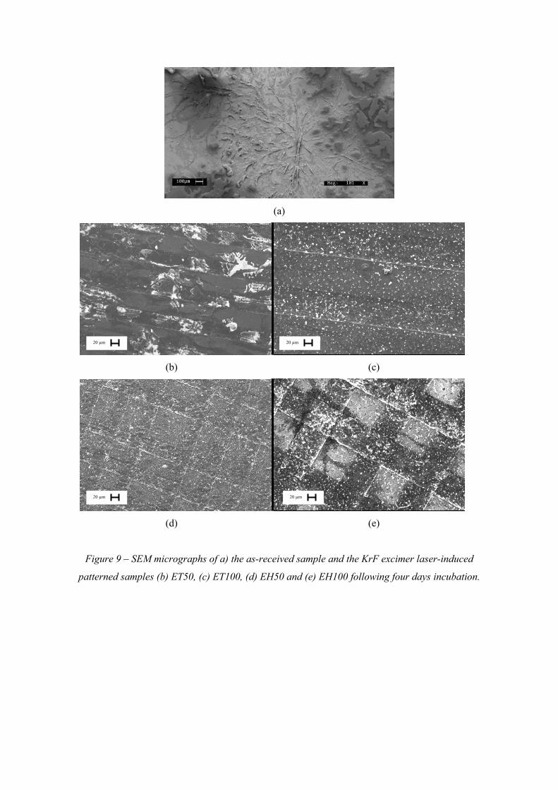
With four days of incubation elapsed, Figure 9 shows that the cell growth was at an advanced stage
for the KrF excimer laser-induced patterned nylon 6,6 samples compared to what was observed
following 24 hrs incubation time (see Figure 6). From Figure 9 it can be seen that the cell morphology
for the as-received sample was radial and coral-like which differed from the KrF excimer laser-
induced patterned samples which gave a more clumped like morphology. It can also be identified
using Figure 9 that there appeared to be more directionality for the KrF excimer laser-induced
patterned samples compared to the as-received sample. As discussed following 24 hrs incubation, this
could potentially be due to the grooves being rougher giving rise to more preferential cell growth in
these areas.
It can be seen from Figure 10 that the KrF excimer laser-induced patterning of the nylon 6,6 samples
gave rise to cover densities of just under 100% which was equivalent to that observed for the as-
received sample (AR). To confirm if the bioactivity had changed for the KrF excimer laser-induced
patterned samples compared to the as-received sample (AR) after four days of incubation cell count
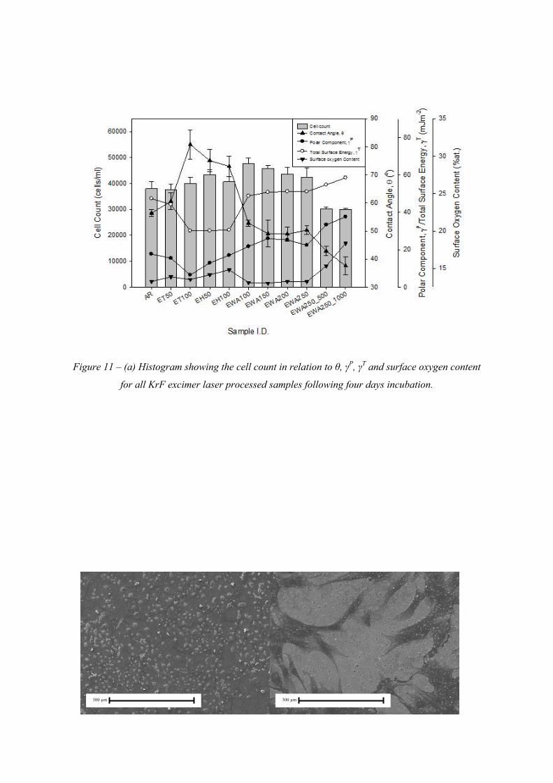
was also taken into account. Figure 11 allows one to realize that the cell count was also equivalent to
that of the as-received sample which was around 40,000 cells/ml. By knowing this, it was possible to
see that the KrF excimer laser-induced patterned nylon 6,6 samples did not give rise to an enhanced
osteoblast cell response when compared to the as-received sample (AR). Having said that, the KrF
excimer laser-induced patterned nylon 6,6 samples did not hinder cell response either and as such can
be seen to give an equivalent response to that seen with the as-received sample. In fact, the only
difference which could be ascertained between the as-received sample and the KrF excimer laser-
induced patterned samples was that of the cell morphology which can be attributed to surface
parameter variations on account of the laser-material interaction.
4.4.2 – KrF Excimer Laser Whole Area Irradiative Processing
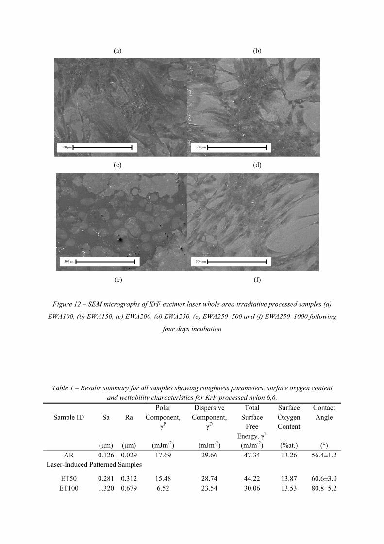
Figure 12 shows the SEM micrographs for the KrF excimer laser whole area irradiative processed
nylon 6,6 samples following four days incubation. As one can see the osteoblast cells were at an
advanced stage of cell growth following 4 days of incubation. Furthermore, it can be seen that the
morphologies appear to have varied somewhat. Sample EWA100 (see Figure 12(a)) gave rise to a
more clumped like whereas the other samples (see Figure 12(b) to Figure 12(f)) gave rise to a more
radial cell morphology. This further suggests that the variation in surface properties as a result of the
laser treatment had a significant impact upon cell signalling and can be seen as an attractive means for
potential use within regenerative medicine.
Figure 10 shows that the cell cover density for all of the KrF excimer laser whole area irradiative
processed samples tended towards 100% after 4 days of incubation. However, it can be said that the
lowest cell cover density was determined for sample EWA250_1000 which had a cell cover density of
around 83%. This is significant as it suggests that a considerably more hydrophilic surface such as
sample EWA250_1000 gave rise to a less enhanced osteoblast cell response as discussed in Section
4.3.2.
To further confirm the effects of the KrF excimer laser whole area irradiative processing on the
osteoblast cell response after four days a cell count was undertaken. Figure 11 shows that most of the
KrF excimer laser whole area irradiative processed samples gave rise to a larger cell count of around
47,000 cells/ml when compared to the as-received sample (AR) which had a cell count of just less
than 40,000 cells/ml. By comparing the KrF excimer laser whole area irradiative processed samples
alone it was found that upon an increasing fluence and incident pulse numbers, the cell count reduced
considerably to the point where sample EWA250_500 and sample EWA250_1000 had a cell count
around 33,000 cells/ml which was less than that of the as-received sample (AR). This can allow one to
see that these samples hindered the osteoblast cell response in comparison to the as-received sample
(AR).
4.4.3 – Wettability Characteristics and Surface Parameters
Figure 11 shows that for the KrF excimer laser-induced patterned samples there did not appear to be
any correlative relationship between the cell count and θ. This can be further accounted for by the
transition in wetting regime which is likely to have had effect on the osteoblast cell response. On the
contrary, it can be seen from Figure 11 that the KrF excimer laser whole area irradiative processed
samples could to some extent have had a correlation between the cell count and θ. Since, the cell
count appears to have reduced on account of a drop in θ.
It can also be seen from Figure 11 that there did not appear to be any correlation between γP and the
cell count for the KrF excimer laser-induced patterned nylon 6,6 samples. In general, for the KrF
excimer laser whole area irradiative processed samples Figure 11 shows that there could have been a
slight correlative trend in that the cell count reduced on account of an increase in γP. In addition to
this, the data presented in Figure 11 to some extent shows an inverse relationship between θ and γP
and as such supports the relationship between θ and γP identified in Section 4.2.
Figure 11 allows one to see that upon a significant reduction in γT for the KrF excimer laser-induced
patterned samples the cell count increased slightly by up to 5,000 cells/ml compared to the as-
received sample (AR). Similarly, for the KrF excimer laser whole area irradiative processed samples
it was found that the cell count decreased dramatically on account of an increase in γT. Furthermore,
from Figure 11 one can see that for all of the KrF excimer laser processed nylon 6,6 samples the cell
count had a strong correlation with surface oxygen content in that an increase in surface oxygen
content brought about a reduction in the cell count, coinciding with what has been observed
previously in Section 4.3.3. However, as stated in Section 4.3.3 this does not necessarily mean that an
increase in the surface oxygen content hinders cell response but could be used as an indirect indicator
of how a KrF excimer laser processed nylon 6,6 will perform when in contact with osteoblast cells.
Similar to the observations following 24 hrs incubation, there were no observations as to any
correlation between the surface roughness parameters Ra and Sa and the osteoblast cell response. This
indicates that the surface roughness following laser surface treatment was not a dominating parameter
governing the biomimetic nature of the laser surface modified nylon 6,6. In Section 4.3.3 it was seen
that there could potentially be an operating threshold window for θ, γP and γT in order for KrF excimer
laser processed nylon 6,6 to give an enhanced osteoblast cell response. However, after 4 days
incubation these trends have not been identified and suggest that the two different KrF excimer laser
processing techniques do not correlate to the same trend when accounting for the osteoblast cell
response. Having said that, it may be the case that the surface parameters and wettability
characteristics played a more significant role in the preliminary stages of cell growth, explaining the
reduction in correlative trends observed after four days of incubation. Even so, trends to some extent
have been observed for the laser whole area irradiative processed samples over both the 24 hrs and
4 day incubation period. As a result of this, one can say that the laser whole area irradiative
processing technique holds more promise in the prediction of cell response and has more potential for
use within regenerative medicine.
5.0 – Conclusions
It has been further demonstrated that UV lasers offer an efficient and convenient means of surface
modifying materials. It has been shown that a KrF excimer laser can be used for two different types of
laser surface treatment: surface patterning and whole area irradiative processing. These different types
of KrF excimer laser surface treatment have been seen to give rise to different topographies and
wettability modifications on the nylon 6,6 samples. For the laser-induced patterned samples θ was
found to have increased by up to 24° when compared to the as-received sample even though Ra and
Sa had increased; whereas the KrF excimer laser whole area processed samples gave θ to either be
equivalent to the as-received sample or have a θ less by up to 20°. The increase in θ for the KrF
excimer laser-induced patterned samples can be explained by the likely presence of a mixed-state
wetting regime. Having said that, even though a change in wetting regime is likely it has still been
observed that γP and γT still play a big role in the wettability of nylon 6,6 such that θ was a decreasing
function of γP and γT.
The KrF excimer laser surface treatment employed throughout this study had a significant impact on
cell response in terms of cell differentiation, cell cover density and cell count. After 24 hrs incubation
it was seen that the osteoblast cells had begun to adhere and proliferate across the surface of each of
the nylon 6,6 samples with the majority of laser-modified samples giving rise to larger cover
densities. Leading on, following 4 days of incubation it was found that all samples had given rise to
an advanced stage of cell growth giving cell cover densities of around 100%. However, it was seen
that the cell count had increased for most of the laser processed samples indicating a more bioactive
surface.
Those samples irradiated with low incident fluences and pulse numbers gave rise to the largest
enhancements in bioactivity. However, on account of the erratic results obtained for the laser-induced
patterned samples due to the likely influence from the mixed-state wetting regime, less correlative
trends between the different parameters were identified. This allows one to see that the laser whole
area irradiative processing technique is a more attractive method for modulating and predicting
osteoblast cell response. Overall, it has been determined that the laser-induced modifications have
given rise to modulated osteoblast cell response in terms of cell proliferation and differentiation.
Therefore, on account of the differing surface parameters giving rise to the modulation of cell
responses one can deduce that laser surface treatment holds a large potential to be widely employed
within regenerative medicine.
6.0 – Acknowledgements
The authors would like to thank their collaborators: Directed Light Inc., East Midlands NHS
Innovation Hub, Nobel Biocare and Photomachining Inc. for all of their much appreciated support.
The authors would also like to thank Chemical Engineering, Loughborough University for use of
their biological laboratory. This study was financially supported by the EPSRC (EP/E046851/1).
7.0 - References
1. W.M. Steen, Laser Material Processing: Third Ed, Springer-Verlag, London, UK, 2005.
2. J.C. Ion, Laser Processing of Engineering Materials: Principles, Procedure and Industrial Application, Elsevier Butterworth-Heinemann, Oxford, UK, 2005.
3. S.E. Nielsen, Laser material processing of polymers, Polym. Test. 3 (1983) 303-310.
4. M. Von Allmen, A. Blatter, Laser-Beam Interactions With Materials: Physical Principles and Applications: Second Ed., Springer-Verlag, New York, USA, 1995.
5. L. Hao, J. Lawrence, Laser Surface Treatment of Bio-Implant Materials, John Wiley & Sons Inc., New Jersey, USA, 2005.
6. K.S. Tiaw, M.H. Hong, S.H. Teoh, Precision laser micro-processing of polymers, J. Alloys Compd. 449 (2008) 228-231.
7. E. Sarantopoulou, Z. Kollia, A.C. Cefalas, A.M. Douvas, M. Chatzichristidi, P. Argitis, S. Kobe, Polymer self-assembled nano-structures and surface relief gratings induced with laser at 157nm, Appl. Surf. Sci. 253 (2007) 7884-7889.
8. K. Callewaert, Y. Martele, L. Breban, K. Naessens, P. Vandaele, R.Baets, G. Geuskens, E. Schacht, Excimer laser induced patterning of polymeric surfaces, Appl. Surf. Sci. 208-209 (2003) 218-225.
9. E.S.A. Hegazy, H.A. Abdel-Rehim, H. Kamal, K.A. Kandeel, Advances in radiation grafting, Nucl. Inst. Meth. Phys. Res. B 185 (2001) 235-240.
10. J. Zhang, J. Khang, P. Hu, Q. Meng, Surface modification of poly(propylene carboante) by oxygen ion implantation, Appl. Surf. Sci. 253 (2007) 5436-5441.
11. S. Dadbin, Surface modification of LDPE film by CO2 pulsed laser irradiation, Euro. Polym. J. 38 (2002) 2489-2495.
12. C. Mao, W. Zhao, C. Zhu, A. Zhu, J. Shen, S. Lin, In vitro studies of platelet adhesion on UV radiation-treated nylon surface, Carbohydr. Polym. 59 (2005) 19-25.
13. F. Yu, F. Mucklich, P. Li, H. Shen, S. Mathur, C.M. Lehr, U. Bakowsky, In vitro cell response to a polymer surface micropatterned by laser interference lithography, Biomacromol. 6 (2005) 1160-1167.
14. W. Pfleging, M. Bruns, A. Welle, S. Wilson, Laser-assisted modification of polystyrene surfaces for cell culture applications, Appl. Surf. Sci. 253 (2007) 9177-9184.
15. H. Mirzadeh, M. Dadsetan, Influence of laser surface modifying of polyethylene terephthalate on fibroblast cell adhesion, Radiat. Phys. Chem. 67 (2003) 381-385.
16. H. Mirzadeh, A.A. Katbab, R.P. Burford, CO2-laser graft copolymerization of HEMA and NVP onto ethylene-propylene rubber (EPR) as biomaterial-(III), Radiat. Phys. Chem. 46 (1995) 859-862.
20. S.R. Paital, N.B. Dahotre, Calcium phosphate coatings for bio-implant applications: Materials, performance factors and methodologies, Mater. Sci. Eng. R 66 (2009) 1-70.
21. J.J. Rajesh, J. Bijwe, B. Venkataraman, U.S. Tewari, Effect of water absorption on erosive wear behaviour of polyamides, J. Mater. Sci. 37 (2002) 5107-5113.
22. K. MacGregor, The ageing population: U.K. focus for biomedical engineering - policy briefing. The Royal Academy of Engineering 2010.
23. D.G. Rance, Chapter 6 - thermodynamics of wetting: From its molecular basis to technological application, in: D.M. Brewis (ed.), Surface Analysis and Pretreatment of Plastics and Metals, Applied Science Publishers, Essex, UK, 1982. p.121.
24. D.G. Waugh, J. Lawrence, The enhancement of biomimetic apatite coatings by means of KrF excimer laser surface treatment of nylon 6,6, Lasers Eng. 21 (2011) 95-114.
25. X. Wu, L. Zheng, D. Wu, Fabrication of superhydrophobic surfaces from microstructured ZnO-based surfaces via a wet-chemical route, Langmuir 21 (2005) 2665-2667.
26. S.M. Lee, T.H. Kwon, Effects of intrinsic hydrophobicity on wettability of polymer replicas of a superhydrophobic lotus leaf, J. Micromech. Microeng. 17 (2007) 687-692.
27. X. Chen, T. Lu, The apparent state of droplets on a rough surface, Sci. China Ser. G.Phys. Mech. Astron. 52 (2009) 233-238.
28. Y.T.Cheng, D.E. Rodak, C.A. Wong, C.A. Hayden, Effects of Micro- and Nano-Structures on the Self-Cleaning Behaviour of Lotus Leaves, Nanotechnol. 17 (2006) 1359-1362.
29. D.G. Waugh, J. Lawrence, D.J. Morgan, C.L. Thomas, Interaction of CO2 laser-modified nylon with osteoblast cells in relation to wettability, Mater. Sci. Eng. C 29 (2009) 2514-2524.
30. D.G. Waugh, J. Lawrence, C.D. Walton, R.B. Zakaria, On the effects of using CO2 and F2 lasers to modify the wettability of a polymeric biomaterial, J. Opt. Laser Technol. 42 (2010) 347-356.
31. D.G. Waugh, J. Lawrence, On the use of CO2 laser induced patterns to modify the wettability of poly(methyl methamecrylate) (PMMA). Opt. Lasers Eng. 48 (2010) 707-715.
32. M.C. Lensen, V.A. Schulte, J. Salber, M. Diez, F. Menges, M. Moller, Cellular response to novel, micropatterned biomaterials. Pure Appl. Chem. 80 (2008) 2479-2487.
33. Sigma-Aldrich. Nylon 6,6 Material Safety Data Sheet - Version 3.0. 2008.
34. S.D. Cook, J.P. Ryaby, J.R.N. McCabe, J.J. Frey, J.D. Heckman, T.K. Kristiansen, Acceleration of tibia and distal radius fracture healing in patients who smoke. Clin. Ortho. Rel. Res. 337 (1997) 198-207.
Laser operating parameters
Surface Chemistry
Surface Topography
Cell Response
Surface Wettability
Figure 1 – Schematic diagram showing the deduced links between the factors arising throughout the
research.
(a)
(b) (c)
(d) (e)
Figure 2 – Continuous axonometric 3-D images and profile extractions for the (a) as-received samples and for the