Page 1
BASIC RESEARCH
What Is Normal Femoral Head/Neck Anatomy? An Analysisof Radial CT Reconstructions in Adolescents
Amir A. Jamali MD, Walter Mak MD, Ping Wang BS, Lynn Tai BS,
John P. Meehan MD, Ramit Lamba MBBS, MD
Received: 7 January 2013 / Accepted: 1 July 2013 / Published online: 7 August 2013
� The Association of Bone and Joint Surgeons1 2013
Abstract
Background Cam morphology in femoroacetabular
impingement has been implicated in the development of
osteoarthritis. The alpha angle and femoral head/neck
offset are widely used to determine femoral head asphe-
ricity. To our knowledge, no study has evaluated the alpha
angle circumferentially using three-dimensional imaging in
a population of healthy individuals of adolescent age.
Questions/purposes We sought to (1) determine normal
values for the alpha angle in adolescents, (2) define the
location along the neck with the highest alpha angle, and
(3) determine normal femoral head and neck radii and
femoral head/neck offset.
Methods Fifty CT scans from a database of scans
obtained for reasons not related to hip pain were studied.
The average age of the subjects was 15 years (range,
14–16 years). Alpha angle and femoral head/neck offset
were measured circumferentially.
Results The alpha angle averaged 40.66 ± 4.46 mm for
males and 37.77 ± 5.65 mm for females. The alpha angle
generally was highest between the 11:40 and 12:40 o’clock
and between the 6:00 and 7:40 o’clock positions. The
femoral head radius was 24.53 ± 1.74 mm for males and
21.94 ± 1.13 mm for females, and the femoral neck radius
was 16.14 ± 2.32 mm for males and 13.82 ± 2.38 mm
for females. The mean femoral head/neck offset was
8.39 ± 1.97 mm for males and 8.13 ± 2.27 mm for
females.
Conclusions In this healthy population of 14- to 16-year-
old subjects, the highest alpha angle was at the superior and
inferior aspects of the heads rather than at the anterosu-
perior aspect. This information will provide benchmark
values for distinction between normal and abnormal mor-
phologic features of the femoral head.
Level of Evidence Level III, diagnostic study. See
Guidelines for Authors for a complete description of levels
of evidence.
Introduction
Femoroacetabular impingement (FAI) is a pathomechani-
cal process that has been linked to the development of
osteoarthritis of the hip [2, 3]. It has been defined as
Each author certifies that he or she, or a member of his or her
immediate family, has no funding or commercial associations
(eg, consultancies, stock ownership, equity interest, patent/licensing
arrangements, etc) that might pose a conflict of interest in connection
with the submitted article.
All ICMJE Conflict of Interest Forms for authors and Clinical
Orthopaedics and Related Research editors and board members
are on file with the publication and can be viewed on request.
Each author certifies that his or her institution approved the human
protocol for this investigation, that all investigations were conducted
in conformity with ethical principles of research, and that informed
consent for participation in the study was obtained.
A. A. Jamali (&), L. Tai
Joint Preservation Institute, Orthopaedic Surgery, 2825 J Street,
#440, Sacramento, CA 95816, USA
e-mail: [email protected]
W. Mak
St Michael’s Hospital, Toronto, Ontario, Canada
P. Wang
UC Davis School of Medicine, Sacramento, CA, USA
J. P. Meehan
Sacramento Knee and Sports Medicine, Sacramento, CA, USA
R. Lamba
Department of Radiology, UC Davis, Sacramento, CA, USA
123
Clin Orthop Relat Res (2013) 471:3581–3587
DOI 10.1007/s11999-013-3166-5
Clinical Orthopaedicsand Related Research®
A Publication of The Association of Bone and Joint Surgeons®
Page 2
abnormal contact between the femoral head and neck and
the rim of the hip socket during normal activities such as
sitting. FAI has been classified into two broad categories—
the cam and pincer types. Cam impingement typically is
seen in young males with an abnormal-shaped femoral
head, whereas pincer type generally is the result of ace-
tabular rim issues such as an excessively deep socket as
seen in coxa profunda or localized overhang in the setting
of acetabular retroversion.
Cam-type FAI has been noted in younger patients [2]
and has been associated with a higher risk of osteoarthritis.
In cam-type FAI, the femoral head has an increased radius
in certain dimensions leading to an aspherical shape [6].
The area of greatest prominence is most often in the
anterolateral aspect of the femoral head and therefore is not
well observed using simple AP or lateral images [9]. Radial
sequence imaging using CT or MRI has been developed as
a method to obtain anatomic information regarding mor-
phologic features of the femoral head and the acetabular
rim in a 360� arc [1, 8].
The asphericity of the head in cam-type FAI has been
quantified using numerous methods, the most common
being the alpha angle [10]. Some studies have been dedi-
cated to characterizing abnormal morphologic features
using the alpha angle [1, 4, 5, 10, 11, 13]. Rakhra et al. [11]
evaluated abnormal hips using radial sequence imaging but
limited it to the anterosuperior aspect (1–3 o’clock only)
based on the premise that this is where more impingement
occurs. They found that in pathologic FAI, the maximum
alpha angle is at the anterosuperior quadrant at the 1 or
2 o’clock position. Despite these efforts, our current
understanding of morphologic features of the head and
neck is still lacking. Up to now, most research in this area
has been performed in cohorts of patients who have FAI or,
alternatively, in presumably healthy volunteers of various
ages [1, 4, 5, 10, 11, 13]. Much of the literature has
experienced selection bias of the symptomatic and control
groups, limited numbers of images analyzed, limited
locations analyzed on any given femoral head, and varia-
tions in subject age. The paucity of information in the
literature compelled us to perform the current study limited
to adolescent patients who presented for CT at our insti-
tution. We expected that in a population of asymptomatic
adolescent patients, the maximum alpha angles would be
consistent with those published in the literature and that the
same regions (1 and 2 o’clock) would have the highest
alpha angles and lowest femoral neck offsets as seen in the
cases described by Rakhra et al. [11].
Using three-dimensional imaging, we sought to
(1) determine normal values for the alpha angle in adolescent
boys and girls, (2) define at what point on the circumference
of the femoral neck the alpha angle is generally highest, and
(3) determine normal femoral head and neck radii and fem-
oral head/neck offset values.
Materials and Methods
Patients included in the study had a mean chronologic age
of 15.6 years old (range, 14.3–15.9 years). We selected this
age group based on our clinical observations that relatively
few sports and activity-related changes would likely have
taken place in this age group. Additionally, at this chrono-
logic age, subjects have reached or have nearly reached
skeletal maturity. All had undergone CT for issues not
related to hip pain. We established CT-based radial recon-
structions of the proximal femora using a standardized
technique and then analyzed the images using a specific
image analysis module based on point registration by an
observer (LT) with secondary automatic calculation of
numerous parameters involving each image. Fifty CT scans
were obtained from our institutional radiographic database.
All scans were obtained from patients treated for issues not
related to hip pain. We selected 50 subjects based on a
previous pilot study in a series of 20 trauma patients of all
ages in which the morphologic data had limited variability.
The indications for CT scanning were abdominal pain in 20
patients; high-energy trauma including motor vehicle
accidents, bicycle accidents, and motorcycle accidents in 20
patients; assault in five patients; and other diagnoses not
related to the hip in five patients. All scans were reviewed
by faculty radiologists and the senior author (AAJ) con-
firming the absence of proximal femoral deformity or
trauma. The raw imaging data in the format of Digital
Imaging and Communication in Medicine (DICOM) ima-
ges were entered into an imaging reconstruction program
(TeraReconTM; TeraRecon Inc, Foster City, CA, USA).
Radial reconstructions then were prepared (Fig. 1) using
this software with a total of 18 images for each head. These
reflected 36 positions around the head. Each image then was
entered into a custom imaging analysis algorithm written in
MATLAB1 (MathWorks, Natick, MA, USA). Multiple
measurements were performed by selecting various land-
mark points in the MATLAB1 software. From these points,
multiple parameters for that image were generated and were
automatically output into a spreadsheet document (Excel,
Microsoft, Redmond, WA, USA). These parameters
included head diameter, neck diameter, head-neck offset,
and alpha angle (Fig. 2). This information was analyzed
using a clock system widely used clinically in the field of
hip arthroscopy (Fig. 3).
Differences in each measurement were compared based
on sex using the unpaired t-test. Interobserver and intra-
observer reliability analysis was performed using intraclass
3582 Jamali et al. Clinical Orthopaedics and Related Research1
123
Page 3
correlation coefficients (ICCs) for two observers (LT and
AAJ) at two times a minimum of 4 weeks apart for five
specimens (total of 360 analyses). Linear regression was
performed using Excel (Microsoft). ICC and ANOVA were
performed with SPSS (Version 9; IBM, Chicago, IL, USA)
and StatView software (SAS Inc, Cary, NC, USA),
respectively. Statistical significance was set at a probability
less than 0.05.
Results
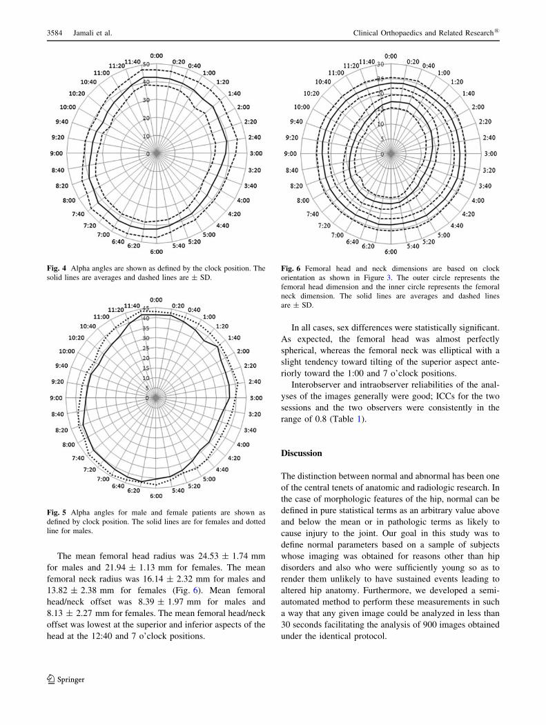
The alpha angle for all subjects and at all locations was
39.25� ± 5.288 (Fig. 4). Based on sex (Fig. 5), the alpha
angle measured 40.66� ± 4.468 for males and 37.77� ±
5.658 for females (p \ 0.001).The alpha angle generally
was highest between the 11:40 and 12:20 o’clock positions
and between 6:00 and 7:40 o’clock positions. In both of
these regions, it measured approximately 428.
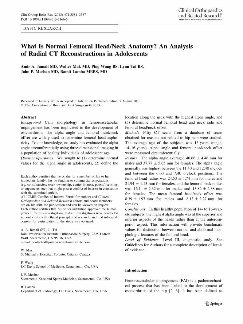
Fig. 1A–B The method for acquisition of radial reconstructions is
shown using TeraReconTM software (TeraRecon Inc, Foster City, CA,
USA). (A) The axis of rotation is placed down the femoral neck in the
axial and coronal (not shown) views. (B) A three-dimensional view of
radial acquisitions is shown.
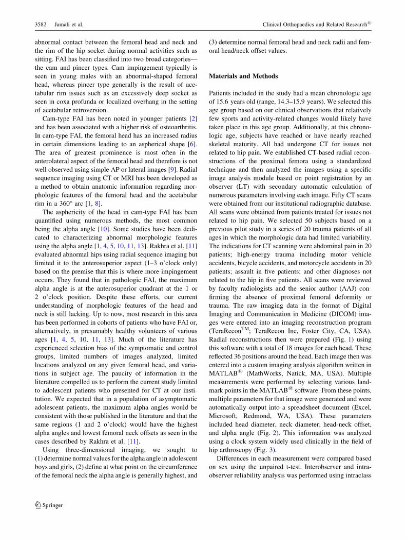
Fig. 2A–B Sample MATLAB1 screen images are shown. (A) Points
and lines of the hip are shown with red x’s, which define the femoral
head circumference. The white arrows show the superior and inferior
extents of the femoral neck with the center point calculated as the
femoral neck center. The large black arrow defines the acetabular rim
line extending from one edge of the rim to the next. The black
arrowhead shows the head/neck axis, extending from the femoral
neck center to the femoral head center. (B) The head/neck axis (black
arrow) is shown with the calculation of the alpha angles (white
arrowheads).
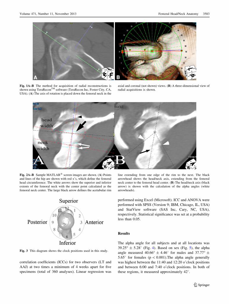
Fig. 3 This diagram shows the clock positions used in this study.
Volume 471, Number 11, November 2013 Femoral Head/Neck Anatomy 3583
123
Page 4
The mean femoral head radius was 24.53 ± 1.74 mm
for males and 21.94 ± 1.13 mm for females. The mean
femoral neck radius was 16.14 ± 2.32 mm for males and
13.82 ± 2.38 mm for females (Fig. 6). Mean femoral
head/neck offset was 8.39 ± 1.97 mm for males and
8.13 ± 2.27 mm for females. The mean femoral head/neck
offset was lowest at the superior and inferior aspects of the
head at the 12:40 and 7 o’clock positions.
In all cases, sex differences were statistically significant.
As expected, the femoral head was almost perfectly
spherical, whereas the femoral neck was elliptical with a
slight tendency toward tilting of the superior aspect ante-
riorly toward the 1:00 and 7 o’clock positions.
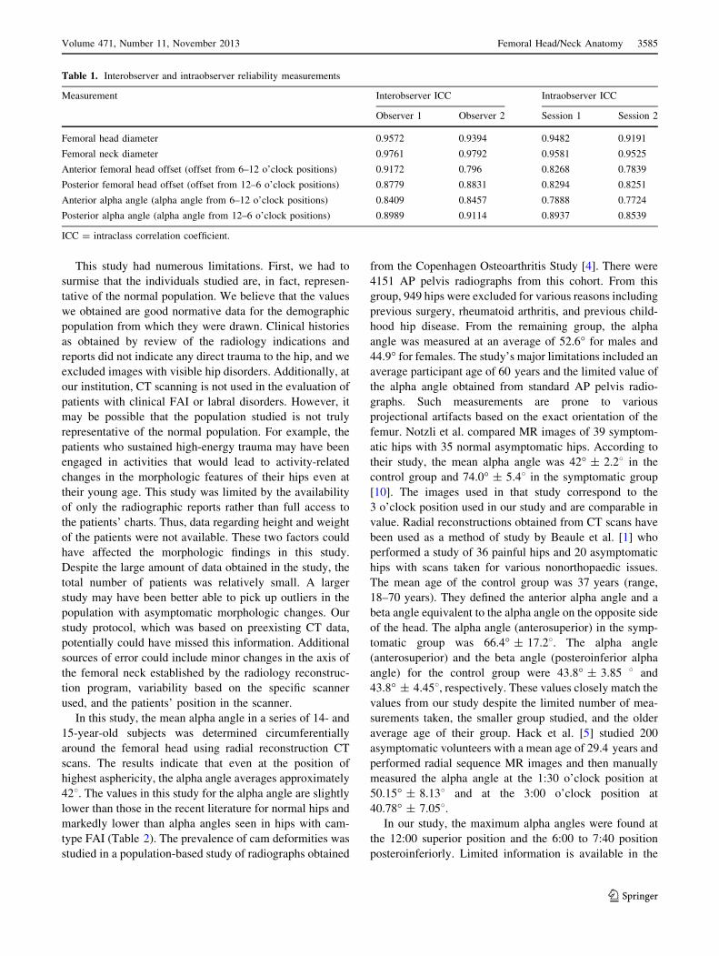
Interobserver and intraobserver reliabilities of the anal-
yses of the images generally were good; ICCs for the two
sessions and the two observers were consistently in the
range of 0.8 (Table 1).
Discussion
The distinction between normal and abnormal has been one
of the central tenets of anatomic and radiologic research. In
the case of morphologic features of the hip, normal can be
defined in pure statistical terms as an arbitrary value above
and below the mean or in pathologic terms as likely to
cause injury to the joint. Our goal in this study was to
define normal parameters based on a sample of subjects
whose imaging was obtained for reasons other than hip
disorders and also who were sufficiently young so as to
render them unlikely to have sustained events leading to
altered hip anatomy. Furthermore, we developed a semi-
automated method to perform these measurements in such
a way that any given image could be analyzed in less than
30 seconds facilitating the analysis of 900 images obtained
under the identical protocol.
Fig. 4 Alpha angles are shown as defined by the clock position. The
solid lines are averages and dashed lines are ± SD.
Fig. 5 Alpha angles for male and female patients are shown as
defined by clock position. The solid lines are for females and dotted
line for males.
Fig. 6 Femoral head and neck dimensions are based on clock
orientation as shown in Figure 3. The outer circle represents the
femoral head dimension and the inner circle represents the femoral
neck dimension. The solid lines are averages and dashed lines
are ± SD.
3584 Jamali et al. Clinical Orthopaedics and Related Research1
123
Page 5
This study had numerous limitations. First, we had to
surmise that the individuals studied are, in fact, represen-
tative of the normal population. We believe that the values
we obtained are good normative data for the demographic
population from which they were drawn. Clinical histories
as obtained by review of the radiology indications and
reports did not indicate any direct trauma to the hip, and we
excluded images with visible hip disorders. Additionally, at
our institution, CT scanning is not used in the evaluation of
patients with clinical FAI or labral disorders. However, it
may be possible that the population studied is not truly
representative of the normal population. For example, the
patients who sustained high-energy trauma may have been
engaged in activities that would lead to activity-related
changes in the morphologic features of their hips even at
their young age. This study was limited by the availability
of only the radiographic reports rather than full access to
the patients’ charts. Thus, data regarding height and weight
of the patients were not available. These two factors could
have affected the morphologic findings in this study.
Despite the large amount of data obtained in the study, the
total number of patients was relatively small. A larger
study may have been better able to pick up outliers in the
population with asymptomatic morphologic changes. Our
study protocol, which was based on preexisting CT data,
potentially could have missed this information. Additional
sources of error could include minor changes in the axis of
the femoral neck established by the radiology reconstruc-
tion program, variability based on the specific scanner
used, and the patients’ position in the scanner.
In this study, the mean alpha angle in a series of 14- and
15-year-old subjects was determined circumferentially
around the femoral head using radial reconstruction CT
scans. The results indicate that even at the position of
highest asphericity, the alpha angle averages approximately
428. The values in this study for the alpha angle are slightly
lower than those in the recent literature for normal hips and
markedly lower than alpha angles seen in hips with cam-
type FAI (Table 2). The prevalence of cam deformities was
studied in a population-based study of radiographs obtained
from the Copenhagen Osteoarthritis Study [4]. There were
4151 AP pelvis radiographs from this cohort. From this
group, 949 hips were excluded for various reasons including
previous surgery, rheumatoid arthritis, and previous child-
hood hip disease. From the remaining group, the alpha
angle was measured at an average of 52.6� for males and
44.9� for females. The study’s major limitations included an
average participant age of 60 years and the limited value of
the alpha angle obtained from standard AP pelvis radio-
graphs. Such measurements are prone to various
projectional artifacts based on the exact orientation of the
femur. Notzli et al. compared MR images of 39 symptom-
atic hips with 35 normal asymptomatic hips. According to
their study, the mean alpha angle was 42� ± 2.28 in the
control group and 74.0� ± 5.48 in the symptomatic group
[10]. The images used in that study correspond to the
3 o’clock position used in our study and are comparable in
value. Radial reconstructions obtained from CT scans have
been used as a method of study by Beaule et al. [1] who
performed a study of 36 painful hips and 20 asymptomatic
hips with scans taken for various nonorthopaedic issues.
The mean age of the control group was 37 years (range,
18–70 years). They defined the anterior alpha angle and a
beta angle equivalent to the alpha angle on the opposite side
of the head. The alpha angle (anterosuperior) in the symp-
tomatic group was 66.4� ± 17.28. The alpha angle
(anterosuperior) and the beta angle (posteroinferior alpha
angle) for the control group were 43.8� ± 3.85 8 and
43.8� ± 4.458, respectively. These values closely match the
values from our study despite the limited number of mea-
surements taken, the smaller group studied, and the older
average age of their group. Hack et al. [5] studied 200
asymptomatic volunteers with a mean age of 29.4 years and
performed radial sequence MR images and then manually
measured the alpha angle at the 1:30 o’clock position at
50.15� ± 8.138 and at the 3:00 o’clock position at
40.78� ± 7.058.In our study, the maximum alpha angles were found at
the 12:00 superior position and the 6:00 to 7:40 position
posteroinferiorly. Limited information is available in the
Table 1. Interobserver and intraobserver reliability measurements
Measurement Interobserver ICC Intraobserver ICC
Observer 1 Observer 2 Session 1 Session 2
Femoral head diameter 0.9572 0.9394 0.9482 0.9191
Femoral neck diameter 0.9761 0.9792 0.9581 0.9525
Anterior femoral head offset (offset from 6–12 o’clock positions) 0.9172 0.796 0.8268 0.7839
Posterior femoral head offset (offset from 12–6 o’clock positions) 0.8779 0.8831 0.8294 0.8251
Anterior alpha angle (alpha angle from 6–12 o’clock positions) 0.8409 0.8457 0.7888 0.7724
Posterior alpha angle (alpha angle from 12–6 o’clock positions) 0.8989 0.9114 0.8937 0.8539
ICC = intraclass correlation coefficient.
Volume 471, Number 11, November 2013 Femoral Head/Neck Anatomy 3585
123
Page 6
Ta
ble
2.
Lit
erat
ure
rev
iew
Stu
dy
Nu
mb
ero
fh
ips
Mea
nal
ph
aan
gle
Ty
pe
of
stu
dy
Rad
iog
rap
hic
met
ho
dR
elia
bil
ity
Go
svig
etal
.[4
]
32
02
Alp
ha
ang
les
on
AP
pel
vis
rad
iog
rap
hs:
52
.6�
for
mal
esan
d4
4.9
�fo
rfe
mal
es;
mea
nag
e
app
rox
imat
ely
60
yea
rs.
Po
pu
lati
on
bas
edco
ho
rtst
ud
yM
anu
alm
easu
rem
ents
of
stan
dar
dra
dio
gra
ph
s
Rep
ort
edin
terc
lass
of
0.8
3
and
ICC
of
0.9
to0
.96
.
Ste
pp
ach
er
etal
.
[13]
50
(25
dy
spla
stic
and
25
dee
pac
etab
ulu
m)
37
.4�
(ran
ge,
19
�–7
8�)
for
dy
spla
stic
,3
6.5
�(r
ang
e,1
9�–
89�)
for
dee
pac
etab
ulu
mg
rou
p
Stu
dy
of
clin
ical
lysy
mp
tom
atic
pat
ien
ts
pre
sen
tin
gw
ith
eith
erh
ipd
ysp
lasi
ao
ra
dee
pac
etab
ulu
m
Cli
nic
alM
RI
arth
rog
ram
s
wit
hra
dia
lse
qu
ence
imag
ing
ICC
Intr
aob
serv
er1
=0
.86
,
ICC
Intr
aob
serv
er
2=
0.7
9,
ICC
inte
rob
serv
er0
.81
Hac
ket
al.
[5]
40
0h
ips
40
.78�
±7
.05�
at3
o’c
lock
po
siti
on
,
50
.15
�±
8.1
3�
at1
:30
po
siti
on
;m
ean
age,
29
yea
rs(r
ang
e,2
1–
51
yea
rs)
MR
Iar
thro
gra
ph
yev
alu
atio
nin
no
rmal
vo
lun
teer
sfr
om
ah
osp
ital
and
med
ical
sch
oo
l
Mea
sure
men
tsp
erfo
rmed
ina
PA
CS
syst
emb
y
two
ob
serv
ers,
each
per
form
ing
eval
uat
ion
of
20
0h
ips
On
lyin
terr
ater
reli
abil
ity
pre
sen
ted
:0
.81
0IC
Cfo
r
axia
lv
iew
(3o
’clo
ck)
and
0.7
96
ICC
for
rad
ial
vie
w
(1:3
0o
’clo
ck)
Bea
ule
and
Zar
ago
za
[1]
56
hip
s(3
6p
ain
ful
and
20
fro
mas
ym
pto
mat
ic
vo
lun
teer
s)
66
.4�
±1
7.2
�(a
nte
rio
ral
ph
aan
gle
)an
d
40
.2�
±5
.4�
(po
ster
ior
also
term
edb
eta
ang
le)
insy
mp
tom
atic
hip
s,4
3.8
�±
3.8
5�
(an
teri
or
alp
ha
ang
le)
and
43
.8�
±4
.45�
(po
ster
ior
also
term
edb
eta
ang
le)
in
asy
mp
tom
atic
con
tro
lh
ips.
Stu
dy
of
CT
-bas
edra
dia
lre
con
stru
ctio
ns
Mea
sure
men
tsp
erfo
rmed
ina
PA
CS
syst
em
Inte
rcla
ssco
rrel
atio
n0
.60
,
ICC
was
0.8
0an
d0
.54
for
the
two
ob
serv
ers.
Rak
hra
etal
.
[11]
41
70
.5�
±1
4�
on
max
imal
rad
ial
imag
e,
53
.4�
±1
1.1
�o
no
bli
qu
eax
ial
imag
e
An
aly
sis
of
41
pat
ien
tsu
nd
erg
oin
gM
R
arth
rog
rap
hy
for
FA
I;ev
alu
ated
the
3
o’c
lock
po
siti
on
(ob
liq
ue
axia
l)an
dth
e
max
imal
rad
ial
of
the
1o
’clo
cko
r2
o’c
lock
po
siti
on
.
Man
ual
mea
sure
men
tso
f
MR
arth
rog
rap
hy
imag
es
No
tp
erfo
rmed
No
tzli
etal
.
[10]
74
(39
sym
pto
mat
ic,
35
con
tro
ls)
74�
±5
.4�
for
sym
pto
mat
icg
rou
p,
42�
±2
.2�
inth
eco
ntr
ols
MR
Iar
thro
gra
ph
yev
alu
atio
nin
sym
pto
mat
icp
atie
nts
and
no
rmal
vo
lun
teer
s
Man
ual
mea
sure
men
tso
f
MR
arth
rog
rap
hy
imag
es
No
ICC
pro
vid
ed
Cu
rren
t
stu
dy
50
hip
so
bta
ined
fro
man
inst
itu
tio
nal
dat
abas
e,
read
asw
ith
ou
tin
jury
,
all
pat
ien
ts1
4–
16
yea
rso
ld.
40
.66
±4
.46
mm
for
mal
esan
d
37
.77
±5
.65
mm
for
fem
ales
Stu
dy
of
CT
-bas
edra
dia
lre
con
stru
ctio
ns
Sem
iau
tom
ated
mea
sure
men
tsu
sin
g
cust
om
soft
war
eo
nC
T
scan
rad
ial
reco
nst
ruct
ion
s
Inte
rob
serv
erIC
C=
0.7
72
4,
intr
aob
serv
erfo
rO
bse
rver
1IC
C=
0.8
40
9,
intr
aob
serv
erfo
rO
bse
rver
2IC
C=
0.8
45
7
ICC
=in
trac
lass
corr
elat
ion
coef
fici
ent;
PA
CS
=P
ictu
rear
chiv
ing
and
com
mu
nic
atio
nsy
stem
;F
AI
=fe
mo
roac
etab
ula
rim
pin
gem
ent.
3586 Jamali et al. Clinical Orthopaedics and Related Research1
123
Page 7
literature regarding the circumferential alpha angle in the
normal population. Steppacher et al. [13] performed a
comprehensive 360� evaluation of the head sphericity,
epiphyseal extension, and the alpha angle using radial MRI
arthrography in a series of hips with either deficient or
excessive acetabular coverage, similar in principal to our
study performed in normal hips. In their entire study, the
alpha angle averaged 408 in the anterosuperior quadrant
and 338 in the posterosuperior quadrant. Interestingly, there
was no difference between the two groups in the antero-
superior region, the most likely area for clinical FAI to
occur in flexion activity.
The shape of the femoral head and neck have been
described qualitatively in numerous studies. Ranawat et al.
[12] studied 100 hips with unilateral FAI and compared the
radiographic predictors of hip pain between the symp-
tomatic and asymptomatic sides. Overall, in their series,
the male femoral head diameter was 58 mm and the female
head was 52 mm [12]. Their study was performed with
standard radiographs with no calibration template. Young
et al. examined the symmetry between the right and left
femoral heads in a series of 160 paired cadaveric femurs
measured using calibrated digital photographs [14]. In their
series, the mean femoral head diameter averaged 55.8 mm
for males and 48.3 mm for females, and the mean femoral
neck diameter was 39.3 and 33.7 mm for males and
females respectively in the AP view and 23.9 and 20.1 mm
for males and females respectively in the craniocaudal
direction. The head and neck sizes were larger in their
study compared with sizes in our study. This may be
related to the average age of 32 years for their subjects and
the measurement technique they used.
We have provided a comprehensive quantitative analy-
sis of proximal femoral morphologic features in 50 subjects
between 14 and 16 years old, and values that can be taken
as normative of this population. The methodology used
allows for efficient and consistent analysis of multiple
radial reconstructions from each subject in a semiautomatic
manner, thus minimizing bias. Despite the sophistication of
the analysis, all measurements described in this study can
be performed with essentially any radiology picture
archiving and communication system or various commer-
cially available imaging programs [7].
Acknowledgments We thank the staff of the UC Davis, Department
of Radiology for assistance with image acquisition and providing use
of their software workstations.
References
1. Beaule PE, Zaragoza E, Motamedi K, Copelan N, Dorey FJ.
Three-dimensional computed tomography of the hip in the
assessment of femoroacetabular impingement. J Orthop Res.
2005;23:1286–1292.
2. Ganz R, Leunig M, Leunig-Ganz K, Harris WH. The etiology of
osteoarthritis of the hip: an integrated mechanical concept. Clin
Orthop Relat Res. 2008;466:264–272.
3. Ganz R, Parvizi J, Beck M, Leunig M, Notzli H, Siebenrock KA.
Femoroacetabular impingement: a cause for osteoarthritis of the
hip. Clin Orthop Relat Res. 2003;417:112–120.
4. Gosvig KK, Jacobsen S, Sonne-Holm S, Gebuhr P. The preva-
lence of cam-type deformity of the hip joint: a survey of 4151
subjects of the Copenhagen Osteoarthritis Study. Acta Radiol.
2008;49:436–441.
5. Hack K, Di Primio G, Rakhra K, Beaule PE. Prevalence of cam-
type femoroacetabular impingement morphology in asymptom-
atic volunteers. J Bone Joint Surg Am. 2010;92:2436–2444.
6. Ito K, Minka MA 2nd, Leunig M, Werlen S, Ganz R. Femoro-
acetabular impingement and the cam-effect: a MRI-based
quantitative anatomical study of the femoral head-neck offset.
J Bone Joint Surg Br. 2001;83:171–176.
7. Jamali AA. Digital templating and preoperative deformity anal-
ysis with standard imaging software. Clin Orthop Relat Res.
2009;467:2695–2704.
8. Leunig M, Podeszwa D, Beck M, Werlen S, Ganz R. Magnetic
resonance arthrography of labral disorders in hips with dysplasia
and impingement. Clin Orthop Relat Res. 2004;418:74–80.
9. Meyer DC, Beck M, Ellis T, Ganz R, Leunig M. Comparison of
six radiographic projections to assess femoral head/neck asphe-
ricity. Clin Orthop Relat Res. 2006;445:181–185.
10. Notzli HP, Wyss TF, Stoecklin CH, Schmid MR, Treiber K,
Hodler J. The contour of the femoral head-neck junction as a
predictor for the risk of anterior impingement. J Bone Joint Surg
Br. 2002;84:556–560.
11. Rakhra KS, Sheikh AM, Allen D, Beaule PE. Comparison of
MRI alpha angle measurement planes in femoroacetabular
impingement. Clin Orthop Relat Res. 2009;467:660–665.
12. Ranawat AS, Schulz B, Baumbach SF, Meftah M, Ganz R,
Leunig M. Radiographic predictors of hip pain in femoroace-
tabular impingement. HSS J. 2011;7:115–119.
13. Steppacher SD, Tannast M, Werlen S, Siebenrock KA. Femoral
morphology differs between deficient and excessive acetabular
coverage. Clin Orthop Relat Res. 2008;466:782–790.
14. Young EY, Gebhart J, Cooperman D, Ahn NU. Are the left and
right proximal femurs symmetric? Clin Orthop Relat Res.
2013;471:1593–1601.
Volume 471, Number 11, November 2013 Femoral Head/Neck Anatomy 3587
123