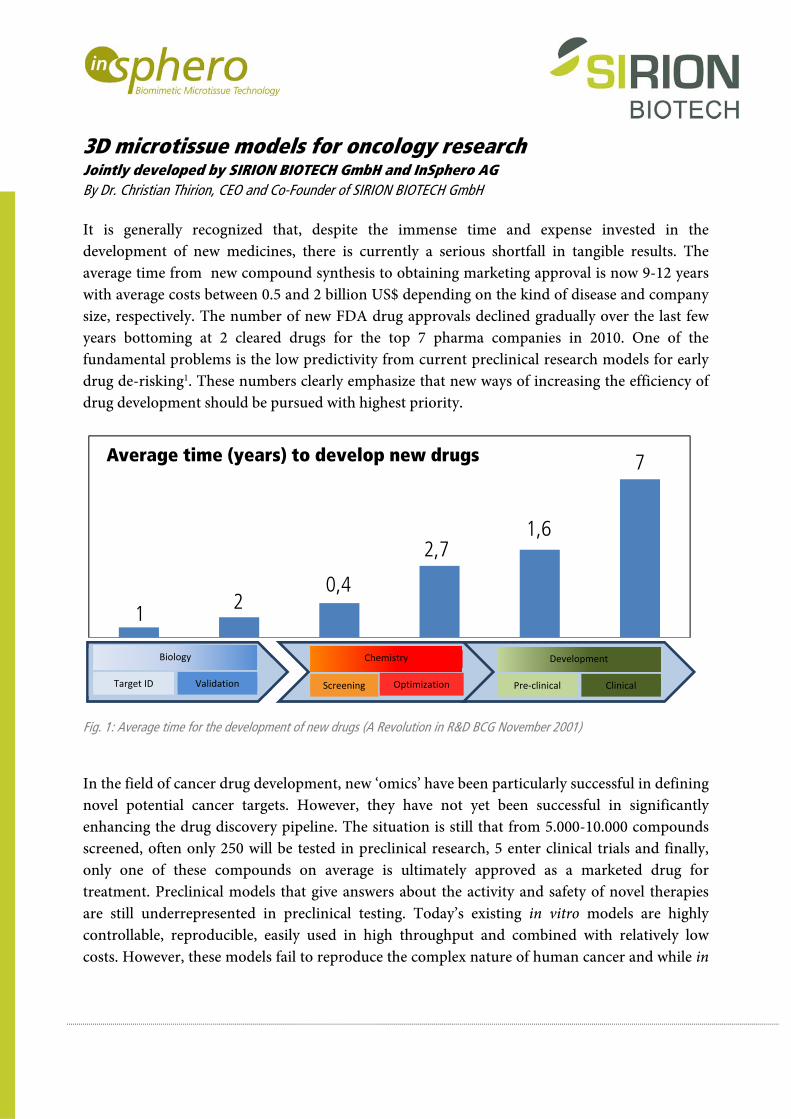
3D microtissue models for oncology research Jointly developed by SIRION BIOTECH GmbH and InSphero AG By Dr. Christian Thirion, CEO and Co-Founder of SIRION BIOTECH GmbH It is generally recognized that, despite the immense time and expense invested in the development of new medicines, there is currently a serious shortfall in tangible results. The average time from new compound synthesis to obtaining marketing approval is now 9-12 years with average costs between 0.5 and 2 billion US$ depending on the kind of disease and company size, respectively. The number of new FDA drug approvals declined gradually over the last few years bottoming at 2 cleared drugs for the top 7 pharma companies in 2010. One of the fundamental problems is the low predictivity from current preclinical research models for early drug de-risking 1 . These numbers clearly emphasize that new ways of increasing the efficiency of drug development should be pursued with highest priority. Fig. 1: Average time for the development of new drugs (A Revolution in R&D BCG November 2001) In the field of cancer drug development, new ‘omics’ have been particularly successful in defining novel potential cancer targets. However, they have not yet been successful in significantly enhancing the drug discovery pipeline. The situation is still that from 5.000-10.000 compounds screened, often only 250 will be tested in preclinical research, 5 enter clinical trials and finally, only one of these compounds on average is ultimately approved as a marketed drug for treatment. Preclinical models that give answers about the activity and safety of novel therapies are still underrepresented in preclinical testing. Today’s existing in vitro models are highly controllable, reproducible, easily used in high throughput and combined with relatively low costs. However, these models fail to reproduce the complex nature of human cancer and while in 1 2 0,4 2,7 1,6 7 Average time (years) to develop new drugs Biology Target ID Validation Chemistry Screening Optimization Development Pre‐clinical Clinical
Transcript
3D microtissue models for oncology research Jointly developed by SIRION BIOTECH GmbH and InSphero AG By Dr. Christian Thirion, CEO and Co-Founder of SIRION BIOTECH GmbH It is generally recognized that, despite the immense time and expense invested in the development of new medicines, there is currently a serious shortfall in tangible results. The average time from new compound synthesis to obtaining marketing approval is now 9-12 years with average costs between 0.5 and 2 billion US$ depending on the kind of disease and company size, respectively. The number of new FDA drug approvals declined gradually over the last few years bottoming at 2 cleared drugs for the top 7 pharma companies in 2010. One of the fundamental problems is the low predictivity from current preclinical research models for early drug de-risking1. These numbers clearly emphasize that new ways of increasing the efficiency of drug development should be pursued with highest priority.
Fig. 1: Average time for the development of new drugs (A Revolution in R&D BCG November 2001)
In the field of cancer drug development, new ‘omics’ have been particularly successful in defining novel potential cancer targets. However, they have not yet been successful in significantly enhancing the drug discovery pipeline. The situation is still that from 5.000-10.000 compounds screened, often only 250 will be tested in preclinical research, 5 enter clinical trials and finally, only one of these compounds on average is ultimately approved as a marketed drug for treatment. Preclinical models that give answers about the activity and safety of novel therapies are still underrepresented in preclinical testing. Today’s existing in vitro models are highly controllable, reproducible, easily used in high throughput and combined with relatively low costs. However, these models fail to reproduce the complex nature of human cancer and while in
1 20,4
2,71,6
7Average time (years) to develop new drugs
Biology
Target ID Validation
Chemistry
Screening Optimization
Development
Pre‐clinical Clinical
vivo models address some of these limitations, the non-human biology, ethical concerns and long experimental duration represent significant disadvantages. The advancement of cell culture technology represented by InSphero’s 3D microtissue technology combined with SIRION BIOTECH’s expertise in genetic cell modification provides a new innovative preclinical approach leading to faster results with far greater in vitro predictive power. 3D tumor microtissues mimic a tumor in vitro and – as an unbeatable advantage – can be derived from primary human origin. Cytology and morphology of tumor microtissues resembles that of natural tumors in humans before neovascularization. Moreover, gene expression of microtissues is much closer to xenografts than monolayer culture. Furthermore, they can be handled easily in high throughput in a cost-effective manner. The combination of 3D cell technology with genetic modification makes this product the perfect tool for functional gene analysis in oncology and cancer drug development. Case study: Target validation of Pim-2 kinase in a hepatic cancer cell HepG2 More than 85 % of the cell lines from the NCI cell panel can be transformed into homotypic 3D microtissues by InSphero using its GravityPLUSTM production process. Microtissues are uniform in size with less than 8% SD and are delivered in a standard 96-well format compatible with automated and manual liquid handling and tissue culture.
Fig. 2: Production of microtissues using a Hamilton Robotics NIMBUS system (source: InSphero AG). The kinase Pim-2 has been shown by two studies to regulate primary hepatic cancer growth and to promote survival2,3. In this case study, HepG2 cells were grown in 2D and the target gene quantitatively silenced by SIRION BIOTECH by means of viral gene transfer prior to 3D microtissue formation. Within one week a significant 42% reduction of the microtissue area compared to the control microtissues transduced with a non-target shRNA was specifically
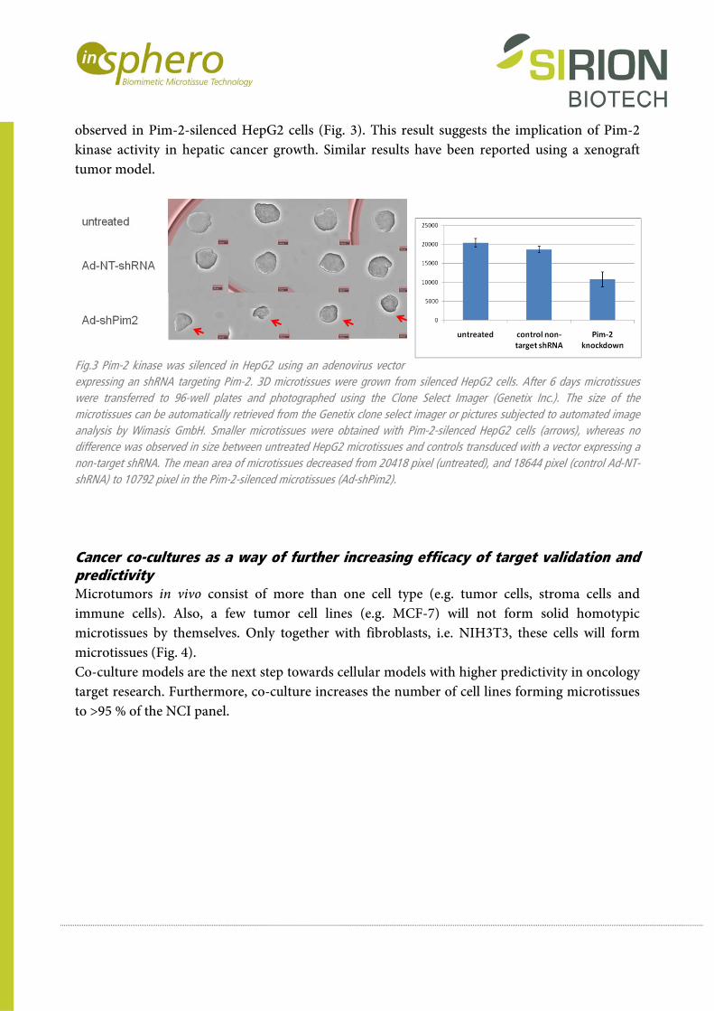
observed in Pim-2-silenced HepG2 cells (Fig. 3). This result suggests the implication of Pim-2 kinase activity in hepatic cancer growth. Similar results have been reported using a xenograft tumor model.
Fig.3 Pim-2 kinase was silenced in HepG2 using an adenovirus vector expressing an shRNA targeting Pim-2. 3D microtissues were grown from silenced HepG2 cells. After 6 days microtissues were transferred to 96-well plates and photographed using the Clone Select Imager (Genetix Inc.). The size of the microtissues can be automatically retrieved from the Genetix clone select imager or pictures subjected to automated image analysis by Wimasis GmbH. Smaller microtissues were obtained with Pim-2-silenced HepG2 cells (arrows), whereas no difference was observed in size between untreated HepG2 microtissues and controls transduced with a vector expressing a non-target shRNA. The mean area of microtissues decreased from 20418 pixel (untreated), and 18644 pixel (control Ad-NT-shRNA) to 10792 pixel in the Pim-2-silenced microtissues (Ad-shPim2).
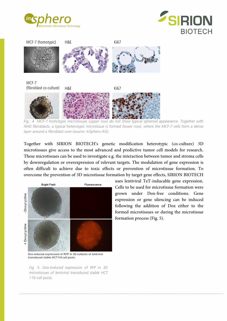
Cancer co-cultures as a way of further increasing efficacy of target validation and predictivity Microtumors in vivo consist of more than one cell type (e.g. tumor cells, stroma cells and immune cells). Also, a few tumor cell lines (e.g. MCF-7) will not form solid homotypic microtissues by themselves. Only together with fibroblasts, i.e. NIH3T3, these cells will form microtissues (Fig. 4). Co-culture models are the next step towards cellular models with higher predictivity in oncology target research. Furthermore, co-culture increases the number of cell lines forming microtissues to >95 % of the NCI panel.
Fig.: 4 MCF-7 homotypic microtissues (upper row) do not show typical spheroid appearance. Together with NHD fibroblasts, a typical heterotypic microtissue is formed (lower row), where the MCF-7 cells form a dense layer around a fibroblast core (source: InSphero AG).
Together with SIRION BIOTECH’s genetic modification heterotypic (co-culture) 3D microtissues give access to the most advanced and predictive tumor cell models for research. These microtissues can be used to investigate e.g. the interaction between tumor and stroma cells by downregulation or overexpression of relevant targets. The modulation of gene expression is often difficult to achieve due to toxic effects or prevention of microtissue formation. To overcome the prevention of 3D microtissue formation by target gene effects, SIRION BIOTECH
uses lentiviral TeT-inducable gene expression. Cells to be used for microtissue formation were grown under Dox-free conditions. Gene expression or gene silencing can be induced following the addition of Dox either to the formed microtissues or during the microtissue formation process (Fig. 5).
Fig. 5: Dox-induced expression of RFP in 3D microtissues of lentiviral transduced stable HCT 116 cell pools.
About InSphero AG InSphero AG is a leading supplier of organotypic, biological in-vitro 3D microtissues for highly predictive drug testing. The company, headquartered in Zurich, Switzerland, currently counts 6 of the top ten global pharmaceutical and cosmetics companies as customers, and is helping them implement the company’s patent-pending microtissue technology in their development work-flow. InSphero’s 3D microtissues are scaffold free, highly reproducible and delivered in an automation-compatible 96-well format to replace conventional 2D cell assays for better biological relevance and predictivity. The 3D cancer microtissues reflect tumor physiology and are used routinely for screening. For toxicology and metabolics applications, rat and human 3D liver microtissues predict even rare cases of idiosyncratic toxicology and remain viable for more than 5 weeks for chronic studies. InSphero’s off-the-shelf portfolio of assay-ready microtissues is complemented by custom-made 3D microtissues with a development time of 4-6 weeks and production turnaround of less than 10 days. InSphero is a spin-off company of the Swiss Federal Institute of Technology (ETH) Zurich and the University Zurich. About SIRION BIOTECH GmbH SIRION BIOTECH GmbH produces genetically modified cells and is a technology provider in the area of viral vector systems. The company was founded in 2005 and is located in the Innovations- und Gründerzentrum Biotechnologie IZB in Martinsried near Munich. The expert in RNAi technology, SIRION BIOTECH offers lead-through service projects in target validation, screening and drug discovery using front running techniques and assay systems. With its strong expertise in mammalian cell culture and viral vector technology SIRION BIOTECH serves as a contract research partner for preclinical R&D, and licenses vector technology for vaccine development. The academic team has more than 20 years experience in mammalian cell culture and viral vector technology. Close cooperation with leading research institutes puts SIRION BIOTECH at the forefront for development of cell systems for pharmaceutical research. SIRION BIOTECH currently operates projects for major pharmaceutical and biotech companies in Europe and USA and has ongoing collaborations with leading academic and governmental research institutes. InSphero AG SIRION BIOTECH GmbH Technoparkstrasse 1 Am Klopferspitz 19 CH-8005 Zurich D-82152 Martinsried Switzerland Germany Phone: +41-44-5150490 Phone: +49-89-7009619913 e-Mail: [email protected] e-Mail: [email protected] www.insphero.com www.sirion-biotech.de 1 Bunnage ME. Getting pharmaceutical R&D back on target. Nat Chem Biol. 2011; 7:335-339. 2 Ren K et al. Pim-2 activates API-5 to inhibit the apoptosis of hepatocellular carcinoma cells through NF-kappaB pathway. Pathol Oncol Res. 2010; 16:229-37. 3 Gong J et al. Serine/threonine kinase Pim-2 promotes liver tumorigenesis induction through mediating survival and preventing apoptosis of liver cell. J Surg Res. 2009; 153:17-22.