FACULTEIT GENEESKUNDE EN GEZONDHEIDSWETENSCHAPPEN DEPARTMENT OF RADIATION ONCOLOGY AND EXPERIMENTAL CANCER RESEARCH Thesis submitted in fulfillment of the requirements for the degree of Doctor in Medical Sciences. WHOLE BREAST IRRADIATION IN THE PRONE POSITION: A PARADIGM SHIFT TOWARDS STANDARD PRACTICE? Thomas Mulliez Promotor: Prof. Dr. Wilfried De Neve Co-promotor: Prof. Dr. Rudy Van den Broecke
Transcript
FACULTEIT GENEESKUNDE EN GEZONDHEIDSWETENSCHAPPEN
Department of raDiation oncology anD experimental cancer research
Thesis submitted in fulfillment of the requirements for the degree of Doctor in Medical Sciences.
Whole Breast irraDiation in the prone position: a paraDigm shift toWarDs stanDarD practice?
Thomas Mulliez
Promotor: Prof. Dr. Wilfried De Neve
Co-promotor: Prof. Dr. Rudy Van den Broecke
Department of raDiation oncology anD experimental cancer re-search
Whole Breast irraDiation in the prone position: a paraDigm shift toWarDs stanDarD practice?
Thomas Mulliez
Promotor: Prof. Dr. Wilfried De Neve
Co-promotor: Prof. Dr. Rudy Van den Broecke
Promotor:
Prof. Dr. Wilfried De Neve Ghent University
Co-promotor:
Prof. Dr. Rudy Van den Broecke Ghent University
Chairman of the examination commission:
Prof. Dr. Johan Vande Walle Ghent University
Examination commission:
Prof. Dr. Anna Kirby Royal Marsden, Sutton, UK
Prof. Dr. Caroline Weltens University of Leuven (KU Leuven)
Prof. Dr. Veronique Cocquyt Ghent University
Prof. Dr. Herman Depypere Ghent University
Prof. Dr. Luc Vakaet Ghent University
Prof. Dr. Gert De Meerleer Ghent University
Dean of the Faculty of Medicine and Health Sciences:
Chapter 3: Publication 1………………………………………………………………………......…….…….………………......………………... .. 25Whole breast radiotherapy in prone and supine position:
Is there a place for multi-beam IMRT?
Chapter 4: Publication 2………………………………………………………………………......…….…….………………......………………... .. 38Improved cone-beam computed tomography in supine and prone breast
radiotherapy: surface reconstruction, radiation exposure and clinical workflow.
Chapter 5: Publication 3………………………………………………………………………......…….…….………………......………………... .. 50Hypofractionated whole breast irradiation for patients with large breasts:
a randomized trial comparing prone and supine positions.
Chapter 6: Publication 4………………………………………………………………………......…….…….………………......………………... .. 63Heart dose reduction by prone deep inspiration breath hold in left-sided
breast irradiation.
Chapter 7: Publication 5………………………………………………………………………......…….…….………………......………………... .. 77Reproducibility of prone deep inspiration breath hold for left-sided
8.1.3.3 The cardiac dilemma.…. .............................................................................. 93
8.1.4 Limitations of prone WBI…………………………………………………………………… ................ 95
8.2 Current and future research ................................................................................................................................96
8.2.1 Use of prone DIBH in a simultaneous integrated vs. sequential
CBCT Cone beam computed tomographyCMSE Clinique et maternité Sainte-Elisabeth
CT Computed tomographyCTCAEv3 Common terminology criteria for adverse events version 3.0
CTDIw Weighted computed tomography dose index CTVWBI Clinical target volume for whole breast irradiation
DIBH Deep inspiration breath holdDmax Maximum dose
Dmean Mean doseDmin Minimum doseDVH Dose-volume histogramFDK Feldkamp–Davis–Kress FOV Field of viewGUH Ghent university hospital
Gy GrayI Instability
ICRU International commission on radiation units & measurements IMRT Intensity modulated radiotherapyLAD Left anterior descending coronary artery
M Population systematic setup error MB Multi-beam
OARs Organs at riskPD Prescription dose
PTVoptim Planning target volume for optimizationPTVWBI Planning target volume for whole breast irradiation
QOL Quality of lifeRCT Randomized controlled trial
RT RadiotherapySB Shallow or normal breathing
SEER Surveillance, epidemiology and end resultsSeqB Sequential boost
SG Study groupSIB Simultaneous integrated boost
TDP Topographical dose painting TF Tangential fieldTT Treatment time
U-BH Unilateral breast holderV105/107 Volumes receiving ⩾105% and ⩾107% of the PD, respectivelyV5/10/20 Partial volumes receiving ⩾5Gy, ⩾10Gy and ⩾20Gy, respectively
VG Validation groupW-TF Wedged tangential fieldsWBI Whole breast irradiation
Σ Population standard deviation of the systematic setup error σ Population random setup error
A B B R E V I AT I O N S
4
SU M M A RY
SUMMARY
Breast cancer is the most frequently diagnosed cancer and the leading cause of cancer death among females. Radiotherapy (RT) halves the recurrence risk and reduces the risk of breast cancer death with nearly 20% after breast conserving surgery. However, evidence accumulates that breast RT is associated with major, even lethal side effects like cardiac disease and cancer induction, especially to lungs and heterolateral breast. The purpose of this thesis is to optimize the treatment technique for whole breast irradiation (WBI) with the aim to reduce irradiation of organs at risk (OARs) while providing an adequate and homogeneous dose to the target. Attention was given to feasibility of new techniques and portability to other centres.
In standard supine position, the breast spreads over the thoracic wall enwrapping heart and lungs leading to suboptimal anatomy to achieve a homogeneous dose to the breast and to reduce dose to the OARs. Prone position provides advantages; it elongates the breast away from the intra-thoracic region, narrows the breast and stretches skin folds. In the first part of this thesis, our aim was optimizing radiation techniques for both positions. Multi-beam intensity modulated RT was the best technique in supine position to provide a conformal dose to the target while avoiding heart and lungs. Differences between various techniques were less pronounced in prone position. Overall, prone positioning yielded better results: better dose conformity to the target, superior lung sparing and improved heart dose metrics in all patients with large breasts (chapter 3). Our further research focused on the validation of prone techniques with special attention to patients who required left-sided WBI.
The second part of this thesis involves the comparative clinical assessment of the best prone and supine technique. Of concern was the daily reproducibility of the CT-simulat-ed position during treatment. Daily cone beam CT (CBCT) was the desired technique but the existing CBCT parameters weren’t optimized for breast RT. Therefore, we optimized the CBCT acquisition parameters to improve surface reconstruction, decrease radiation exposure and enhance clinical practicality (chapter 4).
A comparative assessment was performed using a randomized controlled trial comparing prone and supine positions in 100 large breasted patients. The trial confirmed earlier dosimetric studies. Prone WBI was able to improve target dose distribution, heart and lung sparing. Prone position was associated with significantly less acute toxicity. Moist desqua-mation was reduced from 20% in supine to 6% in prone position. Dermatitis, edema and pain were less frequent and less severe in the prone treated cohort. This study provides level-I evidence of replacing supine by prone WBI for large breasted patients (chapter 5).
5
SU M M A RY
In the third part of the thesis, we focused on optimizing heart dose metrics in the prone po-sition. First, we investigated the effects of deep inspiration breath hold (DIBH), a technique successfully used in supine position, to lower heart dose in prone left-sided WBI. In silico assessment showed that prone DIBH reduced heart dose while maintaining the benefits of prone position on lung sparing (chapter 6). The reproducibility of prone DIBH was studied in 30 patients. CT-simulation and respiratory monitoring data were analysed, all pointing to the feasibility and good reproducibility of the DIBH manoeuvre in prone position (chapter 7).
This thesis confirms the benefits of prone positioning on lung sparing and target dose distri-bution. Moreover, prone WBI results in a strong reduction of acute toxicity in large breasted patients. We demonstrated the feasibility of prone DIBH enabling to minimize heart dose metrics while preserving the advantages of prone positioning. Further refinement of prone DIBH is ongoing research. This thesis strengthens the evidence towards a paradigm shift, to replace the standard supine by prone WBI.
6
SA M E N VAT T I NG
SAMENVATTING
Borstkanker is de meest gediagnosticeerde kanker en de belangrijkste oorzaak van kanker- sterfte bij vrouwen. Radiotherapie (RT) na borstsparende heelkunde halveert de kans op herval en vermindert het risico op borstkankersterfte met ongeveer 20%. Toch neemt het bewijs toe dat borstbestraling wordt geassocieerd met belangrijke, zelfs lethale bijwer-kingen. Borstbestraling is gerelateerd met hartziekte en kanker inductie, voornamelijk ter hoogte van de longen en heterolaterale borst. Het doel van dit proefschrift is om de be-stralingstechniek te optimaliseren voor volledige borstbestraling door de risico-organen maximaal uit te sparen en een adequate homogene dosis te geven aan de volledige borst. Dit met speciale aandacht voor de klinische toepasbaarheid van nieuwe technieken en overdraagbaarheid naar andere centra.
In de standaard ruglig positie spreidt de borst uit over de thoraxwand waarbij het hart en de longen worden omhuld. Dit leidt tot een suboptimale anatomie om een homogene dosis voor de borst te verkrijgen en om de dosis op de risico-organen te beperken. Buiklig biedt een aantal voordelen; het elongeert de borst weg van de intra-thoracale regio en het ver-nauwt de borst terwijl de huidplooien verdwijnen. In het eerste deel van dit proefschrift is het ons doel om de bestralingstechniek in beide posities te optimaliseren. In ruglig bleek de meerdere bundels intensiteit gemoduleerde RT de beste techniek om een conforme dosis te verkrijgen op de borst en om hart en longen uit te sparen. In buiklig bleek het effect van behandelingstechniek veel minder uitgesproken. Doch buiklig resulteerde in betere resultaten: een betere conformiteit van de bestraalde borst, superieure longdosis-sen en een betere hartsparing voor alle patiënten met volumineuze borsten (hoofdstuk 3). Ons verder onderzoek focuste op het valideren van buiklig borstbestraling met aandacht voor linkszijdigen.
Het tweede deel van dit proefschrift betreft een vergelijkende klinische beoordeling van de beste buik- en ruglig techniek. Van belang was de dagelijkse reproduceerbaarheid van de CT-gesimuleerde positie tijdens de behandeling. Dagelijkse cone beam CT (CBCT) was de gewenste techniek, doch de bestaande CBCT parameters werden niet geoptimaliseerd voor borstbestraling. Daarom werden de CBCT parameters aangepast om de oppervlakte- reconstructie en klinische toepasbaarheid te verbeteren en de stralingsbelasting te ver-minderen (hoofdstuk 4).
Een gerandomiseerde gecontroleerde studie werd uitgevoerd waarin buik- met ruglig werd vergeleken bij 100 patiënten met volumineuze borsten. De studie bevestigt eerdere dosi-metrische resultaten. Buiklig kon de dosis distributie binnen de borst en uitsparen van hart
7
SA M E N VAT T I NG
en longen verbeteren. Buiklig is tevens geassocieerd met significant minder acute toxiciteit. Vochtige desquamatie werd teruggebracht van 20% in ruglig tot 6% in de buiklig cohorte. Dermatitis, oedeem en pijn waren minder frequent en minder ernstig in de buiklig groep. Deze studie geeft niveau-I bewijs voor het vervangen van ruglig door buiklig bij patiënten met volumineuze borsten (hoofdstuk 5).
In het derde deel van dit proefschrift hebben we ons gericht op het optimaliseren van de hartdosis in buiklig. Eerst hebben we onderzoek gedaan naar de effecten van diepe adem-halingsblokkage, een techniek die met succes gebruikt wordt in ruglig, om de hartdosis te verlagen voor linkszijdige bestraling in buiklig. In silico analyse toonde aan dat diepe ademhalingsblokkage in buiklig de hartdosis beperkte met behoud van de voordelen van buiklig op longsparing (hoofdstuk 6). De reproduceerbaarheid van deze techniek werd bestudeerd in 30 patiënten. CT-simulatie en ademhalingsgegevens werden geanalyseerd, allen wijzend op de haalbaarheid en de goede reproduceerbaarheid van diepe adem- halingsblokkage in buiklig (hoofdstuk 7).
Dit proefschrift bevestigt de voordelen van buiklig op long sparing en dosisverdeling binnen de borst. Bovendien zorgt het voor een sterke reductie van de acute toxiciteit bij patiën-ten met volumineuze borsten. Tevens toonden we de klinische uitvoerbaarheid van diepe ademhalingsblokkage in buiklig aan; een techniek waarbij hartdosis wordt geminimaliseerd met behoud van de voordelen van buiklig. Verder onderzoek is lopende ter optimalisatie van deze nieuwe techniek. Dit proefschrift versterkt het bewijs voor een paradigmaver-schuiving van de standaard ruglig naar buiklig voor volledige borstbestraling.
8
§ I . BAC KGROU N D
CHAPTER 1: BACKGROUND
1.1 ROLE OF RADIOTHERAPY IN BREAST CANCER
Breast cancer is the most frequently diagnosed cancer and the leading cause of cancer death among females, accounting for 23% of total cancer cases and 14% of cancer deaths [1]. In 2010, 9908 Belgian women were newly diagnosed with breast cancer, which is ap-proximately one third of newly diagnosed cancers in females [2]. For early stage breast can-cer patients, breast-conserving surgery (BCS) followed by adjuvant whole breast irradiation (WBI) has replaced mastectomy as standard of care, since both treatments have shown to be equivalent in several randomized controlled trials [3-6]. The advantages of BCS include less invasive surgery with shorter recovery time and breast preservation, which is important for a women’s self image. However microscopic tumor foci might remain in the treated breast leading to locoregional recurrences and/or distant metastases. The role of adjuvant radiotherapy (RT) after breast sparing surgery is to eradicate these cancer deposits.
The need of radiotherapy (RT) after BCS was clearly demonstrated by the “Early Breast Cancer Trialists’ Collaborative Group” [7, 8]. In the meta-analysis of 2005 [7] the 5-year risk of local recurrence was 7% among those allocated for RT versus 26% in the non-RT group, corresponding to an absolute risk reduction of 19%. An update in 2011 [8] based on 10081 patients showed that RT nearly halves the 10-year recurrence risk (absolute risk reduction = 16%), reduces the 15-year risk of breast cancer death with almost 20% (abso-lute risk reduction = 4%) leading to a 15-year absolute overall survival benefit of 3%. For every four recurrences avoided by year 10, about one breast cancer death was avoided by year 15. This meta-analysis is based on patients irradiated between 1976 and 1999 and it is not clear whether these benefits can be translated to patients treated in more recent years with more contemporary systemic therapies. However, a recent retrospective trial performed by Wockel et al. [9] confirms the benefit of guideline-adherent adjuvant RT in patients treated between 1992 and 2008.
These data provide level I evidence of adjuvant RT after BCS. Still WBI is associated with severe acute and late side effects to the treated breast and organs at risk (OARs).
1.2 RADIOTHERAPY INDUCED SIDE EFFECTS:
Breast RT was considered harmless; but evidence accumulates in recent years that RT correlates with severe, even lethal side effects. Long-term epidemiological data revealed that patients who received RT had an increased risk of non-breast cancer death especially
9
§ I . BAC KGROU N D
due to cardiac events and secondary cancer induction [10-21]. Due to the increased awareness of these major complications of breast RT, research on prevention of radia-tion-induced side effects has been intense and several entities including radiation tech-niques, respiration-related RT and position alterations have been explored. In this chapter, examples of radiation-induced side effects are reported per organ (A) as well as possible strategies to reduce/prevent these effects (B).
1.2.1 Ipsilateral breast
A) Impaired cosmesis, fibrosis and skin changes
Severe acute breast toxicity is reported in 40-50% of patients using standard techniques in supine position. Skin desquamation, dermatitis, edema, pruritus and pain to the treated breast are often reported during or shortly after breast RT [22-32]. These side effects have been shown to negatively influence physical, emotional and functional well-being, body image and treatment satisfaction, moreover to negatively affect quality of life (QOL) [27, 30].
Prolonged follow-up demonstrated RT-induced cosmetic alterations to the treated breast including breast fibrosis, skin atrophy, telangiectasia and pigmentation changes. Using photographic assessments, Donovan et al. [26] reported changes in breast appearance 5 years after irradiation in 40-58% of patients depending on the used radiotherapy tech-nique. Hopwood et al. [31] published that 4 out of 10 women report moderate to marked changes of the treated breast 5 years after completing their RT treatment using non- intensity modulated techniques.
Acute and late breast toxicity has been related with target dose distribution parameters. Dose heterogeneity and the presence of high dose regions (hot spots) within the treated breast have been associated with breast toxicity [25-29]. Therefore it is warranted to obtain an optimal dose distribution covering the target volume while maximizing dose homogeneity.
B) Radiation techniques and position alterations
The ability of traditionally used wedged tangential fields (W-TF) to provide a homogenous dose distribution is rather restricted due to the complex shape of the breast in supine position. Tangential field intensity modulated RT (TF-IMRT) has been introduced to improve target dose distribution and has translated in reduced breast toxicity as shown in random-ized controlled trials [26-29]. Pignol et al. [27] performed a randomized multicenter, double- blind trial involving 358 patients treated either by IMRT or by W-TF. Moist desquamation occurred in 47.8% of the standard treatment group compared to 31.2% in the IMRT group;
10
§ I . BAC KGROU N D
resulting in a significant (p=0.002) absolute reduction of 16.6%. The presence of moist desquamation was associated with pain and a decreased QOL. Donovan et al. [26] used photographs to assess cosmesis and observed that W-TF are 1.7 times more likely to cause a change in breast appearance 5 year after breast irradiation compared to IMRT. A recent trial of Mukesh et al. [28] confirmed the superiority of IMRT techniques in terms of telangiectasia and cosmesis compared to conventional techniques.
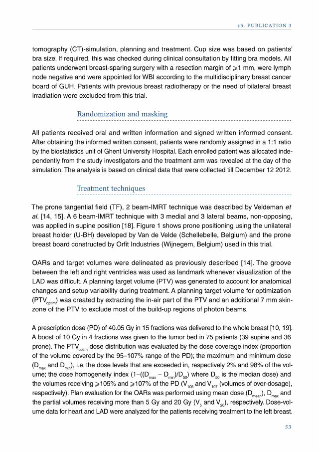
The width and shape of the supine breast makes it difficult to obtain a homogeneous dose distribution in the target. Furthermore, skin folds like the axillary and inframammary fold prevent the skin-protective build-up region of photon beams. As shown in figure 1.1, prone position provides some theoretical advantages due to the gravity-induced anatomical changes: (1) it elongates the breast away from the intra-thoracic region and is therefore able to reduce intra-thoracic irradiation; (2) it narrows the breast enabling to reduce the radiological pathlenghts traversing the breast and (3) it opens the skin folds and is there-fore able to restore build-up effect in this region [33-35].
Figure 1.1: Anatomical modification by executing prone (B) compared to supine (A) position. Prone position elon-gates the breast away from the intra-thoracic region (marked with solid arrow), it narrows the breast and unfolds the skin folds (marked with dotted arrow).
1.2.2 Heterolateral breast
� A) Carcinogenesis
After breast irradiation, an excess of 1.8% in contralateral breast cancer incidence has been reported over a period of 15 years in a meta-analysis of RT vs. non-RT trials for patients treated from the seventies till the nineties [15]. The risk of developing a second primary con-tralateral breast cancer after breast RT is related to patient age, family history, obesity, alco-hol, smoking, heterolateral breast dose, hormonal therapy and systemic therapy [15, 36-38].
A) Supine position. B) Prone position.
11
§ I . BAC KGROU N D
� B) Radiation techniques
Current gantry beam angles are chosen in order to completely avoid heterolateral breast dose, though avoidance of the heterolateral breast might be at cost of an increased intra-thoracic dose.
1.2.3 Lungs
� A) Carcinogenesis, inflammation and functional changes
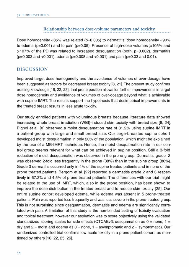
Breast irradiation has been associated with acute, subacute and chronic side effects to the ipsilateral lung including pneumonitis and lung fibrosis. These iatrogenic effects are related to irradiated lung volume, lung dose, systemic therapy, chronic obstructive pulmo-nary disease, smoking and age [39-43]. Figure 1.2 shows a RT-induced pneumonitis 3 months after treatment and its relation to the dose distribution on the treatment plan.
Figure 1.2: Radiation-induced pneumonitis (B) after breast irradiation and its relation to the dose distribution (A).
Patients irradiated in older breast cancer trials (treated between 1973 and 2008) showed an increased risk of mortality from lung cancer at the side of the irradiated breast with a rate ratio of 1.30 [10]. In the meta-analysis [15] published by the “Early Breast Cancer Trialist’s Collaborative Group; 3666 non-breast cancer deaths were reported, 156 died of lung cancer with a rate ratio of 1.78 for irradiated patients. The physiopathology or the dose/volume relationship is not clear; though the available data suggest a linear association between dose and incidence of lung cancer without a threshold dose and no evidence of a downturn risk [44]. In a recent trial by Grantzua et al. [45] the risk rate of lung cancer increased linearly with 8.5% per Gy for non-smoking and 17.3% per Gy for smoking
A) Patient treatment plan. The colored lines (isodoses) indicate the different dose levels in Gy.
B) Radiation induced pneumonitis (indicated with red arrow) 3 months after RT.
12
§ I . BAC KGROU N D
patients. Still the absolute amount of lung cancer patients after breast RT remains low, in this cohort of 23627 early breast cancer patients treated from 1982 till 2007 only 187 cases were documented. Still vigilance is required since odds ratios of mortality from radiation induced lung cancer (ipsilateral versus contralateral) increase with time from treatment [10].
� B) Radiation techniques and position alterations
Multi-beam IMRT (MB-IMRT), tomotherapy and arc therapies are able to reduce high dose regions within the lung, though often at the expense of an increased low dose spread to heart, lungs and contralateral breast [46-51].
Prone as compared to supine position elongates the breast away frow the intra-thoracic region (as shown in figure 1.1) resulting in a spectacular decrease in lung dose. In the New York University trial [52], prone position was associated with a reduction of the in-field lung volume of 86% for right-sided patients and 91% for left-sided patients. This spectacular decrease in lung dose was consistently reported in trials in which individual comparative prone versus supine planning was made [35, 52-58].
1.2.4 Heart
� A) Cardiac disease
Heart disease after breast irradiation is well documented and of major concern [10-16, 20]. Cardiac disease by radiation injury is caused by an interplay of inflammation, fibrosis and atherosclerosis especially due to micro- and macrovascular damage. Different types of heart disease by RT have been identified: (1) Myocardial infarction is a consequence of coronary artery sclerosis. (2) Congestive heart failure is mainly related with microvascular radiation injury causing interstitial myocardial fibrosis. (3) Valvular disease is also related with fibrotic changes. (4) Pericarditis is a result of an exudative inflammatory response. (5) Arrhythmias or conduction defects are related with ischemia or fibrosis to the sinus, the AV node or to the conduction system [20, 21, 59]. The trial of McGale et al. [14] involv-ing 34825 women irradiated during 1976-2006 demonstrated increased left versus right- sided incidence ratios of 1.22 for acute myocardial infarction, 1.25 for angor, 1.54 for val-vular heart disease and 1.61 for pericarditis. US Surveillance, Epidemiology and End Results (SEER) data [10] showed that cardiac mortality increases over time, moreover cardiac mortality ratios of left versus right-sided breast cancer patients irradiated during 1973-1982 are 1.19 at <10 years, 1.35 at 10-14 years, 1.64 at 15-19 years and 1.90 at >20 years; cardiac mortality was observed in 3117 of the 130285 left-sided compared to 2743 of the 126691 right-sided irradiated patients. In unirradiated patients, the cardiac mortality risk was equal for left- and right-sided breast cancer.
13
§ I . BAC KGROU N D
The clinical dose/volume-effect relationship for radiation induced cardiac mortality is not entirely understood. It is most probably an interaction between dose and irradiated cardiac volume. Darby et al. [16] observed a linear relationship between mean heart dose and relative risk of ischemic heart disease starting within a few years and continuing decades after breast RT. Rates of major coronary events increased with 7.4% per Gy mean heart dose, with no apparent threshold dose. The left anterior descending coronary artery (LAD) is especially exposed by left-sided irradiation. Dose to the LAD is associated with coronary disease, leading to excess radiation-induced mortality after prolonged follow-up [14, 60, 61].
Data of more recently treated patients [10, 13, 62, 63] show a trend of a decrease of car-diovascular disease, which can probably be attributed to adjusted treatment techniques, better positioning verification and modified target volume definitions. Still, it is well docu-mented that modern treatment techniques cause cardiac injury [63-66] and recent data must be interpreted with caution because odds ratios of radiation-related cardiac mortality still increase after 20 years of follow-up [10].
� B) Radiation techniques, position alterations
and respiration-related RT
The ability of TF-IMRT compared to conventional techniques to reduce intra-thoracic irradi-ation is rather limited. MB-IMRT, intensity modulated arc therapy and helical tomotherapy can reduce high dose regions to heart though often at cost of low or intermediate dose spread [46-51].
Prone position provides gravity-induced anatomical changes (cfr figure 1.1). It elongates the ipsilateral breast away from the intra-thoracic region, but it induces also an anterior shift of the heart; which could be detrimental for prone left-sided breast irradiation [67, 68]. Formenti et al. [52] scanned 400 patients in prone and supine position and made an indivi- dual dosimetric comparison. Prone position was able to reduce the in-field heart volume in 85% of patients with significantly better LAD-sparing. Formenti et al. [52], Lymberis et al. [54] and Kirby et al. [53] correlated the benefit of prone position to breast-volume. Reduced heart doses are observed in all large-breasted patients treated in prone position, while for small breasted patients results are indistinct.
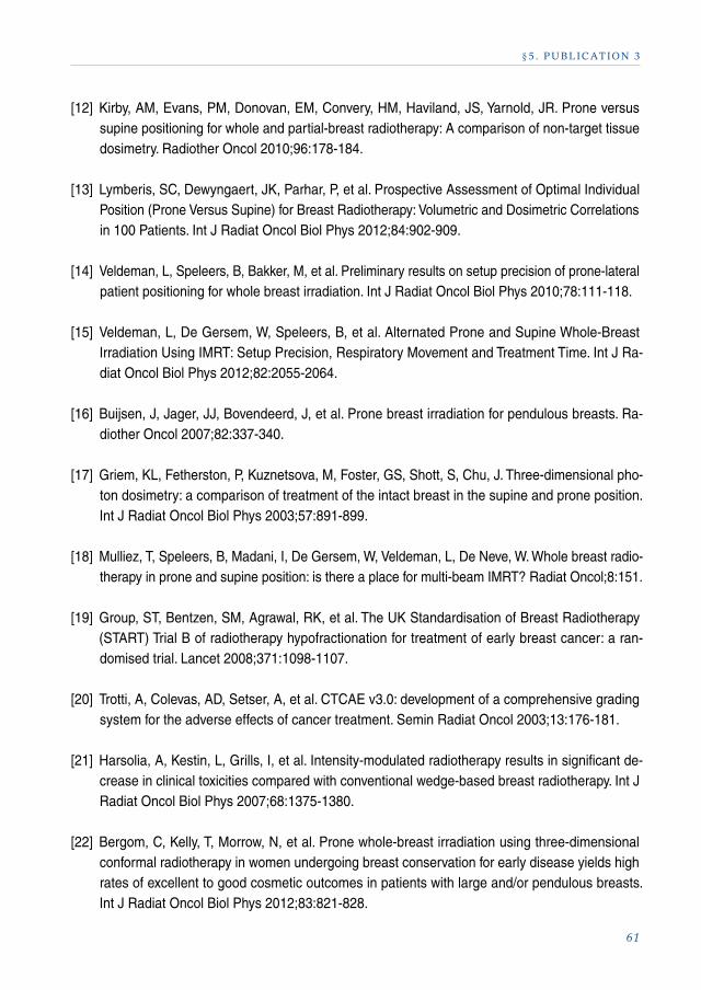
Inspiration causes an increased distance between the heart/LAD and the irradiated anterior chest wall/breast region. In left-sided breast cancer patients these anatomical modifica-tions are used to decrease heart and LAD dose by respiration-related methods. Voluntary deep inspiration breath hold (DIBH) is irradiation while the patient is in a deep inspiratory apnea phase and is performed in several cycles during treatment in supine position [69-74]. Figure 1.3 demonstrates the increased breast-heart distance by performing DIBH
14
§ I . BAC KGROU N D
compared to normal or shallow breathing (SB). A planning comparison by Swanson et al. [73] in 87 left-sided breast cancer patients found a relative mean heart dose reduction of 40% and a relative mean lung dose reduction of 13% with DIBH compared to SB in supine position. A more recent study of Nissen et al. [74] comparing 144 left-sided pa-tients treated in DIBH with 83 patients in SB demonstrated a mean heart dose reduction of 48% in the DIBH cohort.
It might be difficult to evaluate the benefit of modern treatment techniques on cardiac mor-bidity or death since early stage breast cancer patients are long-term survivors and radia-tion induced cardiac disease might occur several decades after breast RT [10]. Therefore it is warranted to have short-term surrogates of cardiac toxicity. Several methods have been developed including the strain rate imaging technique developed by the Leuven group [66] or single-photon emission CT scans [75].
Figure 1.3: Increased breast-heart distance by deep inspiration breath hold (B) compared to shallow breathing (A) in the supine position, based on the transversal slide of the nipple (red arrow).
B) Deep inspiration breath hold.A) Shallow breathing.
15
§ I . BAC KGROU N D
1.3 REFERENCES
[1] Jemal, A, Bray, F, Center, MM, Ferlay, J, Ward, E, Forman, D. Global cancer statistics.
and lung cancer more than 20 years after radiotherapy for breast cancer. Br J Cancer
2013;108:179-182.
16
§ I . BAC KGROU N D
[11] Darby, SC, McGale, P, Taylor, CW, Peto, R. Long-term mortality from heart disease and lung
cancer after radiotherapy for early breast cancer: prospective cohort study of about 300,000
women in US SEER cancer registries. Lancet Oncol 2005;6:557-565.
[12] Cuzick, J, Stewart, H, Rutqvist, L, et al. Cause-specific mortality in long-term survivors of breast
cancer who participated in trials of radiotherapy. J Clin Oncol 1994;12:447-453.
[13] Giordano, SH, Kuo, YF, Freeman, JL, Buchholz, TA, Hortobagyi, GN, Goodwin, JS. Risk of car-
diac death after adjuvant radiotherapy for breast cancer. J Natl Cancer Inst 2005;97:419-424.
[14] McGale, P, Darby, SC, Hall, P, et al. Incidence of heart disease in 35,000 women treated with
radiotherapy for breast cancer in Denmark and Sweden. Radiother Oncol 2011;100:167-175.
[15] Clarke, M, Collins, R, Darby, S, et al. Effects of radiotherapy and of differences in the extent of
surgery for early breast cancer on local recurrence and 15-year survival: an overview of the
randomised trials. Lancet 2005;366:2087-2106.
[16] Darby, SC, Ewertz, M, McGale, P, et al. Risk of ischemic heart disease in women after radio-
therapy for breast cancer. N Engl J Med 2013;368:987-998.
[17] Lorigan, P, Califano, R, Faivre-Finn, C, Howell, A, Thatcher, N. Lung cancer after treatment for
breast cancer. Lancet Oncol 2010;11:1184-1192.
[18] Schaapveld, M, Visser, O, Louwman, MJ, et al. Risk of new primary nonbreast cancers after
breast cancer treatment: a Dutch population-based study. J Clin Oncol 2008;26:1239-1246.
[19] Wickberg, A, Holmberg, L, Adami, HO, Magnuson, A, Villman, K, Liljegren, G. Sector resection
with or without postoperative radiotherapy for stage I breast cancer: 20-year results of a rand-
omized trial. J Clin Oncol 2014;32:791-797.
[20] Shah, C, Badiyan, S, Berry, S, et al. Cardiac dose sparing and avoidance techniques in breast
cancer radiotherapy. Radiother Oncol 2014.
[21] Duma, MN, Molls, M, Trott, KR. From heart to heart for breast cancer patients - cardiovascular
toxicities in breast cancer radiotherapy. Strahlenther Onkol 2014;190:5-7.
[22] Ambrosone, CB, Tian, C, Ahn, J, et al. Genetic predictors of acute toxicities related to radia-
tion therapy following lumpectomy for breast cancer: a case-series study. Breast Cancer Res
2006;8:R40.
17
§ I . BAC KGROU N D
[23] Turesson, I, Nyman, J, Holmberg, E, Oden, A. Prognostic factors for acute and late skin reac-tions in radiotherapy patients. Int J Radiat Oncol Biol Phys 1996;36:1065-1075.
[24] Group, ST, Bentzen, SM, Agrawal, RK, et al. The UK Standardisation of Breast Radiotherapy (START) Trial B of radiotherapy hypofractionation for treatment of early breast cancer: a ran-domised trial. Lancet 2008;371:1098-1107.
[25] Harsolia, A, Kestin, L, Grills, I, et al. Intensity-modulated radiotherapy results in significant decrease in clinical toxicities compared with conventional wedge-based breast radiotherapy. Int J Radiat Oncol Biol Phys 2007;68:1375-1380.
[26] Donovan, E, Bleakley, N, Denholm, E, et al. Randomised trial of standard 2D radiotherapy (RT) versus intensity modulated radiotherapy (IMRT) in patients prescribed breast radiotherapy. Radiother Oncol 2007;82:254-264.
[27] Pignol, JP, Olivotto, I, Rakovitch, E, et al. A multicenter randomized trial of breast intensity-mod-ulated radiation therapy to reduce acute radiation dermatitis. J Clin Oncol 2008;26:2085-2092.
[28] Mukesh, MB, Barnett, GC, Wilkinson, JS, et al. Randomized Controlled Trial of Intensity-Modu-lated Radiotherapy for Early Breast Cancer: 5-Year Results Confirm Superior Overall Cosmesis. J Clin Oncol 2013;31:4488-4495.
[29] Veldeman, L, Madani, I, Hulstaert, F, De Meerleer, G, Mareel, M, De Neve, W. Evidence behind use of intensity-modulated radiotherapy: a systematic review of comparative clinical studies. Lancet Oncol 2008;9:367-375.
[30] Schnur, JB, Ouellette, SC, Dilorenzo, TA, Green, S, Montgomery, GH. A qualitative analysis of acute skin toxicity among breast cancer radiotherapy patients. Psychooncology 2010;20:260-268.
[31] Hopwood, P, Haviland, JS, Sumo, G, et al. Comparison of patient-reported breast, arm, and shoulder symptoms and body image after radiotherapy for early breast cancer: 5-year follow- up in the randomised Standardisation of Breast Radiotherapy (START) trials. Lancet Oncol 2010;11:231-240.
[32] Tortorelli, G, Di Murro, L, Barbarino, R, et al. Standard or hypofractionated radiotherapy in the postoperative treatment of breast cancer: a retrospective analysis of acute skin toxicity and dose inhomogeneities. BMC Cancer 2013;13:230.
[33] Bergom, C, Kelly, T, Morrow, N, et al. Prone whole-breast irradiation using three-dimensional conformal radiotherapy in women undergoing breast conservation for early disease yields high rates of excellent to good cosmetic outcomes in patients with large and/or pendulous breasts. Int J Radiat Oncol Biol Phys 2012;83:821-828.
18
§ I . BAC KGROU N D
[34] Merchant, TE, McCormick, B. Prone position breast irradiation. Int J Radiat Oncol Biol Phys
1994;30:197-203.
[35] Buijsen, J, Jager, JJ, Bovendeerd, J, et al. Prone breast irradiation for pendulous breasts.
Radiother Oncol 2007;82:337-340.
[36] Stovall, M, Smith, SA, Langholz, BM, et al. Dose to the contralateral breast from radiotherapy
and risk of second primary breast cancer in the WECARE study. Int J Radiat Oncol Biol Phys
2008;72:1021-1030.
[37] Hooning, MJ, Aleman, BM, Hauptmann, M, et al. Roles of radiotherapy and chemotherapy in
the development of contralateral breast cancer. J Clin Oncol 2008;26:5561-5568.
[40] Wennberg, B, Gagliardi, G, Sundbom, L, Svane, G, Lind, P. Early response of lung in breast
cancer irradiation: radiologic density changes measured by CT and symptomatic radiation
pneumonitis. Int J Radiat Oncol Biol Phys 2002;52:1196-1206.
[41] Blom Goldman, U, Wennberg, B, Svane, G, Bylund, H, Lind, P. Reduction of radiation pneu-
monitis by V20-constraints in breast cancer. Radiat Oncol 2010;5:99.
[42] Kahan, Z, Csenki, M, Varga, Z, et al. The risk of early and late lung sequelae after conformal
radiotherapy in breast cancer patients. Int J Radiat Oncol Biol Phys 2007;68:673-681.
[43] Erven, K, Weltens, C, Nackaerts, K, Fieuws, S, Decramer, M, Lievens, Y. Changes in pulmo-
nary function up to 10 years after locoregional breast irradiation. Int J Radiat Oncol Biol Phys
2012;82:701-707.
[44] Berrington de Gonzalez, A, Gilbert, E, Curtis, R, et al. Second solid cancers after radiation
therapy: a systematic review of the epidemiologic studies of the radiation dose-response
relationship. Int J Radiat Oncol Biol Phys 2013;86:224-233.
19
§ I . BAC KGROU N D
[45] Grantzau, T, Thomsen, MS, Vaeth, M, Overgaard, J. Risk of second primary lung cancer in women after radiotherapy for breast cancer. Radiother Oncol 2014.
[46] Fogliata, A, Clivio, A, Nicolini, G, Vanetti, E, Cozzi, L. A treatment planning study using non- coplanar static fields and coplanar arcs for whole breast radiotherapy of patients with concave geometry. Radiother Oncol 2007;85:346-354.
[47] Lohr, F, El-Haddad, M, Dobler, B, et al. Potential effect of robust and simple IMRT approach for left-sided breast cancer on cardiac mortality. Int J Radiat Oncol Biol Phys 2009;74:73-80.
[48] Schubert, LK, Gondi, V, Sengbusch, E, et al. Dosimetric comparison of left-sided whole breast irradiation with 3DCRT, forward-planned IMRT, inverse-planned IMRT, helical tomotherapy, and topotherapy. Radiother Oncol 2011;100:241-246.
[49] Fong, A, Bromley, R, Beat, M, Vien, D, Dineley, J, Morgan, G. Dosimetric comparison of intensity modulated radiotherapy techniques and standard wedged tangents for whole breast radiotherapy. J Med Imaging Radiat Oncol 2009;53:92-99.
[50] Abo-Madyan, Y, Aziz, MH, Aly, MM, et al. Second cancer risk after 3D-CRT, IMRT and VMAT for breast cancer. Radiother Oncol 2014;110:471-476.
[51] Coon, AB, Dickler, A, Kirk, MC, et al. Tomotherapy and Multifield Intensity-Modulated Radio-therapy Planning Reduce Cardiac Doses in Left-Sided Breast Cancer Patients with Unfavorable Cardiac Anatomy. Int J Radiat Oncol Biol Phys 2010;72:104-110.
[52] Formenti, SC, DeWyngaert, JK, Jozsef, G, Goldberg, JD. Prone vs supine positioning for breast cancer radiotherapy. JAMA 2012;308:861-863.
[53] Kirby, AM, Evans, PM, Donovan, EM, Convery, HM, Haviland, JS, Yarnold, JR. Prone versus supine positioning for whole and partial-breast radiotherapy: A comparison of non-target tissue dosimetry. Radiother Oncol 2010;96:178-184.
[54] Lymberis, SC, Dewyngaert, JK, Parhar, P, et al. Prospective Assessment of Optimal Individual Position (Prone Versus Supine) for Breast Radiotherapy: Volumetric and Dosimetric Correla-tions in 100 Patients. Int J Radiat Oncol Biol Phys 2012;84:902-909.
[55] Veldeman, L, Speleers, B, Bakker, M, et al. Preliminary results on setup precision of prone-later-al patient positioning for whole breast irradiation. Int J Radiat Oncol Biol Phys 2010;78:111-118.
[56] Veldeman, L, De Gersem, W, Speleers, B, et al. Alternated Prone and Supine Whole-Breast Irradiation Using IMRT: Setup Precision, Respiratory Movement and Treatment Time. Int J Radiat Oncol Biol Phys 2012;82:2055-2064.
20
§ I . BAC KGROU N D
[57] Griem, KL, Fetherston, P, Kuznetsova, M, Foster, GS, Shott, S, Chu, J. Three-dimensional pho-
ton dosimetry: a comparison of treatment of the intact breast in the supine and prone position.
Int J Radiat Oncol Biol Phys 2003;57:891-899.
[58] Varga, Z, Hideghety, K, Mezo, T, Nikolenyi, A, Thurzo, L, Kahan, Z. Individual positioning:
a comparative study of adjuvant breast radiotherapy in the prone versus supine position.
Int J Radiat Oncol Biol Phys 2009;75:94-100.
[59] Prosnitz, RG, Marks, LB. Radiation-induced heart disease: vigilance is still required. J Clin
[66] Erven, K, Florian, A, Slagmolen, P, et al. Subclinical cardiotoxicity detected by strain rate imaging
up to 14 months after breast radiation therapy. Int J Radiat Oncol Biol Phys 2013;85:1172-1178.
[67] Chino, JP, Marks, LB. Prone positioning causes the heart to be displaced anteriorly within the
thorax: implications for breast cancer treatment. Int. J. Radiat. Oncol. Biol. Phys. 2008;70:916-920.
[68] Hannan, R, Thompson, RF, Chen, Y, et al. Hypofractionated Whole-Breast Radiation Therapy:
Does Breast Size Matter? Int J Radiat Oncol Biol Phys 2012;84:894-901.
21
§ I . BAC KGROU N D
[69] McIntosh, A, Shoushtari, AN, Benedict, SH, Read, PW, Wijesooriya, K. Quantifying the repro-ducibility of heart position during treatment and corresponding delivered heart dose in voluntary deep inhalation breath hold for left breast cancer patients treated with external beam radiother-apy. Int J Radiat Oncol Biol Phys 2011;81:e569-576.
[70] Remouchamps, VM, Vicini, FA, Sharpe, MB, Kestin, LL, Martinez, AA, Wong, JW. Significant reductions in heart and lung doses using deep inspiration breath hold with active breathing control and intensity-modulated radiation therapy for patients treated with locoregional breast irradiation. Int J Radiat Oncol Biol Phys 2003;55:392-406.
[71] Sixel, KE, Aznar, MC, Ung, YC. Deep inspiration breath hold to reduce irradiated heart volume in breast cancer patients. Int J Radiat Oncol Biol Phys 2001;49:199-204.
[72] Remouchamps, VM, Letts, N, Vicini, FA, et al. Initial clinical experience with moderate deep-in-spiration breath hold using an active breathing control device in the treatment of patients with left-sided breast cancer using external beam radiation therapy. Int J Radiat Oncol Biol Phys 2003;56:704-715.
[73] Swanson, T, Grills, IS, Ye, H, et al. Six-year Experience Routinely Using Moderate Deep Inspira-tion Breath-hold for the Reduction of Cardiac Dose in Left-sided Breast Irradiation for Patients With Early-stage or Locally Advanced Breast Cancer. Am J Clin Oncol 2012;36:24-30.
[74] Nissen, HD, Appelt, AL. Improved heart, lung and target dose with deep inspiration breath hold in a large clinical series of breast cancer patients. Radiother Oncol 2013;106:28-32.
[75] Zellars, R, Bravo, PE, Tryggestad, E, et al. SPECT analysis of cardiac perfusion changes after whole-breast/chest wall radiation therapy with or without active breathing coordinator: results of a randomized phase 3 trial. Int J Radiat Oncol Biol Phys 2014;88:778-785.
22
§ 2 . OB J EC T I V E S
CHAPTER 2: OBJECTIVES
2.1 PURPOSE
This research includes improving radiation techniques, applying respiration-control meth-ods and studying other patient positions than the standard supine. The unifying principle of this research is to provide a dose distribution which serves a dual goal: to achieve the intended therapeutic effect while avoiding side effects in the target volume as well as in the surrounding organs. The purpose of WBI:
1) Homogeneous prescription dose to the ipsilateral breast - securing therapeutic objectives - avoiding cosmetic changes, fibrosis and skin alterations2) Avoiding irradiation of contralateral breast3) Avoiding lung irradiation4) Avoiding heart and LAD irradiation
2.2 STARTING POINT: What was realized at Ghent university
hospital before the onset of this thesis work?
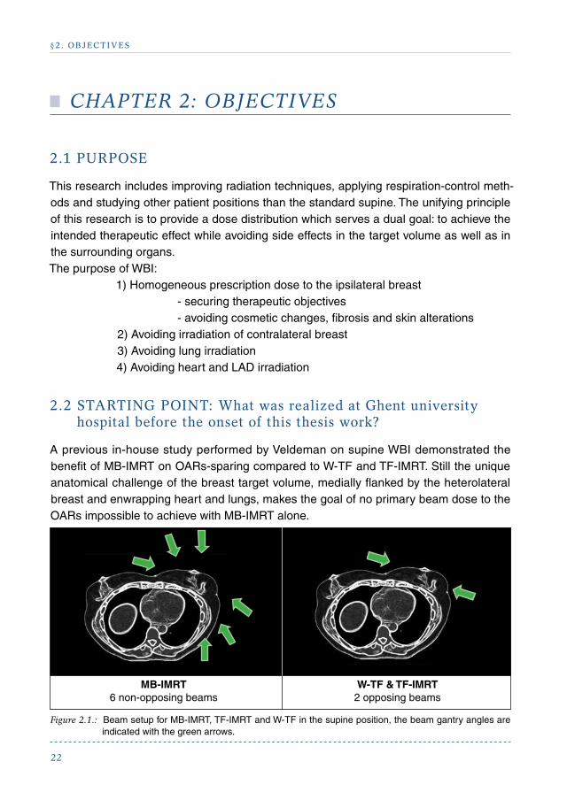
A previous in-house study performed by Veldeman on supine WBI demonstrated the benefit of MB-IMRT on OARs-sparing compared to W-TF and TF-IMRT. Still the unique anatomical challenge of the breast target volume, medially flanked by the heterolateral breast and enwrapping heart and lungs, makes the goal of no primary beam dose to the OARs impossible to achieve with MB-IMRT alone.
Figure 2.1.: Beam setup for MB-IMRT, TF-IMRT and W-TF in the supine position, the beam gantry angles are indicated with the green arrows.
W-TF & TF-IMRT 2 opposing beams
MB-IMRT 6 non-opposing beams
23
§ 2 . OB J EC T I V E S
Therefore an alternative approach by altering the patient’s position into prone position was performed and in silico assessment demonstrated the potential of prone position to per-form a spectacular lung dose reduction and improve population characteristics for dose inhomogeneity [1, 2]. A routinely applicable prone IMRT technique was developed using a modified commercially available breast board and a unilateral breast holder to retract the heterolateral breast away from the treated zone [1]. Using this technique a setup precision comparable to supine position could be obtained not only by comparing with other supine cohorts [1] but also on an intra-patient based assessment [2]. Moreover, prone WBI was able to reduce respiration-related breast movement during treatment.
Daily reproducibility of the CT-simulated position during treatment is crucial. Patient- positioning errors are corrected for by rigid image co-registration between cone beam CT (CBCT) and planning CT. The CBCT parameters were yet improved [3], though not fully optimized for breast RT.
2.3 OBJECTIVES OF THIS THESIS
The first objective of this thesis was to perform a comparative clinical assessment and vali-dation of prone versus supine IMRT. This was done in a phase II trial randomizing patients with at least a cup size C between prone and supine IMRT (chapter 5). The primary end-point was acute skin toxicity (moist desquamation). Secondary endpoints included dose/volume parameters for lung, heart and contralateral breast to estimate long-term risk of cardiac insults, lung cancer and heterolateral breast cancer induction. Before initiation of the randomized trial, the best prone radiation technique to compare with supine MB-IMRT was identified (chapter 3).
The second objective of this thesis was to enhance the clinical feasibility of prone WBI in view of implementation of the technique in daily routine. Therefore a number of issues had to be resolved. Optimization of the unilateral breast holder was done and a new breast board was constructed in collaboration with Orfit Industries (Wijnegem, Belgium) using their commercially available breast board as a starting point with the aim to improve patient comfort and setup reproducibility. The venue of a new linear accelerator at GUH with a more recent CBCT version, created the possibility to further improve the acquisi-tion protocol regarding surface reconstruction, decrease radiation exposure and enhance clinical feasibility (chapter 4).
The third objective of this thesis was to address the issue of heart sparing in prone posi-tion. Patients with small cup size (A or B) were not included in the randomized trial for 2 reasons. First, the risk of acute moist desquamation, the primary endpoint, is correlated with cup size with large-breasted patients being at higher risk. If small-breasted patients
24
§ 2 . OB J EC T I V E S
were included the effect size was expected to be smaller and the sample size had to be in-creased. The second reason was the fear of increased heart dose in prone position in small breasted patients. Cooperation with Vincent Remouchamps at CMSE Namur was start-ed to investigate the possibility of combining prone position with DIBH to maximize heart sparing. The feasibility and reproducibility of prone DIBH is described in chapters 6 and 7.
2.4 REFERENCES
[1] Veldeman, L, Speleers, B, Bakker, M, et al. Preliminary results on setup precision of prone-lateral patient positioning for whole breast irradiation. Int J Radiat Oncol Biol Phys 2010;78:111-118.
[2] Veldeman, L, De Gersem, W, Speleers, B, et al. Alternated Prone and Supine Whole-Breast Irradiation Using IMRT: Setup Precision, Respiratory Movement and Treatment Time. Int J Radiat Oncol Biol Phys 2012;82:2055-2064.
[3] De Puysseleyr, A, Veldeman, L, Bogaert, E, De Wagter, C, De Neve, W. Optimizing image ac-quisition settings for cone-beam computed tomography in supine and prone breast radiotherapy. Radiother Oncol 2011;100:227-230.
25
§ 3. PU BL ICAT ION 1
CHAPTER 3: PUBLICATION 1
Whole Breast raDiotherapy in prone anD supine position: is there a place for multi-Beam imrt?
Thomas Mulliez#, Bruno Speleers#, Indira Madani, Werner De Gersem, Liv Veldeman and Wilfried De Neve Department of Radiotherapy, Ghent University Hospital, Ghent, Belgium. #T.M. and B.S. contributed equally to the design and writing of the manuscript.
doi:10.1186/1748-717X-8-151
Corresponding author: T.M.
� Radiation Oncology: Accepted June 2013.
26
§ 3. PU BL ICAT ION 1
ABSTRACT
Background: Early stage breast cancer patients are long-term survivors and finding techniques that may lower acute and late radiotherapy-induced toxicity is crucial. We com-pared dosimetry of wedged tangential fields (W-TF), tangential field intensity-modulated radiotherapy (TF-IMRT) and multi-beam IMRT (MB-IMRT) in prone and supine positions for whole-breast irradiation (WBI).
Methods: MB-IMRT, TF-IMRT and W-TF treatment plans in prone and supine positions were generated for 18 unselected breast cancer patients. The median prescription dose to the optimized planning target volume (PTVoptim) was 50 Gy in 25 fractions. Dose-volume parameters and indices of conformity were calculated for the PTVoptim and organs-at-risk.
Results: Prone MB-IMRT achieved (p<0.01) the best dose homogeneity compared to WTF in the prone position and WTF and MB-IMRT in the supine position. Prone IMRT scored better for all dose indices. MB-IMRT lowered lung and heart dose (p<0.05) in supine position, however the lowest ipsilateral lung doses (p<0.001) were in prone position. In left-sided breast cancer patients population averages for heart sparing by radiation dose was better in prone position; though non-significant. For patients with a PTVoptim volume ≥600 cc heart dose was consistently lower in prone position; while for patients with smaller breasts heart dose metrics were comparable or worse compared to supine MB-IMRT. Doses to the contralateral breast were similar regardless of position or technique. Dosimetry of prone MB-IMRT and prone TF-IMRT differed slightly.
Conclusions: MB-IMRT is the treatment of choice in supine position. Prone IMRT is superior to any supine treatment for right-sided breast cancer patients and left-sided breast cancer patients with larger breasts by obtaining better conformity indices, target dose distribution and sparing of the organs-at-risk. The influence of treatment techniques in prone position is less pronounced; moreover dosimetric differences between TF-IMRT and MB-IMRT are rather small.
INTRODUCTION
Conventional radiotherapy (RT) using wedged tangential fields (W-TF) after breast-con-serving surgery improves disease control and breast-cancer related survival. However pro-longed follow-up showed an increased RT-induced risk of cardiac events and secondary lung and breast cancer in long-term survivors [1-3]. Therefore strategies for sparing organs-at-risk (OARs), while maintaining an adequate dose coverage of the target are warranted.
In supine position the whole-breast clinical target volume (CTVWBI) is concave 1) enwrap-
27
§ 3. PU BL ICAT ION 1
ping the lung and heart at the left side, and 2) medially adjoining the contralateral breast. Therefore parts of the ipsilateral lung, heart, and contralateral breast may receive inter-mediate to high doses with W-TF.
Intensity-modulated radiotherapy (IMRT) can provide advantages compared to W-TF. In supine position IMRT using a tangential two-beam set-up (TF-IMRT) can improve dose homogeneity; however its ability to reduce high-dose regions to the underlying heart and lung tissue appear to be limited [4, 5]. Supine multi-beam IMRT (MB-IMRT) may overcome those limitations often at cost of low- or intermediate-dose spread over the contralateral breast and ipsilateral thoracic region [6-10].
Prone position modifies the target volume by gravity and moves the breast away from the chest wall. Prone W-TF has previously been used for large, pendulous breasts [11] to reduce fibrosis and improve cosmesis [12, 13]. There are a few studies reporting improved dosimetry by prone TF-IMRT [14-16], though data on whole-breast MB-IMRT in prone position are lacking. Moreover, all dosimetric studies comparing prone and supine position used only non-multi-beam techniques [16-20]. We performed the present study to estab-lish the effect of treatment technique (W-TF, TF-IMRT or MB-IMRT) and position (prone or supine) on dose coverage and heart and lung sparing.
MATERIALS AND METHODS
Eighteen unselected early stage breast cancer patients - 6 right-sided and 12 left-sided - presenting for whole-breast irradiation (WBI) without nodal irradiation after breast con-serving surgery were included in this study, approved by the ethics committee of Ghent University Hospital. Three-mm thick computer-tomography scans were acquired with an Aquilion scanner (Toshiba Medical Systems, Tokyo, Japan) in all patients in prone and supine position. Patient set-up and delineation of the clinical and planning target volumes for WBI (CTVWBI and PTVWBI, respectively) and OARs in both treatment positions can be found elsewhere [16, 17]. Extension of the PTVWBI outside the skin into the air accounted for respiration-related breast movement or swelling of the breast during treatment. A flash region was created outside the patient’s external contour by expanding the PTVWBI with a 10 mm margin followed by subtraction of the patient’s total scanned volume. This flash region was subsequently used in the optimization. A planning target volume for optimiza-tion (PTVoptim), a structure used during plan optimization, was generated by removing the in-air part and a 7 mm-wide build-up region underneath the skin from the PTVWBI.
The dosimetric comparison was made for 6 MV photon beams of an Elekta SLi18 linear ac-celerator (Elekta, Crawley, UK) equipped with a standard 1 cm leaf-width multileaf collimator (MLC). A median prescription dose to the PTVoptim was 50 Gy in 25 fractions of 2.0 Gy with
28
§ 3. PU BL ICAT ION 1
the objective of ≥95% of the PTVoptim receiving >95% of the prescribed dose and minimi-zation of maximum dose, dose heterogeneity and “hot spots”. In both positions TF-IMRT used the same gantry angles as W-TF with the collimator set at 0° and the beams shaped around the PTVWBI with the aid of the MLC. Figure 1 shows the 6-beam setup used in the MB-IMRT plans for right-sided breast tumors in supine position (a) and prone position (b). In both positions MB-IMRT used 6 coplanar beams shaped around the PTVWBI and as in TF-IMRT plans field-in-field segments were created avoiding the ipsilateral lung, heart (in case of left-sided breast tumors) and contralateral breast (for lateral beams in supine position, since medial beams did not traverse the contralateral breast).
Figure 1.: Multi-beam set-up in the prone and supine position. A 6-beam set-up used in the multi-beam intensi-ty-modulated radiotherapy (MB-IMRT) plans for right-sided breast tumors in supine (a) and prone posi-tion (b). Gantry angles expressed in the Elekta coordinate system. The most inclined medial beam has the gantry angle of a tangential beam set by virtual simulation [21]. The gantry angles are 0°, |α|, |2α|, 180° - 0.5|β|, 180° + 0.5|β|, and 180° + 1.5|β| for supine MB-IMRT. The lateral gantry angles in prone MB-IMRT are |β|, |β|+/−24°, the medial gantry angles are |α|, |α|+/− 12°.
A forward planning approach was used for the intensity-modulated and W-TF plans. The convolution-superposition dose engine of a Pinnacle version 9.0 treatment planning sys-tem (Philips Medical Systems, Andover, US) was used for dose computations between optimization cycles of intensity-modulated plans as well as for final plans. Monitor units and MLC shapes were optimized using the optimization tools described before [22]. During op-timization, two patient geometries were taken into account: 1) dose computation for PTVWBI
29
§ 3. PU BL ICAT ION 1
was performed using a density override (1 g/cm3) to the above-mentioned flash region; 2) dose computation for the PTVWBI without build-up and OARs was performed without density overrides. To be able to compute both dose distributions in parallel, the patient data at the Pinnacle treatment planning system were duplicated: for the first patient dataset the flash region was set water-equivalent, while for the second patient dataset, the flash region remained at the density of the CT data (in essence, air outside the patient outline). To avoid hot spots outside regions of interest, a “matroska” sequence of shell structures (isotropical expanded structures outside the target volume) [22] was generated outside the PTVWBI, which were taken into account during optimization. Dose computation for these shell struc-tures was performed using the above-mentioned density override in the flash region. Also the dose update mechanism for changes in leaf positions during optimization took both patient geometries into account. This method was used mainly to account for substantial deformations of the breast during the course of treatments.
D2 and D98, or the dose exceeding 2% and 98% of the dose-volume histogram (DVH) points, respectively, were used as surrogates for maximum and minimum dose. These were evaluated for the PTVoptim, as well as dose homogeneity (1-(D2-D98/median dose)). For the heart and ipsilateral lung D2, mean dose (Dmean), V5, V10, V20 and V25 or the proportion of the volume receiving at least 5 Gy, 10 Gy, 20 Gy and 25Gy, respectively, were extracted from the DVH data. For the contralateral breast D2 and Dmean were evaluated.
The following indices were also calculated for the PTVoptim:Jaccard index = A B / A B Where A is the volume covered by the PTVoptim and B is the volume covered by the 95% isodose, i.e., the volume receiving 47.5 Gy or more. The Jaccard index increases with increase in similarity or overlap between the target volume and the 95% isodose and is a measure of dose conformity of the treatment plan.
Dose-coverage index = A∩B1 / AWhere B1 is the volume covered by the 95-107% isodose, i.e. the volume receiving between 47.5 Gy and 53.5 Gy. The dose-coverage index calculates the proportion of the target, in which the treatment-planning objectives for the target are met.
Mismatch index = B2 / B Where B2 is the volume covered by the 95% isodose and lying outside the PTVoptim. It is the fraction of the 95% isodose non-overlapping the target. If the mismatch index is large, large amounts of normal tissues receive 95% of the prescription dose, i.e., 47.5 Gy.
One-way analysis of variance (ANOVA) was used for a pairwise comparison of dose-volume parameters and indices between MB-IMRT, TF-IMRT and W-TF in the 2 treatment positions.
30
§ 3. PU BL ICAT ION 1
RESULTS
One hundred-and-eight plans were generated. Figure 2 illustrates typical dose distributions obtained with the 3 techniques in prone and supine position.
Figure 2.: Isodose distributions (in Gy) of the 6 treatment plans for a left-sided patient in a transverse plane. Abbreviations: W-TF = wedged tangential fields; TF-IMRT = tangential field intensity-modulated radio-therapy; MB-IMRT = multi-beam intensity-modulated radiotherapy.
31
§ 3. PU BL ICAT ION 1
Dose homogeneity and dose coverage of the target
Table 1 provides numerical data on target coverage and target dose distribution obtained with the 3 techniques in the prone and supine position. D2 is lowered in prone position resulting in improved dose homogeneity since D98 was similar for both positions. Signifi-cance was obtained for prone MB-IMRT versus all supine techniques and a trend (p=0.05) for prone TF-IMRT compared to supine W-TF regarding D2; moreover prone MB-IMRT obtained better (p<0.01) dose homogeneity compared to supine W-TF and MB-IMRT. Intensity-modulated techniques were able to improve dose homogeneity compared to con-ventional techniques in both positions, though significance (p=0.002) was only gained for prone MB-IMRT versus prone W-TF.
Prone WBI scored better for Jaccard and mismatch indices (Table 1). Prone MB-IM-RT achieved better results than any supine treatment technique (p≤0.03, both indices); followed by prone TF-IMRT versus supine TF-IMRT and W-TF (p≤0.001, both indices). In supine position MB-IMRT (p<0.001) was the best and W-TF (p<0.001) was the worst technique for both indices. Prone IMRT improved significantly (p<0.01) dose coverage index: prone TF-IMRT vs. supine MB-IMRT and prone MB-IMRT vs. supine MB-IMRT and TF-IMRT.
Technique D2 [Gy] D98 [Gy] Dose homogeneity [%]
prone supine prone supine prone supine
mean SEM SD mean SEM SD mean SEM SD mean SEM SD mean SEM SD Mean SEM SD
Table 1.a: Dose-volume parameters for the optimized planning target volume (PTVoptim).
Abbreviations: SEM = standard error of the mean; SD = standard deviation; W-TF = wedged tangential fields; TF-IMRT = tangential field intensity-modulated radiotherapy; MB-IMRT = multi-beam intensity- modulated radiotherapy.
Technique Jaccard index [%] Dose-coverage index [%] Mismatch index [%]
prone supine prone supine prone supine
mean SEM SD mean SEM SD mean SEM SD mean SEM SD mean SEM SD mean SEM SD
Table 1.b: Conformity indices for the optimized planning target volume (PTVoptim).
Abbreviations: SEM = standard error of the mean; SD = standard deviation; W-TF = wedged tangential fields; TF-IMRT = tangential field intensity-modulated radiotherapy; MB-IMRT = multi-beam intensity- modulated radiotherapy.
32
§ 3. PU BL ICAT ION 1
Dose-volume parameters in OARs
Figure 3 illustrates cumulative DVHs of the ipsilateral lung (A, all patients) and heart (B, only left-sided patients), numerical data are presented in table 2. Sparing (p<0.001) of the ipsilateral lung by radiation dose was always superior in prone. There was little difference in ipsilateral lung dose between the 3 techniques in prone position, although V10 and V20 were significantly lower in prone MB-IMRT vs. prone W-TF. In supine position treatment technique did alter lung dose (p<0.05), MB-IMRT achieved the best and W-TF the worst lung avoidance by radiation dose. A remarking feature is the modified (p=0.003) ipsilateral lung volume in both positions. Mean ± standard deviation for ipsilateral lung volume is 1504 ± 401cc for prone position versus 1409 ± 431cc for supine position.
Figure 3 A.: Cumulative dose-volume histograms of the ipsilateral lung. Abbreviations: W-TF = wedged tangential fields, TF-IMRT = tangential field intensity-modulated radiotherapy, MB-IMRT = multi-beam intensi-ty-modulated radiotherapy.
Heart dose was lowered with MB-IMRT compared to TF-IMRT (D2, Dmean, V5; p=0.07, 0.05 and 0.03, respectively) and W-TF (D2, V5, p= 0.009 and 0.07, respectively) in supine po-sition. While in prone position the effect of treatment technique on heart dose is less pro-nounced. Population averages for heart dose metrics were non-significantly lowered in prone compared to supine position. Better heart sparing by radiation dose was consistently obtained in prone position for patients with a PTVoptim volume ≥600cc. While for patients with a PTVoptim volume <600cc heart dose metrics were comparable (2/5 patients) or worse (3/5 patients) in prone position compared to supine MB-IMRT.
Neither treatment technique, nor set-up significantly changed doses in the contralateral breast, all procedures achieved a maximum dose <5Gy and mean dose <1.5Gy for all patients.
0 5 10 15 20 25 30 35 40 45 50 55 Dose (Gy)
Supine MB-IMRT
Supine TF-IMRT
Supine W-TF
Prone MB-IMRT
Prone TF-IMRT
Prone W-TF Vo
lum
e (%
)
100
80
60
40
20
0
33
§ 3. PU BL ICAT ION 1
0 5 10 15 20 25 30 35 40 45 Dose (Gy)
Supine MB-IMRT
Supine TF-IMRT
Supine W-TF
Prone MB-IMRT
Prone TF-IMRT
Prone W-TF Vo
lum
e (%
)
100
80
60
40
20
0
Figure 3 B.: Cumulative dose-volume histograms of the heart (only left-sided patients). Abbreviations: W-TF = wedged tangential fields, TF-IMRT = tangential field intensity-modulated radiotherapy, MB-IMRT = multi-beam intensity-modulated radiotherapy.
Table 2.: Mean ± standard deviation for ipsilateral lung (all patients) and heart (left-sided breast cancer patients)
dose metrics.
Abbreviations: Dmean = mean dose; V20 and V25= partial volume receiving at least 20 Gy and 25 Gy, respectively; W-TF = wedged tangential fields; TF-IMRT = tangential field intensity-modulated radiotherapy; MB-IMRT = multi-beam intensity-modulated radiotherapy.
DISCUSSION
In supine position IMRT techniques obtain a higher Jaccard index, i.e. superior dose con-formity, and less mismatch compared to W-TF with MB-IMRT being the superior technique for both indices. Dose conformity, coverage and mismatch are even better for the prone techniques, becoming statistically significant in prone IMRT plans. This is not surprising, since prone position results in a less concave breast volume. Therefore dose to the axil-lary and shoulder region is substantially reduced and less of the prescription dose can be expected to be out of the target. Our results confirm the reduction of dose inhomogeneity, with IMRT-techniques compared to standard W-TF. Though differences were rather small and non-significant in supine position, which could be explained by the use of non-mixture beam energies. Prone as compared to supine IMRT does improve dose homogeneity and hot spots with the best results in prone MB-IMRT plans. Our results are in agreement with
34
§ 3. PU BL ICAT ION 1
other publications on prone IMRT. Goodman et al. [15] demonstrated a maximum dose in the target exceeding 110% with prone W-TF in 16 of 20 patients as compared to 1 patient with prone IMRT (TF-IMRT). Another study comparing MB-IMRT, TF-IMRT and 3D-CRT treatment plans of 5 patients planned in prone position reported significantly higher dose homogeneity of MB-IMRT plans vs. TF-IMRT (p=0.003) and 3D-CRT plans (p=0.03) [23]. Hardee et al. [14] observed a maximum dose reduction and improved median dose homo-geneity in a prone TF-IMRT vs. 3D-CRT patient cohort. Moreover a 11%-decrease of grade 2 dermatitis and a 16%-reduction of grade ≥2 hyperpigmentation were found in the IMRT group. We expect that improved dose homogeneity and hot spots achieved by prone IMRT
– either MB-IMRT or TF-IMRT - will yield lower skin toxicity and better cosmesis [4, 5, 24].
Lung irradiation was lowered with the MB-IMRT technique in supine position, though spar-ing of the ipsilateral lung appeared to be depending more on the treatment position than on the treatment technique. Prone position resulted in a spectacular decrease in lung dose, which is in coherence with other data [16-20]. The decrease in lung dose in prone position might also be attributed by the 7% increase in ipsilateral lung volume, for which we don’t have an explanation. All prone treatment techniques showed similar lung dose metrics.
Left-sided breast cancer patients are at risk of radiation-induced cardiac events [2], emphasizing the importance of using more sophisticated techniques to lower the heart dose. In supine position, MB-IMRT is able to lower the heart dose compared to the other techniques as shown both in our data and in other publications [7-9]. In prone position dif-ferent treatment techniques have less effect on heart dose, especially between IMRT-tech-niques. Even with MB-IMRT, only the minority of patients (3/12) benefitted from supine position; which is in coherence with other data [18, 20]. Moreover consistent better heart dose metrics were achieved in prone position for patients with a PTVoptim volume of ≥600cc. A limitation of this study is the absence of dose parameters of the left descending coronary artery, since this is likely associated with increased cardiac mortality.
The introduction of supine MB-IMRT was not successful because of its complexity, increase in dose to the contralateral breast and higher integral dose [7-9]. In contrast with these studies we selected beams that avoided the contralateral breast and removed beams that included too much lung tissue. In this way reducing the dose in the ipsilateral lung with MB-IMRT, both in supine and prone position, was not at cost of low-dose spread over the lung or heart as illustrated by the DVHs (Figure 3). The dose to the contralateral breast was not increased with MB-IMRT either, moreover a maximum dose <5Gy and mean dose <1.5Gy was obtained for all patients.
As a consequence of the reduced ipsilateral lung and heart dose, better dose distribution and dose coverage, prone IMRT is superior to any supine technique for left-sided
35
§ 3. PU BL ICAT ION 1
patients with larger breasts (PTVoptim≥600cc) and all right-sided patients. While for left- sided patients with smaller breasts individual comparative planning should be made be-tween supine MB-IMRT and prone IMRT in order to choose the best technique for clinical execution. The dosimetric differences between prone TF-IMRT and prone MB-IMRT are rather small. Whether these “small” dosimetric benefits would cause a clinical benefit is unknown. The more complex and time consuming planning procedure and beam delivery of prone MB-IMRT should also be considered.
CONCLUSIONS
MB-IMRT is the preferred technique in supine position by providing better coverage indices of the target and sparing of organs-at-risk. However, prone IMRT is superior to any supine technique for right-sided breast cancer patients and left-sided breast cancer patients with larger breasts. The impact of treatment techniques in prone position is less prominent; moreover dosimetric differences between both IMRT-techniques are rather small.
REFERENCES
[1] Henson, KE, McGale, P, Taylor, C, Darby, SC. Radiation-related mortality from heart disease and lung cancer more than 20 years after radiotherapy for breast cancer. Br J Cancer 2013;108:179-182.
[2] Clarke, M, Collins, R, Darby, S, et al. Effects of radiotherapy and of differences in the extent of surgery for early breast cancer on local recurrence and 15-year survival: an overview of the randomised trials. Lancet 2005;366:2087-2106.
[3] Early Breast Cancer Trialists’ Collaborative, G, Darby, S, McGale, P, et al. Effect of radiother-apy after breast-conserving surgery on 10-year recurrence and 15-year breast cancer death: meta-analysis of individual patient data for 10,801 women in 17 randomised trials. Lancet 2011;378:1707-1716.
[4] Barnett, GC, Wilkinson, JS, Moody, AM, et al. Randomized controlled trial of forward-planned intensity modulated radiotherapy for early breast cancer: interim results at 2 years. Int J Radiat Oncol Biol Phys 2012;82:715-723.
[5] Veldeman, L, Madani, I, Hulstaert, F, De Meerleer, G, Mareel, M, De Neve, W. Evidence behind use of intensity-modulated radiotherapy: a systematic review of comparative clinical studies. Lancet Oncology 2008;9:367-375.
[6] Borca, VC, Franco, P, Catuzzo, P, et al. Does TomoDirect 3DCRT represent a suitable option for post-operative whole breast irradiation? A hypothesis-generating pilot study. Radiat Oncol 2012;7:211.
36
§ 3. PU BL ICAT ION 1
[7] Coon, AB, Dickler, A, Kirk, MC, et al. Tomotherapy and Multifield Intensity-Modulated Radio-
therapy Planning Reduce Cardiac Doses in Left-Sided Breast Cancer Patients with Unfavorable
Cardiac Anatomy. Int J Radiat Oncol Biol Phys 2010;72:104-110.
out a boost to the tumor bed: comparable toxicity of IMRT versus a 3D conformal technique.
Int J Radiat Oncol Biol Phys 2012;82:e415-423.
[15] Goodman, KA, Hong, L, Wagman, R, Hunt, MA, McCormick, B. Dosimetric analysis of a simpli-
fied intensity modulation technique for prone breast radiotherapy. Int J Radiat Oncol Biol Phys
2004;60:95-102.
[16] Veldeman, L, Speleers, B, Bakker, M, et al. Preliminary results on setup precision of prone-lateral
patient positioning for whole breast irradiation. Int J Radiat Oncol Biol Phys 2010;78:111-118.
[17] Veldeman, L, De Gersem, W, Speleers, B, et al. Alternated Prone and Supine Whole-Breast
Irradiation Using IMRT: Setup Precision, Respiratory Movement and Treatment Time. Int J
Radiat Oncol Biol Phys 2012;82:2055-2064.
37
§ 3. PU BL ICAT ION 1
[18] Kirby, AM, Evans, PM, Donovan, EM, Convery, HM, Haviland, JS, Yarnold, JR. Prone versus supine positioning for whole and partial-breast radiotherapy: a comparison of non-target tissue dosimetry. Radiother Oncol 2010;96:178-184.
[19] Varga, Z, Hideghety, K, Mezo, T, Nikolenyi, A, Thurzo, L, Kahan, Z. Individual positioning: a com-parative study of adjuvant breast radiotherapy in the prone versus supine position. Int J Radiat Oncol Biol Phys 2009;75:94-100.
[20] Lymberis, SC, Dewyngaert, JK, Parhar, P, et al. Prospective Assessment of Optimal Individual Position (Prone Versus Supine) for Breast Radiotherapy: Volumetric and Dosimetric Correlations in 100 Patients. Int J Radiat Oncol Biol Phys 2012;84:902-909.
[21] Van Vaerenbergh K, De Gersem W, Vakaet L, et al. Automatic generation of a plan optimization volume for tangential field breast cancer radiation therapy. Strahlenther Onkol 2005;181:82-8.
[22] De Neve W, Wu Y, Ezzel G. Practical IMRT planning. In: Bortfeld T, Schmidt-Ulrich R, De Neve W, Wazer D, editors. Image-guided IMRT. Berlin, Heidelberg: Springer. 2006;47-59
[23] Ahunbay, EE, Chen, GP, Thatcher, S, et al. Direct aperture optimization-based intensity-modu-lated radiotherapy for whole breast irradiation. Int J Radiat Oncol Biol Phys 2007;67:1248-1258.
[24] Harsolia, A, Kestin, L, Grills, I, et al. Intensity-modulated radiotherapy results in significant decrease in clinical toxicities compared with conventional wedge-based breast radiotherapy. Int J Radiat Oncol Biol Phys 2007;68:1375-1380.
38
§ 4. PU BL ICAT ION 2
CHAPTER 4: PUBLICATION 2
improveD cone-Beam computeD tomography in supine anD prone Breast raDiotherapy: surface reconstruction, raDiation exposure anD clinical WorkfloW.
Annemieke De Puysseleyr1#, Thomas Mulliez1#, Akos Gulyban1, Evelien Bogaert1, Tom Vercauteren1, Tom Van Hoof2, Joris Van de Velde2, Rudy Van den Broecke3, Carlos De Wagter1 and Wilfried De Neve1
1Department of Radiotherapy, Ghent University Hospital, Ghent, Belgium. 2Department of Basic Medical Sciences, Ghent University, Ghent, Belgium. 3Department of Gynaecology, Ghent University Hospital, Ghent, Belgium. #A.D.P. and T.M. contributed equally to the design and writing of the manuscript.
doi: 10.1007/s00066-013-0435-x
Corresponding author: T.M.
� Strahlentherapie und Onkologie: Accepted November 2013.
39
§ 4. PU BL ICAT ION 2
ABSTRACT
Background: Cone-beam computerized tomography (CBCT) enables three-dimensional information of the scanned region and provides soft tissue images with good spatial resolu- tion. Our aim was to optimize image acquisition settings for prone and supine breast radio- therapy with respect to contour accuracy, clinical practicalities, and radiation dose.
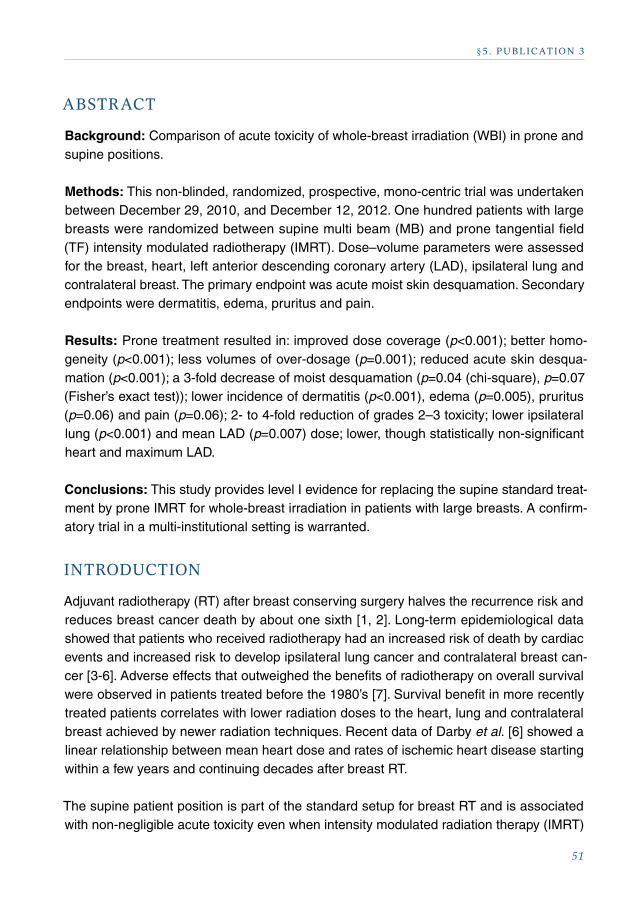
Methods: CBCT images were acquired for both prone and supine anthropomorphic phan-toms and a female cadaver in supine and prone set-up. CBCT protocols were investigated by altering the tube current, exposure time, range of projection views, field of view (FOV), and starting angle. For clinical practicalities, the frequency of the use of an offset CBCT isocenter was evaluated at 558 205°-CBCTs (37 patients; 13 prone and 24 supine) and 1272 360°-CBCTs (102 patients; 13 prone and 89 supine).
Results: Prone and supine breast CBCT images acquired with a bowtie filter, a small FOV, a range of projection views equaling 180°, a tube current of 20 mA and an exposure time of 32 ms, demonstrated adequate contour accuracy and an elimination of the offset CBCT isocenter procedure, while this occurred in 40.7% for the old full-rotation protocol. Further-more a 4.3-fold dose reduction was observed for the Computed Tomography Dose Index (CTDIw) compared to the preset Chest M20 protocol.
Conclusion: The established 180° protocol demonstrated acceptable contour accuracy, eliminated the CBCT isocenter offset procedure and reduced patient radiation exposure.
ZUSAMMENFASSUNG
Hintergrund und Zielsetzung: Die Cone-beam-Computertomographie (CBCT) ermöglicht 3-dimensionale Informationen der gescannten Region und CT-Bilder von Weichteilgewebe in guter räumlicher Auflösung. Unsere Zielsetzung war die Optimierung der Bildakquise für die Einstellungen bei Brustbestrahlungen in Bauch- und Rückenlage in Bezug auf Kontrast, Praktikabilität und Strahlendosis.
Patienten und Methodik: CBCT-Bilder wurden mit einem anthropomorphen Phantom und an einer weiblichen Leiche in Rücken- und Bauchlage aufgenommen. Verschiedene CBCT-Protokolle mit unterschiedlichem Röhrenstrom, unterschiedlichen Expositionszeiten, Projektionsbereichen, Bildausschnitten (FOV) und Anfangswinkeln wurden untersucht. Für die klinische Praxis wurde die Häufigkeit von erforderlichen CBCT-Isozentrum-Anpassun-gen anhand von 558 205°-CBCT (37 Patienten; 13 in Bauchlage, 24 in Rückenlage) und 1272 360°-CBCT (102 Patienten; 13 in Bauchlage, 89 in Rückenlage) überprüft.
40
§ 4. PU BL ICAT ION 2
Ergebnisse: Die mit einem Bowtie-Filter, kleinem FOV, einer 180°-Rotation, 20 mA Röhrenstrom und einer Belichtungszeit von 32 ms erhaltenen CBCT-Bilder in Bauch- und Rückenlage gewährleisteten eine adäquate Konturgenauigkeit und vermeiden eine CBCT in Isozentrumsposition, die bei 40,7% in den alten Vollrotationsprotokollen erforderlich war. Weiterhin ergab sich eine 4,3-fache Reduktion des Computered-Tomography-Dose-Index (CTDIw) verglichen mit dem Chest-M20-Standard-Protokoll.
Schlussfolgerungen: Das 180°-Protokoll zeigt eine akzeptable Konturierungsgenauigkeit, vermeidet CBCT in Isozentrum-Position und reduziert die Strahlenbelastung für die Patientin.
INTRODUCTION
Cone-beam computerized tomography (CBCT) is used in daily practice for setup correction in multiple radiotherapy domains, including prone and supine breast radiotherapy [1-6]. Patient-positioning errors are corrected for by rigid image co-registration between daily CBCT and planning CT. CBCT provides three-dimensional (3D) information of the scanned region and provides soft tissue images with good spatial resolution, both potentially in-creasing the geometric accuracy of radiotherapy delivery [7-10]. However, it is also a more complex and time-consuming imaging modality and the additional radiation exposure might increase the risk of secondary cancer [11].
Due to the absence of distinct anatomical features in breast tissue, the CBCT to planning CT alignment mainly relies on the breast contours, chest wall and, if present, surgical clips. In this respect, a previous study demonstrated the breast contours on CBCT image sets, acquired with the clinically standard settings, to be affected by multiple artefacts [12]. These contour inaccuracies were ascribed to reconstruction artefacts and panel satura-tion effects. Lowering the CBCT tube current and exposure time eliminated this artefact. However, one of the limitations of the above-mentioned study was the absence of a bowtie filter [12]. This filter has been described to improve general image quality and reduce panel saturation effects [13]. Consequently, it has now become the new standard in CBCT imaging. Furthermore, this study only included full-rotation protocols, which are found to be challenging in clinical practice. In order to avoid collisions, a 10 cm translation of the patient in the ipsilateral direction (offset position) was often necessary prior to and after imaging. This intervention complicates procedures and increases treatment times. An-other limitation was the use of polystyrene phantoms, as the described artefacts strongly depend on patient geometry.
In this study, the contour accuracy investigation is expanded to CBCT imaging with bowtie filtration. Moreover, we now investigate CBCT protocols using a limited range of projection
41
§ 4. PU BL ICAT ION 2
views up to 205°. This configuration is intended to eliminate the ipsilateral patient transla-tion, as well as reducing both the imaging time and radiation dose to the patient [14]. For clinical relevance, evaluation was not only performed on polystyrene phantoms, but also on a human cadaver. Additionally, absorbed dose measurements were performed for all investigated protocols.
MATERIALS AND METHODS
Planning CT and CBCT images were acquired for both prone and supine anthropomor-phic phantoms and a female cadaver in supine and prone set-up. As the polystyrene phan-toms provided an excellent geometric stability, they allowed for the detection of very small volumetric discrepancies. The female cadaver, on the other hand, served as a clinically relevant model.
In this study, a Thiel cadaver was used since the Thiel embalming technique authentically preserves the different tissue layers (skin, subcutaneous fat and collagen tissue, fascia, muscular, visceral and glandular tissue) and keeps the complete range-of-motion of all joints intact, allowing for a precise setup (arrangement) of the body’s posture in accord-ance with the corresponding clinical treatment position [15, 16]. The use of the cadaver was approved by the local ethics committee. An extensive description of the prone and supine thoracic phantoms (Polystyrol 495F, BASF, Germany, ρ=1.02 g/cm³) has already been published [12]. Prone cadaver positioning was accomplished using a prone–lateral breast board (Orfit Industries, Wijnegem, Belgium). Supine cadaver set-up was supported by a supine arm support tool (Civco Medical Solutions, Orange City, IA, USA). The polysty- rene phantoms and cadaver were equipped with four and five titanium surgical clips respectively. These clips were positioned in three different planes in the central breast region. Image acquisition was performed for the left breast in all cases.
Helical planning CT images of the phantoms and the cadaver were acquired on a Toshiba Aquillion CT scanner (Toshiba Medical Systems, Tokyo, Japan) with a slice thickness of 2 mm, a tube potential of 120 kVp and a tube current of 150 mA. Set-up reproducibility was optimized by marking the laser line positions on the phantoms and the cadaver.
Cone-beam CT acquisition was performed on the Elekta XVI CBCT imaging module of an Elekta Synergy linear accelerator (Elekta, Crawley, West-Sussex, UK). This module utilizes a flat panel imager consisting of an amorphous silicon detector and fluorescent screen (Perkin-Elmer, Waltham, MA, USA). An aluminum bowtie filter (Elekta, Filter Cassette Assembly, F1) can be mounted below the collimator. This filter modulates the X-ray flux at the skin–air interface and thus reduces panel saturation effects [13]. Image reconstruction in the accompanying XVI software (version 4.5.) is based on the Feldkamp–Davis–Kress (FDK)
42
§ 4. PU BL ICAT ION 2
cone-beam algorithm. An accurate set-up of the phantoms and cadaver was obtained by aligning the planning CT surface markings with the treatment room lasers.
Multiple CBCT protocols with varying mAs, range of projection views and starting angle were studied. A bowtie filter, an angular projection density of 1.85 and a tube potential of 120 kVp was used in all imaging protocols. As the maximum range of projection views amounted to only 205°, all protocols employed a small field of view (FOV) with a recon-struction diameter of 270 mm. Preliminary research demonstrated extremely poor image quality using a medium FOV (reconstruction diameter 410 mm) for a limited range of projection views. As the medium FOV set-up is obtained by asymmetrically collimating the beam and shifting the detector panel in the corresponding direction, each projection view only partly samples the reconstructed FOV. Consequently, any reduction in range of projection views using a medium FOV results in severely affected image quality.
Firstly, contour accuracy was investigated for CBCT protocols using a varying tube cur-rent and exposure time (40 mA–40 ms, 20 mA–32 ms and 20 mA–25 ms) while keeping the range of projection views constant (i.e. 205°). The X-ray source rotation started at 110 and 70° for the prone and supine set-up respectively. The initial tube current and expo-sure time values (40 mA–40 ms) are taken from the preset protocols using a bowtie filter. Whereas these preset bowtie filtration protocols always employ a medium FOV, they have now been adopted as a starting point for the small FOV investigation. Secondly, we as-sessed the influence of a reduction in range of projection views (200, 180, 160 and 140°) and the X-ray source starting angle. The decrease in ranges was equally spread over the start- and endpoint of the original range. This means that the central angle was kept at the same physical location. This resulted in different starting angles for the different ranges. This evaluation was only performed for CBCT protocols using tube current and exposure time of 20 mA and 32 ms.
For all protocols, the conformity of planning CT and CBCT breast contours was visually inspected on the phantom and cadaver images in the XVI viewing and registration soft-ware (version 4.5, Elekta, Crawley, West-Sussex, UK). Registration of CBCT to planning CT images was manually performed on the surgical clips and, for the female cadaver, the chest wall. As in clinical practice, all images were visualized using the soft window settings.Computed Tomography Dose Index (CTDI) measurements were performed to estimate the influence of various CBCT acquisition settings (field of view, tube current and exposure time and angular range of projection views) on patient radiation dose. Measurements were performed using an Unfors XI Platinum CT detector (100 mm long pencil beam CT-ioniza-tion chamber, type 146358, Unfors Instruments AB, Billdal, Sweden) in a cylindrical CTDI phantom (PMMA, diameter 32 cm, length 15 cm). In order to provide scattered dose along the complete CBCT field width, the phantom’s length was increased by adding at least
43
§ 4. PU BL ICAT ION 2
15 cm of polystyrene slabs (Polystyrol 495F, BASF, Germany, ρ=1.02 g/cm³) at the phan-tom’s superior and inferior side. The phantom center was positioned in the imaging module isocenter. CTDI measurements were performed at the center and four equally distributed peripheral positions in the phantom (at 0, 90, 180 and 270°, distance to the center 10 cm). For all CBCT protocols, the bisecting line of the range of projection views was always centered at 0°. The weighted CTDI (CTDIw) was then computed as (Elekta: XVI R4.5):
CTDIw= 1_3 (CTDI
100,center) + 2_
3 (1_4 ΣCTDI
100,peripheral)
In order to investigate the need for patient offset positioning, the frequency of this inter-vention was compared between the old full-rotation protocol and a 205°-rotation proto-col. To that purpose, 558 205°-CBCTs (37 patients; 13 prone and 24 supine) and 1272 360°-CBCTs (102 patients; 13 prone and 89 supine) were included. The 205° protocol employs the maximum range of projection views employed in this investigation. As all further reductions in range of projection views were equally spread over the start- and endpoint of the original range, this 205° protocol served as the most critical protocol with respect to possible collisions.
RESULTS
This study investigated contour accuracy, radiation dose and the need for offset patient positioning for CBCT protocols using bowtie filtration, a small FOV and a limited range of projection views.
Firstly, we evaluated the dependence of contour accuracy on CBCT tube current and expo-sure time for CBCT acquisitions using a bowtie filter, a small FOV and a range of projection views equaling 205° (Figure 1 a, b). The images represent a transversal slice through the center of the breast. Prone and supine CBCT phantom and cadaver images acquired with a tube current and exposure time of 40 mA and 40 ms, demonstrated an important periareolar tissue deficit (Figure 1 a, marked with dotted lines). An additional tissue deficit in the lateral region of the breast was detected in the supine CBCT images. As in the previous study, these CBCT contour inaccuracies were completely eliminated by lowering the tube current and exposure time to 20 mA and 32 ms (Figure 1 b, marked with dotted lines). The cranio- caudal tissue excesses, as detected in the previous phantom study, were still apparent in the polystyrene phantoms, but were hardly detectable in the female cadaver images.
44
§ 4. PU BL ICAT ION 2
Figure 1.: Dependence of contour accuracy on the CBCT tube current and exposure time (a, b). The correspond-ing planning CT contours, automatically generated by the treatment planning system, are shown as solid red lines. The areas of interest showing the absence or presence of the artefacts are marked using dotted lines. The phantom images were acquired using a bowtie filter, a range of projection views of 205° and a tube current and exposure time equaling 40 mA–40 ms (a) and 20 mA–32 ms (b).
Secondly, the influence of a further reduction in range of projection views and the X-ray source starting angle was investigated for CBCT protocols using a tube current and exposure time of 20 mA and 32 ms respectively. This evaluation was uniquely performed on the female cadaver’s images (Figure 2). Contour accuracy was found to be adequate for protocols using a range of projection views equaling 180° or higher (Figure 2 I , II). When further reducing the angular range of projection views, several image artefacts affected the breast contour in itself, as well as the contrast of the matching features (i.e. the chest wall and surgical clips), thus compromising rigid registration.
The corresponding results of the CTDIw measurements are illustrated in Table 1. As can be seen, switching from a medium FOV to a small FOV for a given protocol generally in-creases the CTDIw (by 3.4 % and 1.4% for a full-rotation acquisition with a tube current and exposure time of 40mA - 40 ms and 20 mA - 32 ms respectively). However, in contrast to the asymmetrical M20 acquisition, the S20 set-up never uses a 360° acquisition, as this would oversample the projections. Consequently, the concomitant reduction in range of projection views strongly reduces the measured CTDIw: for a tube current and exposure time of 20 mA and 32 ms, the CTDIw of a 205°-S20 protocol amounted to only 66.3 % of the full-rotation M20 acquisition. As expected, lowering the mAs settings proportionally decreases the CTDIw. The applied 2.5-fold reduction (from 40 mA–40 ms to 20 mA–32 ms)
45
§ 4. PU BL ICAT ION 2
resulted in a 2.5-fold CTDIw decrease (26.8 mGy versus 10.6 mGy). Combination of a 180° rotation, use of small FOV, 20 mA, 32 ms, with use of bowtie filter results in a 4.3-fold dose reduction in CTDIw compared with the preset Chest M20 protocol.
Figure 2.: Dependence of contour accuracy on the CBCT range of projection views. The corresponding planning
CT contours, automatically generated by the treatment planning system, are shown as solid red lines. The areas of interest showing the absence or presence of the artefacts are marked using dotted lines. The cadaver images settings comprised a tube current and exposure time of 20 mA–32 ms and a range of projection views of 200° (I), 180° (II), 160° (III) and 140° (IV).
Filtration Field of view Tube current (mA)
Exposure time (ms)
Range of projec-tion views (°) CTDIw (mGy)
F1 M20 40 40 360 23.4
F1 S20 40 40 360 26.8
F1 M20 20 32 360 9.2
F1 S20 20 32 360 10.6
F1 S20 20 32 205 6.1
F1 S20 20 32 180 5.4
Table 1.: The influence of CBCT acquisition settings on the weighted computed tomography dose index (CTDIw). The first protocol corresponds to the preset Chest M20 protocol meaning medium field of view1. S20 means small field of view. F1 means bowtie filter in place.
1 Elekta Synergy XVI version 4.5. ‘Instructions for Use’ Manual, Elekta, UK, 2009
46
§ 4. PU BL ICAT ION 2
The frequency of offset patient positioning amounted to 40.7% for the old full-rotation pro-tocol, while this procedure was completely eliminated for the 205°-180°-rotation protocols.
DISCUSSION
In contrast to the previously studied protocols [12], the investigated protocols now involve a bowtie filter, a small FOV and a maximum range of projection views of 205°. Each of these factors is expected to affect body-contour accuracy, radiation dose to the patient and the need for offset patient positioning. For such special consideration performing a test is quite challenging as there are no existing image quality phantoms which could properly access reconstruction of superficial contours. Using anthropomorphic phantom (e.g. Alderson- Rando) for such purpose would not be feasible in prone position as the changes in breast shape could not be simulated representatively.
Firstly, our examination using the female cadaver clearly illustrated the suitability of a small FOV for breast CBCT, as all images included all main features for planning CT to CBCT registration: the breast contours, surgical clips and adjacent chest wall. Clinical implemen-tation of the half-rotation rotation CBCT (for 558 acquisitions) did not reveal any relevant clinical differences compared to our test findings.
Moreover, our results clearly show that, even when using a bowtie filter that has been described to reduce panel saturation effects [13], attention regarding these artefacts and their effect on contour accuracy is required. Adoption of the preset tube current and expo-sure time for CBCT imaging with bowtie filtration, only available for a medium FOV, resulted in an important periareolar tissue deficit in both prone and supine breast imaging with a small FOV. Our investigation showed that, even for CBCT protocols using a limited range of projection views, a reduction of tube current and exposure time completely eliminates these artefacts. A detailed description of such panel saturation effects and their relation to the CBCT tube current and exposure time is provided in [12]. The presence of these artefacts in the cadaver images clearly demonstrate the significance of panel saturation artefacts in a clinically relevant set-up.
As in the previous study [12], the phantom images still suffered from craniocaudal tissue excesses. These tissue excesses were ascribed to errors inherent to the FDK-based re-construction algorithm and are most pronounced for flat object edges parallel to the imag-ing module rotation plane [17]. In contrast, they were hardly found in the cadaver images, thus emphasizing the value of using phantoms that adequately reflect human anatomy for studying contour accuracy. Overall, the Thiel embalmed cadaver generally served as a clinically relevant phantom for CBCT acquisition: both clips, soft and hard tissue features were clearly visible and strongly resembled real patient geometry and composition. The
47
§ 4. PU BL ICAT ION 2
only limitation was the seeping of embalmment fluid into the cadaver lungs, resulting in a denser region near the chest wall on both the CBCT and planning CT images.
Additionally, the use of a small FOV has an important impact on patient radiation dose. In contrast to a medium FOV, the use of a small FOV allows for a reduction in range of projection views. While the transition from a medium to a small FOV for a given protocol resulted in a slight increase in CTDIw, lowering the range of projection views from 360° to 205–180° allowed for a more important CTDIw reduction.
Note, however, that the concept of CTDIw was originally developed for reporting doses from CT acquisitions, representing the integral dose of a single CT slice [18]. These measure- ments are generally performed by integrating the dose measured in a 100 mm-long ion-ization chamber, a technique that has also been adopted for CBCT by Amer et al. [19]. They showed that this approach results in a conservative overestimate of the weighted dose across the complete FOV, representing an estimate of the dose in the central 100 mm of the FOV [19]. Moreover, the concept of CTDIw was designed for full-rotation acqui-sitions with an axial-symmetric dose distribution [18]. CBCT acquisitions using a limited range of projection views obviously do not preserve this axial symmetry. The absorbed dose throughout the imaged volume will strongly depend on the location of the narrower entrance beams and the wider but attenuated exit beam, especially in the volume’s periphery [19]. In order to ensure a fair comparison between the different view ranges, the bisecting line of the range was always centered at 0°. Nevertheless, the application of the CTDIw concept in this paper was only used to provide a direct comparison between the investigated protocols, rather than quantifying absorbed doses for comparison with other imaging modules. To that purpose, a more elaborate investigation of the spatial distribution of radiation dose to the patient is required.
Furthermore, the use of a bowtie filter in the investigated protocols has been described to strongly reduce radiation dose to the patient [13]. This effect was found to be the most im-portant in the peripheral regions of the scanned object, but was not investigated in this study.
An additional benefit of the use of a limited range of projection views is the avoidance of the offset procedure. As this intervention was completely eliminated with a range of protection views of 205°, it is also avoided in all protocols using a smaller range of projec-tion views and the same physical central angle.
Stock et al. [8] observed a reasonable difference in image quality between CT and CBCT images. A limitation of this study is that the image quality is not objectively assessed. How-ever the proposed protocol, currently used in our center, provides image quality that is sufficient for all radiation oncologists in our department to define translational positioning
48
§ 4. PU BL ICAT ION 2
errors. Current research can only be extrapolated to Elekta imaging modules and should
not been generalized for other imaging entities.
CONCLUSIONS
The following CBCT acquisition parameters for supine and prone breast radiotherapy
have been established: a 180° rotation, a small FOV, bowtie filtration, a tube current of 20
mA and an exposure time of 32 ms. The use of this protocol results in adequate contour
accuracy and eliminated the offset patient positioning procedure. Moreover, it allows for a
decrease in radiation exposure, a 4.3-fold reduction in CTDIw was observed compared to
the preset Chest M20 protocol.
REFERENCES
[1] Jozsef, G, DeWyngaert, JK, Becker, SJ, Lymberis, S, Formenti, SC. Prospective study of cone-beam computed tomography image-guided radiotherapy for prone accelerated partial breast irradiation. Int J Radiat Oncol Biol Phys 2011;81:568-574.
[2] Fatunase, T, Wang, Z, Yoo, S, et al. Assessment of the residual error in soft tissue setup in patients undergoing partial breast irradiation: results of a prospective study using cone-beam computed tomography. Int J Radiat Oncol Biol Phys 2008;70:1025-1034.
[3] Boda-Heggemann, J, Lohr, F, Wenz, F, Flentje, M, Guckenberger, M. kV Cone-Beam CT-Based IGRT A Clinical Review. Strahlenther Onkol 2011;187:284-291.
[4] Guckenberger, M, Ok, S, Polat, B, Sweeney, RA, Flentje, M. Toxicity after Intensity-Modulated, Image-Guided Radiotherapy for Prostate Cancer. Strahlenther Onkol 2010;186:535-543.
[5] Guckenberger, M, Meyer, J, Wilbert, J, Baier, K, Sauer, O, Flentje, M. Precision of image-guided radiotherapy (IGRT) in six degrees of freedom and limitations in clinical practice. Strahlenther Onkol 2007;183:307-313.
[6] Polat, B, Wilbert, J, Baier, K, Flentje, M, Guckenberger, M. Nonrigid patient setup errors in the head-and-neck region. Strahlenther Onkol 2007;183:506-511.
[7] Topolnjak, R, Sonke, JJ, Nijkamp, J, et al. Breast patient setup error assessment: comparison of electronic portal image devices and cone-beam computed tomography matching results. Int J Radiat Oncol Biol Phys 2010;78:1235-1243.
[8] Stock, M, Pasler, M, Birkfellner, W, Homolka, P, Poetter, R, Georg, D. Image quality and stability of image-guided radiotherapy (IGRT) devices: A comparative study. Radiother Oncol 2009;93:1-7.
49
§ 4. PU BL ICAT ION 2
[9] Oldham, M, Letourneau, D, Watt, L, et al. Cone-beam-CT guided radiation therapy: A model for on-line application. Radiother Oncol 2005;75:271-278.
[10] Smitsmans, MH, de Bois, J, Sonke, JJ, et al. Automatic prostate localization on cone-beam CT scans for high precision image-guided radiotherapy. Int J Radiat Oncol Biol Phys 2005;63:975-984.
[11] Kan, MW, Leung, LH, Wong, W, Lam, N. Radiation dose from cone beam computed tomography for image-guided radiation therapy. Int J Radiat Oncol Biol Phys 2008;70:272-279.
[12] De Puysseleyr, A, Veldeman, L, Bogaert, E, De Wagter, C, De Neve, W. Optimizing image ac-quisition settings for cone-beam computed tomography in supine and prone breast radiotherapy. Radiother Oncol 2011;100:227-230.
[13] Mail, N, Moseley, DJ, Siewerdsen, JH, Jaffray, DA. The influence of bowtie filtration on cone-beam CT image quality. Med Phys 2009;36:22-32.
[14] Islam, MK, Purdie, TG, Norrlinger, BD, et al. Patient dose from kilovoltage cone beam computed tomography imaging in radiation therapy. Med Phys 2006;33:1573-1582.
[15] De Crop, A, Bacher, K, Van Hoof, T, et al. Correlation of contrast-detail analysis and clini-cal image quality assessment in chest radiography with a human cadaver study. Radiology 2012;262:298-304.
[16] Thiel, W. The Preservation of Complete Cadavers without Loss of Natural Color. Ann Anat 1992;174:185-195.
[17] Feldkamp, LA, Davis, LC, Kress, JW. Practical Cone-Beam Algorithm. J Opt Soc Am A 1984;1:612-619.
[18] EU. European Guidelines on Quality Criteria for Computed Tomography Brussels: EU. 1999.
[19] Amer, A, Marchant, T, Sykes, J, Czajka, J, Moore, C. Imaging doses from the Elekta Synergy X-ray cone beam CT system. Brit J Radiol 2007;80:476-482.
50
§ 5. PU BL ICAT ION 3
CHAPTER 5: PUBLICATION 3
hypofractionateD Whole Breast irraDiation for patients With large Breasts: a ranDomizeD trial comparing prone anD supine positions.
Thomas Mulliez1, Liv Veldeman1, Annick van Greveling1, Bruno Speleers1, Simin Sadeghi1, Dieter Berwouts1, Frederik Decoster1, Tom Vercauteren1, Werner De Gersem1, Rudy Van den Broecke2 and Wilfried De Neve1
1Department of Radiotherapy, Ghent University Hospital, Ghent, Belgium.2Department of Gynaecology, Ghent University Hospital, Ghent, Belgium.
doi: 10.1016/j.radonc.2013.08.040.
Corresponding author: T.M.
� Radiotherapy and Oncology: Accepted August 2013.
51
§ 5. PU BL ICAT ION 3
ABSTRACT
Background: Comparison of acute toxicity of whole-breast irradiation (WBI) in prone and supine positions.
Methods: This non-blinded, randomized, prospective, mono-centric trial was undertaken between December 29, 2010, and December 12, 2012. One hundred patients with large breasts were randomized between supine multi beam (MB) and prone tangential field (TF) intensity modulated radiotherapy (IMRT). Dose–volume parameters were assessed for the breast, heart, left anterior descending coronary artery (LAD), ipsilateral lung and contralateral breast. The primary endpoint was acute moist skin desquamation. Secondary endpoints were dermatitis, edema, pruritus and pain.
Results: Prone treatment resulted in: improved dose coverage (p<0.001); better homo-geneity (p<0.001); less volumes of over-dosage (p=0.001); reduced acute skin desqua-mation (p<0.001); a 3-fold decrease of moist desquamation (p=0.04 (chi-square), p=0.07 (Fisher’s exact test)); lower incidence of dermatitis (p<0.001), edema (p=0.005), pruritus (p=0.06) and pain (p=0.06); 2- to 4-fold reduction of grades 2–3 toxicity; lower ipsilateral lung (p<0.001) and mean LAD (p=0.007) dose; lower, though statistically non-significant heart and maximum LAD.
Conclusions: This study provides level I evidence for replacing the supine standard treat-ment by prone IMRT for whole-breast irradiation in patients with large breasts. A confirm-atory trial in a multi-institutional setting is warranted.
INTRODUCTION
Adjuvant radiotherapy (RT) after breast conserving surgery halves the recurrence risk and reduces breast cancer death by about one sixth [1, 2]. Long-term epidemiological data showed that patients who received radiotherapy had an increased risk of death by cardiac events and increased risk to develop ipsilateral lung cancer and contralateral breast can-cer [3-6]. Adverse effects that outweighed the benefits of radiotherapy on overall survival were observed in patients treated before the 1980’s [7]. Survival benefit in more recently treated patients correlates with lower radiation doses to the heart, lung and contralateral breast achieved by newer radiation techniques. Recent data of Darby et al. [6] showed a linear relationship between mean heart dose and rates of ischemic heart disease starting within a few years and continuing decades after breast RT.
The supine patient position is part of the standard setup for breast RT and is associated with non-negligible acute toxicity even when intensity modulated radiation therapy (IMRT)
52
§ 5. PU BL ICAT ION 3
is used. Randomized controlled trials, comparing IMRT with non-IMRT, showed significant reduction of acute toxicity [8, 9], but still up to 40% of the IMRT-treated patients developed acute and late skin toxicity or breast fibrosis leading to discomfort and altered body image. Causes include volumes of over-dosage [dose levels higher than the prescribed range, i.e. between 95% and 107% of the prescription dose (PD)] in the breast and in folds that shield skin tissue from the protective build-up effects (in unfolded skin the maximum dose lays about 1.5 cm beneath the skin/air interface for 6 MV photon beams); both occur especially in larger breasts. Prone breast irradiation exploits gravity to elongate the treat-ed breast away from the heart and lung. Formenti et al. [10, 11] demonstrated the clinical feasibility of this procedure resulting in further dose-reductions to heart and lungs, which have been confirmed by others [12-17]. As compared to supine, the prone position opens the infra-mammary fold, thereby restoring protective build-up, and reduces the radiological path-length i.e. the length of breast tissue traversed by tangential X-rays which helps reducing volumes of over-dosage. With this rationale for prone breast RT we initiated a randomized controlled trial compar-ing prone with supine position for large breasted patients. The primary objective was to test the hypothesis of decreased incidence of acute moist desquamation with prone IMRT, compared to our best supine technique: non-opposed multi-beam (MB) IMRT. Secondary endpoints included the analysis of acute and late skin toxicity, breast fibrosis, cosmetic alterations, quality of life and dose-volume parameters. Dose-volume parameter analysis included volumes of over-dosage in the treated breast and radiation doses delivered to volumes of organs-at-risk (OARs: heart, left anterior descending coronary artery (LAD), contra-lateral breast and ipsilateral lung). We, hereby, report on the primary objective of the study and on dose-volume parameters of treated breast and OARs.
MATERIALS AND METHODS
The study was designed as a non-blinded, randomized, prospective, mono-centric trial comparing prone and supine setup in patients who receive hypo-fractionated IMRT after breast-sparing surgery. Under the hypothesis of a 40% rate of moist acute skin desqua-mation for supine-IMRT [8, 9] and 10% for prone-IMRT, at least 42 patients in each arm were needed for α = 0.05 and β = 0.1. The study was approved by the Ethics Committee of Ghent University Hospital (GUH) on June 10 2010 and registered under number 2009184 (http://www.clinicaltrials.gov/ct2/show/NCT00887523).
Patients
Between December 29, 2010 and October 25, 2012, 100 female adult patients with Eu-ropean cup size C or more were randomized to receive either supine or prone computed
53
§ 5. PU BL ICAT ION 3
tomography (CT)-simulation, planning and treatment. Cup size was based on patients’ bra size. If required, this was checked during clinical consultation by fitting bra models. All patients underwent breast-sparing surgery with a resection margin of ⩾1 mm, were lymph node negative and were appointed for WBI according to the multidisciplinary breast cancer board of GUH. Patients with previous breast radiotherapy or the need of bilateral breast irradiation were excluded from this trial.
Randomization and masking
All patients received oral and written information and signed written informed consent. After obtaining the informed written consent, patients were randomly assigned in a 1:1 ratio by the biostatistics unit of Ghent University Hospital. Each enrolled patient was allocated inde-pendently from the study investigators and the treatment arm was revealed at the day of the simulation. The analysis is based on clinical data that were collected till December 12 2012.
Treatment techniques
The prone tangential field (TF), 2 beam-IMRT technique was described by Veldeman et al. [14, 15]. A 6 beam-IMRT technique with 3 medial and 3 lateral beams, non-opposing, was applied in supine position [18]. Figure 1 shows prone positioning using the unilateral breast holder (U-BH) developed by Van de Velde (Schellebelle, Belgium) and the prone breast board constructed by Orfit Industries (Wijnegem, Belgium) used in this trial.
OARs and target volumes were delineated as previously described [14]. The groove between the left and right ventricles was used as landmark whenever visualization of the LAD was difficult. A planning target volume (PTV) was generated to account for anatomical changes and setup variability during treatment. A planning target volume for optimization (PTVoptim) was created by extracting the in-air part of the PTV and an additional 7 mm skin-zone of the PTV to exclude most of the build-up regions of photon beams.
A prescription dose (PD) of 40.05 Gy in 15 fractions was delivered to the whole breast [10, 19]. A boost of 10 Gy in 4 fractions was given to the tumor bed in 75 patients (39 supine and 36 prone). The PTVoptim dose distribution was evaluated by the dose coverage index (proportion of the volume covered by the 95–107% range of the PD); the maximum and minimum dose (Dmax and Dmin), i.e. the dose levels that are exceeded in, respectively 2% and 98% of the vol-ume; the dose homogeneity index (1−((Dmax − Dmin)/D50) where D50 is the median dose) and the volumes receiving ⩾105% and ⩾107% of the PD (V105 and V107 (volumes of over-dosage), respectively). Plan evaluation for the OARs was performed using mean dose (Dmean), Dmax and the partial volumes receiving more than 5 Gy and 20 Gy (V5 and V20), respectively. Dose-vol-ume data for heart and LAD were analyzed for the patients receiving treatment to the left breast.
54
§ 5. PU BL ICAT ION 3
Figure 1.: Patient set-up devices for prone CT-simulation and treatment: (A) Unilateral breast holder to retract the
contra-lateral breast away from the treated breast. (B) Prone breast board: 1: hand grip; 2: head rest; 3: carbon fiber wedge support of the contra-lateral breast; 4: numeric scale (at both sides of the cau-dal part) to adjust table height using the in-room laser system; 5: safety belt. (C) Ipsilateral (left panel) and contralateral (right panel) views of a patient positioned on the breast board.
Toxicity
Acute toxicity was evaluated before treatment, weekly during irradiation and 1–2 weeks after completion of RT by a radiation nurse and a radiation oncologist. The following pa-rameters were scored: dermatitis, pruritus and pain using the common terminology criteria for adverse events v3.0 (CTCAEv3) [20]: desquamation was evaluated as 0 = none, 1 = dry and 2 = moist; edema as 0 = none, 1 = asymptomatic and 2 = symptomatic. General recommendations during the first clinical consultation (no bathing, rubbing or application of cosmetic products other than the ones prescribed according to departmental guidelines) were made to avoid bias of results due to differences in skin care. Pruritus, dry desquama-tion and faint to brisk erythema were treated with local application of a cold cream with 5% urea. If insufficient a corticoid cream was added for a maximum period of 3 weeks. Moist desquamation was handled with Mepilex®, a self-adhesive silicon-containing material.
55
§ 5. PU BL ICAT ION 3
Statistics
Statistical analysis was performed using SPSS 20.0 software (SPSS Inc., Chicago, IL). A p-value of <0.05 was considered statistically significant. Frequencies of toxicity param-eters between the prone and supine cohort were analyzed using a chi-square test if as-sumptions were satisfied (expected cell counts >5); otherwise Fisher’s exact test was per-formed, except for the primary endpoint where both tests were performed; an independent two sample t-test was used to compare dosimetric means and chi-square/Fisher’s exact test to relate dose–volume parameters with toxicity frequencies.
RESULTS
100 patients were randomized: 50 patients were allocated to prone treatment, 50 to supine treatment. Characteristics of both patient cohorts are presented in table 1. Patient groups were well balanced: there were no statistically significant differences in age, body mass index (BMI), cup size, breast volume, T-stage, systemic therapy or boost irradiation.
Breast volume (cc) 975 (412-2097) 1011 (434-2294) 0.93
T-stage
Is 2 2
0.73
1a 0 3
1b 12 11
1c 23 23
2 13 11
SideLeft 29 31
0.68Right 21 19
ChemotherapyNo 38 35
0.50Yes 12 15
Hormone therapyNo 7 9
0.59Yes 43 41
TrastuzumabNo 48 45
0.44Yes 2 5
BoostNo 11 14
0.49Yes 39 36
Table 1.: Patient characteristics.
Numerical values indicate number of patients, unless otherwise indicated. Mean (range) was used to express BMI, age and breast volume.
Abbreviations: BMI = body mass index, T-stage = tumor stage, boost = sequential boost.
56
§ 5. PU BL ICAT ION 3
Acute Toxicity
Prone positioning reduced desquamation, dermatitis, edema, and pain significantly com-pared to the supine patient cohort (Figure 2). Three prone treated patients versus 10 su-pine treated patients had moist desquamation (p=0.04 (chi-square), p=0.07 (Fisher’s exact test)). Dermatitis grade 2–3 was halved (19/50 prone patients vs. 40/50 supine patients; p<0.001 (chi-square)) in prone position. Confluent moist desquamation outside the skin folds, i.e. grade 3 dermatitis, was absent in the prone cohort while it occurred in 2 patients of the supine cohort. Prone position resulted in a 3-fold decrease (6/50 prone patients versus 18/50 supine patients; p=0.005 (chi-square)) of grade 2 edema. Differences were not statistically significant for pruritus. Pain occurred less frequently in prone compared to supine patients (28 versus 37 patients; p=0.06 (chi-square)); in particular severe pain (2 versus 8 patients: p=0.09 (Fisher’s exact test)).
Figure 2.: Maximum grade of acute toxicity. The vertical axis indicates number of patients. The horizontal axis shows grade of desquamation (scored as none (0), dry (1) or moist (2)), dermatitis, pain, pruritus (scored using the common terminology criteria for adverse events v3.0) and edema (scored as none (0), asymptomatic (1) or symptomatic (2)) for the supine-(grey) and prone-treated (black) patient cohorts.
Plan evaluation
Table 2 shows the dosimetric evaluation of the PTVoptim and OARs. The dose distribution in the treated breast was significantly different between treatment arms: higher dose cover-age index (p<0.001), dose homogeneity (p<0.001) and smaller volumes of over-dosage
57
§ 5. PU BL ICAT ION 3
(p=0.001) were observed in prone position. Moreover, doses exceeding V105 and V107 occurred in 48/50 and 38/50 patients of the supine cohort versus 34/50 and 17/50 pa-tients of the prone cohort, respectively. The International Commission on Radiation Units & Measurements (ICRU Reports 50 and 62) recommends considering over-dosage vol-umes with a diameter ⩾15 mm (≈1.75 cc) as clinically significant. Over-dosage volumes ⩾1.75 cc occurred in 7/50 and 25/50 patients in the prone and supine cohorts, respec-tively (p<0.001). Heart dose parameters (Dmean, Dmax, V5, V20) showed lower averages for the prone-treated cohort, though non-significant. The LAD dose parameters (Dmean, Dmax) showed lower averages in the prone-treated cohort position reaching significance for Dmean. Lung dose parameters were drastically reduced (p<0.001) in prone position. The contralat-eral breast was spared in both procedures: none of the patients received a Dmax of ⩾5 Gy or a Dmean ⩾1.0 Gy.
Mean ± standard deviation for dose coverage index (Coverage), dose homogeneity (Homogeneity) and volume receiving ⩾105% (V105) and ⩾107% (V107) of the prescription dose in the planning target volume for optimiza-tion (PTVoptim); heart, left anterior descending coronary artery (LAD), ipsilateral lung mean dose (Dmean), max-imum dose (Dmax) and partial volume receiving ⩾5 Gy (V5) and ⩾20 Gy (V20). Dose–volume data for heart and LAD were analyzed for patients treated to the left breast. Statistically significant results (p<0.05) are highlighted.
58
§ 5. PU BL ICAT ION 3
Relationship between dose-volume parameters and toxicity
Dose homogeneity <85% was related (p=0.005) to dermatitis; dose homogeneity <90% to edema (p=0.001) and to pain (p=0.05). Presence of high-dose volumes ⩾105% and ⩾107% of the PD was related to increased desquamation (both, p=0.002), dermatitis (p=0.003 and <0.001), edema (p=0.008 and <0.001) and pain (p=0.03 and 0.01).
DISCUSSION
Improved target dose homogeneity and the avoidance of volumes of over-dosage have been suggested as factors for decreased breast toxicity [8, 21]. The present study confirms existing knowledge [16, 22, 23], that prone position allows for further improvements in target dose homogeneity and avoidance of volumes of over-dosage beyond what is achievable with supine IMRT. The results support the hypothesis that dosimetrical improvements in the treated breast results in less acute toxicity.
Our study enrolled patients with voluminous breasts because literature data showed increasing whole breast irradiation (WBI)-induced skin toxicity with breast size [8, 24]. Pignol et al. [8] observed a moist desquamation rate of 31.2% using supine IMRT in a patient group with large and small breast size. Our large-breasted supine cohort developed moist desquamation in only 20% of the population, which might be explained by the use of a MB-IMRT technique. Hence, the moist desquamation rate in our con-trol group seems relevant for what can be achieved in supine position. Still a 3-fold reduction of moist desquamation was observed in the prone group. Dermatitis grade ⩾2 was observed 2-fold less frequently in the prone (38%) than in the supine group (80%). Grade 3 dermatitis occurred only in 4% of the supine treated patients and in none of the prone treated patients. Bergom et al. [22] reported a dermatitis grade 2 and 3 respec-tively in 67.3% and 4.5% of prone treated patients. The differences with our trial might be related to the use of IMRT, which, also in the prone position, has been shown to improve the dose distribution in the treated breast and to reduce skin toxicity [25]. Our entire supine cohort developed edema, while edema was absent in 5 prone-treated patients. Pain was reported less frequently and was less severe in the prone-treated group. This is not surprising since desquamation, dermatitis and edema are significantly corre-lated with pain. A limitation of this study is the non-blinded setting of toxicity evaluation and topical treatment, however our aspiration was to score objectively using the validated standardized scoring scales for side effects (CTCAEv3; desquamation as 0 = none, 1 = dry and 2 = moist and edema as 0 = none, 1 = asymptomatic and 2 = symptomatic). Our randomized controlled trial confirms low acute toxicity in a prone patient cohort, as men-tioned by others [10, 22, 25, 26].
59
§ 5. PU BL ICAT ION 3
In cohorts of patients treated after 1983 no increase of radiation-related cardiac mortality was reported [3]. However, it is well documented that modern radiation techniques still cause cardiac injury [27-29] and that odds ratios of radiation-related cardiac mortality may still increase after 20 years of follow-up [3, 6]. Moreover, recent data [6] suggest that the relative risk of major coronary events increase linearly with mean heart dose by 7.4% per Gy, with no apparent threshold dose. The MB-IMRT technique used in the supine-treated patients of this study was designed to reduce lung and heart irradiation [18]. Mean heart doses in the supine-treated patients were low as compared to literature data [30]. Formenti et al. [10, 11] observed that heart dose reduction by prone positioning remained possible for the majority of patients, which was confirmed by others [12-14, 16, 17, 24, 31]. More-over Formenti et al. [11], Lymberis et al. [13] and Kirby et al. [12] correlated the benefit of prone position to breast-volume; reduced heart doses are observed in all large-breasted patients treated in prone position, while in some patients with small breast size the advan-tage of prone positioning on heart dose might be absent. Extrapolation of Darby’s findings [6] to our data results in a decrease of 3.7% of major coronary events-risk in our prone cohort compared to the supine cohort. This effect might be less pronounced when other supine techniques to improve heart dose metrics like moderate deep inspiration breath hold (mDIBH) or respiratory gating are used [32, 33]. However, we obtained similar dose-volume parameters for heart in another study comparing shallow breathing in prone with voluntary mDIBH in supine position (paper submitted).
In the most recent analysis of Darby, the increase in number of lung cancer deaths (ipsilat-eral versus contralateral) exceeded the increase in number of cardiac deaths (left versus right) for the cohorts treated from 1983 on [3]. Reports on the ipsilateral lung mean dose show average values between 7 and 18 Gy [3]. With a group-average of 3.8 ± 1.3 Gy in the supine treated patients, ipsilateral mean lung dose was comparable to the values reported in study by Kirby (4.4 (3.5–5.2) Gy) [12]. For prone-treated patients ipsilateral lung mean dose was more than 3-fold reduced to a group-average of 1.1 ± 0.9 Gy in this study and 0.8 (0.3–1.4 Gy) in the study of Kirby [12]. A strong dose reduction in lung dose by switch-ing to prone is a robust observation since it cannot only be demonstrated on a population basis but also for each individual patient in whom comparative plans are made [11-17, 31].
In the present era of high-tech radiation therapy, it is a remarkable finding that a simple prone technique employing 2 tangential IMRT fields yields better target dose-distributions and OARs-sparing and is associated with less acute toxicity than a more sophisticated MB-IM-RT technique in supine position. Taking all evidence together, a technique change from the supine standard to prone IMRT could be considered in all patients with right-sided tumors, in patients with left-sided tumors and large breasts and in patients with left-sided tumors and small breasts in whom comparative planning shows an advantage for prone position.
60
§ 5. PU BL ICAT ION 3
REFERENCES
[1] Early Breast Cancer Trialists’ Collaborative, G, Darby, S, McGale, P, et al. Effect of radiother-apy after breast-conserving surgery on 10-year recurrence and 15-year breast cancer death: meta-analysis of individual patient data for 10,801 women in 17 randomised trials. Lancet 2011;378:1707-1716.
[2] Vinh-Hung, V, Verschraegen, C. Breast-conserving surgery with or without radiotherapy: pooled-analysis for risks of ipsilateral breast tumor recurrence and mortality. J Natl Cancer Inst 2004;96:115-121.
[3] Henson, KE, McGale, P, Taylor, C, Darby, SC. Radiation-related mortality from heart disease and lung cancer more than 20 years after radiotherapy for breast cancer. Br J Cancer 2013;108:179-182.
[4] Darby, SC, McGale, P, Taylor, CW, Peto, R. Long-term mortality from heart disease and lung cancer after radiotherapy for early breast cancer: prospective cohort study of about 300,000 women in US SEER cancer registries. Lancet Oncol 2005;6:557-565.
[5] Cuzick, J, Stewart, H, Rutqvist, L, et al. Cause-specific mortality in long-term survivors of breast cancer who participated in trials of radiotherapy. J Clin Oncol 1994;12:447-453.
[6] Darby, SC, Ewertz, M, McGale, P, et al. Risk of ischemic heart disease in women after radio-therapy for breast cancer. N Engl J Med 2013;368:987-998.
[7] Cuzick, J, Stewart, H, Peto, R, et al. Overview of randomized trials of postoperative adjuvant radiotherapy in breast cancer. Cancer Treat Rep 1987;71:15-29.
[8] Pignol, JP, Olivotto, I, Rakovitch, E, et al. A multicenter randomized trial of breast intensity-mod-ulated radiation therapy to reduce acute radiation dermatitis. J Clin Oncol 2008;26:2085-2092.
[9] Donovan, E, Bleakley, N, Denholm, E, et al. Randomised trial of standard 2D radiotherapy (RT) versus intensity modulated radiotherapy (IMRT) in patients prescribed breast radiotherapy. Ra-diother Oncol 2007;82:254-264.
[10] Formenti, SC, Gidea-Addeo, D, Goldberg, JD, et al. Phase I-II trial of prone accelerated inten-sity modulated radiation therapy to the breast to optimally spare normal tissue. J Clin Oncol 2007;25:2236-2242.
[11] Formenti, SC, DeWyngaert, JK, Jozsef, G, Goldberg, JD. Prone vs supine positioning for breast cancer radiotherapy. JAMA 2012;308:861-863.
61
§ 5. PU BL ICAT ION 3
[12] Kirby, AM, Evans, PM, Donovan, EM, Convery, HM, Haviland, JS, Yarnold, JR. Prone versus supine positioning for whole and partial-breast radiotherapy: A comparison of non-target tissue dosimetry. Radiother Oncol 2010;96:178-184.
[13] Lymberis, SC, Dewyngaert, JK, Parhar, P, et al. Prospective Assessment of Optimal Individual Position (Prone Versus Supine) for Breast Radiotherapy: Volumetric and Dosimetric Correlations in 100 Patients. Int J Radiat Oncol Biol Phys 2012;84:902-909.
[14] Veldeman, L, Speleers, B, Bakker, M, et al. Preliminary results on setup precision of prone-lateral patient positioning for whole breast irradiation. Int J Radiat Oncol Biol Phys 2010;78:111-118.
[15] Veldeman, L, De Gersem, W, Speleers, B, et al. Alternated Prone and Supine Whole-Breast Irradiation Using IMRT: Setup Precision, Respiratory Movement and Treatment Time. Int J Ra-diat Oncol Biol Phys 2012;82:2055-2064.
[16] Buijsen, J, Jager, JJ, Bovendeerd, J, et al. Prone breast irradiation for pendulous breasts. Ra-diother Oncol 2007;82:337-340.
[17] Griem, KL, Fetherston, P, Kuznetsova, M, Foster, GS, Shott, S, Chu, J. Three-dimensional pho-ton dosimetry: a comparison of treatment of the intact breast in the supine and prone position. Int J Radiat Oncol Biol Phys 2003;57:891-899.
[18] Mulliez, T, Speleers, B, Madani, I, De Gersem, W, Veldeman, L, De Neve, W. Whole breast radio-therapy in prone and supine position: is there a place for multi-beam IMRT? Radiat Oncol;8:151.
[19] Group, ST, Bentzen, SM, Agrawal, RK, et al. The UK Standardisation of Breast Radiotherapy (START) Trial B of radiotherapy hypofractionation for treatment of early breast cancer: a ran-domised trial. Lancet 2008;371:1098-1107.
[20] Trotti, A, Colevas, AD, Setser, A, et al. CTCAE v3.0: development of a comprehensive grading system for the adverse effects of cancer treatment. Semin Radiat Oncol 2003;13:176-181.
[21] Harsolia, A, Kestin, L, Grills, I, et al. Intensity-modulated radiotherapy results in significant de-crease in clinical toxicities compared with conventional wedge-based breast radiotherapy. Int J Radiat Oncol Biol Phys 2007;68:1375-1380.
[22] Bergom, C, Kelly, T, Morrow, N, et al. Prone whole-breast irradiation using three-dimensional conformal radiotherapy in women undergoing breast conservation for early disease yields high rates of excellent to good cosmetic outcomes in patients with large and/or pendulous breasts. Int J Radiat Oncol Biol Phys 2012;83:821-828.
62
§ 5. PU BL ICAT ION 3
[23] Merchant, TE, McCormick, B. Prone position breast irradiation. Int J Radiat Oncol Biol Phys 1994;30:197-203.
[24] Hannan, R, Thompson, RF, Chen, Y, et al. Hypofractionated Whole-Breast Radiation Therapy: Does Breast Size Matter? Int J Radiat Oncol Biol Phys 2012;84:894-901.
[25] Hardee, ME, Raza, S, Becker, SJ, et al. Prone hypofractionated whole-breast radiotherapy with-out a boost to the tumor bed: comparable toxicity of IMRT versus a 3D conformal technique. Int J Radiat Oncol Biol Phys 2012;82:e415-423.
[26] Stegman, LD, Beal, KP, Hunt, MA, Fornier, MN, McCormick, B. Long-term clinical outcomes of whole-breast irradiation delivered in the prone position. Int J Radiat Oncol Biol Phys 2007;68:73-81.
[27] Lind, PA, Pagnanelli, R, Marks, LB, et al. Myocardial perfusion changes in patients irradiated for left-sided breast cancer and correlation with coronary artery distribution. Int J Radiat Oncol Biol Phys 2003;55:914-920.
[28] Prosnitz, RG, Hubbs, JL, Evans, ES, et al. Prospective assessment of radiotherapy-associated cardiac toxicity in breast cancer patients: analysis of data 3 to 6 years after treatment. Cancer 2007;110:1840-1850.
[29] Marks, LB, Yu, X, Prosnitz, RG, et al. The incidence and functional consequences of RT-asso-ciated cardiac perfusion defects. Int J Radiat Oncol Biol Phys 2005;63:214-223.
[30] Taylor, CW, Bronnum, D, Darby, SC, et al. Cardiac dose estimates from Danish and Swedish breast cancer radiotherapy during 1977-2001. Radiother Oncol 2011;100:176-183.
[31] Varga, Z, Hideghety, K, Mezo, T, Nikolenyi, A, Thurzo, L, Kahan, Z. Individual positioning: a com-parative study of adjuvant breast radiotherapy in the prone versus supine position. Int J Radiat Oncol Biol Phys 2009;75:94-100.
[32] Remouchamps, VM, Vicini, FA, Sharpe, MB, Kestin, LL, Martinez, AA, Wong, JW. Significant reductions in heart and lung doses using deep inspiration breath hold with active breathing control and intensity-modulated radiation therapy for patients treated with locoregional breast irradiation. Int J Radiat Oncol Biol Phys 2003;55:392-406.
[33] Sixel, KE, Aznar, MC, Ung, YC. Deep inspiration breath hold to reduce irradiated heart volume in breast cancer patients. Int J Radiat Oncol Biol Phys 2001;49:199-204.
63
§ 6 . PU BL ICAT ION 4
CHAPTER 6: PUBLICATION 4
heart Dose reDuction By prone Deep inspiration Breath holD in left-siDeD Breast irraDiation.
Thomas Mulliez1, Liv Veldeman1, Bruno Speleers1, Khalil Mahjoubi2, Vincent Remouchamps2, Annick van Greveling1, Monique Gilsoul2, Dieter Berwouts1, Yolande Lievens1, Rudy Van den Broecke3 and Wilfried De Neve1
1Department of Radiotherapy, Ghent University Hospital, Ghent, Belgium.2Department of Radiotherapy, Clinique et Maternité Sainte Elisabeth, Namur, Belgium.3Department of Gynaecology, Ghent University Hospital, Ghent, Belgium.
Corresponding author: T.M.
� Radiotherapy and Oncology: Submitted May 2014.
64
§ 6 . PU BL ICAT ION 4
ABSTRACT
Background and purpose: Feasibility of deep inspiration breath hold (DIBH) in the prone position to reduce heart dose for left-sided whole breast irradiation (WBI).
Material and methods: Twelve patients underwent CT-simulation in supine shallow breathing (SB), supine DIBH, prone SB and prone DIBH. A validation cohort of 38 patients received prone SB and prone DIBH CT-scans; the last 30 patients were accepted for prone DIBH treatment. WBI was planned with a prescription dose of 40.05Gy.
Results: DIBH was able to reduce (p<0.001 by the Friedman test) heart dose in both positions. Results for prone DIBH were at least as favorable as for supine DIBH, while preserving the lung sparing ability of prone positioning. Pooling the data for prone, a max-imum heart dose <10Gy was observed in 18/50, 47/50, 1/12 and 7/12 patients for prone SB, prone DIBH, supine SB and supine DIBH, respectively. Mean heart dose was low-ered from 2.2Gy for prone SB to 1.3Gy for prone DIBH (p<0.001 by the Wilcoxon test). All patients were able to perform the simulation procedure, 28/30 patients were treated with prone DIBH.
Conclusions: This trial demonstrates the ability and feasibility of prone DIBH to acquire optimal heart and lung sparing for left-sided WBI.
INTRODUCTION
A significant overall survival benefit is observed when whole breast irradiation (WBI) is added to breast conserving surgery in the primary treatment of early-stage breast cancer [1]. However, it has also been recognized that breast radiotherapy is associated with an increase in non-breast cancer related deaths [2-8]. Excess radiation-induced mortality is primarily attributed to cardiovascular disease and in early trials the gain in breast cancer specific survival was even offset by the increase in cardiac deaths [3]. Heart and left ante-rior descending coronary artery (LAD) dose have been related to cardiovascular disease in patients irradiated for left-sided breast cancer [2-6]. Darby et al. [2] demonstrated that relative risk of major coronary artery events increase linearly by 7.4% per Gy mean heart dose, with no apparent threshold dose. Long-term epidemiological data also showed that patients who received radiotherapy for breast cancer had an increased risk to develop contralateral breast cancer, ipsilateral lung cancer, pneumonitis and lung fibrosis [4-8].
The anatomical advantages associated with a shift from supine to prone position – i.e. the breast elongates and falls away from the intra-thoracic organs at risk (OARs) – have been published in pioneering work by the New York University group and Royal Marsden group
65
§ 6 . PU BL ICAT ION 4
of London. Studies comparing supine and prone WBI have demonstrated the ability of prone position to reduce lung volume exposed to radiation [9-15]. A drawback of prone WBI is the gravity-induced anterior displacement of the heart towards the irradiated region [16]. Still, Formenti et al. [10] demonstrated that prone WBI seems to be beneficial for 85% of the patients regarding heart irradiation. However, increased heart doses are of concern in a substantial fraction of patients, especially those with small breast volume [9-12]. Irradia-tion during deep inspiration has been successfully implemented in supine WBI to reduce heart dose without increasing dose to other OARs [17-25]. Deep inspiration breath hold (DIBH) increases the distance between heart and breast. If this effect occurs in prone po-sition, DIBH might further reduce heart dose in the majority of patients and may address the problem of higher heart dose in prone than supine for the specific subgroup of patients.
This study is the first to report on DIBH in prone position for left-sided WBI. The primary endpoint was the effect of prone DIBH on heart dose metrics; secondary objectives were non-heart tissue dosimetry, clinical feasibility and acute toxicity.
MATERIALS AND METHODS
Two (consecutive) trials were performed. A first CT-simulation and planning study was con-ducted at Clinique et Maternité Sainte-Elisabeth (CMSE), Namur to investigate if DIBH could lower heart dose as efficiently in prone position as in supine while keeping its su-periority regarding lung dose. CMSE has solid experience in supine DIBH [25]. Using the experience acquired at CMSE the second trial was conducted at Ghent University Hospital (GUH) where the prone position is used as a standard treatment option for WBI [12]. First, it aimed at reproducing the CT-simulation and planning results of prone DIBH obtained at CMSE. The second objective was to investigate the feasibility of prone DIBH treatment.
Patients
The first (CMSE) and second (GUH) trial enrolled 12 and 38 patients, respectively. All 50 patients underwent breast-sparing surgery for left-sided breast cancer and were eligible for WBI according to the multidisciplinary breast cancer board at CMSE or GUH. The CMSE study group received 4 computed tomography (CT) scans for radiotherapy planning: in supine and prone position, both with and without the DIBH maneuver. The GUH study group received only two planning CT scans: prone SB and prone DIBH. The CMSE study group was treated in supine position with the breath hold maneuver if indicated. The first eight patients of the GUH study were part of a learning-phase of the CT-simulation and planning procedure and were treated in prone SB; the last 30 patients were accepted for prone DIBH treatment.
66
§ 6 . PU BL ICAT ION 4
During clinical consultation, the maneuver of the voluntary DIBH was explained, demon-strated and rehearsed as described elsewhere [25]. In brief, patients were educated to execute two “preparatory“ deep inspirations before holding their breaths at a level of deep inspiration which they could maintain for 15-20 seconds. This training took five to ten min-utes. At GUH, a figure of the prone setup and an audio-file containing the sequence of the breath hold technique were mailed to the patients for practicing at home. The same audio sequence was used during simulation and treatment. The DIBH maneuver was briefly rehearsed before the start of the simulation procedure.
Simulation procedure
At CMSE, supine positioning was executed on a Breast Step System® (Elekta, Crawley, UK); prone positioning was previously described by Veldeman et al. [14, 15] and performed on a prone-lateral Horizon breast board (Civco Medical Solutions, Orange City, Iowa, USA). The breathing cycles were additionally monitored using a Varian Real-time Position Manage-ment system (RPMTM) positioned at the dorsal side of the thorax. After positioning, the DIBH was rehearsed with audio-coaching using a telecom system. Thorax expansion was visually checked and breathing cycles were documented with the RPMTM. When needed, verbal feedback was given to the patient. First supine SB and supine DIBH CT-acquisition were performed, afterwards prone SB and prone DIBH; the CT-acquisition time did not exceed 15-20 seconds. Neither scan range nor patient position were altered between SB and DIBH.
Figure 1A shows the workflow during simulation at GUH. A modified prone-lateral breast board fabricated by Orfit Industries (Wijnegem, Belgium) was used for prone positioning [26]; the breathing curves were registered using an emitting and receiving magnetic probe (Respisens magnetic sensors, Nomics, Angleur, Belgium) positioned at the lateral dorsum of the thorax and breast board [15, 25].
Figure 1A.: Chain of procedures executed during simulation. 1) Prone positioning and placement of the Respisens probes (arrows). The upper probe is taped to the lower lateral thoracic wall. The lower probe is taped to the breastboard. The Respisens system continuously measures the distance between upper and lower probes. 2) Rehearsal of the deep inspiration breath hold (DIBH)-procedure and feedback to the patient. 3) Prone shallow breathing (SB) scan with isocenter definition and drawing of skin marks for positioning. 4) Prone DIBH scan (DIBH) for treatment planning.
67
§ 6 . PU BL ICAT ION 4
Planning
Delineation of the target volumes and OARs (heart, LAD, heterolateral breast and lungs) was done as reported in previous publications [12, 14, 15, 26]. A two-beam (CMSE) and two-arc (GUH) intensity modulated technique were used. A median dose of 40.05Gy was prescribed to the whole breast in 15 fractions of 2.67Gy. Plan evaluation for heart, LAD, hetero-lateral breast and ipsilateral lung was performed using mean dose (Dmean) and the dose that is exceeded in 2% of the volume as surrogate for maximum dose (Dmax) [12, 26]. For patients to be treated with prone DIBH the beam-on time for each treatment field was computed when the treatment plan was finished. The beam was divided into parts of less than 18 seconds if the beam-on time exceeded the predefined breath hold limit of 18 sec-onds. This duration was empirically chosen in order to avoid shortness of breath during treatment.
Treatment and acute toxicity
Prone DIBH treatment was performed on an Elekta Synergy linear accelerator (Elekta, Crawley, West-Sussex, United Kingdom). If required, a sequential boost was given in four to six fractions according to the department’s guidelines. Figure 1B shows the workflow executed during treatment. Prone positioning was done in SB and vertical, lateral and longi- tudinal setup errors were corrected on a daily basis using cone beam CT with adapted parameters [27]. The CBCT-scan wasn’t taken in prone DIBH since CT-acquisition takes at least 20-25 seconds and might be too long for patients to hold their breath. Afterwards, a lateral kV-image was acquired during DIBH and vertical and longitudinal errors were corrected based on the fusion with a DRR generated from the DIBH-scan. The systematic and random setup error for each individual patient was defined as the mean and standard deviation of all shifts in the vertical and longitudinal directions. The population systematic setup error (M) was calculated as the average of all means; the population standard deviation
Figure 1B.: Chain of procedures executed during treatment. 1) Prone positioning and placement of the Respis-ens probes. 2) Rehearsal of the DIBH-procedure and feedback to patient. 3) Correction of the trans-lational errors of prone SB using cone beam CT (CBCT). 4) Adjustment of prone DIBH setup errors using lateral kV-kV imaging. 5) Prone DIBH-treatment (Tn).
68
§ 6 . PU BL ICAT ION 4
of the systematic setup error (Σ) was computed as the standard deviation of all means; the population random setup error (σ) as the root mean square of all individual standard deviations [28].
Elekta synergy linear accelerators impose a delay of 4 to 5 seconds between pressing the beam on-button on the console and the actual start of irradiation. Therefore we generated a movie with the input of the moment of pushing the button in the DIBH audio-sequence. In brief, while the patient heard the DIBH-sequence in the treatment room, there was a down timer which the nurses could see on the computer screen and approximately 2 seconds before the patient had to hold their deep breath the nurses had to push the beam on-button. If the patient wasn’t able to reach the DIBH in time an interrupt button could be used, which wasn’t necessary in our cohort.
Acute toxicity was evaluated before, weekly during and 1-2 weeks after completion of RT. Desquamation was scored as 0=none, 1=dry and 2=moist; edema as 0=none, 1=asymp-tomatic and 2= symptomatic; dermatitis and pain were reported using the using the com-mon terminology criteria for adverse events v3.0 [26].
Statistics
Statistical evaluation was performed using SPSS 20.1 software (SPSS Inc., Chicago, IL). A p value ≤0.05 was considered statistically significant. The non-normal distribution of dosimetric parameters was documented by the Shapiro-Wilk test (p<0.05) and by Q-Q plots. The 2-sided Wilcoxon signed rank test (GUH study group) and Friedman’s test (CMSE study group) were conducted to evaluate dose metrics for heart, LAD, ipsilateral lung and both breasts.
RESULTS
Patient characteristics for the whole population are: median age = 55 years (range, 39-72); median body mass index = 27 (range, 17-39) and median breast volume (prone SB) = 899cc (range, 176-3693). Figure 2 shows the location of the heart observed with prone DIBH compared to prone SB. As in supine position, the heart is shifted caudally, medially and posteriorly (arrow number 3), with almost no displacement of the treated breast.
The effect of the DIBH maneuver in prone and supine position is illustrated in Figure 3, which shows the typical dose distributions in a transverse plane of a patient in supine SB, supine DIBH, prone SB and prone DIBH.
69
§ 6 . PU BL ICAT ION 4
Figure 2.: Rigid coregistration, based on couch and breast board, of CT-scans in a transverse and a sagittal plane during shallow breathing (dark grey) and deep inspiration breath hold (light grey) in prone position. (1) thoracic expansion, (2) caudal shift of the diaphragm and (3) narrowing and caudal motion of the heart.
Figure 3.: Typical isodose distribution of a patient in supine shallow breathing (SB), supine deep inspiration breath hold (DIBH), prone SB and prone DIBH in the transverse plane through a radio-opaque clip (indicated with red arrow).
70
§ 6 . PU BL ICAT ION 4
Table 1 provides population averages for heart, LAD and ipsilateral lung dose metrics. In the CMSE study, we observed that prone DIBH was at least as favorable as supine DIBH for heart and LAD sparing. The heart dose/volume results of the CMSE study were repro-duced in the GUH study. Figure 4 provides an overview of the individual prone heart Dmean and Dmax of the whole patient group cohort ranked according to decreasing dose-difference between prone SB and prone DIBH. Prone DIBH was able to lower the heart Dmax to less than 10Gy in 47/50 patients while for prone SB this was only achieved in 18/50 patients (the figures for supine (CSME study group) are 7/12 and 1/12 patients, respectively). 2/12 patients had a lower heart Dmax in supine DIBH, however in dose ranges <10Gy. In only two patients prone DIBH failed to obtain a heart Dmean of <2Gy. The first patient present-ed with an extremely medial, nearly presternal located tumor bed, while the other patient had the largest breast volume of our study population (3693cc). As mentioned above, with prone DIBH a Dmax of ≥10Gy was observed in 3/50 patients. Two of these patients had a total lung volume expansion of 22.6% and 23.0% while the average for the whole popula-tion was 58% (± 27%), suggesting that they had difficulties to perform a deep inspiration in the prone position. Both women were obese with a BMI of 35 and 31, respectively and the former patient was actively smoking and had a 30 pack year history. The third patient was the one with the presternal located tumor volume. Reductions in heart Dmax with prone DIBH compared to prone SB according to breast volume <750cc (18 patients), 750-1500cc (22 patients) and >1500cc (10 patients) were 15.9Gy (± 10.4Gy), 9.0Gy (± 8.2Gy) and 5.0Gy (± 5.7Gy), respectively. Heart Dmean reductions were 1.3 (± 0.9Gy), 0.7 (± 0.7Gy) and 0.4 (± 0.4Gy) in the three groups, respectively. Prone radically achieved better ipsilateral lung dose metrics compared to supine position (CMSE cohort). Slightly better (p<0.01) lung dose metrics were observed in prone SB compared to DIBH (GUH cohort); however results were in similar low dose regions. The planning goal of a Dmax <5Gy and a Dmean <1Gy to the contralateral breast was obtained for all techniques.
Figure 4.: Plots of individual heart mean (Dmean) and maximum (Dmax) dose for prone shallow breathing (SB) and prone deep inspiration breath hold (DIBH) ranked according to decreased effect of the DIBH maneuver.
71
§ 6 . PU BL ICAT ION 4
All patients were able to perform prone positioning while holding their breaths for repeti-tive periods of at least 15 seconds during simulation. Two of the 30 patients addressed for prone DIBH treatment were re-simulated and treated in supine position; one patient due to severe abdominal pain caused by prone positioning during the first treatment fraction, the other was the above mentioned patient with the presternal located tumorbed. All other patients were able to perform repetitive prone DIBH cycles during treatment, no treatment interruptions had to be made.
Table 2 shows individual and population systematic and random setup errors in the vertical and longitudinal axis for prone DIBH after correction of translational errors during SB. A positive value indicates an anterior or cranial shift for the vertical or longitudinal axis, respectively. The population systematic error was close to zero (0.1mm and -0.4mm in the vertical and longitudinal directions, respectively). The population random error was 2.0mm for the vertical and 1.7mm for the longitudinal direction. None of the prone DIBH treated patients developed acute grade III toxicity. Moist desquamation occurred in 2/28 patients, while 11/28 patients developed dry desquamation. Grade I and II acute toxicity occurred in 14/28 and 13/28 patients for dermatitis, 13/28 and 4/28 patients for edema and 6/28 and 4/28 patients for pain, respectively.
Supine SBSupine
DIBHProne SB Prone DIBH
Friedman’s
test
Wilcoxon
test
CMSE CMSE CMSE GUH All CMSE GUH All p-value p-value
Mean ± standard deviation for heart, left anterior descending coronary artery (LAD) and ipsilateral lung (lung) dose metrics for the CMSE study group involving 12 patients who underwent shallow breathing (SB) and deep inspiration breath hold (DIBH) in the supine and prone position and for the GUH study group involving 38 patients who underwent prone SB and prone DIBH. The Friedman’s test and Wilcoxon test was used to analyze dose-vol-ume parameters for the CMSE and GUH cohort, respectively. Abbreviations: CMSE = Clinique et Maternité Sainte-Elisabeth, Belgium; GUH = Ghent University Hospital, Belgium; Dmax = maximum dose; Dmean = mean dose.
Table 2.: Individual and population systematic and random setup errors of prone deep inspiration breath hold af-ter translational error correction of prone shallow breathing. A positive value indicates an anterior or cranial shift in the vertical or longitudinal axis, respectively. Abbreviations: m = individual systematic setup error; SD = individual random setup error; M = population systematic setup error; Σ = standard deviation of M; σ = population random setup error.
DISCUSSION
Breast cancer patients are long-term survivors and using techniques that potentially reduce radiotherapy-related toxicity is crucial. Heart disease after breast irradiation is well documented and of major concern [2-6]. Darby et al. [2] demonstrated a linear relationship between mean heart dose and relative risk of ischemic heart disease starting within a few years and continuing decades after breast RT, with no apparent threshold dose. In agree-ment with others [17-24], our results demonstrated a reduction of heart dose with DIBH in the supine position. Lymberis et al. [11] reported that the majority of left-sided breast cancer patients benefits from prone compared to supine position regarding heart dose.
73
§ 6 . PU BL ICAT ION 4
Our shallow breathing data indeed demonstrated better population averages for heart sparing in prone compared to supine position.
In prone position, deep inspiration might be difficult because thoracic expansion is hin-dered in the anterior direction by the breast board. The average ipsilateral lung volume increase was about 30% smaller in prone than in supine position. In 2 of the patients (both obese) with a heart Dmax >10Gy the lung volume expansion with breath hold was less than half of the population average, suggesting that these patients had difficulties to perform a deep inspiration in prone position. Still, the population average effect of DIBH on heart Dmean was 0.9Gy in prone position, which would translate to a risk reduction of major car-diac ischemic events of 6.7% (range, 21.9% - 0.1%) [2]. Moreover population averages for heart and LAD dose metrics of prone DIBH were at least as favorable as supine DIBH (table 1). Though, in some patients, the advantages of DIBH in the prone position might be limited (figure 4). It has been reported that for patients of smaller breast volume prone position might result in worse cardiac dosimetry than supine position [9-11]. In our patient group, even in patients of breast volume <750cc a heart Dmax of <10Gy and Dmean of <2Gy could be achieved with prone DIBH. Patients with smaller breast volume seem to benefit the most from prone DIBH.
A concern of DIBH is replacing heart by lung tissue inside the irradiated volume. Slightly better lung dose metrics were obtained in prone SB compared to prone DIBH (GUH group), though in similar low dose ranges in which the contribution of scatter and transmission becomes significant. The lung volume increase seems to partially compensate for the eventual dose increase at a small volume part. Clearly, prone DIBH remains superior to any supine technique for lung sparing.
Prone DIBH was clinically applicable without treatment interruptions; only one patient wasn’t able to perform prone DIBH due to abdominal pain during treatment. Random and systematic errors of prone DIBH after setup correction were in the order of 1 to 2 mm, which has also been observed for supine DIBH [29, 30]. Still for some patients higher setup errors up to 4.9 mm are documented (patient 27 in table 2). The current setup procedure for prone DIBH is quite intensive with daily CBCT in prone SB and kV-imaging in prone DIBH, fine-tuning of this procedure is currently investigated. Our results and others [13, 26, 31, 32] confirm the ability of prone (DIBH) WBI to achieve an excellent acute toxicity profile, only 2/28 patients developed moist desquamation.
In conclusion, prone DIBH combines the heart sparing effect of DIBH and the lung sparing ability of prone positioning, pointing to lower rates of heart toxicity and a lower potential of ipsilateral lung cancer induction for left-sided WBI. Fine-tuning and confirmation of prone DIBH WBI is needed to extrapolate this novel treatment approach into standard practice.
74
§ 6 . PU BL ICAT ION 4
REFERENCES
[1] Early Breast Cancer Trialists’ Collaborative, G, Darby, S, McGale, P, et al. Effect of radiother-apy after breast-conserving surgery on 10-year recurrence and 15-year breast cancer death: meta-analysis of individual patient data for 10,801 women in 17 randomised trials. Lancet 2011;378:1707-1716.
[2] Darby, SC, Ewertz, M, McGale, P, et al. Risk of ischemic heart disease in women after radio-therapy for breast cancer. N Engl J Med 2013;368:987-998.
[3] Cuzick, J, Stewart, H, Rutqvist, L, et al. Cause-specific mortality in long-term survivors of breast cancer who participated in trials of radiotherapy. J Clin Oncol 1994;12:447-453.
[4] Darby, SC, McGale, P, Taylor, CW, Peto, R. Long-term mortality from heart disease and lung cancer after radiotherapy for early breast cancer: prospective cohort study of about 300,000 women in US SEER cancer registries. Lancet Oncol 2005;6:557-565.
[5] Henson, KE, McGale, P, Taylor, C, Darby, SC. Radiation-related mortality from heart disease and lung cancer more than 20 years after radiotherapy for breast cancer. Br. J. Cancer 2013;108:179-182.
[6] Clarke, M, Collins, R, Darby, S, et al. Effects of radiotherapy and of differences in the extent of surgery for early breast cancer on local recurrence and 15-year survival: an overview of the randomised trials. Lancet 2005;366:2087-2106.
[7] Lorigan, P, Califano, R, Faivre-Finn, C, Howell, A, Thatcher, N. Lung cancer after treatment for breast cancer. Lancet Oncol 2010;11:1184-1192.
[8] Berrington de Gonzalez, A, Gilbert, E, Curtis, R, et al. Second solid cancers after radiation therapy: a systematic review of the epidemiologic studies of the radiation dose-response rela-tionship. Int J Radiat Oncol Biol Phys 2013;86:224-233.
[9] Kirby, AM, Evans, PM, Donovan, EM, Convery, HM, Haviland, JS, Yarnold, JR. Prone versus supine positioning for whole and partial-breast radiotherapy: A comparison of non-target tissue dosimetry. Radiother Oncol 2010;96:178-184.
[10] Formenti, SC, DeWyngaert, JK, Jozsef, G, Goldberg, JD. Prone vs supine positioning for breast cancer radiotherapy. JAMA 2012;308:861-863.
[11] Lymberis, SC, Dewyngaert, JK, Parhar, P, et al. Prospective Assessment of Optimal Individual Position (Prone Versus Supine) for Breast Radiotherapy: Volumetric and Dosimetric Correlations in 100 Patients. Int J Radiat Oncol Biol Phys 2012;84:902-909.
75
§ 6 . PU BL ICAT ION 4
[12] Mulliez, T, Speleers, B, Madani, I, De Gersem, W, Veldeman, L, De Neve, W. Whole breast radio- therapy in prone and supine position: is there a place for multi-beam IMRT? Radiat Oncol 2013;8:151.
[13] Formenti, SC, Gidea-Addeo, D, Goldberg, JD, et al. Phase I-II trial of prone accelerated inten-sity modulated radiation therapy to the breast to optimally spare normal tissue. J Clin Oncol 2007;25:2236-2242.
[14] Veldeman, L, Speleers, B, Bakker, M, et al. Preliminary results on setup precision of prone-lateral patient positioning for whole breast irradiation. Int J Radiat Oncol Biol Phys 2010;78:111-118.
[15] Veldeman, L, De Gersem, W, Speleers, B, et al. Alternated Prone and Supine Whole-Breast Irradiation Using IMRT: Setup Precision, Respiratory Movement and Treatment Time. Int J Radiat Oncol Biol Phys 2012;82:2055-2064.
[16] Chino, JP, Marks, LB. Prone positioning causes the heart to be displaced anteriorly within the thorax: implications for breast cancer treatment. Int. J. Radiat. Oncol. Biol. Phys. 2008;70:916-920.
[17] Swanson, T, Grills, IS, Ye, H, et al. Six-year Experience Routinely Using Moderate Deep Inspira-tion Breath-hold for the Reduction of Cardiac Dose in Left-sided Breast Irradiation for Patients With Early-stage or Locally Advanced Breast Cancer. Am J Clin Oncol 2012;36:24-30.
[18] Nissen, HD, Appelt, AL. Improved heart, lung and target dose with deep inspiration breath hold in a large clinical series of breast cancer patients. Radiother Oncol 2013;106:28-32.
[19] Remouchamps, VM, Vicini, FA, Sharpe, MB, Kestin, LL, Martinez, AA, Wong, JW. Significant reductions in heart and lung doses using deep inspiration breath hold with active breathing control and intensity-modulated radiation therapy for patients treated with locoregional breast irradiation. Int J Radiat Oncol Biol Phys 2003;55:392-406.
[20] Remouchamps, VM, Letts, N, Vicini, FA, et al. Initial clinical experience with moderate deep- inspiration breath hold using an active breathing control device in the treatment of patients with left-sided breast cancer using external beam radiation therapy. Int J Radiat Oncol Biol Phys 2003;56:704-715.
[21] Giraud, P, Djadi-Prat, J, Morelle, M, et al. Contribution of respiratory gating techniques for optimization of breast cancer radiotherapy. Cancer Invest 2012;30:323-330.
76
§ 6 . PU BL ICAT ION 4
[22] Korreman, SS, Pedersen, AN, Nottrup, TJ, Specht, L, Nystrom, H. Breathing adapted radio-therapy for breast cancer: comparison of free breathing gating with the breath-hold technique. Radiother Oncol 2005;76:311-318.
[23] Remouchamps, VM, Letts, N, Yan, D, et al. Three-dimensional evaluation of intra- and interfrac-tion immobilization of lung and chest wall using active breathing control: a reproducibility study with breast cancer patients. Int J Radiat Oncol Biol Phys 2003;57:968-978.
[24] Korreman, SS, Pedersen, AN, Josipovic, M, et al. Cardiac and pulmonary complication prob-abilities for breast cancer patients after routine end-inspiration gated radiotherapy. Radiother Oncol 2006;80:257-262.
[25] Remouchamps, VM, Huyskens, DP, Mertens, I, et al. The use of magnetic sensors to monitor moderate deep inspiration breath hold during breast irradiation with dynamic MLC compensa-tors. Radiother Oncol 2007;82:341-348.
[26] Mulliez, T, Veldeman, L, van Greveling, A, et al. Hypofractionated whole breast irradiation for patients with large breasts: A randomized trial comparing prone and supine positions. Radiother Oncol 2013;108:203-208.
[27] De Puysseleyr, A, Mulliez, T, Gulyban, A, et al. Improved cone-beam computed tomography in supine and prone breast radiotherapy : Surface reconstruction, radiation exposure, and clinical workflow. Strahlenther Onkol 2013;189:945-950.
[28] van Herk, M. Errors and margins in radiotherapy. Semin Radiat Oncol 2004;14:52-64.
[29] Borst, GR, Sonke, JJ, den Hollander, S, et al. Clinical results of image-guided deep inspiration breath hold breast irradiation. Int J Radiat Oncol Biol Phys 2010;78:1345-1351.
[30] Betgen, A, Alderliesten, T, Sonke, JJ, van Vliet-Vroegindeweij, C, Bartelink, H, Remeijer, P. As-sessment of set-up variability during deep inspiration breath hold radiotherapy for breast cancer patients by 3D-surface imaging. Radiother Oncol 2013;106:225-230.
[31] Hardee, ME, Raza, S, Becker, SJ, et al. Prone hypofractionated whole-breast radiotherapy with-out a boost to the tumor bed: comparable toxicity of IMRT versus a 3D conformal technique. Int J Radiat Oncol Biol Phys 2012;82:e415-423.
[32] Bergom, C, Kelly, T, Morrow, N, et al. Prone whole-breast irradiation using three-dimensional conformal radiotherapy in women undergoing breast conservation for early disease yields high rates of excellent to good cosmetic outcomes in patients with large and/or pendulous breasts. Int J Radiat Oncol Biol Phys 2012;83:821-828.
77
§ 7. PU BL ICAT ION 5
CHAPTER 7: PUBLICATION 5
reproDuciBility of prone Deep inspiration Breath holD for left-siDeD Whole Breast irraDiation
Thomas Mulliez1, Liv Veldeman1, Tom Vercauteren1, Werner De Gersem1, Bruno Speleers1, Annick van Greveling1, Dieter Berwouts1,Vincent Remouchamps2, Rudy Van den Broecke3 and Wilfried De Neve1
1Department of Radiotherapy, Ghent University Hospital, Ghent, Belgium.2Department of Radiotherapy, Clinique et Maternité Sainte Elisabeth, Namur, Belgium.3Department of Gynaecology, Ghent University Hospital, Ghent, Belgium.
Corresponding author: T.M.
� Radiation Oncology: Submitted May 2014.
78
§ 7. PU BL ICAT ION 5
ABSTRACT
Introduction: Investigating reproducibility and instability of deep inspiration breath hold (DIBH) in the prone position to reduce heart dose for left-sided whole breast irradiation.
Material and Methods: Thirty patients were included and underwent 2 prone DIBH CT-scans during simulation. Overlap indices were calculated for the ipsilateral breast, heart and lungs to evaluate the anatomical reproducibility of the DIBH maneuver. The breath-ing motion of 21 patients treated with prone DIBH were registered using magnetic probes. These breathing curves were investigated to gain data on intrafraction reproducibility and instability of the different DIBH cycles during treatment.
Results: Overlap index was 0.98 for the ipsilateral breast and 0.96 for heart and lungs between both DIBH-scans. The magnetic sensors reported population amplitudes of 2.8±1.3mm for SB and 11.7±4.7mm for DIBH, an intrafraction standard deviation of 1.0±0.4mm for DIBH, an intra-breath hold instability of 1.0±0.6mm and a treatment time of 300±69s.
Conclusion: Prone DIBH can be accurately clinically implemented with acceptable repro-ducibility and stability.
INTRODUCTION
Whole breast irradiation (WBI) after surgery in early-stage breast cancer patients can in-duce severe complications including contralateral breast cancer, lung cancer and cardi-ac toxicity [1-12]. These complications may potentially reduce the shown benefits of WBI on overall survival [13]. Therefore, recent research in the field of breast radiotherapy has focused on techniques lowering the dose to the organs at risk (OARs) while maintaining adequate dose coverage to the ipsilateral breast. In supine position, the breast enwraps the heart and ipsilateral lung and is flanked by the contralateral breast permitting only limited beam access without traversing these OARs. Due to this proximity, dose reductions to one OAR without compromising dose to other OARs are only possible to a certain extent. Deep inspiration breath hold (DIBH) has been described in supine position to significant-ly lower heart dose metrics by increasing the heart-breast distance for patients receiving left-sided WBI [14-18]. An alternative to supine setup is prone position, which exploits anatomical changes due to gravitation and has been shown to significantly decrease lung dose in all patients and heart dose in the majority of patients compared to the standard supine position [19-28].
This trial is a part of a phase I-II study combining the advantages of DIBH and prone positioning
79
§ 7. PU BL ICAT ION 5
for left-sided WBI. Prone DIBH enabled to lower mean heart dose to <2Gy in 96% and maximum heart dose to <10Gy in 94% of patients; while, for prone normal or shallow breathing (SB), this was only achieved in 52% and 36%of patients, respectively. Moreover the lung sparing ability of prone positioning was preserved (paper submitted). Dosimetric advantages of a novel treatment technique can only be extrapolated into a clinical benefit when accurate clinical execution can be guaranteed; this trial describes the reproducibility and stability of DIBH in the prone position for left-sided WBI.
MATERIALS AND METHODS
This study was designed as a prospective, mono-centric feasibility trial approved by the Ethics Committee of the Ghent university hospital.
Thirty consecutive female left-sided breast cancer patients were included after informed written consent. All patients underwent breast-conserving surgery, were lymph node neg-ative and eligible for adjuvant left-sided WBI. All patients underwent CT-simulation; the last 22 patients were accepted for prone DIBH WBI treatment. One of the 22 patients wasn’t able to perform prone treatment due to abdominal pain and was re-simulated and treated in supine position.
Prone (-lateral) positioning was performed on a modified prone breast board (Orfit In-dustries, Wijnegem, Belgium) using a unilateral breast holder developed by Van de Velde (Schellebelle, Belgium) [27]. The patient’s breathing motion was registered using 2 Respis-ens magnetic sensors (Nomics, Angleur, Belgium) placed at the breast board and lateral thoracic wall [24, 27, 29]. The voluntary DIBH-maneuver consisting of two introductory non-deep breaths followed by a deep inspiration and a breath hold phase was reported previously by Remouchamps et al. [29]. The different DIBH cycles during simulation and treatment were instructed using verbal audio coaching. During simulation, one prone SB and two prone DIBH CT-scans were acquired as shown in figure 1. Neither patient posi-tioning nor scan range were altered, therefore assuring that the DICOM coordinate sys-tem, indicated by the frame of reference UID of the different scans, remained identical. The first DIBH scan (DIBH1) was used for treatment purposes. The second scan (DIBH2), with adapted CT-scan parameters to minimize radiation exposure to the patient, was used to verify the anatomical reproducibility of prone DIBH. The images were transferred to a Pinnacle planning station (Philips Medical Systems, Andover, US) and delineation of the heart, both breasts and lungs was done on SB, DIBH1 and DIBH2 CT datasets as re-ported in previous publications [23-25, 27]. Rigid registration of the DIBH1 and DIBH2 CT-scans evaluated the reproducibility of the anatomical changes induced by performing the DIBH maneuver. DIBH1 and DIBH2 were fused based on the DICOM coordinates and the overlap index was calculated for the ipsilateral breast, heart and both lungs. The overlap
80
§ 7. PU BL ICAT ION 5
index was defined as the intersection of the volumes on DIBH1 (VDIBH1) and DIBH2 (VDIBH2) divided by the volume on DIBH1 (VDIBH1) [23]: Overlap index = VDIBH1 ∩ VDIBH2 / VDIBH1 The higher the anatomical reproducibility, the higher the overlap index.
Figure 1.: During simulation, one prone shallow breathing (SB) followed by two prone DIBH CT-scans were taken without altering the scan range. The second prone DIBH (DIBH2) CT-scan was acquired with adapted parameters to minimize radiation exposure to the patient. Overlap indices were calculated by rigid registration of DIBH1 and DIBH2.
Twenty-one patients were treated on an Elekta Synergy linear accelerator (Elekta, Crawley, West-Sussex, United Kingdom) using a hypofractionated WBI schedule of 15 fractions. If required a sequential boost was given in 4 to 6 fractions according to the department’s guidelines. Figure 2 (upper part) provides the typical breathing curve during one treat-ment session recorded with the Respisens probes. The breathing curves registered by the Respisens system was used to analyze the reproducibility and stability of the breath hold amplitude. In-house C++ software was used to analyze the Respisens data. The noise of the Respisens dataset was initially reduced using a symmetric 25-points Savitzky-Golay filter [30] and normalized to an average amplitude. A Cholesky decomposition was used to fit a second degree polynomial, which was subtracted from the amplitude to obtain a trend-corrected dataset, which was used to compute the maximum inspiration and expi-ration time per breathing cycle based on the method described by Veldeman et al. [24]. The amplitude of each inspiration and expiration time was determined on the non trend- corrected data.
81
§ 7. PU BL ICAT ION 5
Figure 2.: Graphical output from the Respisens system of a typical sequence of prone deep inspiration breath holds (DIBH).The horizontal axis defines time (t), the vertical axis amplitude (A). The upper part demonstrates a typical breathing curve during treatment. kV-kV imaging (Im) was used to correct trans-lational errors of DIBH (between Im-T1). Afterwards different DIBH-maneuvers (T1-T5) were performed for irradiation, note the gap between T3 and T4 to rotate the gantry. The SB-range was defined to cal-culate the SB-amplitude. The lower part shows the details of a DIBH-maneuver with two preparatory breaths followed by deep inspiration and breath hold during approximately 15 seconds. A systematic peak is visible at the beginning (B) as well as a smaller peak at the end (after E). The following param-eters, A till E, are assessed to gain data on prone DIBH amplitude, instability and time.
Prone SB:A shallow breathing range was selected to compute all minimum and maximum amplitudes. The SB amplitude was calculated as the difference between the average of all minima and maxima.
Prone DIBH:Characteristically, as shown in figure 2 (lower part), within one breath hold phase there are 2 peaks caused by the contraction and relaxation of the thoracic muscles; one prominent at the beginning (B-C) and one less pronounced at the end (after E). The time and ampli-tude of maximum expiration and inspiration (A and B, respectively) was computed based on the minima and maxima as explained above. The duration of the breath hold-range (B till E; without the end-peak) was measured for each non-imaging DIBH phase (Tn). Afterwards, the peak at the beginning (between B and C) of the breath hold was subtract-ed for each breath hold-range.
82
§ 7. PU BL ICAT ION 5
- The intra-breath hold instability was calculated as the difference between the upper (C) and lower (E) values inside a breath hold.
- The DIBH amplitude was defined as the difference between the average (D) of (C) and (E) and the end-expiration through (A) preceding the breath hold.The average and standard deviation of the DIBH amplitudes and intra-breath hold insta-bility were calculated for each treatment session to evaluate intra-fraction DIBH reproduc-ibility and instability. The DIBH-time was registered from A till E for each DIBH-maneuver; the treatment time was recorded from A of the first (T1) till E of the last (Tn) DIBH phase.
Thirty patients were included. All patients underwent deep inspiration breath hold (DIBH) cycles in the prone po-sition during simulation. Twenty-two patients were accepted for prone DIBH treatment; one patient indicated with # wasn’t able to perform prone positioning during treatment. Abbreviations: BMI = body mass index; Pack year = smoking history expressed as pack year; Vbreast = breast volume.
83
§ 7. PU BL ICAT ION 5
All patients underwent CT-simulation including 2 prone DIBH CT-scans. The overlap index (mean ± standard deviation) between DIBH1 and DIBH2 was 0.98±0.04 for the ipsilateral breast, 0.96±0.06 for the heart, 0.96±0.03 for lung left and 0.96±0.04 for lung right. Total lung volumes were 2748±452cc, 4239±810cc and 4228±802cc for SB, DIBH1 and DIBH2, respectively. Lung volumes between both DIBH-scans did not differ (p=0.7 by the paired t-test).
Twenty-one patients were treated with prone DIBH. The Respisens data are presented in table 2. The population amplitude of the DIBH was 4 times larger than the SB, showing the ability of patients to perform a deep breath in prone position. The intra-fraction standard deviation of the DIBH amplitude was 1.0±0.4mm (range 0.5-1.9mm). This illustrates the high reproducibility of breath hold amplitudes during one treatment fraction. The instability of the DIBH, i.e. the difference in breath hold amplitude between the beginning and the end of one breath hold (without the peaks), was <2mm in 19/21 patients (range 0.1 – 2.9 mm) with a mean intra-fraction SD of <1mm in all patients. This corresponds with an intra-breath hold instability of <10% (9±4%) of the total prone DIBH amplitude. The number of breath holds required to deliver the treatment ranged from 4 to 7, each lasting on average 16±1s. For imaging 1 or 2 additional DIBH maneuvers were required. This resulted in a treatment time of 300±69s (range 231-445s).
Population 2.8±1.3 11.7±4.7 1.0±0.4 1.0±0.6 0.4±0.2 5±1 16±1 300±69
Table 2. Respiration data recorded with the Respisens system during treatment. Individual and population averages for mean and intrafraction standard deviation (Ia SD) for shallow breathing (SB) amplitude (ASB), deep inspiration breath hold (DIBH) amplitude (ADIBH) and instability (IDIBH). The number of DIBHs required for treatment is indicated by N, mean and standard deviations are displayed for DIBH (tDIBH) and treatment time (tT).
84
§ 7. PU BL ICAT ION 5
DISCUSSION
RT is part of the standard treatment for early breast cancer after breast conserving sur-gery. Though it is also associated with severe side effects to heart, lungs and contralater-al breast [1-12]. DIBH has been shown to be an effective technique to lower heart dose in supine position [14-18], while prone position is clearly the preferred technique for lung sparing [19-25]. By performing prone DIBH we were able to combine the advantages of both entities. This trial focuses on the accuracy of reproducibility and instability of DIBH in the prone position during simulation and treatment.
Despite the heterogeneous patient group (table 1); all 30 patients were able to perform prone breath hold maneuvers during simulation. One of the 22 patients addressed for prone DIBH treatment couldn’t tolerate prone position during treatment due to discomfort. Since prone DIBH treatment is expected to take more time than a standard prone treat-ment, complaints due to prone positioning [25, 31-33] can be of more importance. Still 21 of the 22 patients were able to perform repetitive breath hold cycles (range 4-7) of on average 14-18 seconds during treatment. A mean increase in total lung volume of approx-imately 50% was seen and breath hold amplitudes were on average 4 times higher than SB amplitudes, illustrating the feasibility of DIBH in the prone position.
Reproducibility and instability of supine DIBH appears to be in the order of a few millime-ters as reported in different studies [34-37]. Our data suggest similar high reproducibility of the prone DIBH technique. Overlap indices of ≥0.96 for breast, heart and lungs indicate a high rate of intra-fractional anatomical reproducibility during simulation. There is a 4-fold increase in amplitude by performing DIBH compared to SB. The intra-fraction SD of the breath hold amplitude was less then 2mm in all patients, illustrating the high reproducibil-ity of the breath hold amplitudes during treatment. The instability of the amplitude during one breath hold was 1.0±0.6mm for the whole population, which is <10% of the DIBH am-plitude. This instability is quite consistent indicated by the very low intrafraction standard deviation of 0.4mm.
CONCLUSIONS
DIBH for prone left-sided WBI is achievable with accurate reproducibility and stability dur-ing simulation and treatment. Further research is needed to validate these results in order to implement prone DIBH in daily practice.
85
§ 7. PU BL ICAT ION 5
REFERENCES
[1] Darby, SC, Ewertz, M, McGale, P, et al. Risk of ischemic heart disease in women after radio-therapy for breast cancer. N Engl J Med 2013;368:987-998.
[2] McGale, P, Darby, SC, Hall, P, et al. Incidence of heart disease in 35,000 women treated with radiotherapy for breast cancer in Denmark and Sweden. Radiother Oncol 2011;100:167-175.
[3] Darby, S, McGale, P, Peto, R, Granath, F, Hall, P, Ekbom, A. Mortality from cardiovascular dis-ease more than 10 years after radiotherapy for breast cancer: nationwide cohort study of 90 000 Swedish women. BMJ 2003;326:256-257.
[4] Henson, KE, McGale, P, Taylor, C, Darby, SC. Radiation-related mortality from heart disease and lung cancer more than 20 years after radiotherapy for breast cancer. Br J Cancer 2013;108:179-182.
[5] Darby, SC, McGale, P, Taylor, CW, Peto, R. Long-term mortality from heart disease and lung cancer after radiotherapy for early breast cancer: prospective cohort study of about 300,000 women in US SEER cancer registries. Lancet Oncol 2005;6:557-565.
[6] Clarke, M, Collins, R, Darby, S, et al. Effects of radiotherapy and of differences in the extent of surgery for early breast cancer on local recurrence and 15-year survival: an overview of the randomised trials. Lancet 2005;366:2087-2106.
[7] Lorigan, P, Califano, R, Faivre-Finn, C, Howell, A, Thatcher, N. Lung cancer after treatment for breast cancer. Lancet Oncol 2010;11:1184-1192.
[8] Oie, Y, Saito, Y, Kato, M, et al. Relationship between radiation pneumonitis and organizing pneu-monia after radiotherapy for breast cancer. Radiat Oncol 2013;8:56.
[9] Cornelissen, R, Senan, S, Antonisse, IE, et al. Bronchiolitis obliterans organizing pneumonia (BOOP) after thoracic radiotherapy for breast carcinoma. Radiat Oncol 2007;2:2.
[10] Appalanaido, GK, Arul, E, Choung, WL. Meta-analysis of incidence of early lung toxicity in 3-dimensional conformal irradiation of breast carcinomas. Radiat Oncol 2013;8:268.
[11] Blom Goldman, U, Wennberg, B, Svane, G, Bylund, H, Lind, P. Reduction of radiation pneumo-nitis by V20-constraints in breast cancer. Radiat Oncol 2010;5:99.
[12] Sioka, C, Exarchopoulos, T, Tasiou, I, et al. Myocardial perfusion imaging with (99 m)Tc-tetrofos-min SPECT in breast cancer patients that received postoperative radiotherapy: a case-control study. Radiat Oncol 2011;6:151.
86
§ 7. PU BL ICAT ION 5
[13] Early Breast Cancer Trialists’ Collaborative, G, Darby, S, McGale, P, et al. Effect of radiother-
apy after breast-conserving surgery on 10-year recurrence and 15-year breast cancer death:
meta-analysis of individual patient data for 10,801 women in 17 randomised trials. Lancet
reductions in heart and lung doses using deep inspiration breath hold with active breathing
control and intensity-modulated radiation therapy for patients treated with locoregional breast
irradiation. Int J Radiat Oncol Biol Phys 2003;55:392-406.
[15] Sixel, KE, Aznar, MC, Ung, YC. Deep inspiration breath hold to reduce irradiated heart volume
in breast cancer patients. Int J Radiat Oncol Biol Phys 2001;49:199-204.
[16] Swanson, T, Grills, IS, Ye, H, et al. Six-year Experience Routinely Using Moderate Deep Inspira-
tion Breath-hold for the Reduction of Cardiac Dose in Left-sided Breast Irradiation for Patients
With Early-stage or Locally Advanced Breast Cancer. Am J Clin Oncol 2012;36:24-30.
[17] Nissen, HD, Appelt, AL. Improved heart, lung and target dose with deep inspiration breath hold
in a large clinical series of breast cancer patients. Radiother Oncol 2013;106:28-32.
[18] Remouchamps, VM, Letts, N, Vicini, FA, et al. Initial clinical experience with moderate deep-in-
spiration breath hold using an active breathing control device in the treatment of patients with
left-sided breast cancer using external beam radiation therapy. Int J Radiat Oncol Biol Phys
2003;56:704-715.
[19] Kirby, AM, Evans, PM, Donovan, EM, Convery, HM, Haviland, JS, Yarnold, JR. Prone versus
supine positioning for whole and partial-breast radiotherapy: A comparison of non-target tissue
dosimetry. Radiother Oncol 2010;96:178-184.
[20] Hannan, R, Thompson, RF, Chen, Y, et al. Hypofractionated Whole-Breast Radiation Therapy:
Does Breast Size Matter? Int J Radiat Oncol Biol Phys 2012;84:894-901.
[21] Lymberis, SC, Dewyngaert, JK, Parhar, P, et al. Prospective Assessment of Optimal Individual
Position (Prone Versus Supine) for Breast Radiotherapy: Volumetric and Dosimetric Correlations
in 100 Patients. Int J Radiat Oncol Biol Phys 2012;84:902-909.
87
§ 7. PU BL ICAT ION 5
[22] Formenti, SC, DeWyngaert, JK, Jozsef, G, Goldberg, JD. Prone vs supine positioning for breast cancer radiotherapy. JAMA 2012;308:861-863.
[23] Mulliez, T, Speleers, B, Madani, I, De Gersem, W, Veldeman, L, De Neve, W. Whole breast radiotherapy in prone and supine position: is there a place for multi-beam IMRT? Radiat Oncol 2013;8:151.
[24] Veldeman, L, De Gersem, W, Speleers, B, et al. Alternated Prone and Supine Whole-Breast Irradiation Using IMRT: Setup Precision, Respiratory Movement and Treatment Time. Int J Ra-diat Oncol Biol Phys 2012;82:2055-2064.
[25] Veldeman, L, Speleers, B, Bakker, M, et al. Preliminary results on setup precision of prone-lateral patient positioning for whole breast irradiation. Int J Radiat Oncol Biol Phys 2010;78:111-118.
[26] Huppert, N, Jozsef, G, Dewyngaert, K, Formenti, SC. The role of a prone setup in breast radi-ation therapy. Front Oncol 2011;1:31.
[27] Mulliez, T, Veldeman, L, van Greveling, A, et al. Hypofractionated whole breast irradiation for patients with large breasts: A randomized trial comparing prone and supine positions. Radiother Oncol 2013;108:203-208.
[28] Krengli, M, Masini, L, Caltavuturo, T, et al. Prone versus supine position for adjuvant breast ra-diotherapy: a prospective study in patients with pendulous breasts. Radiat Oncol 2013;8:232.
[29] Remouchamps, VM, Huyskens, DP, Mertens, I, et al. The use of magnetic sensors to monitor moderate deep inspiration breath hold during breast irradiation with dynamic MLC compensa-tors. Radiother Oncol 2007;82:341-348.
[30] Savitzky, AG, M.J.E Smoothing and Differentiation of Data by Simplified Least Squares Proce-dures. Analytical Chemistry 1964;36:1627–1639.
[31] Kirby, AM, Evans, PM, Helyer, SJ, Donovan, EM, Convery, HM, Yarnold, JR. A randomised trial of Supine versus Prone breast radiotherapy (SuPr study): Comparing set-up errors and respi-ratory motion. Radiother Oncol 2011;100:221-226.
[32] Merchant, TE, McCormick, B. Prone position breast irradiation. Int J Radiat Oncol Biol Phys 1994;30:197-203.
[33] Mahe, MA, Classe, JM, Dravet, F, Cussac, A, Cuilliere, JC. Preliminary results for prone-position breast irradiation. Int J Radiat Oncol Biol Phys 2002;52:156-160.
88
§ 7. PU BL ICAT ION 5
[34] Betgen, A, Alderliesten, T, Sonke, JJ, van Vliet-Vroegindeweij, C, Bartelink, H, Remeijer, P. Assessment of set-up variability during deep inspiration breath hold radiotherapy for breast cancer patients by 3D-surface imaging. Radiother Oncol 2013;106:225-230.
[35] McIntosh, A, Shoushtari, AN, Benedict, SH, Read, PW, Wijesooriya, K. Quantifying the repro-ducibility of heart position during treatment and corresponding delivered heart dose in voluntary deep inhalation breath hold for left breast cancer patients treated with external beam radiother-apy. Int J Radiat Oncol Biol Phys 2011;81:e569-576.
[36] Borst, GR, Sonke, JJ, den Hollander, S, et al. Clinical results of image-guided deep inspiration breath hold breast irradiation. Int J Radiat Oncol Biol Phys 2010;78:1345-1351.
[37] Bartlett, FR, Colgan, RM, Carr, K, et al. The UK HeartSpare Study: Randomised evaluation of voluntary deep-inspiratory breath-hold in women undergoing breast radiotherapy. Radiother Oncol 2013;108:242-247.
89
§ 8 . DI SC US SION
CHAPTER 8: DISCUSSION
8.1 HAVE THE OBJECTIVES OF THIS THESIS BEEN ACHIEVED?
8.1.1 Determining the best radiation technique for prone and supine WBI
In chapter 3 we investigated the consequences of MB-IMRT, TF-IMRT and W-TF by indi-vidual comparison in the prone and supine positions. In agreement with others [1-5], IMRT was able to reduce dose inhomogeneity compared to conventional W-TF, though differ-ences were rather small and more dependent on positioning. MB-IMRT was established as the preferred technique in supine position [6-8], though even with this technique prone enabled better conformity indices, target dose distribution and lung sparing [9-18]. Heart dose metrics were consistently better in prone position for large breasted patients [11-13], while for patients with smaller breasts heart dose metrics were comparable or worse compared to supine MB-IMRT. None of our selected primary beams pass through the het-erolateral breast regardless of technique or positioning. Little differences were observed between the different radiation techniques in prone position. This might be of interest for other centres wanting to implement prone WBI: there is no need for advanced techniques to exploit the superiority of prone WBI.
8.1.2 Enhancing clinical feasibility of prone WBI
Prone WBI was first tested at Ghent University Hospital in 2008. Over the years, a prone IMRT technique was developed which could be routinely used. A commercially available breast board was modified and a unilateral breast holder was developed to retract the het-erolateral breast [14, 15]. However, before implementing the technique in daily practice some problems had to be solved. Patient comfort on the breast board was suboptimal and the performance of the cone-beam CT to assess setup errors was improved but still not ideal. Therefore some adjustments had to be made in order to optimize clinical feasibility.
8.1.2.1 Unilateral breast holder & breast board
We observed in some patients thoracic/abdominal folds under the treated breast in prone position. These folds are at risk of being in the radiation field. Therefore, the submammary elastic belt of the unilateral breast holder was widened and reinforced in cooperation with Van de Velde (Schellebelle, Belgium).
90
§ 8 . DI SC US SION
Figure 8.1A: Unilateral breast holder to retract the contralateral breast away from the treated breast. The sub-mammary elastic red belt reduces thoracic/abdominal skin folds.
A new breast board was developed in collaboration with Orfit Industries (Wijnegem, Belgium) in order to improve patient comfort, setup easiness and reproducibility. Their commercially available breast board was used as a starting point, but a sloping surface was created to force the patient into a prone lateral position by using a carbon fibre wedge support for the contralateral breast and by using wedged-shaped elevation cushions. The head rest was also changed to improve patient comfort. A numeric scale to adjust the table height using the in room laser and a safety belt were added to the design. The most important difference with the modified Horizon breast board we used in the past [14, 15] is the arm position which is more comfortable for the patient and the ipsilateral shoulder which is now supported by the breast board.
Figure 8.1B: Prone breast board: 1: hand grip; 2: head rest; 3: carbon fiber wedge support of the contra-lateral breast; 4: numeric scale (at both sides of the caudal part) to adjust table height using the in-room laser system; 5: safety belt. The wedge support and elevation cushions force the patient into a roll of ≈15°. The treated breast hangs freely between the cranial and caudal parts of the breast board with unobstructed access for radiation beams from both sides.
91
§ 8 . DI SC US SION
8.1.2.2 Cone beam CT
In chapter 4 we updated the current existing CBCT parameters in order to improve surface reconstruction, radiation exposure and clinical workflow for supine and prone breast radio- therapy. The established CBCT acquisition parameters (180° rotation, small FOV, bowtie filtration, tube current of 20 mA and exposure time of 32 ms) result in (1) an adequate con-tour accuracy, (2) decrease the radiation burden to the patient and (3) eliminate the offset patient positioning procedure. It has now evolved into a robust quality control procedure and is used as standard practice for breast RT at GUH.
8.1.3 Clinical and dosimetric validation of prone WBI
8.1.3.1 Ipsilateral breast
Up to 40% of patients develop non-neglectable acute or late ipsilateral breast toxicity due to breast irradiation [1, 2, 19]. Figure 8.2 displays different grades of dermatitis and des-quamation due to breast RT. Desquamation was scored as 0 = none, 1 = dry and, 2 = moist. Dermatitis was documented using the common terminology criteria for adverse events v3.0. Dermatitis was evaluated as 0 = none, 1 = faint erythema or dry desquamation, 2 = moderate to brisk erythema; patchy moist desquamation mostly confined to the skin folds and creases and 3 = moist desquamation other than skin folds and creases, bleeding induced by minor trauma or abrasion.
Figure 8.2.: Desquamation (A; 1=dry, 2=moist) and dermatitis (B; grade 1, 2, 3) due to breast irradiation.
1 2
1 2 3
A) Desquamation.
B) Dermatitis.
92
§ 8 . DI SC US SION
Impaired target dose distribution has been linked with breast toxicity [1-3, 20, 21]. Our clinical randomized trial confirmed the correlation between adverse target dose distribu-tion parameters and acute toxicity. The presence of high dose regions >105% and >107% of the prescription dose was associated with desquamation, dermatitis, edema and pain. Dose homogeneity <85% was related with dermatitis; <90% with edema and pain (chapter 5). The radiological pathlengths traversing the breast are reduced due to narrowing of the breast in prone position; resulting in less dose fluctuations within the target as shown in our results (chapter 3/5) or by others [9, 10, 22]. The antero-medial shift of the lateral border of the breast in prone position allows a more conformal dose and less of the prescription dose in non-target tissue (especially towards the shoulder region). Furthermore prone position restores build-up effect by unfolding the skin folds, especially in the inframamma-ry and axillary region.
The improved target dose distribution due to prone positioning results in less acute toxicity in large breasted patients as shown in our randomized trial (chapter 5). A 3-fold reduction of moist desquamation was observed from 20% (10/50) of the supine treated patients to 6% (3/50) in the prone cohort. Grade 3 dermatitis, i.e. moist desquamation outside the skin folds, was absent in prone position, while it occurred in 4% of patients treated in supine position. Dermatitis grade 2/3 was halved (19/50 versus 40/50 patients) in the prone group. Edema, pruritus and pain occurred less frequently and were less severe in the prone compared to the supine population. Our data (chapter 5/6) are comparable with others [22-25] and confirm the excellent acute toxicity profile of prone WBI. Chronic skin toxicity and cosmesis of these patients is assessed with digital photographs and will be evaluated in a future stage.
8.1.3.2 Lungs
Lung toxicity is an important iatrogenic effect of breast RT (chapter 1.2.3) There is a marked increase of ipsilateral versus contralateral lung cancer mortality after breast irradiation increasing with time from diagnosis [26-29]. In the study of Henson et al. [29] the mortal-ity from radiation induced lung cancer (ipsilateral versus contralateral) exceeds mortality from cardiac radiation injury (left-sided versus right-sided irradiation) for patients treated after 1983.
Prone position elongates the breast away from the chest wall region and reduces the vol-ume of intra-thoracic irradiation. Our results demonstrate an at least 4-fold decrease in mean ipsilateral lung dose based on population (chapter 5) and individual comparisons (chapter 3/6), being consistent with other published data [10-18]. Extrapolation of the data of Grantzau et al. [30] on our randomized trial (chapter 5) results in a decrease of relative
93
§ 8 . DI SC US SION
risk of 153% (for non-smokers) and 311% (smokers) of secondary lung cancer in the highest lung dose region. Still the absolute amount of radiation induced lung cancer after breast RT remains low.
8.1.3.3 The cardiac dilemma
During this thesis we had the opportunity to work at Clinique et Maternité Sainte-Elisabeth (CMSE), Namur; where DIBH is routinely used as standard practice whenever the primary beams intersect the heart for supine WBI [33-35]. Previous dosimetric comparisons [10-18] between supine and prone WBI were only performed using non-respiration-related techniques. Our purpose was to compare prone SB with supine DIBH for left-sided WBI. Our secondary objective was to explore potential effects on heart dose metrics by com-bining the prone experience from the GUH with the DIBH experience of CMSE Namur. To our knowledge, prone DIBH has not been investigated before. We expected only minor effects on heart dose of prone breathing adapted RT compared to prone SB due to the limited respiration related movement of the breast in prone position. In a pilot phase we scanned 12 patients in SB and DIBH in the prone and supine position at CMSE Namur. In both positions DIBH demonstrated to be a powerful technique to reduce
Henson et al. [29] reported that patients treated after 1983 had no increased risk of cardiac mortality; nevertheless vigilance is required since cardiac injury is still reported [31] and risk ratios of heart mortality increase over time since RT [29]. Moreover, a recent trial from Darby et al. [32] observed a linear relationship between rates of major coronary events and mean heart dose, with no apparent threshold dose.
As mentioned above, MB-IMRT is the preferred technique for heart sparing in supine posi-tion [6-8]; however prone WBI, irrespective of radiation technique, enables better population averages for heart dose metrics; though non-significantly. Our results (chapter 3/5/6) are in agreement with other publications [10-12, 14, 16-18] and confirm the ability of prone posi-tion to reduce heart dose metrics for the majority of patients, especially for large breasted patients. Based on extrapolation of Darby’s findings [32], a risk reduction of 3.7% of major coronary events in our large breasted prone cohort is estimated (chapter 5).
Based on the data from the randomized trial, prone WBI became the standard treatment technique at GUH for all right-sided breast cancer patients and for left-sided patients with a cup size C or more. For small-breasted left-sided patients prone IMRT is not consistently better regarding heart dose. For some patients, it results in a significantly higher heart dose compared to supine WBI. Therefore, prone position was not routinely used for left WBI in patients with a cup size A or B.
94
§ 8 . DI SC US SION
cardiac irradiation, while for prone DIBH the advantages of prone positioning on lung spar-ing were preserved. In a second phase we transferred the prone DIBH technique to GUH where we scanned an additional 38 patients in prone SB and DIBH as a validation cohort. Pooling the data for prone position, a mean heart dose reduction of 0.9 Gy was observed with prone DIBH compared to prone SB. A mean heart dose of <2 Gy was achieved in 48/50 patients with prone DIBH, even in patients with a small breast volume. The effect of DIBH is caused by a caudal movement of the heart triggered by the contraction of the diaphragm rather then a displacement of the breast target volume as shown in figure 8.3. The reproducibility of prone DIBH was shown in chapter 7. Thirty patients were evaluated; the first 8 patients were used to get experience with DIBH in a simulation procedure, the next 22 patients were accepted for the prone DIBH treatment protocol. All patients were able to perform the simulation part, one patient wasn’t able to perform prone positioning during treatment and was treated in supine SB. Data on reproducibility were collected from the 2 prone DIBH CT- scans during simulation and magnetic sensors during treat-ment. All measurements pointed to acceptable intrafraction reproducibility and stability of the procedure (chapter 6 & 7). Prone DIBH offers the opportunity to avoid heart and lung tissue in the primary radiation beam. In an ongoing trial at GUH, patients presenting for prone left-sided WBI are scanned with and without DIBH to gain more experience with the procedure and to identify patients that benefit from the technique.
Figure 8.3.: Anatomical consequences of DIBH in the prone position. The heart and ipsilateral breast are delineated in red and yellow for prone SB, in green and purple for prone DIBH, respectively.
95
§ 8 . DI SC US SION
8.1.4 Limitations of prone WBI
Presently, patients requiring WBI without lymph node irradiation are offered prone treatment at GUH, either in daily routine or in ongoing trials. Introduction of WBI in the prone position over the past 6 years was a steep learning curve. However there is much room for improvement.
Prone position and positioning might be occasionally difficult, especially for older rigid patients with large breasts. Despite the introduction of a more comfortable breast board, neck/shoulder/rib discomfort is reported. This especially for procedures that expand treat-ment time, like DIBH. Currently we are constructing a novel breast board. Our aim is to: (1) allow prone whole breast + lymph node irradiation, (2) enhance patient comfort and setup reproducibility.
Setup precision is comparable with supine WBI except for lateral errors that are often higher in prone position. Moreover we use daily CBCT in order to compensate for these setup inaccuracies.
The current prone DIBH procedure is quite intensive and time consuming evoking the assumption of a higher treatment cost. Treatment delivery is the most important component of treatment-related activities; being responsible for the biggest share in radiotherapy resource cost. Consequently prolongation of total treatment time is detrimental from a financial point of view for RT departments [36]. Treatment slots for supine/prone SB WBI are 20 minutes; while for prone DIBH WBI this is 25 minutes or even 30 minutes in the simultaneously integrated boost (SIB) trial. Currently we use a hypofractionated schedule of 15 fractions for WBI according to the START B trial [37] compared to normofractiona-tion schedule of 25 fractions, which we used several years ago. This translates into a de-crease of total treatment time of 125 minutes for DIBH and 200 minutes for SB compared to previous years, which might offset the assumed higher treatment cost of prone DIBH compared to the older regime. Moreover we are investigating SIB (chapter 8.2.1) with the assumption to further decrease the amount of fractions and thus treatment cost. In this trial evaluation of the cost effectiveness will be objectively assessed [38].
We are also fine-tuning the DIBH procedure to make it more applicable in other centres. Currently we are investigating whether we could reduce the CBCT imaging time to <15-20 seconds in order to perform the CBCT in prone DIBH and avoid one additional DIBH cycle. It is also investigated whether daily DIBH kV-imaging is needed since random and systematic errors were in the order of 1 to 2 mm for the majority of patients (chapter 6). Still, prone DIBH will be hard to implement for all patients. We are currently investigating which patients benefit the most from the technique.
96
§ 8 . DI SC US SION
8.2 CURRENT AND FUTURE RESEARCH
8.2.1 Use of prone DIBH in a simultaneous integrated vs. sequential boost randomized trial
The use of prone DIBH technique is further applied and evaluated in left-sided patients included in a randomized trial comparing a sequential boost (SeqB) with a simultaneous integrated boost (SIB). Left-sided patients are simulated in prone SB and DIBH and when-ever the mean heart dose exceeds 2Gy and/or maximum heart dose exceeds 10Gy in SB, prone DIBH is performed during treatment.
After WBI, a SeqB dose to the tumour bed further improves local control [39]. A SeqB is typically given in 4 to 8 extra fractions, which prolongs the overall treatment time. Prone IMRT offers the possibility to deliver the boost dose within the 15 fractions of WBI, the so-called SIB. SIB shortens the overall treatment time, which is convenient for the patient and the radiotherapy department. In the randomized trial the hypothesis of acceptable skin toxicity and reduced cost with SIB using prone IMRT with topographical dose painting (TDP) is tested. TDP is a technique recently developed in our group in which the dose is modulated across the breast depending on the risk of microscopic tumor spread. TDP is a variant of signal-intensity dose-painting [40].
8.2.2 The use of prone gated techniques for WBI
At CMSE Namur we wish to explore respiratory gating for prone WBI: • In a first phase we evaluated the feasibility of prone gating in a simulation pro-
cedure resulting in 80% of patients having a maximum heart dose of <10Gy in the end-inspiratory phase [41].
• In a second phase, prone SB, prone DIBH and prone inspiratory gating will be evaluated regarding heart dose. Our hypothesis is that different respirato-ry techniques could lower heart dose metrics in the prone position, however it is unclear which method is the most powerful. Whenever mean heart dose exceeds 2Gy and/or maximum heart dose exceeds 10Gy in SB, prone DIBH will be executed during treatment.
• In a third phase prone DIBH and inspiratory gating will be compared with respect to patient comfort, setup accuracy and reproducibility during treat-ment using a daily alternating scheme.
97
§ 8 . DI SC US SION
8.2.3 Prone nodal irradiation
The advantages of prone WBI are accumulating though the role of prone positioning for nodal irradiation is unclear. Our hypothesis is that the dosimetric benefits of prone WBI can be translated to patients needing nodal irradiation. Prone whole breast with lymph node irradiation necessitates the development of a new positioning device. Our goal is to devel-op a dedicated breast board appropriate for prone nodal irradiation using Thiel-embalmed cadavers. The technique of DIBH for prone whole breast and lymph node irradiation will be investigated.
8.2.4 Documentation of endpoints
Dosimetric advantages of novel techniques are only useful when this can be translat-ed into clinical benefits. It is our goal to prospectively follow the patients and document co-morbidities. Patients our monitored one-to-two weeks after completion of their RT then every six month’s during the first year and afterwards yearly. It is our purpose to document acute and late breast ipsilateral toxicity using photographs. As pointed out by Professor De Meerleer, we might not have documented all radiotherapy induced side effects. Rib cage problems including rib fractures are reported in literature after breast radiotherapy [42-48], we will document this toxicity in the future. We also wish to evaluate late toxicity including secondary cancers and heart toxicity; still the method of evaluation is under discussion.
8.3 CONCLUSIONS
This thesis strengthens the evidence of replacing the standard supine treatment by prone (DIBH) WBI. It confirms the advantages of prone WBI on target dosimetry and lung spar-ing [9-18]. Moreover a reduced frequency and severity of acute toxicity was observed in a prone cohort compared to a supine cohort in a randomized trial with large breasted patients. Improvement of heart dose could not be demonstrated in all patients by prone positioning [10-12, 14, 16-18]. By uniting DIBH and prone positioning we were able to com-bine the benefits of both entities resulting in better heart and lung dosimetry for left-sided WBI. Fine-tuning of prone DIBH is needed to extrapolate this novel treatment approach into standard practice. Registration of late breast toxicity, cardiac comorbidities and secondary cancer induction is warranted in order to document the clinical advantages of prone (DIBH) WBI. We are collecting data prospectively in order to analyse these statements in the future.
98
§ 8 . DI SC US SION
8.4 REFERENCES
[1] Pignol, JP, Olivotto, I, Rakovitch, E, et al. A multicenter randomized trial of breast intensity-mod-ulated radiation therapy to reduce acute radiation dermatitis. J Clin Oncol 2008;26:2085-2092.
[2] Donovan, E, Bleakley, N, Denholm, E, et al. Randomised trial of standard 2D radiotherapy (RT) versus intensity modulated radiotherapy (IMRT) in patients prescribed breast radiotherapy. Ra-diother Oncol 2007;82:254-264.
[3] Veldeman, L, Madani, I, Hulstaert, F, De Meerleer, G, Mareel, M, De Neve, W. Evidence behind use of intensity-modulated radiotherapy: a systematic review of comparative clinical studies. Lancet Oncol 2008;9:367-375.
[4] Goodman, KA, Hong, L, Wagman, R, Hunt, MA, McCormick, B. Dosimetric analysis of a simpli-fied intensity modulation technique for prone breast radiotherapy. Int J Radiat Oncol Biol Phys 2004;60:95-102.
[5] Ahunbay, EE, Chen, GP, Thatcher, S, et al. Direct aperture optimization-based intensity-modu-lated radiotherapy for whole breast irradiation. Int J Radiat Oncol Biol Phys 2007;67:1248-1258.
[6] Coon, AB, Dickler, A, Kirk, MC, et al. Tomotherapy and Multifield Intensity-Modulated Radio-therapy Planning Reduce Cardiac Doses in Left-Sided Breast Cancer Patients with Unfavorable Cardiac Anatomy. Int J Radiat Oncol Biol Phys 2010;72:104-110.
[7] Beckham, WA, Popescu, CC, Patenaude, VV, Wai, ES, Olivotto, IA. Is multibeam IMRT better than standard treatment for patients with left-sided breast cancer? Int J Radiat Oncol Biol Phys 2007;69:918-924.
[8] Fogliata, A, Clivio, A, Nicolini, G, Vanetti, E, Cozzi, L. A treatment planning study using non-co-planar static fields and coplanar arcs for whole breast radiotherapy of patients with concave geometry. Radiother Oncol 2007;85:346-354.
[9] Merchant, TE, McCormick, B. Prone position breast irradiation. Int J Radiat Oncol Biol Phys 1994;30:197-203.
[10] Buijsen, J, Jager, JJ, Bovendeerd, J, et al. Prone breast irradiation for pendulous breasts. Ra-diother Oncol 2007;82:337-340.
[11] Kirby, AM, Evans, PM, Donovan, EM, Convery, HM, Haviland, JS, Yarnold, JR. Prone versus supine positioning for whole and partial-breast radiotherapy: A comparison of non-target tissue dosimetry. Radiother Oncol 2010;96:178-184.
99
§ 8 . DI SC US SION
[12] Lymberis, SC, Dewyngaert, JK, Parhar, P, et al. Prospective Assessment of Optimal Individual Position (Prone Versus Supine) for Breast Radiotherapy: Volumetric and Dosimetric Correlations in 100 Patients. Int J Radiat Oncol Biol Phys 2012;84:902-909.
[13] Formenti, SC, DeWyngaert, JK, Jozsef, G, Goldberg, JD. Prone vs supine positioning for breast cancer radiotherapy. JAMA 2012;308:861-863.
[14] Veldeman, L, Speleers, B, Bakker, M, et al. Preliminary results on setup precision of prone-lateral patient positioning for whole breast irradiation. Int J Radiat Oncol Biol Phys 2010;78:111-118.
[15] Veldeman, L, De Gersem, W, Speleers, B, et al. Alternated Prone and Supine Whole-Breast Irradiation Using IMRT: Setup Precision, Respiratory Movement and Treatment Time. Int J Radiat Oncol Biol Phys 2012;82:2055-2064.
[16] Griem, KL, Fetherston, P, Kuznetsova, M, Foster, GS, Shott, S, Chu, J. Three-dimensional pho-ton dosimetry: a comparison of treatment of the intact breast in the supine and prone position. Int J Radiat Oncol Biol Phys 2003;57:891-899.
[17] Varga, Z, Hideghety, K, Mezo, T, Nikolenyi, A, Thurzo, L, Kahan, Z. Individual positioning: a com-parative study of adjuvant breast radiotherapy in the prone versus supine position. Int J Radiat Oncol Biol Phys 2009;75:94-100.
[18] Hannan, R, Thompson, RF, Chen, Y, et al. Hypofractionated Whole-Breast Radiation Therapy: Does Breast Size Matter? Int J Radiat Oncol Biol Phys 2012;84:894-901.
[19] Hopwood, P, Haviland, JS, Sumo, G, et al. Comparison of patient-reported breast, arm, and shoulder symptoms and body image after radiotherapy for early breast cancer: 5-year follow-up in the randomised Standardisation of Breast Radiotherapy (START) trials. Lancet Oncol 2010;11:231-240.
[20] Harsolia, A, Kestin, L, Grills, I, et al. Intensity-modulated radiotherapy results in significant decrease in clinical toxicities compared with conventional wedge-based breast radiotherapy. Int J Radiat Oncol Biol Phys 2007;68:1375-1380.
[21] Mukesh, MB, Barnett, GC, Wilkinson, JS, et al. Randomized Controlled Trial of Intensity- Modulated Radiotherapy for Early Breast Cancer: 5-Year Results Confirm Superior Overall Cosmesis. J Clin Oncol 2013;31:4488-4495.
[22] Bergom, C, Kelly, T, Morrow, N, et al. Prone whole-breast irradiation using three-dimensional conformal radiotherapy in women undergoing breast conservation for early disease yields high rates of excellent to good cosmetic outcomes in patients with large and/or pendulous breasts. Int J Radiat Oncol Biol Phys 2012;83:821-828.
100
§ 8 . DI SC US SION
[23] Formenti, SC, Gidea-Addeo, D, Goldberg, JD, et al. Phase I-II trial of prone accelerated inten-
sity modulated radiation therapy to the breast to optimally spare normal tissue. J Clin Oncol
heart dose reduction using end-inspiratory versus end-expiratory gating. Cancer Radiother 2014.
[42] Lei, RY, Leonard, CE, Howell, KT, et al. Four-year clinical update from a prospective trial of ac-
celerated partial breast intensity-modulated radiotherapy (APBIMRT). Breast Cancer Res Treat
2013;140:119-133.
[43] Renoult, F, Marchal, C, Brunaud, C, Harter, V, Peiffert, D. [Safety and efficacy of whole breast
irradiation with a concomitant boost: Analysis of 121 cases treated at the institut de cancerol-
ogie de Lorraine]. Cancer Radiother 2014;18:165-170.
[44] Zissiadis, Y, Langlands, AO, Barraclough, B, Boyages, J. Breast conservation: long-term results
from Westmead Hospital. Aust N Z J Surg 1997;67:313-319.
[45] Smith, GL, Xu, Y, Buchholz, TA, et al. Association between treatment with brachytherapy vs
whole-breast irradiation and subsequent mastectomy, complications, and survival among older
women with invasive breast cancer. JAMA 2012;307:1827-1837.
102
§ 8 . DI SC US SION
[46] Meric, F, Buchholz, TA, Mirza, NQ, et al. Long-term complications associated with breast- conservation surgery and radiotherapy. Ann Surg Oncol 2002;9:543-549.
[47] Fung, MC, Schultz, DJ, Solin, LJ. Early-stage bilateral breast cancer treated with breast- conserving surgery and definitive irradiation: the University of Pennsylvania experience. Int J Radiat Oncol Biol Phys 1997;38:959-967.
[48] Pierce, SM, Recht, A, Lingos, TI, et al. Long-term radiation complications following conservative surgery (CS) and radiation therapy (RT) in patients with early stage breast cancer. Int J Radiat Oncol Biol Phys 1992;23:915-923.
103
§ 9. DA N K WO OR D
CHAPTER 9: DANKWOORD
Het maken van een doctoraat is zoals het maken van een trektocht, soms liggen de paden drassig en lijkt het een fysische en psychische uitputtingsslag. Doch de onwezenlijke schoonheid van het bereiken van doelen door het nemen van reeds bestaande of onbe-staande wegen maakt deze beproeving echter een prachtige belevenis. Zonder de hulp van een aantal mensen was deze tocht echter niet mogelijk geweest:
Vooreerst wil ik graag Prof. Dr. Wilfried De Neve bedanken, die ontegensprekelijk mijn weten- schappelijke mentor is. U hebt mij het fundament aangereikt en mij de vrijheid gegeven om mij wetenschappelijk te ontplooien. Het is een ware eer en plezier om deel uit te maken van uw team en hoop dit in de toekomst te mogen verder zetten. Graag had ik ook mijn co-pro-moter Prof. Dr. Rudy Van den Broecke bedankt voor de begeleiding tijdens mijn doctoraat.
Liv, Annick en Bruno, het is een voorrecht om met gemotiveerde mensen als jullie te mo-gen samenwerken. Een speciaal dankwoord voor Liv om het buiklig-pad voor borstbestra-ling te effenen in het UZ Gent. Ik kan jou niet genoeg bedanken voor de input die jij hebt gegenereerd in elk facet van dit doctoraat, superbedankt! Mijn dank is tevens groot voor Annick en Bruno, medesteunpilaren van het borstteam. Annick, uit naam van de patiënten wil ik je enorm bedanken, het was een plezier om samen met jou de bundelrichtingen uit te zoeken. Bruno, onze planningsgoeroe, bedankt voor al je planningspareltjes! Chris ik wens jou oprecht een goede verderzetting van dit prachtige project toe.
Werner, Tom en Frederic, hartelijk dank voor jullie expertise, jullie zijn onmisbaar voor het wetenschappelijke onderzoek op onze dienst. Ook Akos, Annemieke en Sophie be-dankt voor de vruchtbare samenwerking. Een speciale dank voor Wendy en Sophie voor de administratie en receptie.
Dr Remouchamps et l’équipe de CMSE Namur, je voudrais vous remercier pour notre coopération, c’était un plaisir de travailler avec vous. Khalil, Monique et Philippe merci de m’aider dans mon travail scientifique.
I would like to thank the members of the examination committee for their valuable com-ments and suggestions.
This work would not have been possible without financing from the Cancer Plan, an initiative of minister Laurette Onkelinx. We thank S. Van den Bogaert, A. Lejeune and the Cell Cancer Plan for their support and guidance during the execution of the projects Action 29-015 (2009) and Action 29-008 (2012).
104
§ 9. DA N K WO OR D
Graag bedank ik ook Orfit Industries en Van de Velde voor het ontwerpen van een nieuwe breast board en het verbeteren van de unilaterale BH.
Prof. Lievens, Prof. Mareel, Prof. Vakaet, Prof. van Eijkeren, Prof. De Meerleer, Prof. Boterberg, Sabine, Valérie, Fréderic, Katrien, Piet, Lien, Patrick, Chloë, Chris, Simin, Bruno, Pieter, Martijn, Chantal, Marlies, Diederik, fysici, verpleegkundigen, planners en mensen van het secretariaat, bedankt om mij bij te staan waar nodig en jullie onvoorwaardelijke inzet voor de patiënt. Een speciale dank aan mijn wetenschaps-broeder Diet, ik vind het enorm jammer van je transfer desalniettemin you rule!
Papa en mama ik kan jullie niet voldoende bedanken voor alle kansen die jullie mij heb-ben geboden en de steun die jullie mij hebben gegeven. Jullie zijn het fundament van de persoon die ik geworden ben, hartelijk dank! Alex, Katrien, Philippe, Ann, Laurent, Mieke, familie, vrienden van Gent en Halle bedankt om mijn leven zo fantastisch te maken, ik ga eindelijk meer tijd met jullie kunnen spenderen.
En dan last but certainly not least, mijn allerliefste schattiebollie met wie ik reeds 3 dagen gehuwd ben. Jij bent de zon in mijn leven, ik zie jou supergraag en wil de rest van mijn leven met jou delen. Bedankt voor alles!!!
- Everaert K, de Waard WI, Van Hoof T, Kiekens C, Mulliez T, D’herde C. Neuroanatomy and neu-rophysiology related to sexual dysfunction in male neurogenic patients with lesions to the spinal cord or peripheral nerves. Spinal Cord. 2010 Mar;48(3):182-91.
- Mulliez T, Speleers B, Madani I, De Gersem W, Veldeman L, De Neve W. Whole breast irradiation in prone and supine position: is there a place for multi-beam IMRT? Radiat Oncol. 2013;8:151.
- De Puysseleyr A, Mulliez T, Gulyban A, Bogaert E, Vercauteren T, Van Hoof T, Van de Velde J, Van den Broecke R, De Wagter C, De Neve W. Improved cone-beam computed tomography in supine and prone breast radiotherapy : Surface reconstruction, radiation exposure, and clinical workflow. Strahlenther Onkol 2013;189:945-950.
- Mulliez T, Veldeman L, van Greveling A, Speleers B, Sadeghi S, Berwouts D, Decoster F, Vercauteren T, De Gersem W, Van den Broecke R, De Neve W. Hypofractionated whole breast irradiation for patients with large breasts: A randomized trial comparing prone and supine positions. Radiother Oncol 2013;108:203-208.
- Van de Velde J, Audenaert E, Speleers B, Vercauteren T, Mulliez T, Vandemaele P, Achten E, Kerckaert I, D’Herde K, De Neve W, Van Hoof T. An anatomical validated brachial plexus contouring method for intensity modulated radiation planning. Int J Radiat Oncol Biol Phys 2013;87:802-808.
- Mulliez T, Speleers B, Mahjoubi K, Remouchamps V, Gilsoul M, Veldeman L, Van den Broecke R, De Neve W. Prone left-sided whole breast irradiation: Significant heart dose reduction using end-in-spiratory versus end-expiratory gating. Cancer Radiother 2014 Epub ahead of print.
PresentAties / Posters:- Mulliez T. Comparison of IMRT techniques for whole breast irradiation in the prone and supine
position. Presentatie Spring meeting ABRO/BVRO; 2011: eerste prijs.
- Mulliez T. Hypofractionated whole breast irradiation for patients with large breasts: A randomized trial comparing prone and supine positions. Presentatie Autumn meeting ABRO/BVRO; 2013: eerste prijs.
- Van de Velde J, Audenaert E, Speleers B, Vercauteren T, Mulliez T, Vandemaele P, Achten E, Kerckaert I, D’Herde K, De Neve W, Van Hoof T. Anatomically validated brachial plexus contouring method in IMRT treatment planning. 175 congres Nederlandse Anatomen Vereniging te Lunteren (nl); 2013: Prijs beste Poster.
- Speleers B, De Neve W, Madani I, Veldeman L, Mulliez T. Whole breast radiotherapy in prone and supine position: what treatment technique to choose? ESTRO, 2014. Eposter.
- Mulliez T, Veldeman L, Speleers B, Mahjoubi K, Remouchamps V, van Greveling A, Gilsoul M, Berwouts D, Lievens Y, Van den Broecke R, De Neve W. The effect of moderate deep inspiration breath hold on heart dose for prone whole breast irradiation in left-sided breast cancer patients. ESTRO, 2014. Eposter.