!Slocum B. Slocum TD: Cranial Tibial Wedge Osteotomy: a technique for eliminating cranial tibial thrust in cranial cruciate ligament repair. JAVMA 1984
!Slocum B. Slocum TD: Cranial Tibial Thrust: a primary force in the canine stifle. JAVMA 1983
My comment: “Crazy Americans, cutting a bone to fix a ligament!”
7
1993 - 1997
!Slocum B. Slocum TD: Tibial Plateau Levelling Osteotomy for repair of cranial cruciate ligament rupture in the canine. Vet Clin North Am 1993
!Slocum B. ACVS San Francisco 1996: meet the forces, do not oppose to them
8
November 1997, Dresden Germany!Barclay Slocum and Theresa Devine Slocum !First TPLO wet-lab in Europe organized by Ingo Pfeil !Attended by 18 curious and interested european veterinary specialists
9
November 1997, Dresden Germany!Barclay Slocum and Theresa Devine Slocum !Outstanding experience, revolutionary technique !Very interested to apply it!
10
January 1998 - December 2014
!17 years !2432 TPLOs !Dogs from 1.5 kg to 106 kg
Weight No. %< = 10 kg (min. 1.5 kg) 97 411- 20 kg 267 1121 - 30 kg 657 2731- 40 kg 852 3541 - 60 kg 486 20> = 61 kg (max. 106 kg) 73 3
TTA!TPLO moves the plateau to meet the forces !TTA moves the forces to meet the plateau S. Tepic, 2001
Nisell, 1985
12
May 2004 - December 2013
!TTA vs TPLO, case selection:
‣ Narrow proximal tibia, to
increase the lever arm
‣ Moderate tibial plateau slope
‣ < 25°
‣ No malalignment
13
May 2004 - December 2013
!TTA vs TPLO, case selection:
‣ Narrow proximal tibia, to
increase the lever arm
‣ Moderate tibial plateau slope
‣ < 25°
‣ No malalignment
Rottweiler, F. 3 yrs.
14
May 2004 - December 2013
!TTA
!346 cases (+ 1224 TPLOs)
!Decreasing TTAs year by year after a peak of 72 cases in 2007
!Down to the last 5 cases done in 2013
!Good to excellent results in general
!But few unpredictable poor results
!Undercorrection?
!Wrong case selection?
!Uncertain planning?
!Lost of confidence?
15
TTA FU 1 mo.
May 2004 - December 2013
!Olmo, Labrador M, 3 yrs.,
Left ACL
16
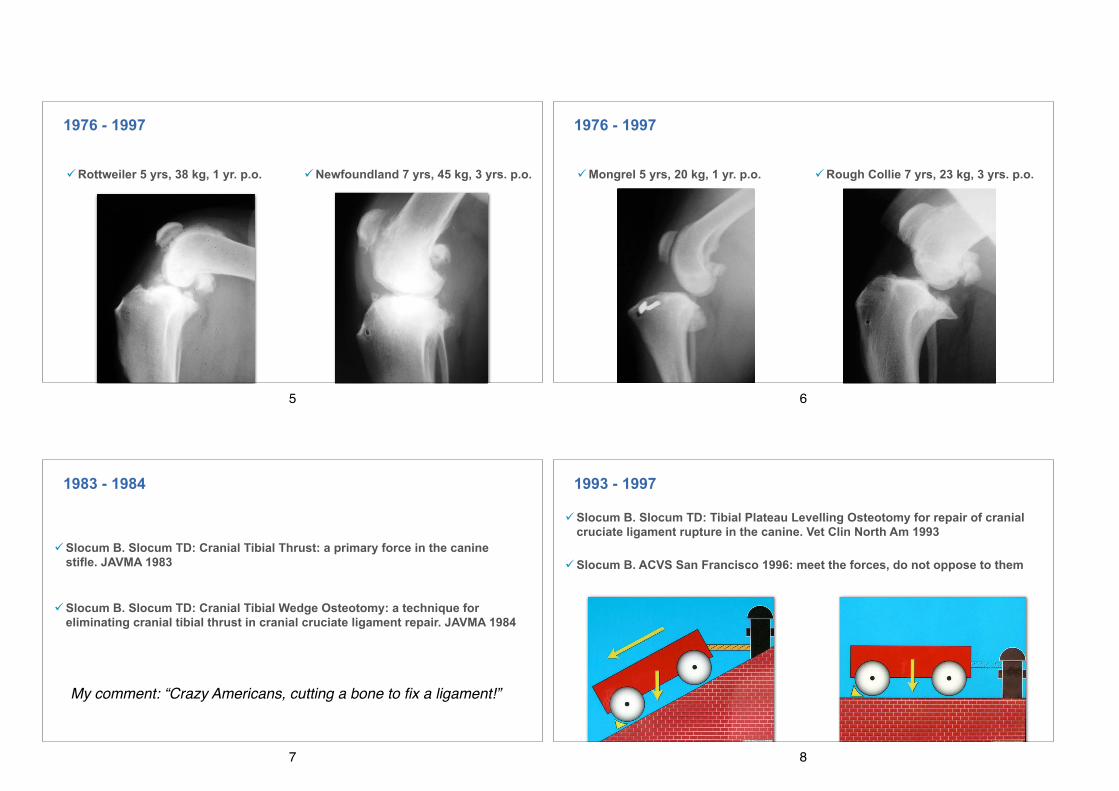
May 2004 - December 2013
!Momb, Labrador F, 6 yrs.,
bilateral TTA, FU 3 yrs.
17
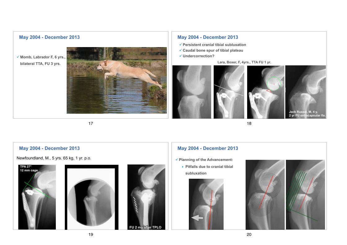
May 2004 - December 2013!Persistent cranial tibial subluxation !Caudal bone spur of tibial plateau !Undercorrection?
Long term FU of TTABoxer F, 4 yrs. TTA 6 mm (undercorrected) FU 1 yr., lame,
31
Jack Russel, M, 4 y, 2 yr FU extracapsular fix.
Lara, Boxer, F, 4yrs., TTA FU 1 yr.
18
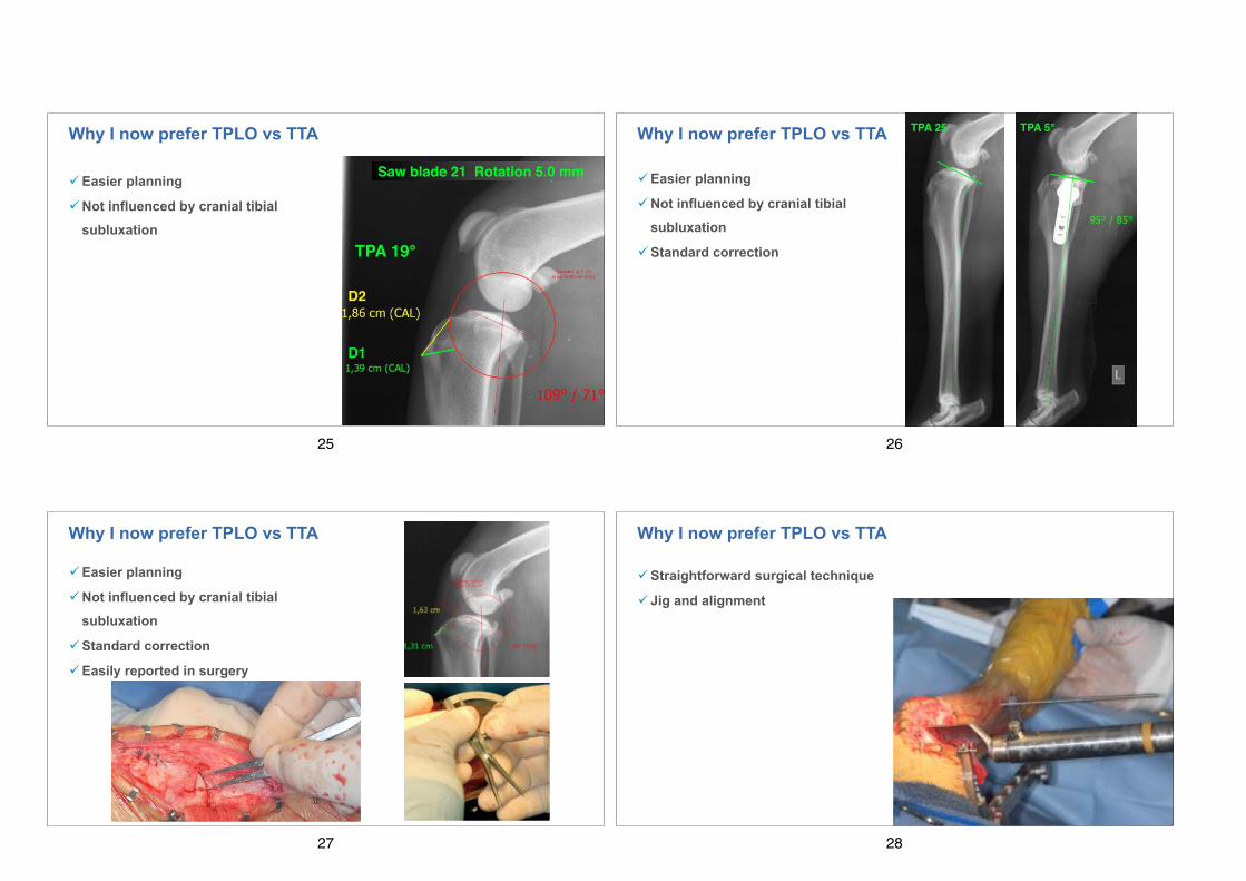
May 2004 - December 2013
Newfoundland, M., 5 yrs. 65 kg, 1 yr. p.o.
FU 2 mo after TPLO
TPA 27°12 mm cage
19
May 2004 - December 2013
!Planning of the Advancement:
‣ Pitfalls due to cranial tibial
subluxation
20
May 2004 - December 2013
!Planning of the Advancement:
‣ Pitfalls due to tibial tuberosity morphology and patella tendon insertion
21
May 2004 - December 2013
!Effective Advancement:
‣ Influenced by cage position
22
May 2004 - December 2013
!Effective Advancement:
‣ attempts to improve planning
● B: most distal point of the margo cranial-is tibiae
● C: most caudal point of the tibial plateau, represented by the midpoint between the medial and lateral tibial condyles
● D: most cranial point of the tibial plateau The following points, lines and angles were calculated by geometric measures: ● E: cross point of a circle with the center C
and the radius CD, and the line AC ● F: cross point of the circle with the center
D and the radius 2 x DB, and the cranial border of the tibial cortex
● G: cross point of the circle with the center D and the radius 2 x DB, and the caudal border of the tibial cortex
● Proximal tibial tuberosity angle (PTTA): α, angle formed by DAE
● Tibial plateau angle (TPA): β, angle formed by ACD
● Tibia width (TW): distance between F and G
● Tibial plateau length (TPL): distance be-tween C and D
● Relative tibial tuberosity width (rTTW): AE/EC
The relation of tibial width to tibial tuberos-ity width was calculated by the quotient TW/AE. The relation of tibial width to the caudal tibial plateau length was calculated by the quotient TW/EC.
The body weight (BW) of each dog was measured and the relative body weight (rBW) was calculated by the quotient BW/TW.
Clinical data collection The radiographs were assigned to three main groups: healthy dogs (H), dogs with confirmed partial or complete CrCL rupture (R), and dogs with a TTA treatment (T). The diagnosis of the partial or complete CrCL in the R and T groups was made by clinical testing, or by surgical exploration. Subdivi-sion was made into dogs younger than five years (Y), and dogs five years of age and older (O), resulting in six subgroups. Age and body weight (BW) were noted.
Statistical analysis The analysis was performed with StatView 5.1 (SAS: StatView 5.0 Statistical Software, Cary, NC, USA, 2002). A factorial analysis
of variance (ANOVA) was calculated. The Bonferroni/Dunn analysis was made for the subgroups HY, HO, RY, RO, TY, TO with the following parameters: BW, age, PTTA, TPA, rTTW, rBW, TW/EC and TW/AE. Level of significance was set to p < 0.05 and to p < 0.0033 with Bonferroni/Dunn correction.
Results The data collection is given in Table 1 and the Bunferroni/Dunn analysis in Table 2. The most important differences were as fol-lows: the dogs with stifles from Group H (Fig. 2) had a greater rTTW, a lower BW, a lower rBW, a smaller PTTA, a smaller TW/AE and were younger than those patients from Group R (Fig. 3). A comparison of Groups H and T (Fig. 4) revealed that dogs from Group H had a smaller rTTW, a lower BW, a lower rBW, a greater PTTA, a greater TW/AE and were younger.
In each of the comparisons, there was a lack of significant difference in TPA and TW/EC.
Discussion The identification of the described anatomi-cal landmarks was easy. The only problem was that the medial and lateral condyles were not superimposed in the radiographic views. We chose the mid-point, between the
Fig. 1 Schematic illustration of a canine stifle in a medio-lateral view. A: most proximal point of the margo cranialis tibiae; B: most distal point of the margo cranialis tibiae; C: most caudal point of the tibial plateau, repre-sented by the midpoint between the medial and lateral ti-bial condyles; D: most cranial point of the tibial plateau; E: cross point of a circle with the center C and the radius CD, and the line AC; F: cross point of the circle with the center D and the radius 2 x DB, and the cranial border of the tibial cortex; G: cross point of the circle with the center D and the radius 2 x DB, and the caudal border of the tibial cortex
Table 1 Group assignment and data collection of radiographs of 219 stifle joints.
For personal or educational use only. No other uses without permission. All rights reserved.Downloaded from www.vcot-online.com on 2015-04-25 | ID: 1000446089 | IP: 155.48.255.252
Received: August 8, 2007 Vet Comp Orthop Traumatol 1/2009 Accepted: February 21, 2008
Tibial tuberosity conformation as a risk factor for cranial cruciate ligament rupture in the dog R. Inauen1; D. Koch1; M. Bass1; M. Haessig2 1Koch & Bass Referral Practice for Small Animal Surgery, Diessenhofen, Switzerland; 2Large Animal Department, Section for Herd Health, Vetsuisse Faculty, University of Zurich, Zurich, Switzerland
Summary The influence of the tibial tuberosity conformation on cranial cruciate ligament (CrCl) rupture was evaluated and the size of the tibial tuberosity of healthy dogs (group H) was compared with dogs with CrCl rupture (group R) and dogs treated by tibial tuberosity ad-vancement (TTA) (group T). The medio-lateral radio-graphs of 219 stifle joints were evaluated. Relative ti-bial tuberosity width (rTTW), proximal tibial tuberosity angle (PTTA), tibial plateau angle (TPA), tibial width (TW) and tibial plateau length (TPL) were measured on each radiograph. Body weight (BW) was measured and relative body weight (rBW) was calculated. The data from group H was compared with that of group R and group T. Group H had significantly larger rTTW, lower BW, lower rBW and smaller PTTA than group R. A com-parison of groups H and T showed that dogs from group H were significantly younger, had a lower BW, a lower rBW, a greater PTTA and a smaller rTTW. In each of the comparisons, the TPA and the TW/TPL were not signifi-cantly different. The conformation of the canine tibial tuberosity has a significant influence on CrCl rupture. We hypothesized that the smaller the tibial tuberosity width, the larger the cranial tibial thrust, which results in more rapid CrCL degeneration, thus leading to rup-ture in a younger population of dogs. The rTTW could be a helpful measurement for breeding selection. Only dogs with a rTTW of more than 0.90 should be used for breeding.
Prepublished online: April 11, 2008 doi:10.3415/VCOT-07-08-0078
Introduction
Rupture of the cranial cruciate ligament (CrCL) is one of the most common causes of hind limb lameness in dogs (1). In contrast to humans, the pathophysiology is not ex-plained by trauma, but rather by degener-ation (2–4). A review of the literature re-veals many theories about the aetiology, among which are immune-mediated arthro-pathies (5–7), bodyweight (8, 9), breed pre-disposition (9), abnormal bone con-formation (10–14), genetic factors (3, 8, 9) and the tibial plateau slope (15–17)
In 1978, Henderson postulated that two main forces exist between the femur and tibia: one force being a compressive force acting through the stifle, and the other a sheer force acting parallel to the joint sur-faces. The latter force is generated by the ga-strocnemius muscles and is counteracted by the CrCL (15). Slocum (17), re-investigated the forces acting around the stifle joint. He concluded that an increased tibial plateau slope increased the load on the CrCL and promoted degeneration and subsequent par-tial and complete rupture (17). Many au-thors confirmed his ideas (18–22), however, contradictionary investigations (23, 24) demonstrate that the aetiopathogenesis is still not clearly understood. Slocum’s idea concerning the pathogenesis of CrCL rup-ture led to him to develop the tibia plateau levelling osteotomy surgery (25). In this sur-gery, the stifle biomechanics are altered in order to reduce the shear forces to nearly zero, thus making the CrCL redundant.
Another group (26) calculated the stifle forces differently than Slocum did, and pro-posed an advancement of the tibial tuberos-ity (TTA). The quadriceps muscle was identified as the main force in the stifle
joint. Its insertion on the tibial tuberosity is set to 90° by the TTA, thereby neutralizing the cranial tibial thrust. This fact has re-cently been demonstrated (27, 28).
The fact that the tibial tuberosity ge-ometry influences the stifle biomechanics, as well as the clinical observation that the ti-bial tuberosities have individual shapes, led us to the hypothesis that dogs with large ti-bial tuberosities would have a decreased risk of sustaining a CrCL rupture.
The aim of this study was to detect risk factors for cranial cruciate ruptures in the anatomy of the tibial tuberosity. These risk factors could then be used for breeding guidelines. Furthermore, the study should have the potential to be useful in clinical routine situations. Therefore, the investi-gation was made on routine radiographs of the stifle joint.
Material and methods Radiographs The medio-lateral radiographs of 219 stifle joints were evaluated. The radiographs were taken between April 2004 and April 2006. All of the radiographs on which the caudal margin of the medial and lateral tibial con-dyles were separated less than 4 mm were included in the study.
Radiographic analysis (Fig. 1) The following points were identified on the medio-lateral radiographs: ● A: most proximal point of the margo
For personal or educational use only. No other uses without permission. All rights reserved.Downloaded from www.vcot-online.com on 2015-04-25 | ID: 1000446089 | IP: 155.48.255.252
23
!Easier planning
!Straightforward surgical technique
!Repeatability intra and inter operators
!Consistent outcome
!Predictable clinical results
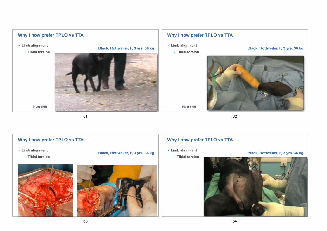
!Versatility
!Limb alignment
!Patellar luxation
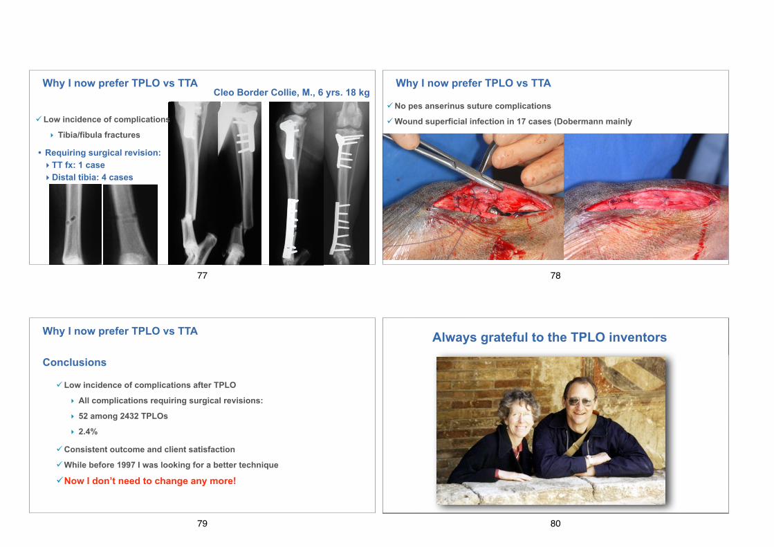
!Low complication rate
Why I now prefer TPLO vs TTA
24
!Easier planning
!Not influenced by cranial tibial
subluxation
Saw blade 21 Rotation 5.0 mm
TPA 19°
D1
D2
Why I now prefer TPLO vs TTA
25
!Easier planning
!Not influenced by cranial tibial
subluxation
!Standard correction
TPA 25° TPA 5°Why I now prefer TPLO vs TTA
26
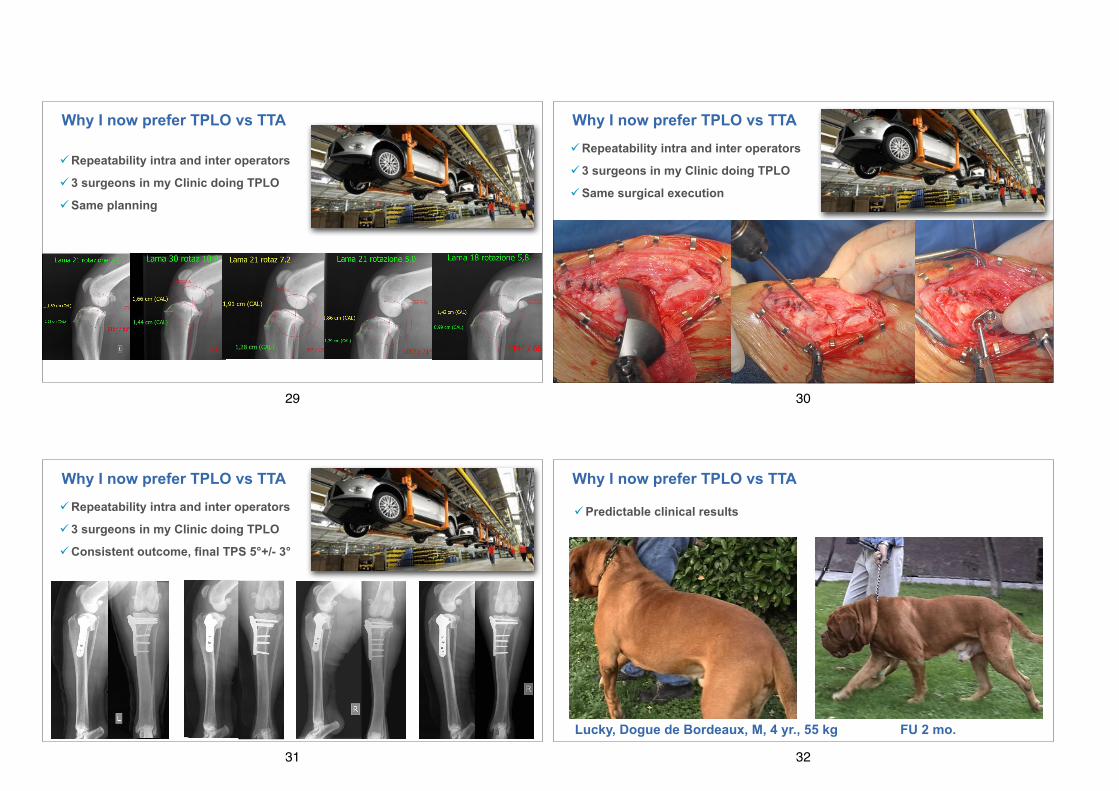
!Easier planning
!Not influenced by cranial tibial
subluxation
!Standard correction
!Easily reported in surgery
Why I now prefer TPLO vs TTA
27
!Straightforward surgical technique
!Jig and alignment
Why I now prefer TPLO vs TTA
28
!Repeatability intra and inter operators
!3 surgeons in my Clinic doing TPLO
!Same planning
Why I now prefer TPLO vs TTA
29
!Repeatability intra and inter operators
!3 surgeons in my Clinic doing TPLO
!Same surgical execution
Why I now prefer TPLO vs TTA
30
!Repeatability intra and inter operators
!3 surgeons in my Clinic doing TPLO
!Consistent outcome, final TPS 5°+/- 3°
Why I now prefer TPLO vs TTA
31
!Predictable clinical results
FU 2 mo.Lucky, Dogue de Bordeaux, M, 4 yr., 55 kg
Why I now prefer TPLO vs TTA
32
!Predictable clinical results
!Client satisfaction
Lucky, Dogue de Bordeaux, M, 4 yr., 55 kg FU 6 mo.
Best in show Barcellona, World Dog Show 2001
Why I now prefer TPLO vs TTA
33
!Predictable clinical results
!Client satisfaction
! Field champion winning again!
Dila, German Shorthaired Pointer, F, 5 yr., 18 kg FU 5 mo.
Why I now prefer TPLO vs TTA
34
Toby, mongrel, M, 7 yr., 4 kg, FU 3 mo.
!Predictable clinical results
!Client satisfaction
Why I now prefer TPLO vs TTA
35
!Predictable clinical results
!Long term FUs Mongrel F, 3 yrs. 23 kg FU 5 yrs.
Why I now prefer TPLO vs TTA
36
!Predictable clinical results
!Long term FUs Labrador M, 7 yrs. 36 kg FU 5 yrs.
Why I now prefer TPLO vs TTA
37
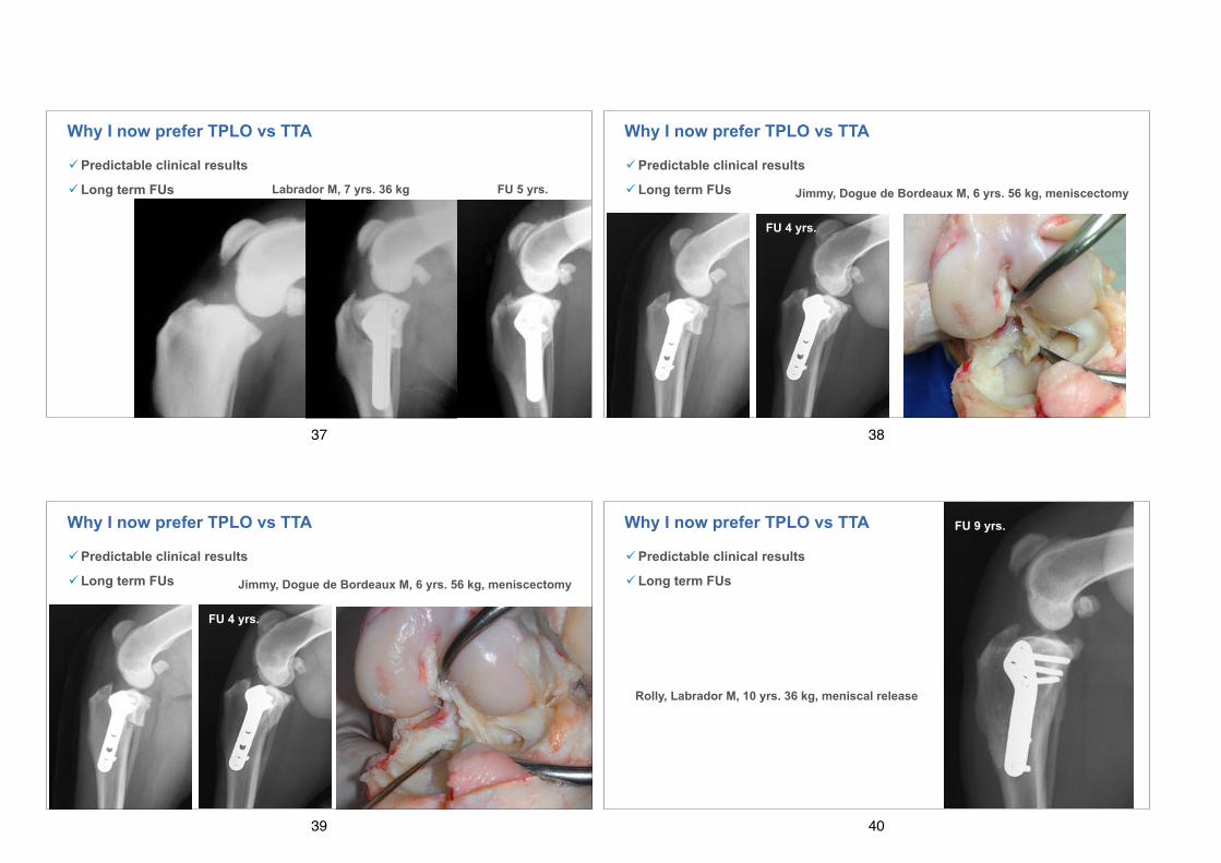
!Predictable clinical results
!Long term FUs Jimmy, Dogue de Bordeaux M, 6 yrs. 56 kg, meniscectomy
Why I now prefer TPLO vs TTA
FU 4 yrs.
38
!Predictable clinical results
!Long term FUs Jimmy, Dogue de Bordeaux M, 6 yrs. 56 kg, meniscectomy
Why I now prefer TPLO vs TTA
FU 4 yrs.
39
!Predictable clinical results
!Long term FUs
Rolly, Labrador M, 10 yrs. 36 kg, meniscal release
Why I now prefer TPLO vs TTA FU 9 yrs.
40
!Versatility
‣ Any dog size and weight Lea, Yorskshire, F, 5 yr., 2 kg
Why I now prefer TPLO vs TTA
41
!Versatility
‣ Any dog size and weight
Jack, JRT, M, 6 yr., 7 kg
Why I now prefer TPLO vs TTA
42
!Versatility
‣ Any dog size and weight
Jack, JRT, M, 6 yr., 7 kg FU 2 mo.
Why I now prefer TPLO vs TTA
43
!Versatility
‣ Any dog size and weight
Nelson, St. Bernard, M, 4 yr., 75 kg, Left complete, Right partial
Why I now prefer TPLO vs TTA
44
!Versatility
‣ Any dog size and weight
Nelson, St. Bernard, M, 4 yr., 75 kg, Left complete, Right partial
FU 4 mo. Right FU 2 mo. Left
Why I now prefer TPLO vs TTA
45
!Versatility
‣ Any tibial plateau slope
Snoopy, mongrel, M, 7 yrs., 6.9 kg
TPA 39° pre op TPA 6° post-op
Why I now prefer TPLO vs TTA
46
!Versatility
‣ Any tibial plateau slope
Peggy, Yorkshire F, 9 yrs., 7 kg
TPA 40° pre op TPA 5° post-op
Why I now prefer TPLO vs TTA
47
!Versatility
‣ Pathologic tibial plateau slope
Lapo, Drahthaar M, 3 yrs., 33 kg TPLO+CCWO
TPA 42° pre op TPA 7° post-op
Why I now prefer TPLO vs TTA
48
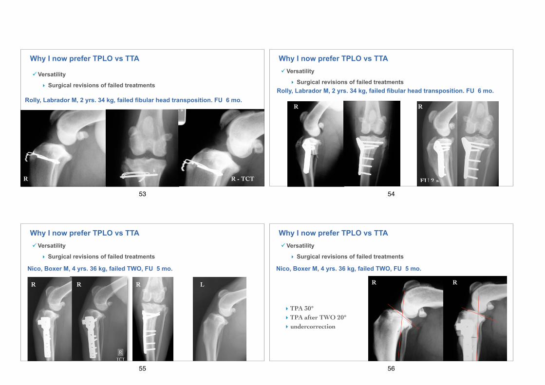
!Versatility
‣ Surgical revisions of failed treatments
Bichon frisé, M, 9 yrs. 4.3 kg, failed extracapsular stabilization
Why I now prefer TPLO vs TTA
49
!Versatility
‣ Surgical revisions of failed treatments
Trilli, Spaniel frisé, M, 4 yrs. 21.5 kg, failed extracapsular stabilization FU 9 mo.