Cell Reports Report Wiskott-Aldrich Syndrome Protein Regulates Leukocyte-Dependent Breast Cancer Metastasis Dan Ishihara, 1,5 Athanassios Dovas, 1,5 Lorena Hernandez, 1 Maria Pozzuto, 2 Jeffrey Wyckoff, 1,3 Jeffrey E. Segall, 1,3 John S. Condeelis, 1,3 Anne R. Bresnick, 2 and Dianne Cox 1,3,4, * 1 Department of Anatomy and Structural Biology 2 Department of Biochemistry 3 Gruss Lipper Biophotonics Center 4 Department of Developmental and Molecular Biology Albert Einstein College of Medicine, Bronx, NY 10461, USA 5 These authors contributed equally to this work *Correspondence: [email protected]http://dx.doi.org/10.1016/j.celrep.2013.07.007 This is an open-access article distributed under the terms of the Creative Commons Attribution License, which permits unrestricted use, distribution, and reproduction in any medium, provided the original author and source are credited. SUMMARY A paracrine interaction between epidermal growth factor (EGF)-secreting tumor-associated macro- phages (TAMs) and colony-stimulating factor 1 (CSF-1)-secreting breast carcinoma cells promotes invasion and metastasis. Here, we show that mice deficient in the hematopoietic-cell-specific Wiskott- Aldrich syndrome protein (WASp) are unable to support TAM-dependent carcinoma cell invasion and metastasis in both orthotopic and transgenic models of mammary tumorigenesis. Motility and invasion defects of tumor cells were recapitulated ex vivo upon coculture with WASp / macrophages. Mechanistically, WASp is required for macrophages to migrate toward CSF-1-producing carcinoma cells, as well as for the release of EGF through metalloprotease-dependent shedding of EGF from the cell surface of macrophages. Our findings sug- gest that WASp acts to support both the migration of TAMs and the production of EGF, which in concert promote breast tumor metastasis. INTRODUCTION Cancer progression is a complex, multistep process consisting of neoplastic cell transformation, tumor growth, invasion, and metastasis. Although metastasis is the major cause of cancer deaths, it remains poorly characterized (Valastyan and Wein- berg, 2011). Malignant dissemination is not an exclusively cancer-cell-autonomous process; other constituents of the tumor microenvironment, such as inflammatory cells, heavily influence cancer progression (Hanahan and Coussens, 2012). Additionally, clinical studies on invasive breast cancer have demonstrated that tumor-associated macrophage (TAM) density correlates with poor prognosis (Qian and Pollard, 2010). Indeed, TAMs promote angiogenesis and extracellular matrix proteolysis, and enhance tumor cell invasion and dissemination (Joyce and Pollard, 2009). Circulating levels of colony-stimulating factor 1 (CSF-1), a pleiotropic macrophage growth factor and chemoattractant, correlate with poor prognosis in breast cancer patients, and high levels of CSF-1 are found in invasive, but not in situ, breast cancer (Qian and Pollard, 2010). In agreement with these observations, CSF-1 deficiency in the polyoma middle-T antigen mouse model of mammary carcinogenesis (MMTV-PyMT) led to a reduction in TAM density, delayed the development of tumors to invasive metastatic carcinoma, and reduced lung metastasis with no change in primary tumor growth (Lin et al., 2001), demon- strating that CSF-1 played a critical role in breast tumor progres- sion and metastasis. Macrophages and breast carcinoma cells depend on an epidermal growth factor (EGF)/CSF-1 paracrine loop for invasion both in vitro (Goswami et al., 2005) and in vivo (Wyckoff et al., 2004). In vivo tumor cell invasion was blocked by inhibiting either the EGF receptor (EGFR) or the CSF-1 receptor (CSF-1R), expressed by breast carcinoma cells and macrophages, res- pectively (Wyckoff et al., 2004). Given that recruitment of higher numbers of TAMs has been reported in EGFR-positive breast tumors (Leek et al., 2000), blockade of the paracrine interaction may have therapeutic benefit in the treatment of breast cancer. Additionally, targeting TAMs is attractive because, unlike cancer cells, they are genetically stable and less likely to develop drug resistance. We demonstrate here that Wiskott-Aldrich syndrome protein (WASp) could be a potential therapeutic target. Wiskott-Aldrich syndrome (WAS) is an X-linked genetic disorder resulting from mutations in the WAS gene (Thrasher and Burns, 2010). WASp is expressed exclusively in none- rythroid hematopoietic cells, unlike its ubiquitously expressed homolog, neural (N)-WASP. WASp is an actin nucleation- promoting factor that regulates chemotaxis (Takenawa and Suetsugu, 2007). Since macrophages of WAS patients are defective in chemotaxis toward CSF-1 (Zicha et al., 1998), we hypothesized that WASp deficiency may block the para- crine interaction between TAMs and breast carcinoma cells, Cell Reports 4, 429–436, August 15, 2013 ª2013 The Authors 429
Transcript
Cell Reports
Report
Wiskott-Aldrich Syndrome Protein RegulatesLeukocyte-Dependent Breast Cancer MetastasisDan Ishihara,1,5 Athanassios Dovas,1,5 Lorena Hernandez,1 Maria Pozzuto,2 Jeffrey Wyckoff,1,3 Jeffrey E. Segall,1,3
John S. Condeelis,1,3 Anne R. Bresnick,2 and Dianne Cox1,3,4,*1Department of Anatomy and Structural Biology2Department of Biochemistry3Gruss Lipper Biophotonics Center4Department of Developmental and Molecular BiologyAlbert Einstein College of Medicine, Bronx, NY 10461, USA5These authors contributed equally to this work
This is an open-access article distributed under the terms of the Creative Commons Attribution License, which permits unrestricted use,
distribution, and reproduction in any medium, provided the original author and source are credited.
SUMMARY
A paracrine interaction between epidermal growthfactor (EGF)-secreting tumor-associated macro-phages (TAMs) and colony-stimulating factor 1(CSF-1)-secreting breast carcinoma cells promotesinvasion and metastasis. Here, we show that micedeficient in the hematopoietic-cell-specific Wiskott-Aldrich syndrome protein (WASp) are unable tosupport TAM-dependent carcinoma cell invasionand metastasis in both orthotopic and transgenicmodels of mammary tumorigenesis. Motility andinvasion defects of tumor cells were recapitulatedex vivo upon coculture with WASp�/� macrophages.Mechanistically, WASp is required for macrophagesto migrate toward CSF-1-producing carcinomacells, as well as for the release of EGF throughmetalloprotease-dependent shedding of EGF fromthe cell surface of macrophages. Our findings sug-gest that WASp acts to support both the migrationof TAMs and the production of EGF, which in concertpromote breast tumor metastasis.
INTRODUCTION
Cancer progression is a complex, multistep process consisting
of neoplastic cell transformation, tumor growth, invasion, and
metastasis. Although metastasis is the major cause of cancer
deaths, it remains poorly characterized (Valastyan and Wein-
berg, 2011). Malignant dissemination is not an exclusively
cancer-cell-autonomous process; other constituents of the
tumor microenvironment, such as inflammatory cells, heavily
influence cancer progression (Hanahan and Coussens, 2012).
Additionally, clinical studies on invasive breast cancer have
demonstrated that tumor-associatedmacrophage (TAM) density
correlates with poor prognosis (Qian and Pollard, 2010).
Indeed, TAMs promote angiogenesis and extracellular matrix
proteolysis, and enhance tumor cell invasion and dissemination
(Joyce and Pollard, 2009).
Circulating levels of colony-stimulating factor 1 (CSF-1), a
pleiotropic macrophage growth factor and chemoattractant,
correlate with poor prognosis in breast cancer patients, and
high levels of CSF-1 are found in invasive, but not in situ, breast
cancer (Qian and Pollard, 2010). In agreement with these
observations, CSF-1 deficiency in the polyoma middle-T antigen
mouse model of mammary carcinogenesis (MMTV-PyMT) led to
a reduction in TAM density, delayed the development of tumors
to invasive metastatic carcinoma, and reduced lung metastasis
with no change in primary tumor growth (Lin et al., 2001), demon-
strating that CSF-1 played a critical role in breast tumor progres-
sion and metastasis.
Macrophages and breast carcinoma cells depend on an
epidermal growth factor (EGF)/CSF-1 paracrine loop for invasion
both in vitro (Goswami et al., 2005) and in vivo (Wyckoff et al.,
2004). In vivo tumor cell invasion was blocked by inhibiting either
the EGF receptor (EGFR) or the CSF-1 receptor (CSF-1R),
expressed by breast carcinoma cells and macrophages, res-
pectively (Wyckoff et al., 2004). Given that recruitment of higher
numbers of TAMs has been reported in EGFR-positive breast
tumors (Leek et al., 2000), blockade of the paracrine interaction
may have therapeutic benefit in the treatment of breast cancer.
Additionally, targeting TAMs is attractive because, unlike cancer
cells, they are genetically stable and less likely to develop drug
resistance.
We demonstrate here that Wiskott-Aldrich syndrome
protein (WASp) could be a potential therapeutic target.
Wiskott-Aldrich syndrome (WAS) is an X-linked genetic
disorder resulting from mutations in the WAS gene (Thrasher
and Burns, 2010). WASp is expressed exclusively in none-
rythroid hematopoietic cells, unlike its ubiquitously expressed
homolog, neural (N)-WASP. WASp is an actin nucleation-
promoting factor that regulates chemotaxis (Takenawa and
Suetsugu, 2007). Since macrophages of WAS patients are
defective in chemotaxis toward CSF-1 (Zicha et al., 1998),
we hypothesized that WASp deficiency may block the para-
crine interaction between TAMs and breast carcinoma cells,
Cell Reports 4, 429–436, August 15, 2013 ª2013 The Authors 429
Figure 1. WASp Is Required for Cell Elongation and Invasion of Both Breast Carcinoma Cells and Macrophages
MTLn3GFP cells were cultured alone or with WASp+/+, WASp�/�, or WASp�/� rescued (WASp-Re) BMMs for 16 hr.
(A) Representative phase-contrast images of the indicated conditions. MTLn3 cells are denoted by a C in coculture images. Scale bar: 10 mm.
(B and C) Quantification of the average elongation of (B) MTLn3GFP and (C) the indicated macrophage types. n R 4 independent experiments. *p < 0.05,
**p < 0.01 compared with control cells.
(D and E) Quantification of the fraction of MTLn3 (D) or BMMs (E) invading above 20 mm in the absence (�) or presence of WASp+/+ or WASp�/� BMMs from three
independent mice.
Error bars signify SEM from five independent fields. ****p < 0.0001 compared with MTLn3 cultured conditions with WASp+/+ BMMs. See also Figure S1 and
Movies S1 and S2.
disabling their comigration and attenuating metastatic
dissemination.
RESULTS
Macrophage WASp Is Required for Breast CarcinomaCell Interaction In VitroTo test whether WASp plays a role in the EGF/CSF-1 paracrine
interaction, we cocultured bone-marrow-derived macrophages
(BMMs) from WASp+/+ or WASp�/� mice with GFP-expressing
MTLn3 cells (MTLn3GFP). Contrary to the WASp�/� BMMs,
the WASp+/+ BMMs induced an increase in MTLn3GFP elonga-
tion (Figures 1A and 1B), indicative of the acquisition of a motile
phenotype (Goswami et al., 2005). Concurrently, the WASp+/+
BMMs elongated in the presence of MTLn3GFP cells, whereas
the WASp�/� BMMs did not (Figures 1A and 1C). Expression
of human WASp in WASp�/� BMMs restored both MTLn3 and
BMM elongation in cocultures (Figures 1B and 1C). Live-cell
imaging of BMMs added to MTLn3GFP cultures showed that
within the first few hours of coculture, the WASp+/+ BMMs elon-
gated robustly toward MTLn3GFP cells, whereas the WASp�/�
BMMs did not (Figures S1A–S1C; Movies S1 and S2).
To test whether WASp was required for macrophage-depen-
dent MTLn3 cell invasion into 3D matrices, we overlaid BMM
and MTLn3GFP cocultures with a collagen I gel and determined
the fraction of invading cells (Goswami et al., 2005). WASp+/+
430 Cell Reports 4, 429–436, August 15, 2013 ª2013 The Authors
BMMs enhanced MTLn3GFP invasion into the matrix 7-fold,
whereas MTLn3GFP invasion was not enhanced by WASp�/�
BMMs (Figures 2A, 2B, and S1D). In the presence of MTLn3GFP
cells, the fraction of coinvading WASp+/+ BMMs was increased
significantly compared with WASp�/� BMMs (Figures 2A, 2C,
and S1D). WASp expression was therefore necessary for
BMMs to adopt a migratory phenotype and to engage in and
maintain the paracrine interaction with breast carcinoma cells,
and for BMMs and breast carcinoma cells to coinvade a three-
dimensional (3D) collagen matrix. Importantly, WASp was not
required for BMM invasion into a fibrous collagen matrix in the
absence of carcinoma cells (A.D. Haein Park, I. Maridonneau-
Parini, and D.C., unpublished data), which suggests that WASp
is required for movement in response to a soluble guidance
cue (in this case, CSF-1 provided by carcinoma cells). This is
consistent with the well-established role of WASp in the chemo-
taxis of macrophages. These results prompted us to investigate
whether WASp is required for macrophage-dependent breast
tumor invasiveness and metastasis in mouse models of breast
cancer.
WASp Is Required for Efficient Breast CancerMetastasisTo specifically test the effect of myeloid WASp on tumor inva-
siveness and metastasis, we crossed WASp+/� or WASp�/�
mice with Rag2�/� mice to generate xenograft-competent
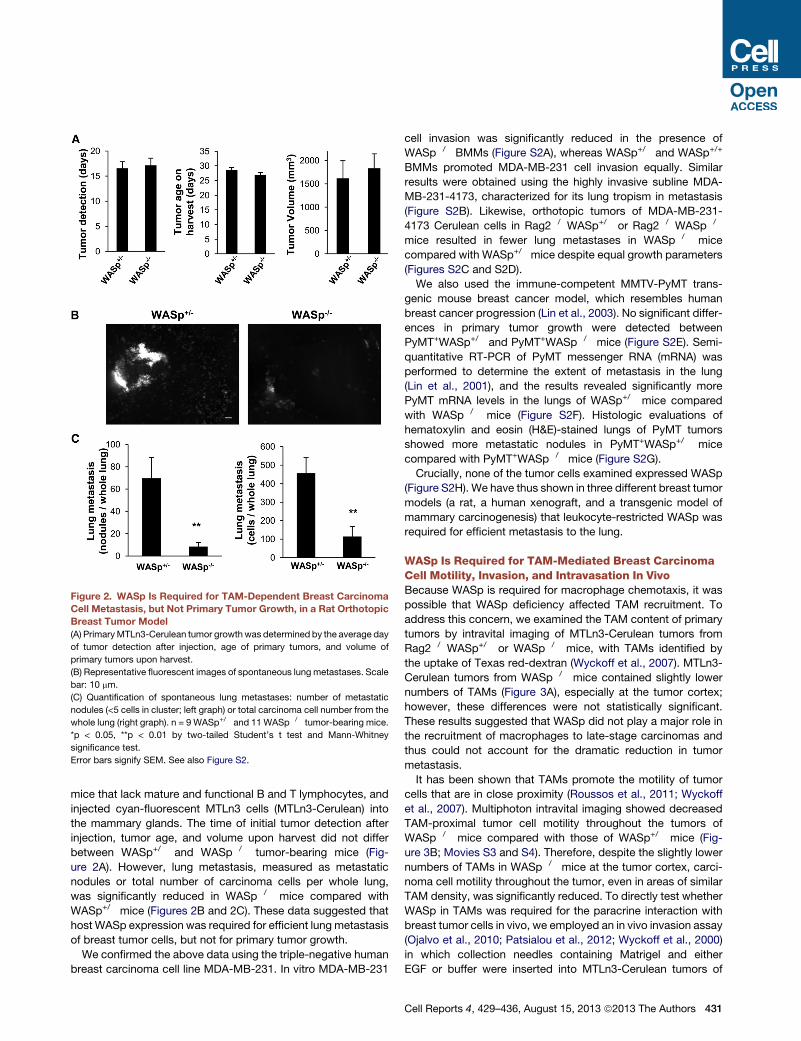
Figure 2. WASp Is Required for TAM-Dependent Breast Carcinoma
Cell Metastasis, but Not Primary Tumor Growth, in a Rat Orthotopic
Breast Tumor Model
(A) PrimaryMTLn3-Cerulean tumor growth was determined by the average day
of tumor detection after injection, age of primary tumors, and volume of
primary tumors upon harvest.
(B) Representative fluorescent images of spontaneous lung metastases. Scale
bar: 10 mm.
(C) Quantification of spontaneous lung metastases: number of metastatic
nodules (<5 cells in cluster; left graph) or total carcinoma cell number from the
whole lung (right graph). n = 9 WASp+/� and 11 WASp�/� tumor-bearing mice.
*p < 0.05, **p < 0.01 by two-tailed Student’s t test and Mann-Whitney
significance test.
Error bars signify SEM. See also Figure S2.
mice that lack mature and functional B and T lymphocytes, and
injected cyan-fluorescent MTLn3 cells (MTLn3-Cerulean) into
the mammary glands. The time of initial tumor detection after
injection, tumor age, and volume upon harvest did not differ
between WASp+/� and WASp�/� tumor-bearing mice (Fig-
ure 2A). However, lung metastasis, measured as metastatic
nodules or total number of carcinoma cells per whole lung,
was significantly reduced in WASp�/� mice compared with
WASp+/� mice (Figures 2B and 2C). These data suggested that
host WASp expression was required for efficient lung metastasis
of breast tumor cells, but not for primary tumor growth.
We confirmed the above data using the triple-negative human
breast carcinoma cell line MDA-MB-231. In vitro MDA-MB-231
cell invasion was significantly reduced in the presence of
WASp�/� BMMs (Figure S2A), whereas WASp+/� and WASp+/+
BMMs promoted MDA-MB-231 cell invasion equally. Similar
results were obtained using the highly invasive subline MDA-
MB-231-4173, characterized for its lung tropism in metastasis
(Figure S2B). Likewise, orthotopic tumors of MDA-MB-231-
4173 Cerulean cells in Rag2�/�WASp+/� or Rag2�/�WASp�/�
mice resulted in fewer lung metastases in WASp�/� mice
compared with WASp+/� mice despite equal growth parameters
(Figures S2C and S2D).
We also used the immune-competent MMTV-PyMT trans-
genic mouse breast cancer model, which resembles human
breast cancer progression (Lin et al., 2003). No significant differ-
ences in primary tumor growth were detected between
PyMT+WASp+/� and PyMT+WASp�/� mice (Figure S2E). Semi-
quantitative RT-PCR of PyMT messenger RNA (mRNA) was
performed to determine the extent of metastasis in the lung
(Lin et al., 2001), and the results revealed significantly more
PyMT mRNA levels in the lungs of WASp+/� mice compared
with WASp�/� mice (Figure S2F). Histologic evaluations of
hematoxylin and eosin (H&E)-stained lungs of PyMT tumors
showed more metastatic nodules in PyMT+WASp+/� mice
compared with PyMT+WASp�/� mice (Figure S2G).
Crucially, none of the tumor cells examined expressed WASp
(Figure S2H). We have thus shown in three different breast tumor
models (a rat, a human xenograft, and a transgenic model of
mammary carcinogenesis) that leukocyte-restricted WASp was
required for efficient metastasis to the lung.
WASp Is Required for TAM-Mediated Breast CarcinomaCell Motility, Invasion, and Intravasation In VivoBecause WASp is required for macrophage chemotaxis, it was
possible that WASp deficiency affected TAM recruitment. To
address this concern, we examined the TAM content of primary
tumors by intravital imaging of MTLn3-Cerulean tumors from
Rag2�/�WASp+/� or WASp�/� mice, with TAMs identified by
the uptake of Texas red-dextran (Wyckoff et al., 2007). MTLn3-
Cerulean tumors from WASp�/� mice contained slightly lower
numbers of TAMs (Figure 3A), especially at the tumor cortex;
however, these differences were not statistically significant.
These results suggested that WASp did not play a major role in
the recruitment of macrophages to late-stage carcinomas and
thus could not account for the dramatic reduction in tumor
metastasis.
It has been shown that TAMs promote the motility of tumor
cells that are in close proximity (Roussos et al., 2011; Wyckoff
et al., 2007). Multiphoton intravital imaging showed decreased
TAM-proximal tumor cell motility throughout the tumors of
WASp�/� mice compared with those of WASp+/� mice (Fig-
ure 3B; Movies S3 and S4). Therefore, despite the slightly lower
numbers of TAMs in WASp�/� mice at the tumor cortex, carci-
noma cell motility throughout the tumor, even in areas of similar
TAM density, was significantly reduced. To directly test whether
WASp in TAMs was required for the paracrine interaction with
breast tumor cells in vivo, we employed an in vivo invasion assay
(Ojalvo et al., 2010; Patsialou et al., 2012; Wyckoff et al., 2000)
in which collection needles containing Matrigel and either
EGF or buffer were inserted into MTLn3-Cerulean tumors of
Cell Reports 4, 429–436, August 15, 2013 ª2013 The Authors 431
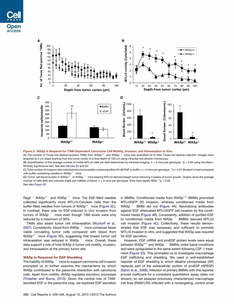
Figure 3. WASp Is Required for TAM-Dependent Carcinoma Cell Motility, Invasion, and Intravasation In Vivo
(A) The number of Texas-red dextran-positive TAMs from WASp+/� and WASp�/� mice was quantified 24 hr after Texas-red dextran injection. Images were
acquired at 5 mm steps starting from the tumor cortex to a final depth of 100 mm using intravital two-photon microscopy.
(B) Quantification of the average number of motile MTLn3 cells per field determined by intravital imaging. n > 3 mice per genotype. *p < 0.05 using the Mann-
Whitney significance test. See also Movies S3 and S4.
(C) Total number of invasive cells collected intomicroneedles containing either 25 nMEGF or buffer. n = 4mice per genotype. **p < 0.01 Student’s t test compared
with buffer-containing needles in WASp+/� mice.
(D) Tumor cell blood burden in WASp+/� or WASp�/� mice bearing MTLn3-derived breast tumor following 3 weeks of tumor growth. Graphs show the average
number of cells (left) and colonies (right) per milliliter of blood. n > 4 mice per genotype. Error bars signify SEM. **p < 0.05.
See also Figure S2.
Rag2�/�WASp+/� and WASp�/� mice. The EGF-filled needles
collected significantly more MTLn3-Cerulean cells than the
buffer-filled needles from tumors of WASp+/� mice (Figure 3C).
In contrast, there was no EGF-induced in vivo invasion from
tumors of WASp�/� mice even though TAM levels were only
reduced by a maximum of 30%.
TAMs also assist tumor cell intravasation (Wyckoff et al.,
2007). Consistently, blood from WASp�/� mice contained fewer
viable circulating tumor cells compared with blood from
WASp+/� mice (Figure 3D), suggesting that breast tumor cell
intravasation was reduced in WASp�/� mice. Overall, these
data support a role of host WASp in tumor cell motility, invasion,
and intravasation at the primary tumor site.
WASp Is Required for EGF SheddingThe inability ofWASp�/�mice to support carcinoma cell invasion
prompted us to further examine the mechanisms by which
WASp contributed to the paracrine interaction with carcinoma
cells. Apart from motility, WASp regulates secretory processes
(Thrasher and Burns, 2010). Given the central role of TAM-
secreted EGF in the paracrine loop, we explored EGF secretion
432 Cell Reports 4, 429–436, August 15, 2013 ª2013 The Authors
in BMMs. Conditioned media from WASp+/+ BMMs promoted
MTLn3GFP 3D invasion, whereas conditioned media from
WASp�/� BMM did not (Figure 4A). Neutralizing antibodies
against EGF attenuated MTLn3GFP cell invasion by the condi-
tioned media (Figure 4B). Consistently, addition of purified EGF
to conditioned media from WASp�/� BMMs rescued MTLn3
cell invasion (Figure 4C). Collectively, these results demon-
strated that EGF was necessary and sufficient to promote
MTLn3 invasion in vitro, and suggested that WASp was required
for EGF secretion.
However, EGF mRNA and proEGF protein levels were equal
between WASp+/+ and WASp�/� BMMs under basal conditions
and were upregulated to the same extent following CSF-1 treat-
ment (Figure S3). This prompted us to investigate macrophage
EGF trafficking and shedding. We used a well-established
reporter of EGF shedding in which alkaline phosphatase (AP)
replaces part of the extracellular portion of proEGF (APEGF)
(Sahin et al., 2006). Infection of primary BMMs with this reporter
proved inefficient for a consistent quantitative assay (data not
shown), so we assayed previously characterized macrophage
cell lines (RAW/LR5) infected with a nontargeting, control small
Figure 4. WASp Is Required for EGF Release
(A) Quantification of in vitro 3D invasion using MTLn3GFP cells cultured alone (�) or with conditioned media (CM) from WASp+/+ or WASp�/� BMMs.
(B) Quantification of 3D invasion of MTLn3GFP cells in the absence (�) or presence of WASp+/+ CM with either a nonspecific immunoglobulin G (IgG) or two
different EGF-neutralizing antibodies (aEGF Ab1 or Ab2).
(C) Addition of exogenous EGF (5 nM) to WASp�/� BMM CM restores invasion of MTLn3GFP cells. n R 3 experiments per condition.
(D) Confocal sections of control sh and shWASp RAW/LR5 cells expressing APEGF-sfGFP. Unpermeabilized fixed cells were stained with an antibody against
extracellular AP. Scale bar: 10 mm.
(E) Equal expression levels of APEGF-sfGFP (arrow) expressed by the control sh and shWASp cells, detected with an antibody against GFP.
(F) CM from the indicated APEGF-sfGFP-expressing cells under the conditions shown were collected and assayed for shed AP activity.
(G) Quantification of MTLn3GFP invasion with CM collected from WASp�/� BMM infected with either a control vector or a vector expressing recombinant
soluble EGF.
Error bars signify SEM. *p < 0.05, **p < 0.01 and ***p < 0.001. See also Figures S3 and S4.
Cell Reports 4, 429–436, August 15, 2013 ª2013 The Authors 433
hairpin RNA (shRNA, hereafter termed control sh) or one that
targets WASp (shWASp) (Dovas et al., 2009; Figure S4A). Similar
to BMMs, these cells engaged in WASp-dependent paracrine
interactions with carcinoma cells (Figures S4B–S4D). Control
sh or shWASp cells were infected with a retrovirus expressing
APEGF fused to superfolder GFP in its cytoplasmic tail
(APEGF-sfGFP). Both control sh and shWASp cells were able
to traffic APEGF-sfGFP to the cell surface, with no observable
differences in subcellular distribution (Figure 4D). Despite equal
expression levels of APEGF-sfGFP (Figure 4E) and basal
shedding of AP activity, the shWASp cells shed significantly
lower amounts of APEGF-sfGFP than the control sh cells
following CSF-1 stimulation (Figure 4F). Treatment of cells with
the broad-spectrum inhibitor GM6001 reduced shedding to its
basal levels (Figure 4F), indicating that WASp regulates CSF-1-
mediated EGF shedding in a metalloprotease-dependent
manner. Consistently, media from WASp�/� BMMs infected
with a construct expressing a secretable form of the mature,
soluble human EGF (Yao et al., 1998) bypassed the require-
ment for EGF shedding and promoted invasion of MTLn3 cells
(Figure 4G).
DISCUSSION
We have demonstrated that WASp regulated macrophage-
mediated tumor cell invasion in vitro and tumor cell motility,
invasion, intravasation, and lung metastasis, but not tumor
growth, in vivo. These results corroborate previous studies
implicating CSF-1 and TAMs in metastasis and suggest that
inhibition of effectors downstream of CSF-1R signaling may be
an effective strategy for antimetastatic therapy.
Additionally, we uncovered a role for WASp in the regulation of
CSF-1R-mediated EGF shedding, but found that EGF mRNA,
protein levels, and cell-surface expression were WASp indepen-
dent. Our data suggest that WASp may bridge CSF-1R signaling
to a cell-surface metalloprotease activity. Importantly, dendritic
cells deficient in WASp interacting protein (WIP), which binds
and regulates WASp levels in vivo, showed reduced secretion
of proMMP2 and proMMP9 (Banon-Rodrıguez et al., 2011).
Interference with the WASp pathway in myeloid cells may thus
lead to systemic defects in pericellular proteolysis.
WASp may regulate EGF shedding from the cell surface via
several, perhaps interdependent, mechanisms: (1) directly via
delivery of vesicles containing the sheddase(s) by F-actin-
mediated propulsion, (2) indirectly via the establishment of cell
polarity and the formation of F-actin conduits for sheddase
delivery, and (3) indirectly via the formation of podosomes,
F-actin-rich structures that concentrate proteolytic activities.
Such mechanisms have been proposed to act in the delivery of
proteases to the plasma membrane and to invadopodia of inva-
sive tumor cells (Poincloux et al., 2009). Further experiments will
be needed to determine the exact mechanism by which WASp
regulates EGF shedding.
The decreased in vivo cell invasion observed inWASp�/�mice
despite the presence of exogenous EGF supplied through the
microneedle (Figure 3C) may appear to contradict the in vitro
data indicating that EGF alone restored breast carcinoma cell in-
vasion (Figure 4C). However, efficient chemotaxis in the tumor
434 Cell Reports 4, 429–436, August 15, 2013 ª2013 The Authors
requires the propagation of chemotactic signals from cell to
cell in a streaming fashion (Roussos et al., 2011; Sharma et al.,
2012) so as to maintain the migration of cells. Consistent with
this, blockade of CSF-1R in the presence of EGF was equally
effective as EGFR blockade in reducing the number of invasive
cells collected (Wyckoff et al., 2004). Given that WASp was
required for the elongation of BMMs toward tumor cells in vitro
(Figure 1), our data suggest that WASp in macrophages contrib-
utes to carcinoma cell invasion by supporting both macrophage
chemotaxis and EGF release.
Our findings suggest thatWASp inhibition may have therapeu-
tic benefit in breast tumors. We argue that inhibition of a protein
downstream of CSF-1R could bypass several issues associated
with CSF-1R blockade, such as the nonspecificity of kinase
inhibitors or elevated levels of circulating CSF-1 in the case of