Interaction of Sulfonamide and Sulfone Compounds with Toxoplasma gondil Dihydropteroate Synthase Carmen J. Allegra,* Donna Boarman,* Joseph A. Kovacs,* Paul Morrison,1 Jacqueline Beaver,$ Bruce A. Chabner,* and Henry Masurt *Medicine Branch, National Cancer Institute, tClinical Center, and §Biomedical Engineering and Instrumentation Branch, Division ofResearch Services, National Institutes ofHealth, Bethesda, Maryland 20892 Abstract Toxoplasma gondii is a common protozoan disease that often causes life-threatening disease, particularly among patients with the acquired immunodeficiency syndrome. This study demonstrates that the dihydropteroate synthase in T. gondii is kinetically distinct from the enzyme characterized from other sources and can be highly purified with a high yield using sequential dye-affinity chromatography. Conditions have been identified that allow for stabilization of the purified enzyme, and its physical characteristics have been elucidated. The mo- lecular weight of the native protein was 125,000 and the pro- tein appeared to contain both dihydropteroate synthase and 6-hydroxymethyl-dihydropterin pyrophosphokinase activities. The sulfonamide class of compounds vary in inhibitory potency by more than three orders of magnitude. Sulfathiazole, sulfa- methoxazole, and sulfamethazine, with 50% inhibitory concen- trations (IC50's) of 1.7, 2.7, and 5.7 MM, respectively, represent the most potent of this class of inhibitors. Several sulfone analogues, including dapsone, were identified as highly potent inhibitors with ICN's < 1 uM. The results of these cell-free experiments were corroborated by investigating the metabolic inhibition produced by the various inhibitors in intact organ- isms. The qualitative and quantitative relations among the in- hibitors were preserved in both the cell-free and intact cell assay systems. These studies suggest that the sulfones may be important therapeutic agents for the treatment of toxoplas- mosis. (J. Clin. Invest. 1990. 85:371-379.) antimetabolites. enzymology- protozoan * sulfonamides Introduction Toxoplasma gondii infects up to 40% of individuals in North America (1). In immunocompetent patients, T. gondli expo- sure can result in a febrile lymphadenopathic syndrome, al- though most acutely infected adults are asymptomatic (1, 2). Fetal infection with T. gondii can occur in women who be- come infected during pregnancy, and may be accompanied by serious sequelae including neurologic abnormalities (1). T. gondii infection is prevalent in immunocompromised pa- tients, particularly those with AIDS. Clinical disease occurs in 5-10% of all patients with AIDS and in - 30% of toxoplasma Address correspondence to Dr. Carmen J. Allegra, National Cancer Institute, National Institutes of Health, Building 10, Room 12N226, Bethesda, MD 20892. Receivedfor publication 10 July 1989 and in revisedform 6 October 1989. The Journal of Clinical Investigation, Inc. Volume 85, February 1990, 371-379 antibody-positive AIDS patients in the United States (1). If the adult who has T. gondii cysts has diminished function of T lymphocytes, especially due to AIDS, fatal disseminated dis- ease or cerebral disease can occur. Folate compounds are essential cofactors for a number of critical metabolic steps including the formation of purines and thymidine nucleotides. Dihydrofolate reductase (DHFR)' is the enzyme responsible for the maintenance of folate pools in their physiologic reduced states. Dihydropteroate synthase (DHPS) is one of the enzymes responsible for the de novo synthesis of the folate molecule. Unlike mammalian cells, which possess a carrier-mediated active transport system re- sponsible for the uptake of performed folates, T. gondii lack this system and must synthesize their required folates de novo (3). Several of the enzymes of the de novo folate pathway, including DHPS, are unique to nonmammalian cells, and as such provide targets for drug therapy with a potentially high therapeutic index. Therapy of T. gondii disease has tradition- ally depended on a combination of pyrimethamine, a DHFR inhibitor, and sulfadiazine, an inhibitor of DHPS. In general, DHFR and DHPS inhibitors are most effective when used in combination (4). While combination therapy is effective, the incidence of adverse side-effects in the AIDS population is substantial (30-70%) (5, 6). Due to the severity of side-effects, this therapy must be discontinued in a large percentage of patients. Clearly, alternative therapy is urgently needed. A limited number of sulfonamide and sulfone compounds have been tested for antitoxoplasma activity in the murine animal model. Sabin and Warren initially demonstrated the utility of these compounds for the treatment of murine toxo- plasmosis (7). These studies were subsequently confirmed by several independent laboratories (8-13). The classic works of Eyles and Coleman assessed the relative in vivo potency of various sulfonamide inhibitors against T. gondii and found sulfapyrazine, sulfamethazine, sulfamerazine, and sulfadiazine to be the most potent of the sulfonamides tested, and approxi- mately equivalent in their ability to prolong the survival of infected mice (9-12). Notably, sulfisoxazole and sulfanilamide were found to be - 20 times less capable of prolonging sur- vival. Subsequent studies from these same laboratories dem- onstrated the relative activity of several sulfone analogues, in- cluding 4,4'-diaminodiphenyl sulfone (dapsone) (13). They found dapsone to be equivalent in activity to sulfadiazine and the most potent of the tested sulfones. Based on the effectiveness of antifolate therapy and the presumption that many of the side-effects are dose- and inhibi- 1. Abbreviations used in this paper: DHFR, dihydrofolate reductase; DHPS, dihydropteroate synthase; H2PtCH20PP, 6-hydroxymethy- 7,8-dihydropterin pyrophosphate; 2-ME, 2-mercaptoethanol; pABA, paraaminobenzoate; Pi, isoelectric focusing point. Toxoplasma gondii Dihydropteroate Synthase 371
Transcript
Interaction of Sulfonamide and Sulfone Compoundswith Toxoplasma gondil Dihydropteroate SynthaseCarmen J. Allegra,* Donna Boarman,* Joseph A. Kovacs,* Paul Morrison,1 Jacqueline Beaver,$Bruce A. Chabner,* and Henry Masurt*Medicine Branch, National Cancer Institute, tClinical Center, and §Biomedical Engineering and Instrumentation Branch,Division ofResearch Services, National Institutes ofHealth, Bethesda, Maryland 20892
Abstract
Toxoplasma gondii is a common protozoan disease that oftencauses life-threatening disease, particularly among patientswith the acquired immunodeficiency syndrome. This studydemonstrates that the dihydropteroate synthase in T. gondii iskinetically distinct from the enzyme characterized from othersources and can be highly purified with a high yield usingsequential dye-affinity chromatography. Conditions have beenidentified that allow for stabilization of the purified enzyme,and its physical characteristics have been elucidated. The mo-lecular weight of the native protein was 125,000 and the pro-tein appeared to contain both dihydropteroate synthase and6-hydroxymethyl-dihydropterin pyrophosphokinase activities.The sulfonamide class ofcompounds vary in inhibitory potencyby more than three orders of magnitude. Sulfathiazole, sulfa-methoxazole, and sulfamethazine, with 50% inhibitory concen-trations (IC50's) of 1.7, 2.7, and 5.7 MM, respectively, representthe most potent of this class of inhibitors. Several sulfoneanalogues, including dapsone, were identified as highly potentinhibitors with ICN's < 1 uM. The results of these cell-freeexperiments were corroborated by investigating the metabolicinhibition produced by the various inhibitors in intact organ-isms. The qualitative and quantitative relations among the in-hibitors were preserved in both the cell-free and intact cellassay systems. These studies suggest that the sulfones may beimportant therapeutic agents for the treatment of toxoplas-mosis. (J. Clin. Invest. 1990. 85:371-379.) antimetabolites.enzymology- protozoan * sulfonamides
Introduction
Toxoplasma gondii infects up to 40% of individuals in NorthAmerica (1). In immunocompetent patients, T. gondli expo-sure can result in a febrile lymphadenopathic syndrome, al-though most acutely infected adults are asymptomatic (1, 2).Fetal infection with T. gondii can occur in women who be-come infected during pregnancy, and may be accompanied byserious sequelae including neurologic abnormalities (1). T.gondii infection is prevalent in immunocompromised pa-tients, particularly those with AIDS. Clinical disease occurs in5-10% of all patients with AIDS and in - 30% oftoxoplasma
Address correspondence to Dr. Carmen J. Allegra, National CancerInstitute, National Institutes of Health, Building 10, Room 12N226,Bethesda, MD 20892.
Receivedfor publication 10 July 1989 and in revisedform 6 October1989.
The Journal of Clinical Investigation, Inc.Volume 85, February 1990, 371-379
antibody-positive AIDS patients in the United States (1). Iftheadult who has T. gondii cysts has diminished function of Tlymphocytes, especially due to AIDS, fatal disseminated dis-ease or cerebral disease can occur.
Folate compounds are essential cofactors for a number ofcritical metabolic steps including the formation ofpurines andthymidine nucleotides. Dihydrofolate reductase (DHFR)' isthe enzyme responsible for the maintenance of folate pools intheir physiologic reduced states. Dihydropteroate synthase(DHPS) is one of the enzymes responsible for the de novosynthesis of the folate molecule. Unlike mammalian cells,which possess a carrier-mediated active transport system re-sponsible for the uptake of performed folates, T. gondii lackthis system and must synthesize their required folates de novo(3). Several of the enzymes of the de novo folate pathway,including DHPS, are unique to nonmammalian cells, and assuch provide targets for drug therapy with a potentially hightherapeutic index. Therapy of T. gondii disease has tradition-ally depended on a combination of pyrimethamine, a DHFRinhibitor, and sulfadiazine, an inhibitor of DHPS. In general,DHFR and DHPS inhibitors are most effective when used incombination (4). While combination therapy is effective, theincidence of adverse side-effects in the AIDS population issubstantial (30-70%) (5, 6). Due to the severity of side-effects,this therapy must be discontinued in a large percentage ofpatients. Clearly, alternative therapy is urgently needed.
A limited number of sulfonamide and sulfone compoundshave been tested for antitoxoplasma activity in the murineanimal model. Sabin and Warren initially demonstrated theutility of these compounds for the treatment of murine toxo-plasmosis (7). These studies were subsequently confirmed byseveral independent laboratories (8-13). The classic works ofEyles and Coleman assessed the relative in vivo potency ofvarious sulfonamide inhibitors against T. gondii and foundsulfapyrazine, sulfamethazine, sulfamerazine, and sulfadiazineto be the most potent of the sulfonamides tested, and approxi-mately equivalent in their ability to prolong the survival ofinfected mice (9-12). Notably, sulfisoxazole and sulfanilamidewere found to be - 20 times less capable of prolonging sur-vival. Subsequent studies from these same laboratories dem-onstrated the relative activity of several sulfone analogues, in-cluding 4,4'-diaminodiphenyl sulfone (dapsone) (13). Theyfound dapsone to be equivalent in activity to sulfadiazine andthe most potent of the tested sulfones.
Based on the effectiveness of antifolate therapy and thepresumption that many ofthe side-effects are dose- and inhibi-
1. Abbreviations used in this paper: DHFR, dihydrofolate reductase;DHPS, dihydropteroate synthase; H2PtCH20PP, 6-hydroxymethy-7,8-dihydropterin pyrophosphate; 2-ME, 2-mercaptoethanol; pABA,paraaminobenzoate; Pi, isoelectric focusing point.
Toxoplasma gondii Dihydropteroate Synthase 371
tor specific, we initiated an investigation of alternate DHPSinhibitors. Previous studies from our laboratory have indi-cated that the interaction of inhibitors with folate-dependentenzymes isolated from T. gondii, such as DHFR, may vary byseveral orders of magnitude when compared with the interac-tions of comparable enzymes isolated from other nonmam-malian sources (4). Thus, we felt it critical to investigate theinteraction ofDHPS inhibitors with the target enzyme specifi-cally isolated from the T. gondii organisms to guide the devel-opment of new therapies for this infection. To this end, wehave developed techniques to isolate, purify, and characterizethe DHPS of T. gondii. Further, we investigated the interac-tion of various DHPS inhibitors with the cell-free target en-zyme and with intact T. gondii organisms using the metabolicactivity of the organisms as a measure of drug activity.
Methods
Materials6-Hydroxymethyl-7,8-dihydropterin pyrophosphate (H2PtCH2OPP)was obtained from the Drug Synthesis and Chemistry Branch, Na-tional Cancer Institute, National Institutes of Health (Bethesda, MD).The compound was synthesized according to the method of Shiota etal. (14) and the structure was confirmed by mass spectroscopy andnuclear magnetic resonance spectroscopy. The purity of the productwas determined to be 82% by HPLC and was used without furtherpurification. The HPLC method consisted ofthe development ofa C-8,g bondapak column with a concave gradient (No. 8) of methanolfrom 0 to 40% with Pic A, pH 5.5, constituting the balance of themobile phase. The gradient was run over 40 min at a flow rate of 2ml/min. Under these conditions, the H2PtCH2OPP substrate had aretention time of 9 min and was easily separable from the monophos-phate (retention time = 4 min). The compound was stored as a powderunder nitrogen at -80'C and found to be stable for at least one year.The starting compounds for the synthesis of H2PtCH2OPP, pyrophos-phoric acid and 6-hydroxymethyl-pterin, were supplied by Fluka Bio-chemicals (Buchs, Switzerland) and Schiricks Laboratories (Jona,Switzerland), respectively. 3',5'-[3H]Paraaminobenzoate (pABA) (spact 50 Ci/mmol) was purchased from Moravek Biochemicals (Brea,CA) and was found to be > 98% pure by HPLC (see below). ATP,DTT, BSA, fraction V, 2-mercaptoethanol (2-ME), and various sul-fonamides including sulfabenzamide, sulfacetamide, sulfadiazine, sul-famerazine, sulfamethazine, sulfapyridine, and sulfathiazole were ob-tained from Sigma Chemical Co. (St. Louis, MO). 3MM paper waspurchased from Whatman Laboratory Products Inc. (Clifton, NJ).Matrix gel blue A and matrix gel green A were obtained from AmiconCorp. (Lexington, MA). A protein assay kit was obtained from Bio-Rad Laboratories (Richmond, CA). Other sulfonamide compoundswere obtained from Hoffman-La Roche Inc. (Nutley, NJ), includingsulfamethoxazole, sulfisoxazole, and sulfadoxine. Sulfanilamide, sul-finpyrazone, and sulfamethizole were obtained from Lilly Researchlaboratories, Ciba-Geigy (Suffern, NY) and Ayerst Laboratories (NewYork, NY), respectively. 4,4'-Diaminodiphenyl sulfone (Dapsone) wasobtained from Jacobus Pharmaceutical Co. (Princeton, NJ). All othersulfone analogues were obtained from Warner-Lambert/Parke-DavisInc. (Ann Arbor, MI) or from the Drug Synthesis and ChemistryBranch, National Cancer Institute, National Institutes of Health. Par-tially purified Escherichia coli DHPS was a generous gift from Dr.Robert Ferone (Burroughs-Wellcome Co., Research TrianglePark, NC).
Enzyme source and purificationT. gondii organisms (RH strain) were passaged every 3-4 d in theperitoneal cavities of Balb/c mice. Tachyzoites were isolated from theperitoneal exudates of 30-40 infected mice and separated from inflam-matory cells by differential centrifugation as previously described (4).
The sedimented toxoplasma organisms were then resuspended in I mlof PBS and either used immediately or frozen at -80'C.
ExtractionPreparations of T. gondii trophozoites were disrupted by a 60-s burstfrom a Branson sonifier 350 (Branson Sonic Power Co., Danbury, CT)equipped with a microtip, and the supernatant was clarified by centrif-ugation (40C) at 2,000 g for 15 min.
Affinity chromatographyDHPS was purified by the sequential use oftwo dye affinity resins, blueand green Sepharose. The enzyme-containing cytosolic supernatantwas first applied at room temperature to a 0.7 X 4-cm glass column(Bio-Rad Laboratories) containing blue Sepharose that had been pre-equilibrated with 100mM potassium chloride (KCI). After loading, thecolumn was washed with 3 ml of 100mM KCG followed by 2 ml of200mM KCl. DHPS activity was then eluted from the column with alinear gradient ofKC1 from 0.2 to 1 M. 1.2-ml fractions were collectedin glass tubes (12 X 75 mm) containing 10 mM Tris/HCI, pH 8.3, 5mM DTT, and 5 mM 2-ME in 120 yd. These fractions were collectedon ice and then assayed for DHPS activity. The fractions containingDHPS activity were pooled and concentrated by ultrafiltration usingan ultrafiltration cell (Amicon Corp.) equipped with a 30,000-mol wtcut-offmembrane (PM-30; Amicon Corp.). The pooled fractions wereconcentrated 10-fold at 4VC then diluted with 40 mM Tris/HCl, pH8.3, to yield a final salt concentration < 200 mM. The concentratedpreparation was then loaded onto a second affinity column containinggreen Sepharose prepared in an identical fashion as the blue dye col-umn. Elution of the DHPS activity was carried out as described above.The active fractions were again pooled and concentrated 10-fold byultrafiltration. The purified enzyme was stored at -80°C.
Molecular weight determinationColumn chromatography. Purified DHPS preparations were applied toa protein sizing column (TSK 2000 SW; Beckman Instruments, Inc.,Palo Alto, CA) having a usable molecular weight range of 12,000-300,000. Protein standards including glutamate dehydrogenase(290,000), lactate dehydrogenase (142,000), bovine albumin (67,000),and adenylate kinase (32,000) were used to calibrate the column(United States Biochemical Corp., Cleveland, OH). Column elutionwas accomplished using an HPLC (LKB-Bromma, Uppsala, Sweden)composed of a model 2150 pump, 2152 system controller, and model2211 fraction collector. The column was developed using a flow rate of0.3 ml/min of PBS (pH 7.0) under isocratic conditions. 0.3-ml frac-tions were collected at 4°C in glass tubes containing 10 mM Tris/HCI,pH 8.3, 5 mM DTT, and 5 mM 2-ME. The molecular weight wasdetermined by comparison of the retention time with standards.
Gel electrophoresis. NaDodSO4 gel electrophoresis was carried outon 10% gels according to the methods ofWeber et al. (15) using myosin(200,000), phosphorylase b (92,500), BSA (68,000), ovalbumin(43,000), and i-chymotrypsinogen (25,700) as molecular weight stan-dards. Protein bands were detected using a silver staining kit (Bio-RadLaboratories).
Isoelectric focusingAmpholine pagplates, pH 3.5-9.5 (LKB-Bromma), were used with aMultiphor apparatus (LKB-Bromma) to determine the isoelectric fo-cusing point (Pi) of the purified DHPS. The focusing procedure wascarried out at 4°C and according to the manufacturer's instructionsincluded with the apparatus. The gel was developed over 60 min at acurrent of 40 mA. Human hemoglobin (Pi 6.8) served as a control foreach experiment. Proteins were considered to have reached their iso-electric points when the movement of hemoglobin, which was loadedon separate lanes at either end of the electrophoretic plate, stabilized atparallel positions. After development, the gel was cut into 0.5-cmstrips. One-half of each strip was placed into 0.5 ml of deionized waterfor pH determination after equilibration at room temperature for 1 h.The remaining portion ofthe strips were equilibrated over a 3-h periodat 40C in a buffered solution containing 50mM Tris/HCI, pH 8.3, and
372 Allegra, Boarman, Kovacs, Morrison, Beaver, Chabner, and Masur
5 mM DTT. DHPS activity was then measured using the eluted en-zyme according to the described methods (see below) with the excep-tion that the reaction time was increased to 18 h and performed at 40C.The DHPS Pi was determined by comparison of the enzyme activitywith a plot of gel pH versus distance from the anode.
DHPS catalytic assayDHPS was assayed using a modification of an assay previously pub-lished (16). Assay tubes contained 5 mM MgCl2, 5 mM DTT, 10 AMH2PtCH20PP, 1 MM [3H]pABA (final sp act, 2 Ci/mmol), and 40 mMTris/HCI, pH 8.3, in a total reaction volume of 100 Ml. Various con-centrations of inhibitors were added when applicable. Reactions wereinitiated by the addition of 0.5 mU of enzyme (1 U = 1 nmol ofproduct formed/min). After a 30-min incubation at 370C the reactiontubes were placed on ice to terminate the reactions, and then spottedonto 3 X 30-cm strips of 3 MM chromatography paper (WhatmanLaboratory Products Inc.). The strips were developed in a descendingchromatography tank using an elution buffer of 0.1 M KH2PO4, pH7.0. The origins containing the labeled products of the reaction werecut from the strips, placed in scintillation vials, and counted in a liquidscintillation counter (Packard Tri-Carb; Packard Instrument Co. Inc.,Downers Grove, IL) 24 h after the addition of 9.5 ml of countingcocktail (3A70b; Research Products International Corp., Mt. Pros-pect, IL).
6-Hydroxymethyl-7,8-dihydropterinpyrophosphokinase assayKinase activity was measured as a coupled reaction that included ki-nase and DHPS activity. The kinase assay was performed in an identi-cal fashion as that for DHPS except: 50 mM ATP was added to thereaction tubes; the substrate was changed to 50MM 6-hydroxymethyl-7,8-dihydropterin; and the final pABA concentration was increased to5 MM. These reactions were allowed to proceed for 1 h at 37°C to allowadequate conversion of the substrate to the dihydropteroate product.
Protein analysisProteins were quantitated according to the methods of Bradford usingBSA as the protein standard (17).
pABA uptake studiesThe potency of the various inhibitors was assessed in intact organismsaccording to published methods (18). These methods quantitated theability of drugs to inhibit the metabolism of [3HjpABA to intracellularfolates. Briefly, T. gondii trophozoites (I10 organisms) were placed intotissue culture flasks containing a monolayer of Wi-38 human lungfibroblast cells and 10 ml of pABA and folate-free RPMI media sup-plemented with 10% fetal bovine serum. Plated organisms were treatedwith various concentrations of the drug under study for 2 h, followedby an 18-h exposure to 0.6MM [3H]pABA (sp act 50 Ci/mmol). At theend of the exposure the cells and organisms were harvested with the aidof a rubber policeman and washed three times with ice-cold PBS.Labeled intraorganism folates were then extracted by a 1-min immer-sion into boiling water after the addition of 2 ml of boiling 2% ascor-bate, pH 6.0, and 2% 2-ME. Cellular debris and denatured proteinwere then removed by centrifugation for 5 min at 2,000 g, followed byhydrolase treatment with 1 ml of partially purified hog kidney hydro-lase (12-15 mg protein/ml) for 30 min at 37°C. Porcine proteins weredenatured in boiling water, followed by clarification at 2,000 g for 5min. The treated folates were concentrated using a C- 18 Sep-Pak(Waters Associates, Milford, MA) and separated by HPLC accordingto previously described methods (19). The separated folates werequantitated by an in-line liquid scintillation counter (Flow-oneBeta, model CR; Radiomatic Instruments & Chemical Co., Inc.,Tampa, FL).
Data analysisAll kinetic parameters were computed using a nonlinear least squareskinetics program designed by Dr. Rodbard and Dr. Lutz (20) and
converted for use on a Macintosh computer. IC50 values for both theintact cell and cell-free experiments were computed using the ALLFITprogram on the National Institutes of Health DEC 10 computer sys-tem (21), except for the values determined for the sulfone analoguespresented in Table V. These IC50's were determined by weighted leastsquares fitting of the pure competitive inhibition equation. Beginningwith the usual expression for this type of inhibition,
V
where v is the enzyme reaction rate, V is the maximal rate (this V alsoincludes second substrate terms as long as this substrate has constantconcentration), K is the Michaelis constant of the inhibited sub-strate (s), Ki is the inhibitor constant, and I is the inhibitor concentra-tion. An expression, A, for fractional remaining activity in the presenceof an inhibitor may be derived from the definition A = v(I)/v(O).Expressed as an inverse,
A-'= 1 + i ( ) I 1 +fAI. (1)
For a given sulfone, # is a constant since K, is inhibitor specific, and S(i.e., pABA) is not varied during the course of inhibition measure-ments. fl-' is equivalent to the IC50. Because Eq. I is linear, a simpleregression to this yielded an IC5o for each ofthe inhibitors investigated.
In performing these regressions, we used a local variance weightingfactor proportional to A2, a relationship holding under the assumptionthat the enzyme activities measured both in the presence and absenceof inhibitor are characterized by a constant coefficient of variation.
Results
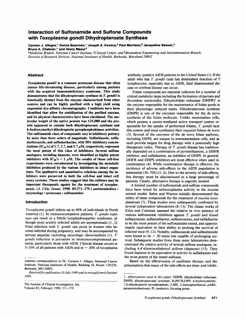
DHPSpreparationPurification and stability. Affinity columns of blue and greenSepharose bound the DHPS enzyme with adequate affinity toallow a high degree of purification with high yield when theaffinity columns were used sequentially. Table I illustrates arepresentative purification and Fig. 1 is typical of the elutionprofile of the enzyme from the green Sepharose column. Elu-tion from the blue and green columns occurred with KCI con-centrations of 0.6 and 0.8 M, respectively. Generally, purifica-tion resulted in the recovery of 5-10-fold more enzyme activ-
Purification scheme for the isolation ofDHPS enzyme from a cyto-solic preparation of T. gondii. The cytosolic fraction from the T.gondii organisms was applied to the blue Sepharose column whichwas developed using a KCI gradient. The active fractions from theblue affinity column were pooled and concentrated before loadingonto the green affinity column, which was also developed using aKCI gradient. The active fractions collected from the green columnwith the highest specific activity had protein concentrations < 1 jsg/ml.
Toxoplasma gondli Dihydropteroate Synthase 373
X2D - Figure 1. Purification ofE-0.207DHPS by green Sepha-00 A\ S R 0 rose chromatography.x X Jo 1 i 05 8 The DHPS protein was
10lo purified using a green0.1- 0.3 E Sepharose dye affinity
column. After elution2-0.1 from the prior affinity
ao-s£JU,4step, the active fractions0 10 20 were pooled, concen-
Elution Volume (ml) trated by ultrafiltration,and diluted to a final
salt concentration of < 200 mM. This preparation was applied to thegreen Sepharose column preequilibrated with 100 mM KCI buffer.After washing with 5 column volumes of the loading buffer, the col-umn was developed at room temperature using a linear KCI gra-dient. Protein elution (*), DHPS activity (o), and KCI gradient (o).
ity than was present in the crude preparations, suggesting theelimination of an inhibitor or enzymatic activity capable ofdegrading the substrate. In addition, 6-hydroxymethyl-7,8-di-hydropterin pyrophosphokinase activity was also assayed andfound to copurify with DHPS such that its fold-purificationafter the final step (green Sepharose) was identical to that ofDHPS. This activity was also found in identical fractions asDHPS using an HPLC gel filtration column to measure themolecular weight of the green Sepharose purified enzyme (seebelow).
While the crude T. gondii cytosolic preparations were rela-tively stable with repeated freeze-thawing, and when stored at-80'C, the purified DHPS activity was extremely unstable,having a half-life of only 1-2 d. Because of the limited avail-ability of T. gondii DHPS, we conducted preliminary stabilityexperiments with DHPS purified from E. colt using the exactmethods outlined above. As shown in Table II, we found littleeffect on DHPS stability with the addition of sucrose or pro-tease inhibitors, or with storage at either 4 or -80'C despitethe presence of2-ME and Tris/HCl buffer. However, while theaddition of either BSA or DTT alone had little effect, thecombination of the two agents resulted in stabilization of en-zyme activity. Table II represents the effect of combiningDTT, BSA, and 2-ME in a buffered solution on the stability ofT. gondii DHPS. The combination of these agents resulted in amarked stabilization of the enzyme for at least 14 d. We foundthat concentrations of BSA of 0.01 or 1% resulted in identicalstabilization. Other potential stabilizers led to no additionalimprovement when tested in samples stored at -80'C as illus-trated in Table II.
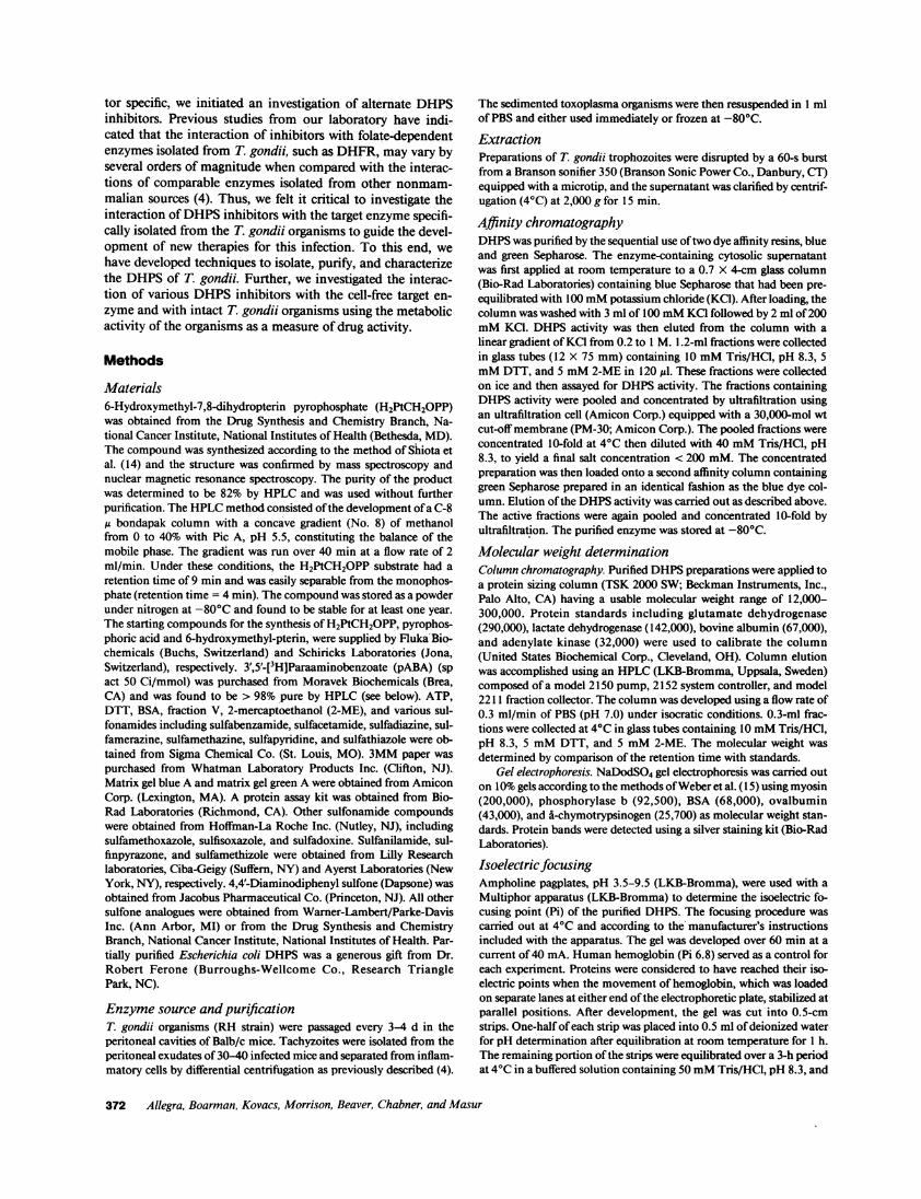
Characterization. The kinetic constants of both substratesfor the crude and purified DHPS enzyme are shown in TableIII. Under the assay conditions used we found the DHPS reac-tion to be linear for up to 60 min and up to an enzyme con-centration of 20 mU. The Michaelis-Menten constant forpABA was found to be highly dependent on the purity of theenzyme with an apparent affinity 10-fold greater for the puri-fied enzyme (0.36 MM) when compared with the crude enzyme(3.6 AM). The molecular weight of the enzyme in its nativestate was determined by molecular sizing chromatographyusing an HPLC system equipped with a protein sizing columncapable of separating proteins with molecular weights in therange of 12,000-300,000. The sizing chromatography revealedthe enzymatic activity to elute at a molecular weight of
Table II. Stability ofPurifiedDHPSfrom E. coli and T. gondii
* All preparations contained Tris/HCl (pH 8.3), 100 mM/2-ME 50mM.t Soybean trypsin inhibitor (50 gg/ml), chymostatin (50 Mg/ml), leu-peptin (20 ,g/ml), benzamidine (1 mg/ml), aprotinin (50 Mg/ml), andphenanthroline (2 mg/ml).§ Concentrations used were 0.01 and 1%.
125,000 (Fig. 2 A). 6-Hydroxymethyl-7,8-dihydropterin pyro-phosphokinase activity was present in the identical fractions asthe DHPS activity. Samples of protein from each ofthe purifi-cation steps, including that from the peak activity fractions ofthe sizing column studies, were applied to 10% SDS polyacryl-amide gels. We noted the consistent appearance of a proteinband of 120,000 mol wt that was not apparent in the crudepreparations but that became more intense with the purifica-tion process. In addition to this protein band, green column
Table III. Kinetic Parameters of T. gondii DHPS
KmSubstrate Crude Purified
IpM
pABA 3.6±0.29 0.36±0.13
H2PtCH2OPP 5.5±1 5.2±1.7
The Michaelis-Menten constants for the substrates were determinedfor both crude and purified enzyme preparations of T. gondii DHPS.The Km value for the pABA substrate was determined over a range of0.1-2 uM for the purified enzyme and 0.5-10MM for the crudepreparation. The pterin substrate was determined over a range of1-40 MM. The second substrate was maintained at saturating concen-trations. The kinetic values represent the mean and SE of three to sixexperiments.
374 Allegra, Boarman, Kovacs, Morrison, Beaver, Chabner, and Masur
purified protein also contained a consistent protein band at74,000 and, in three of the six tested preparations, a band of31,000 mol wt. After the molecular sizing chromatography,only the 120,000 mol wt protein remained apparent in thefractions containing DHPS activity. The isoelectric focusingpoint of the enzyme was found to be 6.3 (Fig. 2 B). As shownin Fig. 2 C, the enzyme retained activity over a broad pH rangeof 6.0-8.0.
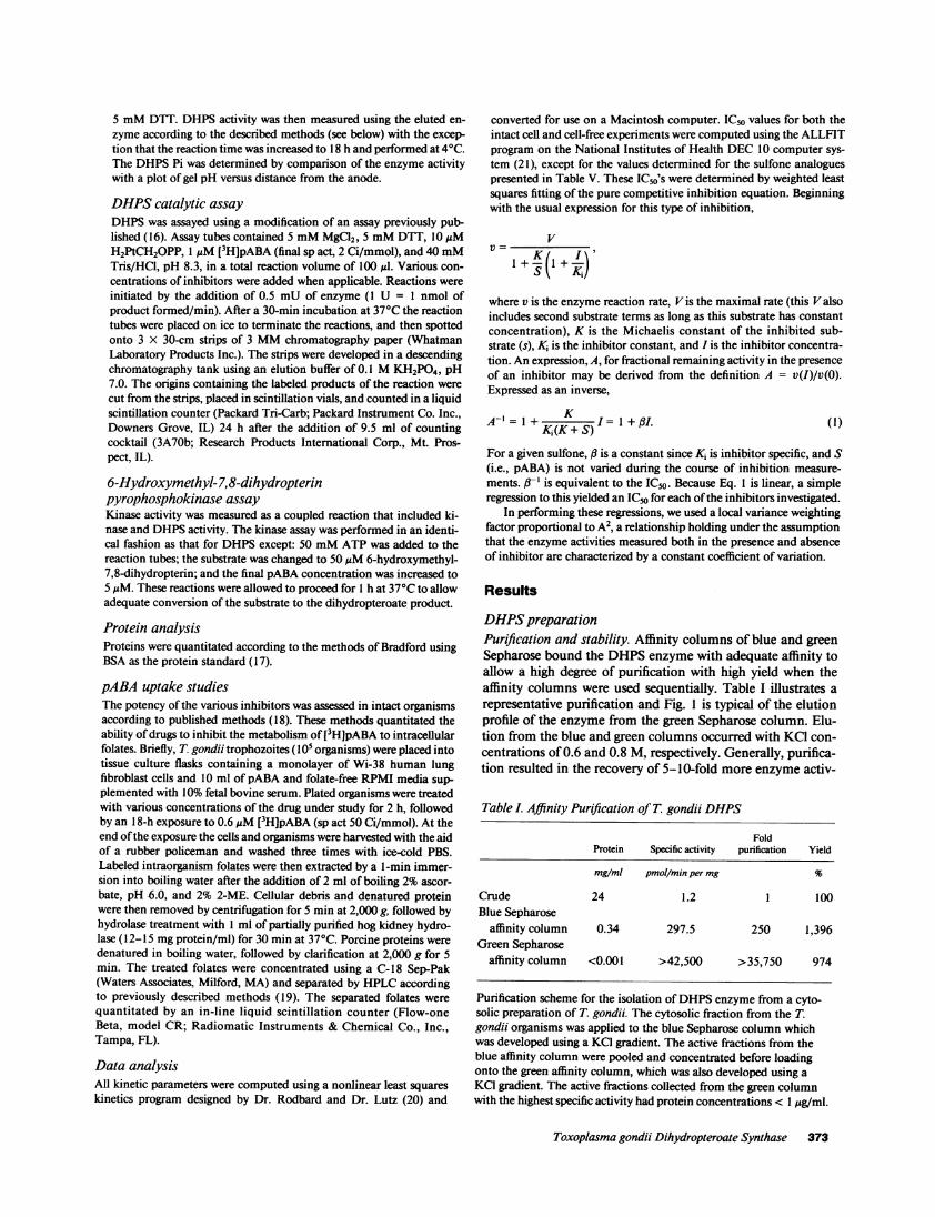
Interaction ofDHPS with paraaminobenzoate analoguesVarious sulfonamides, as shown in Table IV, were tested fortheir ability to inhibit the DHPS enzyme. The sulfonamidesvaried by over a three log range in IC50's with sulfathiazole,sulfamethoxazole, and sulfamethazine being the most potentof the sulfonamides tested. These studies were performed withboth crude and purified DHPS. The purified enzyme provedto be more susceptible to inhibition when compared with thecrude form by variable degrees depending on the potency ofthe sulfonamide. This difference was most apparent for theleast potent agents, wherein the difference exceeded 40-fold,and least apparent for the more potent inhibitors (< 4-fold).K,'s were generated for selected inhibitors (Table IV). In allcases tested, the inhibition was found to follow a pure competi-tive inhibition pattern with respect to pABA. Of interest wasthe finding that the sulfone, dapsone (4,4diamino-diphenylsulfone), was more potent than any of the sulfonamide ana-logues tested with an IC5o value of 1.2 and 0.25 gM for inhibi-tion of the crude and purified enzyme, respectively. Fig. 3illustrates the competitive nature ofinhibition by dapsone andsulfamethoxazole with respect to the pABA substrate.
- 1000 100 Figure 2. Characterization1 N: \ itIl- 80 of the T. gondii DHPS en-' zyme. The purified DHPS
(D ioo enzyme was characterized0tx asillustrated inA-C. The
20 o molecular weight (A) was0 10 0 determined by retention
15 20 25 30 time from an HPLC gel-fil-Retention Time (min) tration column calibrated
30B with proteins of known
molecular weight. o, mo-:tg20 1 1 Pi=6.3 lecular weight standards;*,EeX [ DHPS activity. These de-
I2 10- terminations were per-4 formed on four separate
0 occasions with an average4 5 6 7 8 9 10 weight found to be 125,000
pH (range, 105,000-135,000).1.4 C Determinations with crude
pH to a plot of gel pH versusgel location. A Pi of 6.3
represents the average of three separate determinations (range,6.1-6.4). The effect ofpH on synthetic function (C) was determinedby performing the assay at each of the denoted pH intervals using100 mM Tris/HCI buffer. Assay tubes were tested for pH accuracyafter the inclusion of all reactants, including enzyme.
Table IV. Inhibition of T. gondii DHPS bypABA Analogues
* Mean±SEM represent two to six separate experiments.
Interaction ofDHPS with sulfone analoguesBecause of the strength of dapsone as an inhibitor of DHPS,we investigated the ability of various sulfone analogues to in-hibit the DHPS enzyme relative to dapsone. Using the DHPSassay described above with purified enzyme, enzyme activitieswere determined in the presence of 1- and 10-,gM concentra-tions of various sulfone inhibitors. For the 25 sulfone ana-logues summarized in Table V, all compounds except AA hadat least one of the residual activities lying between 2 and 80%,and nearly halfhad both activities within this range. The IC50'sobtained by the data analysis method described previouslyclosely reproduce the experimental activities for all inhibitorsexamined. Experimental activities were generally within 6% oftheoretical. The IC"o for dapsone was obtained from a separate
Figure 3. Representa-30- / tive double-reciprocal
plots ofDHPS inhibi-tion by dapsone andsulfamethoxazole. A
20 - /representative double-reciprocal plot of the in-teractions of dapsone
10- and sulfamethoxazole is/ illustrated. For these ex-
~ e=periments, the concen-0 tration of the pterin0 1 2 3 4 S substrate was held con-
vs stant (10 MM) and vari-able amounts of the
pABA (0.1-10 MM) were tested against the concentrations of each in-hibitor as noted. ,, No inhibitor, o, 50 MM sulfamethoxazole; ., 10gM sulfamethoxazole; ., 1 MM dapsone; o, 10 MM dapsone. All reac-tions were initiated with 0.5 mU of the purified DHPS enzyme andallowed to proceed for 30 min before processing as outlined inMethods. All points represent the mean of duplicate determinationsfrom a representative experiment. S, Concentration ofpABA X 106;V, moles of product formed per minute X 10'°.
Toxoplasma gondii Dihydropteroate Synthase 375
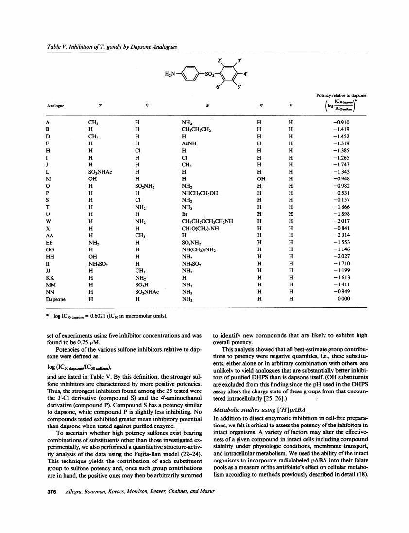
Table V. Inhibition of T. gondii by Dapsone Analogues
H2N c- S02 \ 4
6' 5Potency relative to dapsone
Analogue 2' 3' 4' 5' 6' (lo IC
A CH3 H NH2 H H -0.910B H H CH3CH2CH2 H H -1.419D CH3 H H H H -1.452F H H AcNH H H -1.319H H C1 H H H -1.385I H H Cl H H -1.265J H H CH3 H H -1.747L SO2NHAc H H H H -1.343M OH H H OH H -0.9480 H SO2NH2 NH2 H H -0.982P H H NHCH2CH2OH H H -0.531S H C1 NH2 H H -0.157T H NH2 NH2 H H -1.866U H H Br H H -1.898W H NH2 CH3CH2OCH2CH2NH H H -2.017X H H CH3O(CH2)3NH H H -0.841AA H CH3 H H H -2.314EE NH2 H SO2NH2 H H -1.553GG H H NH(CH2)3NH2 H H -1.146HH OH H NH2 H H -2.027II NH2SO2 H NH2SO2 H H -1.710JJ H CH3 NH2 H H -1.199KK H NH2 H H H -1.613MM H SO3H NH2 H H -1.411NN H SO2NHAc NH2 H H -0.949Dapsone H H NH2 H H 0.000
* -log IC5o dpsone = 0.6021 (IC50 in micromolar units).
set of experiments using five inhibitor concentrations and wasfound to be 0.25 ,uM.
Potencies of the various sulfone inhibitors relative to dap-sone were defined as
log (IC50 dapsone/ICSO sulfone),
and are listed in Table V. By this definition, the stronger sul-fone inhibitors are characterized by more positive potencies.Thus, the strongest inhibitors found among the 25 tested werethe 3'-C1 derivative (compound S) and the 4'-aminoethanolderivative (compound P). Compound S has a potency similarto dapsone, while compound P is slightly less inhibiting. Nocompounds tested exhibited greater mean inhibitory potentialthan dapsone when tested against purified enzyme.
To ascertain whether high potency sulfones exist bearingcombinations of substituents other than those investigated ex-perimentally, we also performed a quantitative structure-activ-ity analysis of the data using the Fujita-Ban model (22-24).This technique yields the contribution of each substituentgroup to sulfone potency and, once such group contributionsare in hand, the positive ones may then be arbitrarily summed
to identify new compounds that are likely to exhibit highoverall potency.
This analysis showed that all best-estimate group contribu-tions to potency were negative quantities, i.e., these substitu-ents, either alone or in arbitrary combination with others, areunlikely to yield analogues that are substantially better inhibi-tors of purified DHPS than is dapsone itself. (OH substituentsare excluded from this finding since the pH used in the DHPSassay alters the charge state of these groups from that encoun-tered intracellularly [25, 26].)
Metabolic studies using [3H]pABAIn addition to direct enzymatic inhibition in cell-free prepara-tions, we felt it critical to assess the potency ofthe inhibitors inintact organisms. A variety of factors may alter the effective-ness of a given compound in intact cells including compoundstability under physiologic conditions, membrane transport,and intracellular metabolism. We used the ability ofthe intactorganisms to incorporate radiolabeled pABA into their folatepools as a measure ofthe antifolate's effect on cellular metabo-lism according to methods previously described in detail (18).
376 Allegra, Boarman, Kovacs, Morrison, Beaver, Chabner, and Masur
The results of these studies are illustrated in Fig. 4 for selectedDHPS inhibitors. Also shown for comparative purposes arethe results using the DHFR inhibitors trimetrexate and pyri-methamine. In general, the inhibitory potency of the DHPSinhibitors in the intact cell assay closely paralleled the findingsin the cell-free experiments. The DHFR inhibitor trimetrexatewas found to be a potent inhibitor of toxoplasma metabolismwith an IC50 of 3.5 nM. Pyrimethamine, the inhibitor cur-rently used to treat T. gondii infections, was > 200-fold lesspotent than trimetrexate. Methotrexate, which requires an ac-tive transmembrane transport system not found in toxoplasmaorganisms, had no metabolic effects at concentrations > 10jiM (data not shown). These results are in concert with thosepreviously reported using the DHFR inhibitors in cell-free en-zymatic studies. The inhibitory potency ofthree sulfonamidesand dapsone in the intact cell assay are also illustrated in Fig.4. Unlike the cell-free experiments, sulfadiazine and sulfa-methoxazole appeared to be two- to threefold better inhibitorsthan dapsone. Finally, since there was a great dependence ofsubstrate and inhibitor affinity on the purity ofthe enzyme, wecompared the inhibitory results of selected sulfonamide andsulfone inhibitors when their potency was assayed using eachof the three assay systems described in this report (Table VI).When the inhibitory potency of these inhibitors was com-pared, we noted that the cell-free results using the crude en-zyme provided a more accurate quantitative indication of in-hibitory potency when either cell-free result was comparedwith the intact cell system. From a qualitative perspective,either set of cell-free data accurately predicted the relative in-hibitory potency of the inhibitors in intact cells. The sulfoneanalogues, S and P, demonstrated similar potency in the intactand cell-free assay systems. In particular, analogue S was five-fold more potent than dapsone and twofold more potent thaneven the most potent sulfonamides in the intact cell assaysystem.
Discussion
We report here a novel, high-yield, and simple purificationscheme capable of purifications of T. gondii DHPS > 30,000-fold. The initial characterization of the T. gondii enzyme re-vealed it to be a 125,000 mol wt protein (native state), which isintermediate between that reported for E. coli (50,000) andplasmodia (190,000-250,000) (27-29). Given the copurifica-
100- Figure 4. Inhibition of in-tact T. gondii organisms by
075 \\° \ antifolates. Various antifo-
lates were tested as meta-50 bolic inhibitors of intact T.
*s \ \\\\ \ gondii organisms. *, Tri-25
* \ .\6 \* metrexate; , sulfamethoxa-0 zole; o, pyrimethamine; o,
0.0o1 0.01 0.1 1 lo loo iooo dapsone; *, sulfisoxazole;Drug Concencation (pM) A, sulfadiazine. In each
case, the organisms wereexposed to various concentrations of the inhibitors for 2 h before theaddition of radiolabeled pABA. After an additional 18 h the organ-isms were harvested and their intracellular folates quantitated. Thesevalues served as a measure of the inhibitory potency ofthe inhibitorswhen total intracellular folates were compared with those in controlorganisms (100%). Each point represents the mean of three to six in-dependent experiments.
Table VI. Comparison ofSelected Inhibitorsin the Three Study Systems
* Mean±SEM represent four to eight separate experiments using fiveinhibitor concentrations for each of the three study systems.
tion of 6-hydroxymethyl-7,8-dihydropterin pyrophosphoki-nase activity, which also appeared in identical fractionsthroughout the purification scheme as the DHPS activity, it isconceivable either that both activities reside on the same pro-tein or that the protein is comprised of separate subunits withdistinct functions. Richey and Brown were able to resolve theDHPS and 6-hydroxymethyl-7,8-dihydropterin pyrophospho-kinase activities in E. coli into proteins with Mr's of 50,000(DHPS) and 15,000 (dihydropterin pyrophosphokinase) usinga Sephadex G-100 column (27). While DHPS activity was alsoseparable from 6-hydroxymethyl-7,8-dihydropterin pyrophos-phokinase activity by DEAE-Sephadex chromatography inPlasmodium berghei, these activities were not separable inPlasmodium chabaudi, nor were they separable in pea seed-lings (16, 30, 31). DHPS protein purified through the greenSepharose affinity column did not appear to be homogeneous,as at least one additional protein (74,000 mol wt) was consis-tently noted to copurify. DHPS further purified by sizing chro-matography contained both synthase and pyrophosphokinaseactivity and appeared as a single protein band on a silver-stained denaturing polyacrylamide gel.
As reported by others, we also noted increased enzymaticyield (up to 10-fold) during purification. This apparent in-crease in activity may be explained by the presence of enzy-matic activity in the crude preparations capable of degradingthe substrate. Upon examination ofthe crude preparations, weidentified enzymatic activity capable of degrading a diphos-phate (ADP) that was not present after the first purificationstep (data not shown). Whether this activity is capable of de-grading the pyrophosphate substrate remains to be elucidated.Purification ofDHPS from potential inhibitors present in thecrude preparations represents an additional possibility that hasnot been excluded. Instability ofthe DHPS enzymatic activityisolated from other organisms has not previously been notedand may be due to differences in the purity or source of thepreparations used in other studies (16, 27-30). The DHPSactivity in crude cytosolic preparations of T. gondii was stableand became unstable only with purification > 100-200-fold.The purified enzyme appears to be highly susceptible to oxi-dation and adherence to inert surfaces given the stability of-
Toxoplasma gondii Dihydropteroate Synthase 377
fered by an inclusion of DTT and BSA. The DHPS enzymewas found to have an acidic Pi of 6.3. and was capable ofmaintaining catalytic activity over a broad pH range. Previousreports have noted that the optimum pH for plastic enzyme is
8.5 (29, 30), and this is consistent with that noted for T.gondii.
In investigating the Michaelis-Menten constants for theDHPS enzyme with respect to both substrates, we found theaffinity constant for pABA to be similar to those reported forenzymes isolated from both P. berghei (0.28 uM) and E. coli(0.21 uM) by Ferone and co-workers (16, 28), but somewhattighter than that reported by others (1.1-2.8 MM) (29, 30).These differences may relate to the dependence of affinity onthe purity of the enzyme preparation. We found that the Kmfor pABA differed by a factor of 10 between crude and pureenzyme, while the Km for the pterine substrate showed littledependence on the purity of the enzyme. The affinity of T.gondii DHPS for the pterin pyrophosphate substrate is slightlyweaker (- 5 uM) than that reported for either P. berghei or E.coli (0.5-1.4 uM) (16, 28).
These kinetic studies allow only speculation as to why thesubstrate and inhibitor affinities are dependent on enzymepurity. It is plausible that the conformation of the enzymechanges with purification to allow greater access to the activesite and thereby enhance the apparent affinity ofthe substratesfor the enzyme. This postulate is additionally supported by thefinding that the potency of various inhibitors depended on theenzyme purity. This difference was most apparent for theweaker inhibitors but is, in general, apparent even for the mostpotent compounds including the sulfones. This postulatewould account for the crude enzyme being a better predictor ofinhibitory activity in intact cells, as the enzyme in the crudepreparations may more closely reflect the native conformationin intact cells.
The inhibition of DHPS isolated from bacterial and plas-modial sources by sulfonamides and sulfones and their utilityas therapeutic agents has previously been reported ( 16, 28, 29).Investigations ofthe interactions of sulfonamides and sulfoneswith the DHPS isolated from T. gondii demonstrated that thesulfonamides exhibited a broad range of inhibitory potency,with sulfadiazine, the DHPS inhibitor of choice for the treat-ment oftoxoplasma encephalitis, being one ofthe most potentof the sulfonamides tested. Of some note was the finding thatsulfadoxine, commonly used in prophylaxis regimens forPneumocystis carinii, was a particularly poor toxoplasmaDHPS inhibitor. These results are in conformity with thosereported from in vivo experiments using the acute murinetoxoplasma model. In the murine model, sulfadiazine, sulfa-methazine, sulfamerazine, and sulfathiazole were noted to beof high potency, while sulfisoxazole, sulfapyridine, and sulfa-nilamide were found to be therapeutically poor compounds(9-12). As others have previously reported using DHPS fromE. coli and Plasmodia, we found both classes ofinhibitors to bepurely competitive with respect to the pABA substrate ( 16). Afinding that was in contrast with previously reported bacterialstudies but consistent with results from the murine model, wasthe potency of the sulfone, dapsone, relative to the sulfon-amides. Generally, the sulfones have been found to be rela-tively poor inhibitors of bacterial DHPS. These studies suggestthat the T. gondii enzyme is substantially different from that ofbacteria in its interaction with these agents and thus probablydistinct in its overall protein structure.
Several investigators have noted that the potency of the
sulfonamides may be directly related to their ability to ionizeat physiologic pH (32-34). Since these drugs penetrate cellularmembranes with difficulty, the best inhibitors of intact organ-isms were those that were half-ionized at physiologic pH, i.e.,their pKa was between 6 and 7.4. The nature of the sulfonestructure does not allow these compounds to be fit into asimilar relationship; however, the activity ofthese compoundsas inhibitors of bacterial growth has been related to the molec-ular charge distribution of the inhibitor with respect to thenatural substrate (pABA). De Benedetti and co-workers inves-tigated such structure-activity relationships for the sulfonesand identified several analogues with activity exceeding that ofdapsone (25). Given the potency of dapsone as an inhibitor ofT. gondii DHPS, we studied additional sulfone analogues forinhibitory potency. Direct determination of the activity of thesulfones in Table V against purified DHPS identified com-pounds S and P as those most similar to dapsone. Quantitativestructure-activity analyses then identified the contributions toinhibitory potency from individual substituent groups in theanalogues investigated. In the mean, none ofthese groups werefound to contribute positively to potency, and thus no strongcandidates for new analogues could be suggested.
Examination of the error factors associated with the var-ious group contributions did reveal the possibility that the3'-Cl, 4'-NH(CH2)20H,2'-NH2, and S02NH2/SO2NHAcgroups could be associated with increased potency. Indeed,detailed examination ofcompound S in the cell-free system ledto a statistically insignificant difference in activity from dap-sone, leaving open the possibility of its 3'-Cl group being inhi-bition enhancing. In contrast, the 4'-NH(CH2)20H group,characteristic of compound P and quite similar to the 4' groupof compound W, failed to significantly enhance the activity ofdapsone. The 2'-NH2 group, when it is the only additionalsubstituent on dapsone, has not been investigated in our assay.However, De Benedetti et al. (25), in presenting the activity ofsulfones against the DHPS from E. co/i, reported the 2'-NH2derivative of dapsone to be twice as active as the parent com-pound. It is consistent with our structure-activity analysis thatthis level of increased activity may also exist against T. gondiiDHPS, and further testing, perhaps involving the 2',4',6'-(NH2)3 compound as well, is warranted.
To further enhance our understanding of the interactionsofpABA analogues with T. gondii DHPS, we investigated theireffects on intact organisms using the metabolism of radiola-beled pABA as a measure of inhibitor potency. Unlike thecell-free experiments, this system accounts for events in addi-tion to enzyme inhibition, including inhibitor stability, mem-brane transport, and intracellular metabolism. We found anexcellent quantitative and qualitative relation between the re-sults obtained in the cell-free system and those from the pABAexperiments, suggesting that enzyme inhibitor interactions arethe critical rate-limiting step for these compounds. Interest-ingly, the cell-free results using crude enzyme correlated mostclosely with those obtained from the intact cell experimentswhen compared with those with purified DHPS, suggestingonce again that purification may alter the native state of theenzyme. While compound S had the same potency as dapsonein the cell-free systems, it was fivefold more potent in theintact cell experiments, suggesting an advantage perhaps dueto improved membrane transport. The results obtained usingDHFR inhibitors, particularly trimetrexate, illustrate the ex-traordinary potency of these compounds compared withDHPS inhibitors in this assay system. These results document
378 Allegra, Boarman, Kovacs, Morrison, Beaver, Chabner, and Masur
the presence of the DHPS enzyme in T. gondii organisms andshow that it is kinetically distinct from the DHPS of othersources. While we have been able to purify and characterizethe enzyme, it appears that kinetic experiments with crudeenzyme preparations are quantitatively more representative ofthe potency that may be expected from pABA analogues inintact organisms. Of importance is the demonstration thatsulfonamides vary in potency by over three orders of magni-tude, supporting the utility of direct enzyme studies in guidingthe selection of sulfonamides for clinical testing. Further, thefinding that dapsone and several analogues are highly potentinhibitors of T. gondii DHPS supports the consideration oftherapeutic trials using dapsone for the treatment of T. gondiiinfections and serves as a focal point for the identification anddesign of new pABA analogues that may potentially have im-proved physical, pharmacokinetic, and therapeutic properties.
Acknowledgments
The authors wish to acknowledge Dr. Robert Ferone for his helpfulsuggestions regarding this work.
References
1. Masur, H. 1985. Toxoplasmosis. In Cecil Textbook ofMedicine.J. P. Wyngaarden and L. H. Smith, editors. W. B. Saunders Company,Philadelphia. 1792-1795.
2. McCabe, R., and J. S. Remington. 1988. Toxoplasmosis: thetime has come. N. Engl. J. Med. 318:313-315.
3. Allegra, C. J., J. A. Kovacs, J. C. Drake, J. C. Swan, B. A.Chabner, and H. Masur. 1987. Potent' in vitro and in vivo antitoxo-plasma activity of the lipid-soluble antifolate trimetrexate. J. Clin.Invest. 79:478-482.
4. Eyles, D. E., and N. Coleman. 1955. An evaluation of the cura-tive effects of pyrimethamine and sulfadiazine alone and in combina-tion on experimental mouse toxoplasmosis. Antibiot. Chemother.(Basel). 5:529-539.
5. Kovacs, J. A., J. W. Heimenz, A. M. Macher, D. Stover, H. W.Murray, J. Shelhamer, H. C. Lane, C. Urmacher, C. Honig, D. L.Longo, M. M. Parker, C. Natanson, J. E. Parrillo, A. S. Fauci, P. A.Pizzo, and H. Masur. 1984. Pneumocystis pneumonia carinii: a com-parison between patients with the acquired immunodeficiency syn-drome and patients with other immunodeficiencies. Ann. Intern. Med.100:663-671.
6. Leport, C., F. Raffi, S. Matheron, C. Katlama, B. Regnier, A.Samoit, C. Marche, C. Vedrenne, and J. L. Vilde. 1988. Treatment ofcentral nervous system toxoplasmosis with pyrimethamine/sulfadizinecombination in 35 patients with the acquired immuno-deficiency syn-drome. Am. J. Med. 84:94-100.
7. Sabin, A. B., and J. Warren. 1942. Therapeutic effectiveness ofcertain sulfonamides on infection by an intracellular protozoan (toxo-plasma). Proc. Soc. Exp. Biol. Med. 51:19-23.
8. Biocca, E., and R. Pasqualin. 1942. Aqao terapeutica de algunscompostos sulfa-nil amidicos na infecao experimental per toxoplasma.Arq. Biol. (Sao Paulo). 26:107-109.
9. Eyles, D. E., and N. Coleman. 1953. The relative activity ofthecommon sulfonamides against experimental toxoplasmosis in themouse. Am. J. Trop. Med. Hyg. 2:54-63.
10. Eyles, D. E. 1953. The present status of the chemotherapy oftoxoplasmosis. Am. J. Trop. Med. Hyg. 2:429-444.
11. Eyles, D. E. 1956. Newer knowledge of the chemotherapy oftoxoplasmosis. Ann. NYAcad. Sci. 64:252-267.
12. Eyles, D. E., and N. Coleman. 1955. The effect of sulfadime-tine, sulfisoxazole, and sulfapyrazine against mouse toxoplasmosis.Antibiot. Chemother. (Basel). 5:525-528.
13. Eyles, D. E., and N. Coleman. 1957. An evaluation ofthe effect
of sulfones on experimental toxoplasmosis in the mouse. Antibiot.Chemother. (Basel). 7:577-585.
14. Shiota, T., M. N. Disraely, and M. P. McCann. 1964. Theenzymatic synthesis of folate-like compounds from hydroxymethyl-pteridine pyrophosphate. J. Biol. Chem. 239:2259-2266.
15. Weber, K., J. R. Pringle, and M. Osborn. 1972. Measurementof molecular weights by electrophoresis on SDS-acrylamide gel.Methods Enzymol. 26:3-27.
16. Ferone, R. 1973. Enzymatic synthesis of dihydropteroate anddihydrofolate by plasmodium berghei. J. Protozool. 20:459-464.
17. Bradford, M. 1976. A rapid and sensitive method for the quan-titation of microgram quantities of protein utilizing the principle ofprotein-dye binding. Anal. Biochem. 72:248-254.
18. Kovacs, J. A., C. J. Allegra, J. Beaver, D. Boarman, M. Lewis,J. E. Parrillo, B. A. Chabner, and H. Masur. 1989. Characterization ofde novo folate synthesis in Pneumocystis carinii and Toxoplasmagondii: potential utilization for screening therapeutic agents. J. Infect.Dis. 160:312-320.
19. Allegra, C. J., R. L. Fine, J. C. Drake, and B. A. Chabner. 1986.The effect of methotrexate on intracellular folate pools in humanMCF-7 breast cancer cells: evidence for direct inhibition of purinesynthesis. J. Biol. Chem. 261:6478-6485.
20. Allegra, C. J., B. A. Chabner, J. C. Drake, R. Lutz, D. Rodbard,and J. Jolivet. 1985. Enhanced inhibition of thymidylate synthase bymethotrexate polyglutamates. J. Biol. Chem. 260:9720-9726.
21. DeLean, A., P. J. Munson, and D. Rodbard. 1978. Simulta-neous analysis of families of sigmoidal curves: application to bioassay,radioligand assay, and physiological dose-response curves. Am. J.Physiol. 235:E97-E102.
22. Fujita, T., and T. Ban. 1971. Structure-activity study of phen-ethylamines as substrates of biosynthetic enzymes of sympathetictransmitters. J. Med. Chem. 14:148-152.
23. Kubinyi, H. 1977. Quantitative structure-activity relationships.Arzneim. Forsch. 27:750-758.
24. Free, S. M., Jr., and J. W. Wilson. 1964. A mathematicalcontribution to structure-activity studies. J. Med. Chem. 7:395-399.
25. De Benedetti, P. G., D. larossi, C. Menaizni, V. Caiolfa, C.Frassineti, and C. Cennamo. 1987. Quantitative structure-activityanalysis in dihydropteroate synthase inhibition by sulfones: compari-son with sulfonamides. J. Med. Chem. 30:459-464.
26. Seydel, J. K., and K. Schaper. 1980. Enzyme Inhibitors asDrugs. M. Sandier, editor. Macmillan Journals Ltd., London. 68.
27. Richey, D. P., and G. M. Brown. 1969. Biosynthesis of folicacid. IX. Purification and properties of the enzymes required for theformation of dihydropteroic acid. J. Biol. Chem. 244:1582-1592.
28. Roland, S., R. Ferone, R. J. Harvey, V. L. Styles, and R. W.Morrison. 1979. Characteristics and significance of sulfonamides assubstrates for Escherichia coli dihydropteroate synthase. J. Biol. Chem.254:10337-10345.
29. McCullough, J., and T. A. Maren. 1974. Dihydropteroate syn-thetase from Plasmodium berghei: isolation, properties, and inhibitionby dapsone and sulfadiazine. Mol. Pharmacol. 10: 140-145.
30. Walter, R. D., and E. Konigk. 1974. Purification and propertiesof the 7,8 dihydropteroate synthesizing enzyme from Plasmodiumchabaudi. Z. Phys. Chem. 335:431-437.
31. Okinaka, O., and K. Iwai. 1970. The biosynthesis of folic acidcompounds in plants. IV. Purification and properties of the dihydro-pteroate-synthesizing enzyme from pea seedlings. J. Vitaminol.(Kyoto). 16:201-209.
32. Schmelkes, F. C., 0. Wyss, H. C. Marks, B. J. Ludwig, and F. B.Stranskov. 1942. Mechanism of sulfonamide action. I. Acidic dissocia-tion and antibacterial effect. Proc. Soc. Ex. Biol. Med. 50:145-148.
33. Bell, P. H., and R. 0. Roblin, Jr. 1942. Studies in chemother-apy. VII. A theory of the relation of structure to activity of sulfanil-amide type compounds. J. Am. Chem. Soc. 64:2905-2917.
34. Yamazaki, M., N. Kakeya, T. Morishita, A. Kamada, and A.Aoki. 1970. Biological activity of drugs. X. Relation of structure to thebacteriostatic activity of sulfonamides (1). Chem. Pharm. Bull.(Tokyo). 18:702-707.


![Primerdesign Ltd TM Toxoplasma gondii - Home : genesig · Toxoplasma gondii is a species of parasitic protozoa in the genus Toxoplasma.[1] The definitivehostofT.gondiiisthecat,buttheparasitecanbecarriedbythevastmajorityof](https://static.documents.pub/doc/80x56/5cc21bb288c993ed078d60da/primerdesign-ltd-tm-toxoplasma-gondii-home-toxoplasma-gondii-is-a-species.jpg)


