From Eye to Insight Concentrating on workflow solutions, we provide a product range that is aligned to your needs in TEM, SEM, LM, and AFM investigations. Leica Nano Technology WORKFLOW SOLUTIONS FOR LIFE SCIENCE RESEARCH
Transcript
From Eye to Insight
Concentrating on workflow solutions, we provide a product range that is aligned to
your needs in TEM, SEM, LM, and AFM investigations.
Leica Nano Technology
WORKFLOW SOLUTIONS
FOR LIFE SCIENCE RESEARCH
2
Great Results with the Workflow of your Choice
Do you need to pay attention to every detail – not just during image acquisition and
analysis, but also sample preparation? Leica Microsystems offers you an extensive
portfolio of high performance instruments for electron microscopy sample preparation.
Designed to achieve effective sample preparation workflows, these solutions enable you
to obtain top results.
In this publication, we have compiled and present in a clear way appropriate workflows
for the most frequently required sample preparation methods. The solutions discussed
are proven to bring about great results, both reliable and reproducible, within a short
time. If you have special requirements for your workflow or any questions regarding the
topics shown here, our Leica experts will be happy to assist you at any time.
High pressure system for freezing aqueous samples delivers optimal sample preservation. Offers the highest flexibility to meet multiple application demands.
EM Cryo CLEM
The system ensures contamination-free sample transfer and loading from cryo sample preparation instruments to a Leica fix stage light microscope. Maintains sample vitrified during cryo imaging.
EM ACE600
Fully automated, versatile high vacuum coater producing very thin, fine-grained, conductive metal and carbon coatings. Up to two angled coating sources configurable. For high resolution analysis, required for FE-SEM and TEM applications.
Explore the knowledge portal on EM sample preparation on
www.leica-microsystems.com/science-lab
and get detailed information on Application Notes, Articles and Tutorials!
See our complete portfolio, browse through our product websites and download all
Versatile vacuum cryo transfer system for contamination-free transfer of specimens between different preparation and analysis instruments.
EM ACE900
High-end sample preparation system to perform freeze fracture, freeze etching, and e-beam coating to create replicas for TEM analysis and to image in the Cryo-SEM using the EM VCT500 connection.
EM UC7
Ultramicrotome for ultrathin sectioning of biological and industrial samples for TEM, SEM, AFM and LM examination. With the ARTOS 3D you can also simplify your preparation for Array Tomography.
Cover images: top: Collembolen (source: Dr. Daniela Gruber, University of Vienna, Austria); bottom left: Cross-fractured axon showing myelin layers, (source: Dr. Andres Käch, Center for Microscopy and Image Analysis, University of Zurich, Switzerland); bottom middle: Euglena gracilis Klebs (source: Dr. Andres Käch, Center for Microscopy and Image Analysis, University of Zurich, Switzerland); bottom right: Ultrastructure of Arabidopsis thaliana primary root cells (source: Dr. Riet de Rycke, Department for Molecular Biomedical Research, 9000 Gent ). Top image page 2: 3D reconstruction of the first mitotic spindle in a C. elegans embryo (Dr. Stefanie Redemann, University of Virgina, Charlotteville, USA); Category image “Correlative Methodologies”: In resin fluorescence overlay of a C1-GFP transfected HeLa cell (source: Dr. Christopher Peddie, The Francis Crick Institute, London, UK); Catergory image “Optogenetics & Electron-Physiology”: Symmetric Synapse (source: Dr. Shuwen Chang, Charité Universitätsmedizin Berlin, Germany); Catergory image “Surface Analysis”: Salivary gland (source: David McCarthy, UCL School of Pharmacy, London, UK); Catergory image “2D Tissue & Cellular Morphology”: C. elegans cross section (source: Dr. Thomas Müller-Reichert, TU Dresden, Dresden, Germany); Catergory image “3D Tissue & Cellular Morphology”: 3D reconstruction of an intercellular bridge in a C. elegans embryo (source: Dr. Julia König, TU Dresden, Dresden, Germany); Catergory image “Suspension & Macromolecules”: Visualization of DNA Molecules (source: Michele Giannattasio, Istituto FIRC di Oncologia Molecolare (IFOM)-Electron Microscopy Facility (Single Molecules Visualisation), Milano, Italia, and Gisela Höflinger, Leica Mikrosysteme, Vienna, Austria); Image page 25: Reconstructed z-slice of an intercellular bridge of a sea urchin one-cell embryo (source: Dr. Julia König, TU Dresden, Dresden, Germany).
This is an ideal workflow to target the cell of interest on the grid by cryo-fluorescence microscopy for subsequent cryo-soft-X-ray-tomography.
TEM
Cryo CLEM
This workflow joins on-grid plunge freezing with cryo-fluorescence microscopy to target, relocate and overlay the structure of interest with the subjacent ultrastructure imaged with the cryo-TEM.
Combining CEMOVIS of high-pressure frozen samples with cryo-fluorescence microscopy allows to target the structure of interest within cryo sec-tions. This is then superimposed with the underlying ultrastructure.
9
In resin CLEM
This workflow is optimised to correlate fluorescently labelled structures in different image modalities to combine the advantages of fluorescence microscopy with the superior ultrastructure of high pressure freezing in RT processing.
To achieve high precision correlation of the fluorescent and electron microscopic image, an integrated light and scanning electron microscope is utilized.
TEM
SEM
Tokuyasu CLEM
Introducing light microscopy into a classic Tokuyasu workflow is used to identify the region of interest on the section followed by a more precise labelling localisation at the ultrastructural level.
TEM
and
10
Live cell CLEM
This workflow aims to understand ultrastructural changes over time by combining live-cell imaging and instant fixation by high pressure freezing.
Post fixation epoxy CLEM
This is another opportunity to identify the structure of interest of tissue samples by using a mild chemical fixation, followed by LM imaging. Afterwards the samples will be further processed for electron microscopy.
TEM
TEM
11
OPTOGENETICS & ELECTRO- PHYSIOLOGY
Light Stimulation
Using light stimulation in combination with high pressure freezing a moment in time can be frozen. Based on optogenetics, this workflow enables to trigger action potentials in excitable cells and arrest the process at any specific time.
Electrical Stimulation
The workflow enables to coordinate electrical stimulation of neuronal tissue with rapid freezing to visualize membrane dynamics at millisecond precision.
TEM
TEM
Related Application Notes downloadable from the following product website:EM AFS2: Sample Preparation of Arabidopsis Thaliana Root Tips Cells Cell envelope structure of a thermotolerant y-proteobacterium Acidithiobacillus sp., strain HV2/2 Targeting of peroxisomal matrix proteins in the diatom Phaeodactylum tricornutum Sample Preparation of Mouse heartEM ICE: Symmetric Synapse Visualization of membrane dynamics with millisecond temporal resolution
Related Application Note downloadable from the following product website:EM ICE: UV light stimulated sun screen lotion
This is the standard SEM workflow for biological samples to investigate surface architecture of chemically fixed samples.
SEM
or
Pc
Freeze Fracturing & Cryo SEM
Cryo SEM imaging is a fast way to receive very high-resolution images of internal structures or surfaces of biological but also industrial samples.
SEM
or
References on Science Lab: Brief introduction to freeze fracture and etchingGiardia lamblia sample preparation for cryo semDrosophila larvae sample preparation for cryo semCryo sem imaging of latex paint
EM CPD300: Bacteria Protocol Black Mould Protocol Clawed Frog Nuclear Envelope Protocol Human Blood Cells Protocol Micro-CT of Book Scorpion Musculature Micro-CT of Insect Brain Protocol
Micro-CT of Insect Larva ProtocolNematode E. dianae ProtocolRice Anther ProtocolRice Hull ProtocolRice Root ProtocolSludge Worm Protocol
Related Application Notes downloadable from the following product website:Tobacco Leaf ProtocolWall Cress Pod ProtocolWall Cress Stigma ProtocolWater Flea ProtocolWrinkled Giant Hyssop Leaf Protocol
Related Application Note downloadable from the following product website:EM ACE900: Cryo-SEM / Giardia lamblia
This is a classic freeze fracture workflow to receive a replica from the fractured surface of cryo fixed samples to get very high-resolution RT TEM images.
TEM
Related Application Note downloadable from the following product website:EM HPM100: Freeze-fracture replication of pyramidal cells
The technique is most commonly used for classic ultrastructural analysis of biological materials. Sample preparation is done at room temperature.
TEM
Hybrid Processing
This standard workflow combines superior ultrastructural fixation by high pressure freezing with room temperature manipulation using freeze substi-tution and resin embedding.
TEM
EM HPM100: Aureococcus (algae) Hep-2 cells infected with Chlamydia pneumoniae Maple (Acer saccharum) leaves Maple (Acer saccharum) leaves Mouse cerebellum Poplar (Populus sp.) stem
Poplar (Populus sp.) stemRictus lutensis (protist)Salmonella sp.Structural study of Caenorhabditis elegansCultured Rat Hippocampal Neurons
Related Application Note downloadable from the following product website:EM TP: Embedding of murine fibroblasts grown on Aclar plastics for TEM
Related Application Notes downloadable from the following product website:
A different way to label the structure of interest on a nanometre scale is to use cryo fixation in combination with low temperature resin embedding to preserve the antigens for subsequent labelling.
Tokuyasu Immunolabelling
It is a widely used method for preparation of biological material for immunolabelling by using chemical fixation, sucrose infiltration and plunge freezing. Because of the well-preserved antigens, this technique is a fast and safe way to acquire results on ultrastructure and immunolabelling.
TEM
ReferenceBasic course cryosectioning and Immuno-gold labeling of thawed frozen sections (Tokuyasu technique) at Cell Microscopy Core Utrecht, The Netherlands held by George Posthuma and others. www.cellbiology-utrecht.nl/em-courses/basic-course-cryosectioning.html
3D electron tomography enables to study the organization and interaction of biological structures within three dimensions in a defined volume. In this workflow the samples are processed at RT, followed by semi-thick serial sectioning on TEM grids.
TEM
The same approach can be applied using high pressure freezing as a fixation method to increase structural preservation and minimize introduction of artefacts.
TEM
CEMOVIS 3D
CEMOVIS enables not only the study of vitrified cells and tissue in 2D but also in 3D. The recently optimized double micromanipulator enables a better control of the cutting process. Combined with cryo electron tomography, it allows to study the supramolecular organization of cells.
TEM
Related Application Note downloadable from the following product website:EM HPM100: CEMOVIS of yeast
Optimised dimensional structural analysis of biological material can be achieved with this technique. In comparison to the workflows above, thin sections are collected on silicon wafers to be imaged in a SEM for higher throughput.
The same approach can be applied using high pressure freezing as a fixation method to increase structural preservation and minimize introduction of artefacts.
SEM
SEM
or
or
18
SEM Serial Blockface
Serial block face imaging is an integrated solution of ultrathin serial sectioning and simultaneous imaging inside a SEM. This workflow is suitable for chemically fixed samples to generate 3D datasets of big volumes.
SEM
Same concept as above, but using a light microscope or a micro CT to target the region of interest within the volume for subsequent targeted trimming and serial block face SEM.
The same approach can be applied using high pressure freezing as a fixation method to increase structural preservation and minimize introduction of artefacts.
or
SEM
SEM
or
or
or
and
and
and
19
FIB SEM
In contrast to serial block face imaging, the slicing of the samples is achieved by using a focused ion beam. Samples are also chemically fixed and processed at RT. The SEM images can be combined to generate 3D datasets.
or
Same concept as above, but using a light microscope or a micro CT to target the region of interest within the volume for subsequent targeted trim-ming and FIB SEM.
The same approach can be applied using high pressure freezing as a fixation method to increase structural preservation and minimize introduction of artefacts.
Related Application Note downloadable from the following product website:EM ACE600: Sample Protection prior to FIB processing
Combining the workflow above with an intermediate cryo-LM step, helps identify regions of interest easier and faster.
TEM
Cryo FIB on grid lamella
From vitrification up to sample transfer under cryogenic condition, this workflow guarantees samples free from contamination for cryo-TEM.
or
or
Using a dedicated cryo FIB to speed up sample retrieving and milling.
TEM
TEM
21
Cryo FIB lift out
Vitrified cells or tissue prepared by high pressure freezing can be prepared for Cryo TEM using a Cryo FIB to mill/produce a lamella which can be transferred onto a grid for Cryo TEM.
TEM
or
or
Cryo FIB SEM
This method has the advantage to image under cryogenic conditions, not introducing any artefacts by any FS and RT sample processing.
or
In combination with an intermediate cryo-LM step, it facilitates to identify regions of interest easier and enables to prepare the lamella at a specific location within the sample.
TEM
22
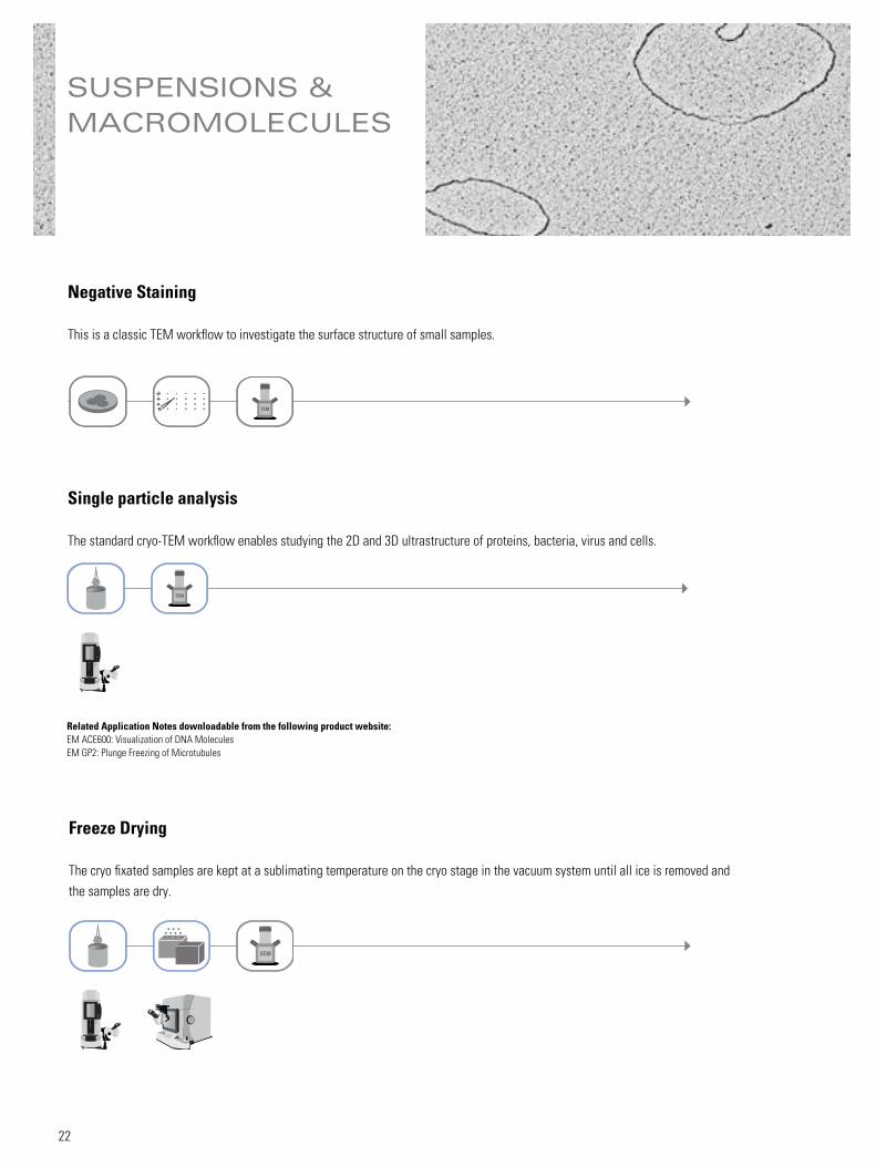
SUSPENSIONS & MACROMOLECULES
Negative Staining
This is a classic TEM workflow to investigate the surface structure of small samples.
Single particle analysis
The standard cryo-TEM workflow enables studying the 2D and 3D ultrastructure of proteins, bacteria, virus and cells.
Freeze Drying
The cryo fixated samples are kept at a sublimating temperature on the cryo stage in the vacuum system until all ice is removed and the samples are dry.
SEM
TEM
TEM
Related Application Notes downloadable from the following product website:EM ACE600: Visualization of DNA MoleculesEM GP2: Plunge Freezing of Microtubules
Coating in very low angles enables to resolve the structure of very small structures such as DNA or proteins.
Related Application Note downloadable from the following product website:EM ACE600: Visualization of DNA Molecules
TEM
Freeze Fracturing & Replica
This is a classic freeze fracture workflow to receive a replica from the fractured surface to image in the TEM. Plunge freezing instead of high pres-sure freezing can be a quicker and easier way to fixate the samples.
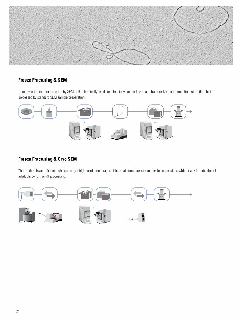
To analyse the interior structure by SEM of RT chemically fixed samples, they can be frozen and fractured as an intermediate step, then further processed by standard SEM sample preparation.
or or
Freeze Fracturing & Cryo SEM
This method is an efficient technique to get high resolution images of internal structures of samples in suspensions without any introduction of artefacts by further RT processing.