Phosphatidylinositol-3-kinase is an important enzyme for intracellularsignaling. The microbial product wortmanain and some of its analogueshave been shown to be potent inhibitors of phosphafidylinositol-3-kinase.The 50% inhibitory concentration for inhibition by wortmannin is 2 to 4flM. Kinetic analysis demonstrates that wortmannin is a noncompetitive,
irreversible inhibitor of phosphatidyllnositol-3-kinase, with inactivationbeing both time- and concentration-dependent. Wortmannin has previously been reported to be an inhibitor of myosin light chain kinase but
with an inhibitory concentration of 0.2 @&M.Wortmannin was found not tobe an inhibitor of phosphatidylinositol-4-hinase, protein kinase C, orprotein tyrosine kinase. Wortmannin inhibited the formation of phosphatidylinositol-3-phosphates in intact cells. The results of the study suggestthat wortmannin and its analogues may have utility as pharmacologicalprobes for studying the actions of phosphatidylinositol-3-kinase.
INTRODUCTION
Ptdlns-3-kinase3 exists as a tightly associated heterodimer of an Mr85,000 regulatory subunit and a Mr 110,000 catalytic subunit (1).Ptdlns-3-kinase is found in cellular complexes with almost all ligandactivated growth factor receptor and oncogene protein tyrosinekinases (2). Ptdlns-3-kinase is also associated with p2l@ (3). The Mr
85,000 regulatory subunit probably acts as an adaptor protein thatallows the Mr 110,000 catalytic subunit of Ptdlns-3-kinase to interactwith growth factor receptors and tyrosine phosphorylated proteins (4).The bovine Ptdlns-3-kinase Mr 110,000 catalytic subunit has sequencehomology to a yeast Ptdlns-3-kinase that is involved in vacuolarprotein sorting (5, 6), suggesting there may be a similar role formammalian Ptdlns-3-kinase.
Ptdlns-3-kinase is an important enzyme for mitogenesis, cell transformation, and other cellular events involving protein tyrosinekinases. Cells transfected with mutant PDGF receptors that retainprotein tyrosine kinase activity but that do not associate with oractivate Ptdlns-3-kinase fail to show a mitogenic response to PDGF,unlike cells transfected with the wild-type PDGF receptor (7). Specific mutation-restoration of Ptdlns-3-kinase binding to a tyrosinemutated PDGF receptor is sufficient to restore a mitogenic response toPDGF (8). Polyoma middle T mutants that activate ppf@Jcsrctyrosinekinase but that fail to activate Ptdlns-3-kinase are nontransforming(9). The levels of cellular Ptdlns-3-phosphates are elevated by transforming mutants of polyoma middle T but not by transformationdefective mutants (10). Transformation-defective pp@Jvsrc with mutations in the src-homology-3 (SH3) domain show decreasedassociation with Ptdlns-3-kinase (11). A mutant CSF-1 receptor with
Received 12/3/93; accepted 2/28/94.The costs of publication of this article were defrayed in part by the payment of page
charges. This article must therefore be hereby marked advertisement in accordance with18 U.S.C. Section 1734 solely to indicate this fact.
1 Work in the laboratories of G.P., R.A., C.A. and L.Z. supported by NC! GrantCA52995.
2To whomrequestsfor reprintsshouldbe addressed,at ArizonaCancerCenter,University of Arizona, 1515 North Campbell Avenue, Tucson, AZ 85724
3 The abbreviations used are: Ptdlns, phosphatidylinositol; PDGF, platelet-derived
a kinase-insert deletion shows a significantly reduced association with
Ptdlns-3-kinase, and while it is capable of conferring CSF-1-dependent transformation to some cells, it has lost the ability in other cells(12, 13). Transforming neu/HER2 is found constitutively coupled to
Ptdlns-3-kinase, whereas nontransforming kinase-defective or carboxyl-terminal deleted versions show no constitutive association withPtdlns-3-kinase (14).
We have tested wortmannin, a microbial secondary metabolitefound in a variety of fungal species and previously reported to be aninhibitor of myosin light chain kinase (15), as an inhibitor of Ptdlns3-kinase. We have found wortmannin and some of its analogues to behighly potent and selectiveinhibitors of the enzyme.
MATERIALS AND METHODS
Cells. Swiss mouse 3T3 cells were obtained from the American TypeCulture Collection (Rockville, MD), and v-sis-transformed NIH mouse 3T3cells were obtained from Dr. D. S. Aaronson (National Cancer Institute,
Bethesda, MD). Cells were maintained in bulk culture in DMEM supplemented with 10% fetal calf serum and were passaged using 0.025% trypsin and
0.02% EDTA.
Chemicals. Wortmannin, compound 1 (Fig. 1), was produced by the aerobic liquid fermentation of a soil-derived Penicillium species designated
A24603.1, which has been deposited in the collection of the AgriculturalResearch Service (Peoria, IL) and given the accession number NRRL 21122.
Isolation of wortmannin was accomplished by previously published methods(16—18).Wortmannin analogues 2, 3, 4, and 5 were synthesized from wortmannin using standard procedures (17, 19).
Agarose bead conjugated antiphosphotyrosine monoclonal antibody(IgG2bk) was obtained from Upstate Biotechnology, Inc. (Lake Placid, NY),and PDGF, a recombinant human BB homodimer, was purchased fromGenzyme (Cambridge, MA). [-y-32P]ATP(10 Ci/mmol) was purchased fromDupont New England Nuclear (Boston, MA), and [3H]Ptdlns(4,5)bisphosphate(5Ci/mmol)andcarrierfree[32P]H3PO4(285CL/mg)werepuchasedfromICNBiomedicals (Irvine, CA).
Preparation of Ptdlns-3-kinase Assay. Ptdlns-3-kinasewas preparedintwo ways. In the first method, Ptdlns-3-kinase was prepared from confluentSwiss 3T3 cells. Cells (24 X 106) on four 100-mm culture plates were washed
with 10 ml HBSS, pH 7.4, and the cells were left in DMEM without fetal calf
serum for 1 h before being stimulated for 15 mm with 100 ng/ml PDGF. The
medium was aspirated, and the cells were washed with 10 ml HBSS beforebeing lysed with 3 ml 137 mM NaC1, 20 mM Tris (pH 8.0), 1 m@i MgCl2, 10%
glycerol, 1% Triton X-100, 2 p@g/mlleupeptin, 2 @g/mlaprotonin, 1 mMphenylmethylsulfonyl fluoride, and 1 mM sodium orthovanadate. The cellswere scraped free from the surface of the dish and centrifuged at 6000 X g for10 mm. The supernatant was mixed with 50 pAwashed IgG2bk antiphosphotyrosine antibody beads in 1.5-mI tubes. The tubes were capped and rotated for2 h at 4°C,and the beads were washed with 2 X 1 ml HBSS containing 2
@Wmlleupeptin, 4 @iWmlaprotonin, 1 mMphenylmethylsulfonyl fluoride, 200@LMadenosine, and 1 mM sodium orthovanadate. The tyrosine-phosphorylated
Ptdlns-3-kinase was eluted from the beads with 200 pA/tube of 10 mM sodium
phenylphosphate, 10 mMTris (pH 7.5), 2 MNaC1, 1 mMEDTA, and 200 @Madenosine. In the second method, Ptdlns-3-kinase was purified from bovinebrain using isoelectric precipitation, QAE-Sepharose anion exchange chroma
change chromatography, and monoQ anion exchange chromatography; details2419
Wortmannin, a Potent and Selective Inhibitor of Phosphatidylinositol-3-kinase'
Garth Powis,2Rosanne Bonjouklian, Margareta M. Berggren, Alfred Gallegos,Robert Abraham, Curtis Ashendel,Leon Zalkow, William F. Matter, Jeffrey Dodge, Gerald Grindey, and Chris J. VlahosArizona Cancer Center, University ofArizona, Tucson, Arizona 85724 [G. P., M. M. B., A. G.J; Lilly Research Laboratories, Eli Lilly and Co., Indianapolis, Indiana 46285[R. B., W. F. M., J. D., G. G., C. J. V.]; Mayo Clinic, Rochester, Minnesota 55905 [R. A.J; Purdue University, West Lafayette, Indiana 47907 (C. A.]; and Georgia Institute ofTechnology, Atlanta, Georgia 30332 [L. 1]
Phosphatidylserine and phorbol ester but no Ca2@were included in the assay.Protein tyrosine kinase activity was assayed using recombinant [email protected], the assay mixture consisted of 6 @genzyme, 10 @gRaytide (OncogeneScience, Long Island, NY), 20 @gbovine serum albumin, and 2 pCifr'2P]ATP in 40 ,.d 20 mM 1,4-piperazinediethanesullonic acid buffer (pH
7.0), 10 mM KCL, 10 mM MgCl2, 0.1 mM dithiothreitol, and 50 @.&MAlP. Themixture was incubated at 30°Cfor 15 mm, and the reaction was terminatedwith 90 p1 cold 3.3% tnchloroacetic acid. After centrifugation at 6000 X g forS mm, a 50-pAaliquot of the supematant was applied to ion exchange paper(Whatman P81; Maidstone, Kent, United Kingdom); the paper was washedfour times with 1% phosphoric acid, and the activity remaining bound to thepaper was determined by liquid scintillation counting.
Ptdlns-3-phosphate Formation by Intact Cells. v-sic NIH 3T3 cells werechosen for measuring of Ptdlns-3-phosphate levels because, unlike Swiss 3T3cells, they exhibit constitutive as well as PDGF-stimulated Ptdlns-3-kinase
activity (24, 25). v-sis NIH 3T3 cells in logarithmic growth in a 75-cm2 culture
flask were placed for 2 h in DMEM without fetal calf serum. The cells werewashed with phosphate-free DMEM and incubated in the same mediumcontaining 0.1% fatty acid free bovine serum albumin and 0.15 mCi/mi[32P]H3P04for 70 min.The cells were stimulatedwith 50 ng/ml PDGFfor 10mm. ET-18-OCH3, when used, was added to the incubation medium 30 mmbefore the addition of PDGF. To measure Ptdlns-3-phosphates in the cells, themedium was removed, and the cells were washed once with phosphatebuffered saline before adding 4 mI/flask ofHCl:methanol(1:1 by volume). Thecells were scraped from the flask, and total lipids were extracted by the methodof Folch et aL (26). Deacylated lipids were prepared using methylamine asdescribed by Clark and Dawson (27) and separated by HPLC using a 10-cmRAC II Partisil S SAX column (Whatman) eluted with an NH4H2PO4gradientat a flow of 0.8 ml/min as described by Auger et a!. (28). Detectionof the eluting peaks was by a radioactive flow detector (Plo-One Beta, ModelMiS; Radiomatic Instruments, Meriden, CT). The reference compounds usedwere [3H]Ptdlns(3,4,5)bisphosphate and [32PJPtdIns(3,4,5)tnsphosphate.[32P]Ptdlns(3,4,5)trisphosphatewas prepared by the [y-32P]ATP-dependentphosphorylation of Ptdlns(4,5)bisphosphate by Ptdlns-3-kinase as describedabove.
RESULTS
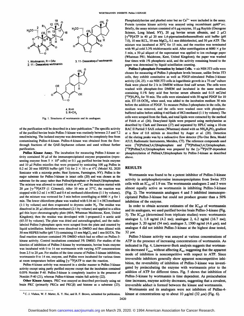
Wortmannin was found to be a potent inhibitor of Ptdlns-3-kinaseactivity in antiphosphotyrosine immunoprecipitates from Swiss 3T3cells with an IC50 of 1.9 nM. The wortmannin analogues 2 and 3 werealmost equally active as wortmannin in inhibiting Ptdlns-3-kinase(Fig. 2). The wortmannin analogues 4 and S inhibited immunoprecipitated Ptdlns-3-kinase but could not produce greater than a 50%inhibition of the enzyme.
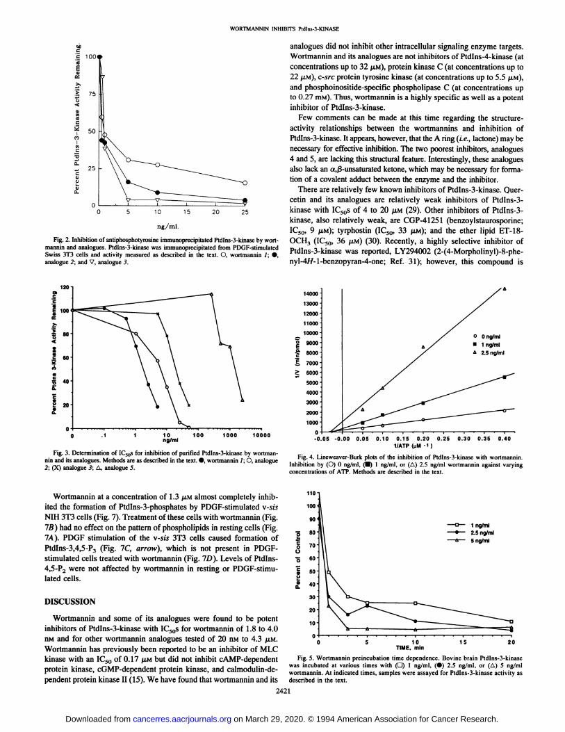
In order to obtain accurate estimates of the IC5@sof wortmanninand its analogues, we used purified bovine brain Ptdlns-3-kinase (Fig.3). The IC50s (determined from triplicate studies) were: wortmannin
analogue 1, 1.8 ng/ml (4.2 nM); analogue 2, 6.2 ng/ml (16.7 aM);analogue 3, 20 ng/ml (54 flM); analogue 5, 1500 ng/ml (4.6 SM); andanalogue 4 did not inhibit Ptdlns-3-kinase at the highest dose tested,32 @LM.
Ptdlns-3-kinase activity was assayed at various concentrations ofAl? in the presence of increasing concentrations of wortmannin. Asindicated in Fig. 4, Lineweaver-Burk analysis suggests that wortmanfin decreased Vm,,y@without affecting Km, suggesting that the apparentmode of inhibition is noncompetitive with respect to AlP. Sinceirreversible inhibitors generally show apparent noncompetitive inhibition, the reversibility of inhibition of Ptdlns-3-kinase was investigated by preincubating the enzyme with wortmannin prior to theaddition of ATP for different times. Fig. 5 shows that inhibition ofPtdlns-3-kinase by wortmannin is time dependent. As preincubationtime increases, enzyme activity decreases, suggesting that a covalent,irreversible adduct is formed between the kinase and wortmannin.
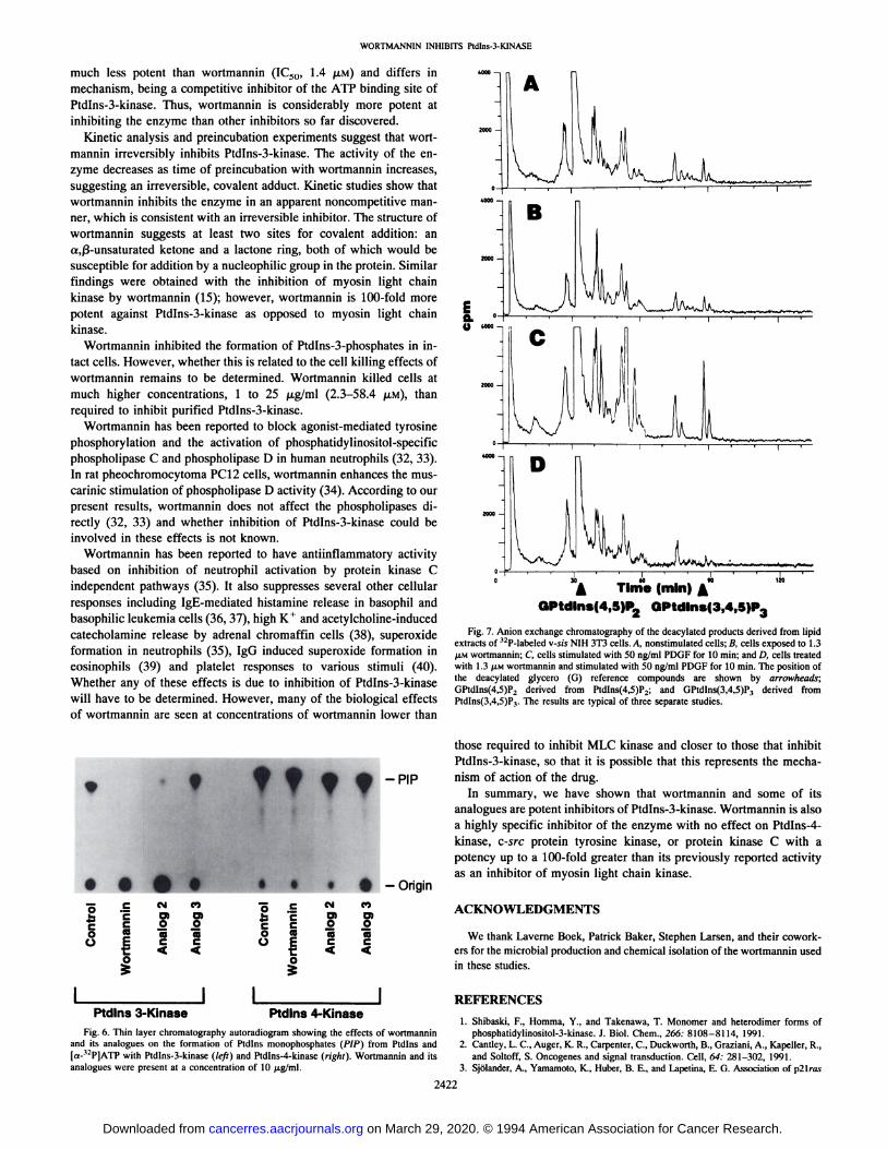
Wortmannin and its analogues were not inhibitors of Ptdlns-4-kinase at concentrations up to about 10 @Wml(32 j.LM)(Fig. 6).
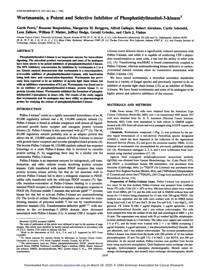
3. 11-DesacetoXy@'-WOrtm@flifl
4. R=H5.R=CH3
Fig. I . Structures of wortmannin and its analogues.
of the purification will be described in a later publication.4The specific activityof the purified bovine brain Ptdlns-3-kinase was routinely between 2.5 and 7.2nmol/min/mg. The isolated enzyme was determined to be completely free fromcontaminating Ptdlns-4-kinase. Ptdlns-4-kinase was obtained from the flowthrough fractions of the QAE-Sepharose column and used without furtherpurification.
Ptdlns Kinase Assay. The incubation for measuring Ptdlns-3-kinase ac
tivity contained 30 @lof the immunoprecipitated enzyme preparation (repre
senting enzyme from S X 106cells) or 0.1 @gpurified bovine brain enzymeand 10 ,.d Ptdlns micelles that were prepared by sonicating 100 @gPtdlns in
0.2 ml 20 mM HEPES buffer (pH 7.6) for 2 X 10 s at 4°C (Model XL 2020
Sonicator with a microtip probe; Heat Systems, Farmington, NY). Ptdlns is themajor substrate for Ptdlns-3-kinase in intact cells (20) and was chosen as thesubstrate for the assay rather than Ptdlns(4)phosphate or Ptdlns(4,5)bisphosphate.The mixture was allowed to stand 10 mm at 4°C,and the reaction started with20 @M[y-32P]ATP (1 Ci/mmol). After 10 mm at 37°C, the reaction wasstopped with 0.2 ml 1 N HCI and 0.4 ml methanol:chloroform (1:1 by volume),and the mixture was vortexed for 3 mm before centrifuging at 6000 X g for 1mm. The lower chloroform phase was washed with 0.16 ml 1 NHCI:methanol(1:1 by volume) and then evaporated to dryness under N2. The residue wasdissolved in 20 pi chloroform:methanol (2:1 by volume) and applied to a silicagel thin layer chromatography plate (60A; Whatman Maidstone, Kent, UnitedKingdom); then the residue was developed with 1-propanol:2 N acetic acid(65:35 by volume). The plate was dried and autoradiographed, and the radio
labeled Ptdlns-3-phosphate spots were scraped from the plate and counted by
liquid scintillation. Inhibitors were dissolved in DMSO and then diluted with
50 mMHEPES buffer (pH 7.5) containing 15 m@iMgCl2and 1 m@iEGTA. Thefinal reaction mixture contained 3% DMSO which had no effect on Ptdlns-3-kinase activity. Control incubations contained 3% DMSO. For studies of the
kinetics of inhibition of Ptdlns-3-kinase by wortmannin, bovine brain enzymewas incubated with 0 to 14 nM wortmannin with varying Al? concentrationsfrom 2.5 to 20 gLM.For studies of the time course of Ptdlns-3-kinase inhibition,wortmannin 0 to 14 nM, enzyme, and Ptdlns were incubated for various times
at room temperature before adding [y-32P]ATPto start the reaction.Ptdlns-4-kinase activity was measured in a similar manner to Ptdlns-3-kinase
activity except using partly purified enzyme except that the incubation contained
0.05% Nonidet P40. Ptdlns-3-kinase is completely inactive in the presence ofNonidet P-40 (21), whereas Ptdlns-4-kinase retains full activity (22).
Other Kinase Assays. PKCwas assayedas describedpreviouslyusing ratbrain PKC (primarily PKCct and PKCj3) and histone as a substrate (23).
4 C. J. Vlahos, W. F. Matter, K. Y. Hui, and R. F. Brown, submitted for publication.
analogues did not inhibit other intracellular signaling enzyme targets.Wortmannin and its analogues are not inhibitors of Ptdlns-4-kinase (atconcentrations up to 32 SM), protein kinase C (at concentrations up to22 SM), c-src protein tyrosine kinase (at concentrations up to 5.5 SM),and phosphoinositide-specific phospholipase C (at concentrations upto 0.27 mM). Thus, wortmannin is a highly specific as well as a potent
inhibitor of Ptdlns-3-kinase.Few comments can be made at this time regarding the structure
activity relationships between the wortmannins and inhibition ofPtdlns-3-kinase. It appears, however, that the A ring (i.e., lactone) may benecessary for effective inhibition. The two poorest inhibitors, analogues
4 and 5, are lacking this structural feature. Interestingly, these analogues
also lack an aj3-unsaturated ketone, which may be necessary for formation of a covalent adduct between the enzyme and the inhibitor.
There are relatively few known inhibitors of Ptdlns-3-kinase. Quercetin and its analogues are relatively weak inhibitors of Ptdlns-3-kinase with IC50s of 4 to 20 p.M (29). Other inhibitors of Ptdlns-3-kinase, also relatively weak, are CGP-41251 (benzoylstaurosporine;IC50, 9 p@M);tyrphostin (IC50, 33 ,.LM);and the ether lipid ET-18-OCH3 (IC50, 36 p.M) (30). Recently, a highly selective inhibitor ofPtdlns-3-kinase was reported, LY294002 (2-(4-Morpholinyl)-8-phenyl-4H-1-benzopyran-4-one; Ref. 31); however, this compound is
Wortmannin and some of its analogues were found to be potentinhibitors of Ptdlns-3-kinase with IC50s for wortmannin of 1.8 to 4.0aM and for other wortmannin analogues tested of 20 flM to 4.3 @.LM. 5 1 0 1 5 20
Wortmannin has previously been reported to be an inhibitor of MLC TIME.mmkinase with an IC50 of 0.17 p.M but did not inhibit cAMP-dependent Fig.5. Wortmanninpreincubationtimedependence.BovinebrainPtdlns-3-kinase
was incubated at various times with (0) 1 ng/ml, (•)2.5 ng/ml, or (@@)5 ng/mlprotein kinase, cGMP-dependent protein kinase, and calmodulin-de- wortmannin.At indicatedtimes,sampleswereassayedfor Ptdlns-3-kinaseactivityaspendent protein kinase II (15). We have found that wortmannin and its describedin the text.
2421
100
0 .1
75
50
100 1000
0
10000
ng/ml.
Fig. 2. Inhibition of antiphosphotyrosine immunoprecipitated Ptdlns-3-kinase by wortmannin and analogues. Ptdlns-3-kinase was immunoprecipitated from PDGF-stimulatedSwiss 3D cells and activity measured as described in the text. 0, wortmannin 1;•,analogue 2; and V, analogue 3.
Fig. 3. Determination of 1C5,,s for inhibition of purified Ptdlns-3-kinase by wortmannm and its analogues. Methods are as described in the text. @,wortmannin 1; 0, analogue2; (X) analogue 3; i@, analogue 5.
Fig. 4. Lineweaver-Burk plots of the inhibition of Ptdlns-3-kinase with wortmannin.Inhibition by (0) 0 ng/ml, J) 1 ng/ml, or (i@) 2.5 ng/ml wortmannin against varyingconcentrations of ATP. Methods are described in the text.
110
100
90
Wortmannin at a concentration of 1.3 ,tM almost completely inhibited the formation of Ptdlns-3-phosphates by PDGF-stimulated v-sisNIH 3T3 cells (Fig. 7). Treatment of these cells with wortmannin (Fig.7B) had no effect on the pattern of phospholipids in resting cells (Fig.7A). PDGF stimulation of the v-sis 3T3 cells caused formation ofPtdlns-3,4,5-P3 (Fig. 7C, arrow), which is not present in PDGFstimulated cells treated with wortmannin (Fig. 7D). Levels of Ptdlns4,5-P2 were not affected by wortmannin in resting or PDGF-stimulated cells.
much less potent than wortmannin (IC50, 1.4 ,LM) and differs inmechanism, being a competitive inhibitor of the ATP binding site ofPtdlns-3-kinase. Thus, wortmannin is considerably more potent atinhibiting the enzyme than other inhibitors so far discovered.
Kinetic analysis and preincubation experiments suggest that wortmannin irreversibly inhibits Ptdlns-3-kinase. The activity of the enzyme decreases as time of preincubation with wortmannin increases,suggesting an irreversible, covalent adduct. Kinetic studies show thatwortmannin inhibits the enzyme in an apparent noncompetitive manncr, which is consistent with an irreversible inhibitor. The structure of
wortmannin suggests at least two sites for covalent addition: ana,j3-unsaturated ketone and a lactone ring, both of which would be
susceptible for addition by a nucleophilic group in the protein. Similar
findings were obtained with the inhibition of myosin light chainkinase by wortmannin (15); however, wortmannin is 100-fold morepotent against Ptdlns-3-kinase as opposed to myosin light chainkinase.
Wortmannin inhibited the formation of Ptdlns-3-phosphates in intact cells. However, whether this is related to the cell killing effects ofwortmannin remains to be determined. Wortmannin killed cells atmuch higher concentrations, 1 to 25 ,tg/ml (2.3—58.4 p.M), thanrequired to inhibit purified Ptdlns-3-kinase.
Wortmannin has been reported to block agonist-mediated tyrosinephosphorylation and the activation of phosphatidylinositol-specificphospholipase C and phospholipase D in human neutrophils (32, 33).In rat pheochromocytoma PC12 cells, wortmannin enhances the muscarinic stimulation of phospholipase D activity (34). According to ourpresent results, wortmannin does not affect the phospholipases di
rectly (32, 33) and whether inhibition of Ptdlns-3-kinase could beinvolved in these effects is not known.
Wortmannin has been reported to have antiinflammatory activitybased on inhibition of neutrophil activation by protein kinase Cindependent pathways (35). It also suppresses several other cellularresponses including IgE-mediated histamine release in basophil andbasophilic leukemia cells (36, 37), high K@and acetylcholine-inducedcatecholamine release by adrenal chromaffin cells (38), superoxideformation in neutrophils (35), IgG induced superoxide formation ineosinophils (39) and platelet responses to various stimuli (40).Whether any of these effects is due to inhibition of Ptdlns-3-kinasewill have to be determined. However, many of the biological effectsof wortmannin are seen at concentrations of wortmannin lower than
5@ -
EU
120
Fig. 7. Anion exchange chromatography of the deacylated products derived from lipidextracts of 32P-labeled v-sis NIH 3T3 cells. A, nonstimulated cells; B, cells exposed to 1.3@LMwortmannin; C, cells stimulated with 50 ng/ml PDGF for 10 mm; and D, cells treated
with 1.3 @.LMwortmannin and stimulated with 50 ng/ml PDGF for 10 mm. The position ofthe deacylated glycero (G) reference compounds are shown by arrowheads;GPtdlns(4,5)P2 derived from Ptdlns(4,5)P2; and GPtdlns(3,4,5)P3 derived fromPtdlns(3,4,5)P3. The results are typical of three separate studies.
those required to inhibit MLC kinase and closer to those that inhibitPtdlns-3-kinase, so that it is possible that this represents the mechanism of action of the drug.
In summary, we have shown that wortmannin and some of itsanalogues are potent inhibitors of Ptdlns-3-kinase. Wortmannin is alsoa highly specific inhibitor of the enzyme with no effect on Ptdlns-4-
kinase, c-src protein tyrosine kinase, or protein kinase C with apotency up to a 100-fold greater than its previously reported activityas an inhibitor of myosin light chain kinase.
ACKNOWLEDGMENTS
We thank Laverne Bock, Patrick Baker, Stephen Larsen, and their coworkera for the microbial production and chemical isolation of the wortmannin usedin these studies.
REFERENCES
1. Shibaski, F., Homma, Y., and Takenawa, T. Monomer and heterodimer forms ofphosphatidylinositol-3-kinase. J. Biol. Chem., 266: 8108—8114, 1991.
2. Cantley, L. C., Auger, K. R., Carpenter, C., Duckworth, B., Graziani, A., Kapeller, R.,and Soltoff, S. Oncogenes and signal transduction. Cell, 64: 281—302,1991.
3. Sjdtander, A., Yamamoto, K., Huber, B. E., and Lapetina, E. G. Association of p2lras
I I I IPtdlns 3-Kinase - -@Ptdlns 4-Kinase
Fig. 6. Thin layer chromatography autoradiogram showing the effects of wortmanninand its analogues on the formation of Ptdlns monophosphates (PIP) from Ptdlns and[a-32P]ATP with Ptdlns-3-kinase (left) and Ptdlns-4-kinase (right). Wortmannin and itsanalogues were present at a concentration of 10 p@g,'ml.
with phosphatidylinositol 3-kinase. Proc. Ned Mad. Sri. USA, 88:7908-7947,1991.4. Margolis, B. Protein with 5H2 domains: transducers in the tyrosine kinase signaling
pathway. Cell Growth & Differ., 3: 73-80, 1992.5. Riles, I. D., Otsu, M., Volinia, S., Fry, M. J., Gout, I., Dhand, R., Panayatou, G.,
Ruiz-Larrea, F., Thompson, A., Tally, N. F., Hsuan, J. J., Courtneidge, S. A., Parker,P.J., andWaterfield,M.D.Phosphatidylinositol3-kinase:structureandexpressionofthe 110Kdcatalyticsubunit.Cell,70:419—429,1992.
6. 5dm, P. V., Takegawa, K., Fry, M. J., Stack, J. H., Waterfield, M. D., and Emr,S.D.Phosphatidylinositol3-kinaseencodedbyyeastVPS34geneessentialforproteinsorting. Science (Washington DC), 260: 88—91,1993.
7. Coughlin, S. R., Escobedo, J. A., and Williams, L T. Role of phosphatidylinositol
kinase in PDGF receptor signal transduction. Science (Washington DC), 243: 1191-1194, 1989.
8. Valius, M., and Kazlauskas, A. Phospholipase C-'yl and phosphatidylinositol 3 kinaseare the downstreammediatorsof the PDOFreceptor'smitogenicsignal.Cell, 73:321—334,1993.
9. Kaplan, D. R., Whitman, M., Schafthausen, B., Pallas, D. C., White, M., Cantley, L,and Roberts, T. M. Common elements in growth factor stimulation and oncogenictransformation: 85 kd phosphoprotein and phosphatidylinositol kinase activity. Cell,50:1021—1029,1987.
10. Serunian, L A., Auger, K. R., Roberts, T., and Cantley, L C. Production of novelpolyphosphoinositides in vivo is linked to cell transformation by polyomavirus middleT-antigcn. J. VIrOL, 64: 4718—4725, 1989.
11. Wages, D. S., Keefer, J., Rail, T. B., and Weber, M. J. Mutations in the 5H3 domainof the src oncogenewhichdecreaseassociationof phosphatidylinositol3'-kinaseactivity with @p@V@SrCand altercellular morphology. J. Virol., 66: 1866-1874, 1992.
12. Shurtleff,S. A., Downing,J. R., Rock,C. 0., Hawkins,S. A., Roussel,M. F., andSherr,C. J. Structuralfeaturesof thecolony-stimulatingfactor1 receptorthataffectits association with phosphatidylinositol 3-kinase. EMBO J., 9: 2415—2421,1990.
13. Nishimura, J., Huang, J. S., and Deuel, T. F. Platelet-derived growth factor stimulatestyrosine specific protein kinase activity in Swiss mouse 313 cell membranes. Proc.Natl. Aced. Sci. USA, 79: 4303—4307, 1982.
14. Peles, E., Lamprecht, R., Ben-Lazy, R., Tzahar, E., and Yarden, Y. Regulatedcouplingof the Neu receptorto phosphatidylinositol3'-kinaseand its releasebyoncogenicactivation.J. BioLChem.,267: 12266—12274,1992.
15. Nakanishi, S., Kakita, S., Takahashi, I., Kawahara, K., TSUkUda, E., Sano, T.,Yamada, K., Yoshida, M., Kase, H., Matsuda, Y., Hashimoto, Y., and Nonomura, Y.Wortmannin, a microbial product inhibitor of myosin light chain kinase. J. Biol.Chem., 267: 2157—2163,1992.
16. Scher, H., Jodrell, D., Iversen, J., Curley, T., Tong, W., Egorin, M., and Forrest, A.Use of adaptive control with feedback to individualize suramin dosing. Cancer Rca.,52:64—70,1992.
17. Hensey, C., Boscoboinik, D., and Azzi, A. Suramin, an anti-cancer dru& inhibits
protein kinase C and induces differentiation in neuroblastoma cell clone NB2A. FEBSLeft., 258: 156—158,1989.
19. Prestia, M., Tibero, L, Rusnati, M., Dell'Era, P., and Ragnotti, F. Basic fibroblastgrowth factor requires a long-lasting activation of protein kinase C to induce cellproliferation in transformed fetal bovine sortie endothelial cells. Cell Regul., 2:719—726,1991.
20. Cunningham, T. W., and Majerus, P. W. Pathway for the formation of 32P phosphatecontaininginositolphospholipidsinPDGFstimulatedNIH3T3fibroblasts.Biochem.Biophys.Rca.Commun.,175:568—576,1991.
21. Carpenter,C.L, Duckworth,B.C.,Auger,K.R.,Cohen,B.,Schaffbausen,B.S.,andCantley, L C. Purification and characterization ofphosphoinositide 3-kinase from ratliver. J. Biol. Chem., 265: 19704, 1990.
22. Ung, L E., Schulz, J. T., and Cantley, L C. Characterization and purification ofmembrane-associated phosphatidylinositol 4-phosphate kinase from human red bloodcells. J. Biol. Chem., 264: 5080—5088,1989.
23. Kumaravel, 0., Ashendel, C. L, and Gandour, R. D. Hemicholinium and relatedlipids: inhibitors of protein kinase C. J. Med. Chem., 36: 177—178,1993.
24. Jackson, 1. R., Stephens, L R., and Hawkins, P. 1. Receptor specificity of growthfactor-stimulated synthesis of 3-phosphorylated inositol lipids in Swiss 3T3 cells.J. Biol. Chem., 267: 16627—16636,1992.
25. Kaplan, D. R., Pallas, D. C., Morgan, W., Schafthausen, B., and Roberts, T. M.Mechanisms of transformation by polyoma virus middle 1-antigen. Biochim. Biophys. Acta, 948:345-364,1988.
26. Folch, J., Lees, M., and Sloane-Stanley, 0. H. A simple method for the isolation andpurification of total lipids from animal tissues. J. Biol. Chem., 226: 497—509,1957.
27. Clark, N. G., and Dawson, R. M. C. Alkaline O-N-transacylation: a new method forquantitative deacylation of phospholipids. Biochem. J., 195: 301—306,1981.
28. Auger, K. R., Serunian, L A., and Cantley, L C. Separation of novel polyphosphoinositides. In: R. F. Irvine (ed), Methods in Inositide Research, pp. 159—166,New York: Raven Press, Ltd. 1990.
29. Matter, W. F., Brown, R. F., and Viahos, C. J. The inhibition of phosphatidylinositol3-kinase by quercetin and analogs. Biochem. Biophys. Rca. Commun., 186: 624-631, 1992.
30. Berggren, M., Gallegos, A., Dressler, L A., Modest, E. J., and Powis, G. Inhibitionof thesignallingenzymephosphatidylinositol-3-kinascby antitumoretherlipidanalogues. Cancer Rca., 53: 4297—4302, 1993.
31. Vlahos, C. J., Matter, W. F., Hod, K. Y., and Brown, R. F. A specific inhibitor ofphosphatidylinositol3-kinase:2-(4-morpholinyl)-8-phenyl-4H-1-benzopyran-4-one(LY294002). J. Biol. Chem., in press, 1994.
32. Naccache, P. H., Coon, A. C., Gilbert, C., Gaudry, M., Roberge, C. J., Poubelle, P. E.,and Bourgoin, S. Inhibition of tyrosine phosphorylation by wortmannin in humanneutrophils: dissociation from its inhibitory effects on phospholipase D. Lab. Invest.,69:19,1993.
33. Reinhold, S. L, Prescott, S. M., Zimmerman, G. A., and McIntyre, 1. M. Activationof human neutrophil phospholipase D by three separable mechanisms. FASEB J., 4:208—214, 1990.
34. Kanoh, H., Ohbayashi, H., Matsuda, Y., Nonomura, Y., and Nozawa, Y. Enhancing effect of wortmannin on muscarinic stimulation of phospholipase D in ratpheochromocytoma PC12 cells. Biochem. Biophys. Res Commun., 188: 510—515, 1992.
35. Dewald, B., Thelen, M., and Baggliolini, M. Two transduction sequences are nearssary for neutrophil activation by receptor agonists. J. Biol. Chem., 263: 16179—16184, 1988.
36. Kitani, S., Teshima, R., Morita, Y., Ito, K., Matsuka, Y., and Nonomura, Y. InhibitionoflgE-mediatedhistaminereleaseby myosinlightchainkinaseinhibitors.Biochem.Biophys. Res. Commun., 183: 48—54,1992.
37. Knol, E. F., Koenderman, L., Mul, F. P., Verhoeven, A. J., and Roos, D. Differentialactivation of human basophils by anti-IgE and formyl-methionyl-leucyl-phenylalanine.Indicationsfor proteinkinaseC-dependentand -independentactivationpathways. Eur. J. Immunol., 21: 881—885,1991.
38. Ohara-Imaizumi, M., Sakurai, T., Nakamura, S., Nakanishi, S., Matsuda, Y., Moorsmatsu, S., Nonomura, Y., and Kumakura, K. Inhibition of Ca2@-dependentcatecholamine release by myosin light chain kinase inhibitor, wortmannin, in adrenal chromaffin cells. Biochem. Biophys. Rca. Commun., 185: 1016—1021, 1992.
39. Bach, M. K., Brashler, I. R., Petzold, E. N., and Sanders, M. E. Superoxide productionby humaneosinophilscanbe inhibitedin an agonist-selectivemanner.AgentsActions, 35: 1—11,1992.
40. Yatomi, Y., Hazeki, 0., Kume, S., and Ui, M. Suppression by wortmannin of plateletresponses to stimuli due to inhibition ofpleckstrin phosphorylation. Biochem. J., 285:745—751,1992.
1994;54:2419-2423. Cancer Res Garth Powis, Rosanne Bonjouklian, Margareta M. Berggren, et al. Phosphatidylinositol-3-kinaseWortmannin, a Potent and Selective Inhibitor of