59
WOUNDS Dr Phillipo L. Chalya MD, M.Med(Surg) Senior Lecturer – Department of Surgery CUHAS
| Date post: | 12-Aug-2015 |
| Category: |
Education |
| Upload: | sadru-mohamed |
| View: | 62 times |
| Download: | 0 times |

WOUNDS
Dr Phillipo L. Chalya MD, M.Med(Surg)
Senior Lecturer – Department of Surgery
CUHAS

Leaning objectives At the end of this topic, you should be able to:-
Define the term “Wound” List the causes of wounds Outline the classification of wounds Define the term “wound healing” Describe the phases of wound healing Highlight the types of wound healing Describe factors affecting wound healing Outline the complications of wound healing Discuss the management of wounds
DEFINITION A wound is a type of physical trauma
whereby the integrity of the skin or of any tissue is compromised
It is a separation or discontinuity of the skin, mucous membrane or tissue caused by physical, chemical or biological insult
ETIOLOGY The etiology of wounds can be
classified as follows:- Blunt injuries Penetrating injuries Surgical insult Burn injuries
Blunt injuries RTA Falls Assault Sport injuries Bite injuries [animal or human]

Penetrating injuries Stab wounds Gunshot wounds
Surgical wounds Wounds caused by a surgical
procedure
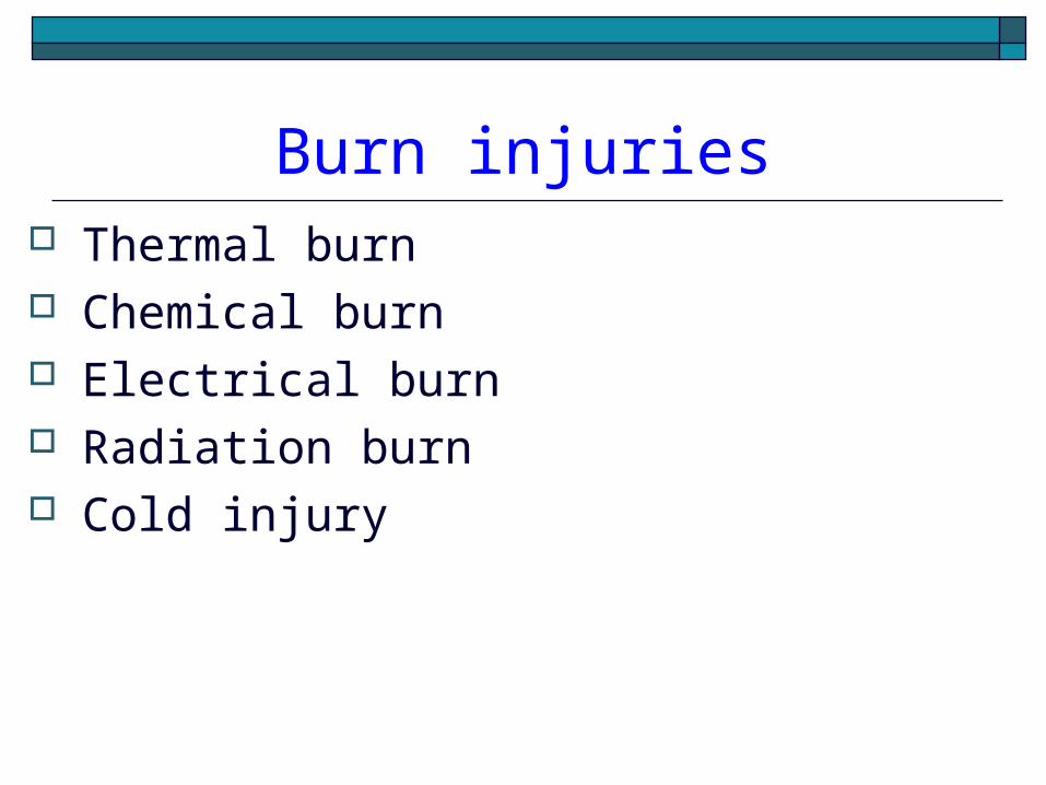
Burn injuries Thermal burn Chemical burn Electrical burn Radiation burn Cold injury
WOUND CLASSIFICATION
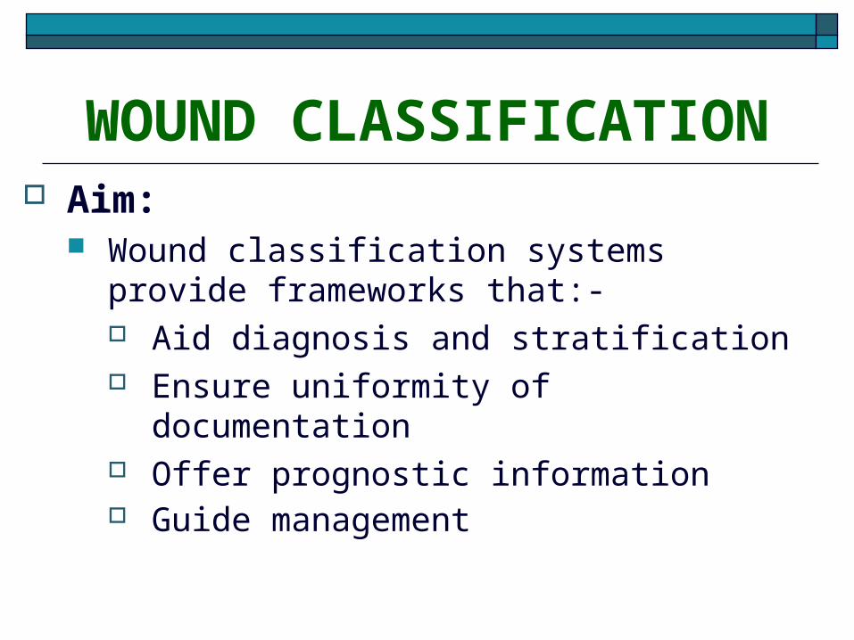
Aim: Wound classification systems provide
frameworks that:- Aid diagnosis and stratification Ensure uniformity of documentation Offer prognostic information Guide management
Types of wound classification
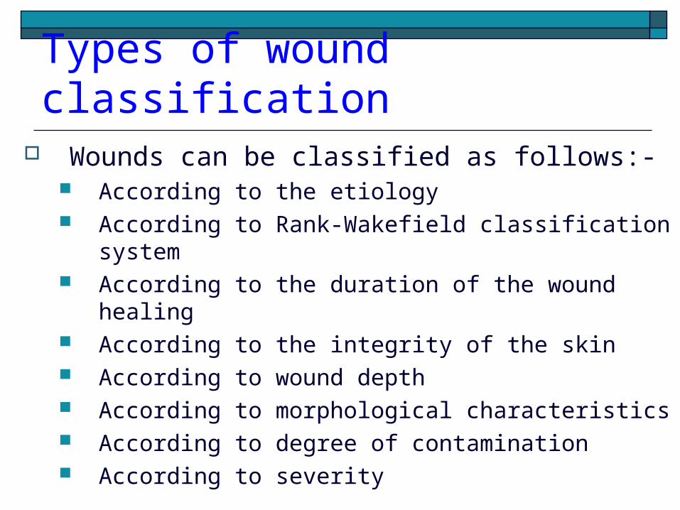
Wounds can be classified as follows:- According to the etiology According to Rank-Wakefield classification system According to the duration of the wound healing According to the integrity of the skin According to wound depth According to morphological characteristics According to degree of contamination According to severity
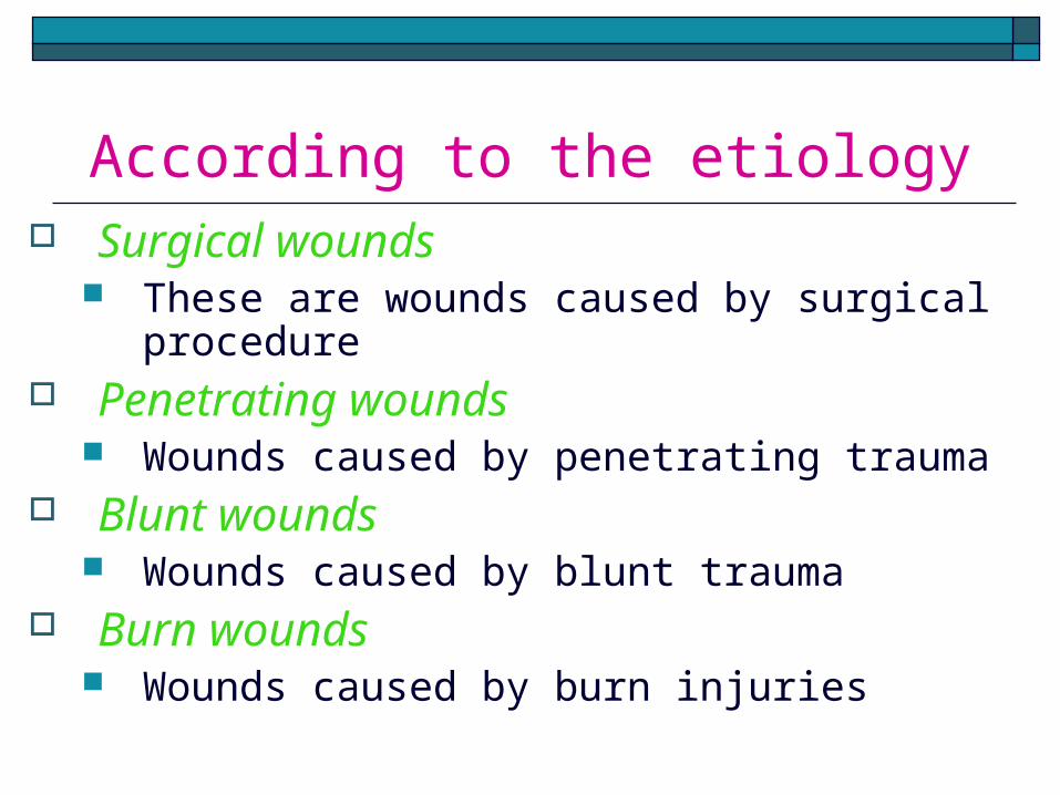
According to the etiology Surgical wounds
These are wounds caused by surgical procedure
Penetrating wounds Wounds caused by penetrating trauma
Blunt wounds Wounds caused by blunt trauma
Burn wounds Wounds caused by burn injuries

According to Rank-Wakefield classification system
Tidy wounds These are wounds inflicted by sharp
instruments and contain no devitalized tissue
Such wounds can be closed primarily with the expectation of quite primary healing
They are usually single with clean cut Associated fractures are uncommon in
tidy wounds Examples: surgical incisions, cuts from
glass and knife wounds

Untidy wounds These are wounds resulting from
crushing, tearing avulsion, vascular injury or burns, and contain devitalized tissue
They are usually multiple and irregular
Commonly associated with fractures Such wounds can not be closed
primarily and therefore should be allowed to heal by second intention

According to the duration of the wound healing
Acute wounds Acute wounds are wounds that usually
heal in the anticipated time frame Duration of the wound: immediately to
few weeks Examples are wounds acquired as a
result of trauma or an operative procedure

Chronic wounds Wounds that fail to heal in the
anticipated time frame and often reoccur
Duration of the wound > 4 weeks to 3 months
Wounds occur as a result of an underlying condition such as extended pressure on the tissues, poor circulation, or even poor nutrition
Pressure ulcers, venous leg ulcers, and diabetic foot ulcers are examples
According to the integrity of the skin
Open wounds Type of wounds in which the skin has
been compromised and underlying tissues are exposed
Open wounds can be classified into a number of different types, according to the object that caused the wound
Examples include incised wounds, laceration, punctured wounds etc

Closed wounds Wounds in which the skin has not been
compromised, but trauma to underlying structures has occurred
Closed wounds have fewer categories, but are just as dangerous as open wounds
Examples of closed wounds are: Contusions - (more commonly known as
a bruise) - caused by blunt force trauma that damages tissue under the skin
Hematoma - (also called a blood tumor) - caused by damage to a blood vessel that in turn causes blood to collect under the skin

According to wound depth Superficial wounds
Only the epidermis is affected and has to be replaced
A truly superficial wound does not bleed and heals within a few days
Examples include most abrasions and blisters

Partial-thickness wounds The epidermis and part of the dermis is
affected A partial-thickness wound does bleed If left uncovered, a blood clot will cover the
wound and a scar will form The missing tissue will then be replaced,
followed by regeneration of the epidermis A partial-thickness wound can take from several
days to several weeks to heal, depending on the patient and the wound treatments chosen
Full-thickness wounds A full-thickness wound involves the
epidermis and the dermis The underlying fatty tissue, bones, muscles,
or tendons may also be damaged If full-thickness wounds cannot be sutured,
the healing process will create new tissue to fill the wound, followed by regeneration of the epidermis
The full-thickness wound takes longer time to heal than does a partial-thickness wound, sometimes as long as several months

According to morphological characteristics
Bruises / contusion These are closed wounds Caused by blunt trauma that damage the
tissue under the skin without breaking the skin
Characterized by skin discoloration due to bleeding into the tissues
Blows to the chest, abdomen, or head with a blunt instrument can cause contusions

Hematoma These are also closed wounds caused
by damage to a blood vessel that in turn causes blood to collect under the skin
Initially this is fluid, but it will clot within minutes or hours later after few days the hematoma will again liquefy increased risk of secondary infection pus formation
Crush wounds Crush wounds are caused by a great or
extreme amount of force applied over a long period of time
These occur when a heavy object falls onto a person, splitting the skin and shattering or tearing underlying structures
They are often accompanied by degloving injuries and compartment syndrome

Abrasions An abrasion is a shearing injury of
the skin I which the surface is rubbed off
Most are superficial and will heal by epitheliazation
Lacerated wound Caused by tearing of tissues Wounds have irregular borders Loss of tissue is limited to skin and
s/c tissue

Penetrated wound Cause by sharp pointed objects like
nails Have relatively small opening May be very deep Infection/ foreign particles might have
been carried deep in to wound opening is inadequate for drainage
eg: punctured wound on foot due to gathered nail

Perforating wound Have two opening one of entrance
and other of exit E.g. gunshot wounds

According to degree of contamination
Clean wounds No break in aseptic technique Incision is made under sterile condions No inflammation is encountered The respiratory tract, alimentary,
genital or uninfected urinary tracts are not entered
Primary closure No drain Eg Herniorrhaphy,

Clean Contaminated wounds Operative wounds in which the
respiratory, alimentary, genital or urinary tract is entered under controlled conditions and without unusual contamination
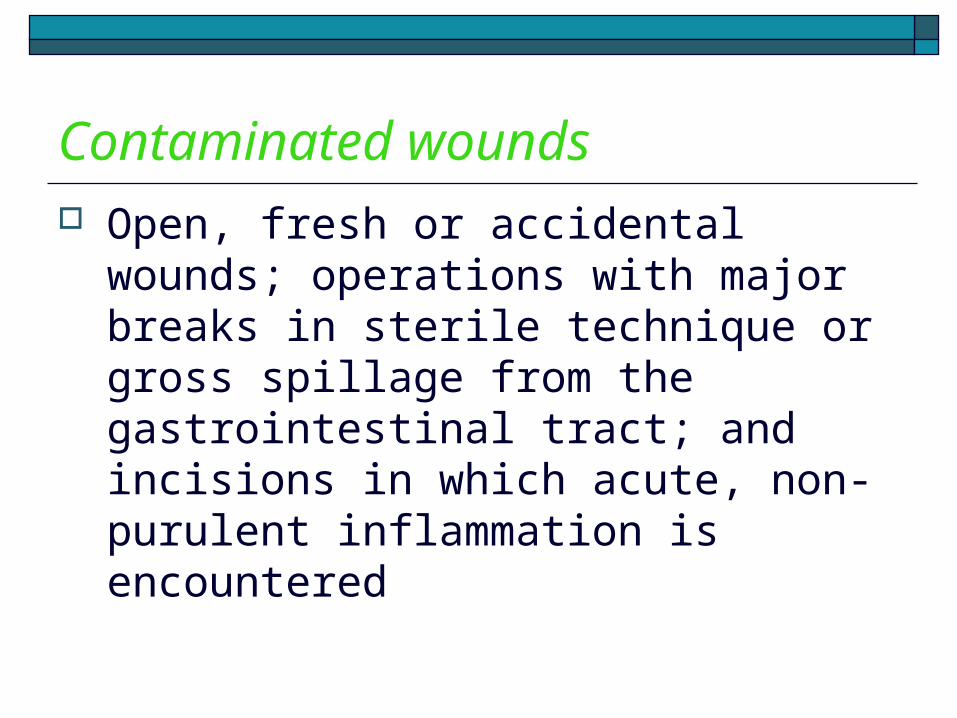
Contaminated wounds Open, fresh or accidental wounds;
operations with major breaks in sterile technique or gross spillage from the gastrointestinal tract; and incisions in which acute, non-purulent inflammation is encountered
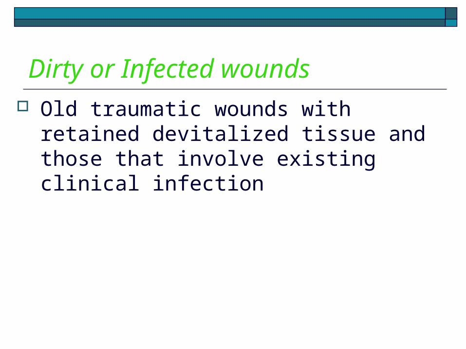
Dirty or Infected wounds Old traumatic wounds with retained
devitalized tissue and those that involve existing clinical infection
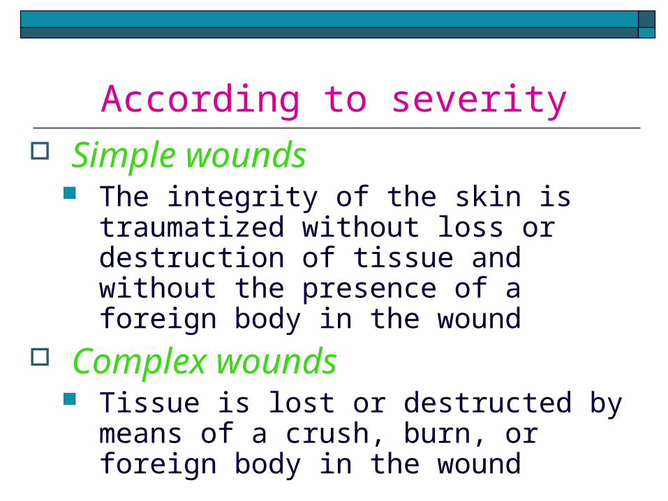
According to severity Simple wounds
The integrity of the skin is traumatized without loss or destruction of tissue and without the presence of a foreign body in the wound
Complex wounds Tissue is lost or destructed by means
of a crush, burn, or foreign body in the wound

WOUND HEALING Definition
Wound healing, or wound repair, is the body's natural process of restoring normal function and structure after injury
The entire wound healing process is a complex series of events that begins at the moment of injury and can continue for months to years

Phases of wound healing Three phases of wound healing
include:- Inflammatory phase Proliferative phase Maturation and remodeling phase

Inflammatory phase Immediate to 2-5 days Aim: to stop bleeding and to prevent
further injury Characterized by :-
Clotting cascade-haemostasis Platelets aggregation Vasoconstriction and vasodilatation Increased polymorphonuclear
neutrophils Increased Macrophages

Clotting cascade Injury to vascular tissue initiates the
extrinsic coagulation cascade by releasing intracellular calcium and tissue factor that activate factor VII
The resulting fibrin plug achieves hemostasis and acts as a lattice for the aggregation of platelets, the most common and “signature” cell type of the early inflammatory phase

Platelets aggregation Within minutes post-injury, platelets
(thrombocytes) aggregate at the injury site to form a fibrin clot
Platelets begin secreting inflammatory factors that serve a lot of functions and also express glycoproteins on their cell membranes that allow them to stick to one another and to aggregate, forming a mass of clot
This clot acts to control active bleeding (hemostasis)

Vasoconstriction and vasodilatation
Immediately after a blood vessel is breached, ruptured cell membranes release inflammatory factors like thromboxanes and prostaglandins that cause the vasoconstriction to prevent blood loss and to collect inflammatory cells and factors in the area
This vasoconstriction lasts 5-10 minutes and is followed by vasodilatation which peaks at about 20 minutes post-wounding

Vasoconstriction and vasodilatation……..
Vasodilatation is the result of factors released by platelets and other cells
The main factor involved in causing vasodilation is histamine
Histamine also causes vascular permeability entry of inflammatory cells like leukocytes into the wound site from the bloodstream

Increased polymorphonuclear neutrophils
Within an hour of wounding, PMNs arrive at the wound site and become the predominant cells in the wound for the first two days after the injury
These PMNs phagocytise debris and bacteria and also kill bacteria by releasing free radicals
They also cleanse the wound by secreting proteases that break down damaged tissue
PMNs usually undergo apoptosis once they have completed their tasks and are engulfed and degraded by macrophages

Increased Macrophages Macrophages are essential to wound
healing They replace PMNs as the predominant
cells in the wound by two days after injury Attracted to the wound site as monocytes
from blood vessels by growth factors released by platelets and other cells
Once they are in the wound site, monocytes mature into macrophages
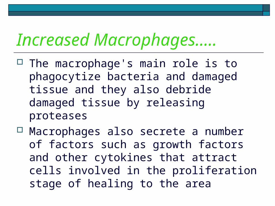
Increased Macrophages….. The macrophage's main role is to
phagocytize bacteria and damaged tissue and they also debride damaged tissue by releasing proteases
Macrophages also secrete a number of factors such as growth factors and other cytokines that attract cells involved in the proliferation stage of healing to the area

Proliferative phase After the inflammatory stage, the proliferative
stage lasts about 3 weeks (or longer, depending on the severity of the wound)
Aim: repair of wounded tissue Characterized by
Angiogenesis Fibroplasia and granulation tissue formation Epithelialization Wound contraction
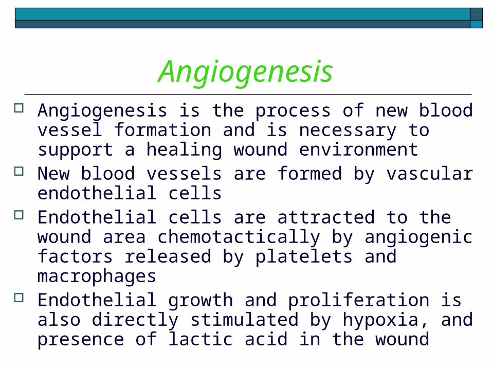
Angiogenesis Angiogenesis is the process of new blood
vessel formation and is necessary to support a healing wound environment
New blood vessels are formed by vascular endothelial cells
Endothelial cells are attracted to the wound area chemotactically by angiogenic factors released by platelets and macrophages
Endothelial growth and proliferation is also directly stimulated by hypoxia, and presence of lactic acid in the wound

Fibroplasia and granulation tissue formation
Fibroblasts begin accumulating in the wound site 2-5 days after wounding and peaks at 1-2 weeks post-wounding
Fibroblasts then deposit ECM into the wound bed, and later collagen and granulation tissue formation
Granulation tissue consists of new blood vessels, fibroblasts, inflammatory cells, endothelial cells, myofibroblasts, and extracellular matrix (ECM)

Epithelialization Epithelial cells migrate across the granulation
tissue to form a barrier between the wound and the environment
Basal keratinocytes from the wound edges and dermal appendages such as hair follicles, sweat glands and sebacious glands are the main cells responsible for the epithelialization phase of wound healing
Epithelialization phase is usually complete within 7-10 days

Wound contraction Contraction is a key phase of wound healing If contraction continues for too long, it can
lead to disfigurement and loss of function Contraction commences approximately a
week after wounding, when fibroblasts have differentiated into myofibroblasts and can last for several weeks
Myofibroblasts, which are similar to smooth muscle cells, are responsible for contraction

Maturation and remodeling phase
The maturation phase of tissue repair begin when the levels of collagen production and degradation equalize
The maturation phase can last for a year or longer, depending on the size of the wound and whether it was initially closed or left open
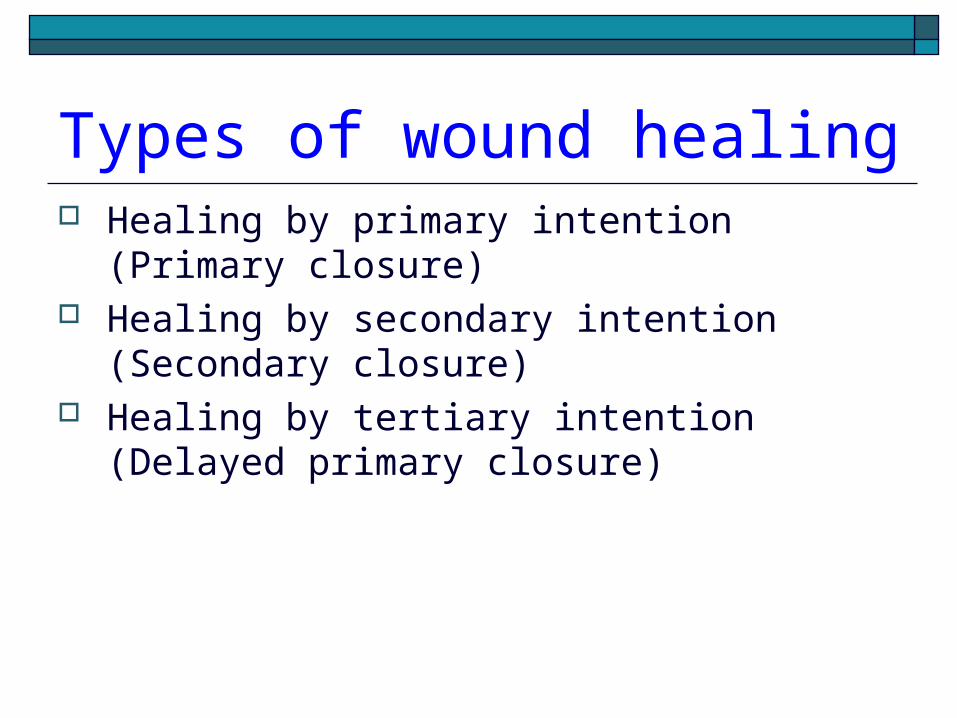
Types of wound healing Healing by primary intention (Primary
closure) Healing by secondary intention
(Secondary closure) Healing by tertiary intention (Delayed
primary closure)

Healing by primary intention (Primary closure)
Healing by primary intention (Primary closure) occurs when a wound is created aseptically with minimal tissue damage
Healing takes place by the approximation of tissue edges with suture, staples, wound sealant etc

Healing by secondary intention (Secondary closure)
Occurs in wounds that are already infected and are usually left open and allowed to heal by epitheliazation and wound contraction
May be caused by infection, excessive trauma, tissue loss, or inability to re-approximate the tissue
It is a slow process

Healing by tertiary intention (Delayed primary closure)
Wounds that are heavily contaminated and are likely to develop an infection if closed primarily may be left open for 3-5 days
This allows the wound to be cleaned and allows the body’s natural defenses to decrease bacterial count
The wound can then be closed and allowed to heal, producing a wound with characteristics similar to primary closure
Factors affecting wound healing
Local factors affecting wound healing Systemic factors affecting wound healing

Local factors affecting wound healing
Infection Surgical Technique Movement Hematoma formation Tissue ischemia Presence of foreign body Exposure to radiation
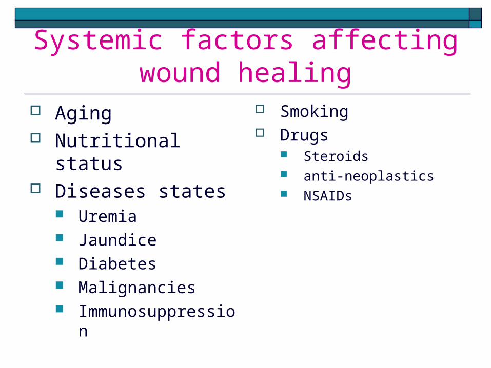
Systemic factors affecting wound healing
Aging Nutritional status Diseases states
Uremia Jaundice Diabetes Malignancies Immunosuppressio
n
Smoking Drugs
Steroids anti-neoplastics NSAIDs

Complications of wound healing
Dehiscence Evisceration Hemorrhage Adhesions Infection Herniation Fistula formation Sinus formation
Suture complications Hypertrophic scar Keloids Malignant changes

MANAGEMENT OF WOUNDS Surgical toilet with:-
Primary closure Delayed closure
Delayed primary closure Skin grafting Flaps
Wound dressing Skin grafting Flaps

SPECIAL THANKS TO SADRU MOHAMED MD STUDENT AT CUHAS BUGANDO 2015 MAKING THESE MATERIA AVAILABLE TO YOU. [email protected] +255759212578


