Page 1
Copy Number Variants (CNVs) Affecting Cancer Predisposing Genes (CPGs) Detected As Incidental
Findings In Routine Germline Diagnostic Chromosomal Micro-Array (CMA) Testing
Josie Innes1, Lisa Reali2, Jill Clayton-Smith1, Georgina Hall1, Derek Lim2, George Burghel1, Kim French1,
Unzela Khan1, Daniel Walker1, Fiona Lalloo1, D. Gareth R. Evans1, Dom McMullan2, Eamonn R. Maher3,
and Emma R. Woodward2
1. Manchester Centre for Genomic Medicine, St Mary's Hospital, Central Manchester University Hospitals
NHS Foundation Trust, Manchester Academic Health Science Centre, Manchester, M13 9WL, UK.
2. West Midlands Regional Genetics Service, Birmingham Women’s and Children’s NHS Foundation Trust,
Birmingham, B15 2TG, UK
3. Department of Medical Genetics, University of Cambridge and NIHR Cambridge Biomedical Research
Centre and Cancer Research UK Cambridge Centre, CB2 OQQ, UK.
Corresponding author:
Dr Emma R. Woodward
Manchester Centre for Genomic Medicine, St Mary's Hospital, Central Manchester University Hospitals
NHS Foundation Trust, Manchester Academic Health Science Centre, Manchester, M13 9WL, UK
[email protected]
Word count: 2,829
Page 2
Abstract
Background
Identification of copy number variations (CNVs) through chromosomal microarray (CMA) testing is first
line investigation in individuals with learning difficulties/congenital abnormalities. Although recognised that
CMA testing may identify CNVs encompassing a cancer predisposition gene (CPG), limited information is
available on the frequency and nature of such results.
Methods
We investigated CNV gains and losses affecting 39 CPGs in 3,366 pilot index case individuals undergoing
CMA testing, and then studied an extended cohort (n=10,454) for CNV losses at 105 CPGs and CNV gains
at 9 proto-oncogenes implicated in inherited cancer susceptibility.
Results
In the pilot cohort, 31/3,366 (0.92%) individuals had a CNV involving one or more of 16/39 CPGs. 30/31
CNVs involved a tumour suppressor gene (TSG), and 1/30 a proto-oncogene (gain of MET). BMPR1A,
TSC2 and TMEM127 were affected in multiple cases. In the second stage analysis, 49/10,454 (0.47%)
individuals in the extended cohort had 50 CNVs involving 24/105 CPGs. 43/50 CNVs involved a TSG and
7/50 a proto-oncogene (4 gains, 3 deletions). The most frequently involved genes, FLCN (n=10) and SDHA
(n=7), map to the Smith-Magenis and cri-du-chat regions respectively.
Conclusion
Incidental identification of a CNV involving a CPG is not rare and poses challenges for future cancer risk
estimation. Prospective data collection from CPG-CNV cohorts ascertained incidentally and through
syndromic presentations is required to determine the risks posed by specific CNVs. In particular,
ascertainment and investigation of adults with CPG-CNVs and adults with learning disability and cancer,
could provide important information to guide clinical management and surveillance.
Page 3
Introduction
The human genome contains marked structural variation and it is over 10 years since the first comprehensive
copy number variant (CNV) map of the human genome was published (1). For children presenting with
developmental delay/learning difficulties and/or congenital abnormalities, diagnostic germline chromosomal
microarray (CMA) for causative CNVs is now a first line investigation and, together with advances in CMA
technology leading to improving resolution (2), there are increasingly numbers of patients identified with
CNVs of uncertain significance or for which the resulting phenotype is unclear. This is particularly pertinent
where an identified CNV encompasses an inherited cancer (cancer predisposition gene/CPG) and there is no
relevant personal or family history, a so-called incidental finding.
With the mainstreaming of modern genomic investigations, CMA testing is often ordered by non-genetics
health care professionals (e.g. paediatricians) who may have limited familiarity with familial cancer
syndromes and are unable to advise on the full significance of the CMA result. Previously, Pichert et al (3)
described the frequency of CNVs affecting 47 CPGs in 4,805 CMA analyses. We report an independent
replication study on a larger patient cohort (two fold increase in cases and a more extensive CPG list
(n=105).
Page 4
Methods
Participants and samples
Samples were referred by paediatricians and clinical geneticists where constitutional diagnostic array
comparative genomic hybridisation (aCGH) was requested to determine causes of developmental delay,
learning difficulties, neurocognitive impairment and/or birth defects. Only arrays pertaining to the index
case in a family were included. The following were excluded (i) patients with clinical features and/or a
family history suggestive of the involvement of a known cancer predisposing gene (CPG) (ii) samples from
prenatal diagnoses; and (iii) results involving whole chromosome or chromosome arm aneuploidy (iv)
results where the CNV identified involving a CPG was present in mosaic form. Monozygotic twins were
counted as one individual for the purposes of this study. A finding was considered positive where the
involvement of a CPG was not suspected before testing (i.e. an incidental finding). CNVs included in the
results were those where the CNV was considered to be causative of the index case’ presenting features, and
also CNVs of either benign or uncertain significance. Approval for the clinical audit study was provided by
Birmingham Women’s NHS Foundation Trust and Central Manchester University Hospitals NHS
Foundation Trust.
Two cohorts of patients were analysed in two stages. Initially, a pilot cohort comprising 3,366 index case
samples investigated between 1 Jan 2009 – 30 Sept 2013 at the West Midlands Regional Genetics
Laboratory and then an extended cohort comprising 10,454 index case samples between 1 Jan 2011 and 31
Dec 2015 at the Manchester Centre for Genomic Medicine.
Gene Search Lists
The pilot cohort (n=3,366) was analysed for CNV gains and losses involving a core set of 39 genes (4
oncogenes, 35 tumour suppressor genes) associated with familial cancer predisposition syndromes (Table
S1). In the 10k extended cohort (10,454 index patients), CNV losses were investigated in a panel of 105
known CPGs (including the 39 genes analysed in the pilot cohort) comprising, 94 genes on the Illumina
Trusight Cancer Panel (4) and 11 further candidate CPGs [CDKN1B, CDKN2B, ESR2, HIF2A, HOXB13,
PDGFRA, POLD1, POLE, SMARCA4, SMARCE1, SDHA (5-15)].
Page 5
Additionally, in the 10k extended cohort, CNV gains at nine of the105 genes [ALK, EGFR, HRAS,
RHBDF2, CDK4, KIT, MET, PDGFRA, RET (10, 16-23)] were investigated as activating alterations have
been described in hereditary cancer predisposition. Partial or whole gene losses and gains were noted and
counted as positive findings.
Laboratory methods and bioinformatics analysis
Testing was undertaken in CPA accredited laboratories. aCGH analysis was carried out using DNA
extracted from peripheral blood or mouthwash samples using standard techniques. For the pilot cohort,
aCGH was undertaken using either the BlueGnome 1Mb BAC array or 8x60k v2.0 (ISCA) design oligo
nucleotide array. Data was analysed in BlueFuse Multi. For the 10k extended cohort, aCGH testing was
carried out using Oxford Gene Technology (OGT) CytoSureTM ISCA v2 (8x60k) arrays for all cases with
the exception of P102 which was tested using OGT CytoSureTM Constitutional v3 Array (8x60k). aCGH
data analysis was performed using OGT CytoSureTM Interpret software. A manual screen for mosaic
aberrations was also performed for all cases. Inheritance studies were performed using karyotype analysis,
targeted aCGH or in situ hybridisation studies, as appropriate where parental samples were available.
Statistical Analysis
Data are displayed as mean ± SD. Continuous data were analysed using a two-tailed Student’s t-test. A
P-value < 0.05 was considered to be statistically significant.
All co-ordinates are GRCh37/hg19 except where otherwise stated.
Page 6
Results
Stage 1: Pilot Cohort Analysis
Within the 3,366 index patients there were 31 individuals (15 males, 16 females) harbouring 31 CNVs
involving one or more of the 39 CPGs analysed (Table S2). The ‘incidental finding’ rate was 0.92%
(31/3366). Mean age at CNV analysis in individuals with a positive finding was 51.9 months (SEM 14.2,
range 0-312 months, median 8 months). In 16/31 cases the CPG-related CNV was considered to be relevant
to the clinical phenotype and in 15 individuals the CNV identified was considered to be either unrelated or
of uncertain clinical significance. In 10 cases the CNV encompassing the cancer gene was de novo and in 14
cases the CNV was inherited (including one where the child inherited the unbalanced form of a parental
balanced translocation). The family history was known in 11 of the 14 cases where the CNV was inherited
(excluding the case with the unbalanced form of the parental translocation), and there were no clinical
features in keeping with a germline pathogenic alteration of the CPG.
Only one of the CNVs involved an oncogene, a gain encompassing MET. The remaining 30 CNVs (20 gains
and 10 losses) involved a tumour suppressor gene and two CNVs affected multiple CPGs: a gain involving
MSH2 and MSH6; and a deletion encompassing BMPR1A and PTEN thought likely causative of the learning
difficulties phenotype. In six cases the CNV arose as consequence of a complex chromosomal
rearrangement and, in all six, resulted in the gain of a tumour suppressor gene (TSC2 x3, PMS2 x1, VHL x2).
The 31 “incidental findings” CNVs involved 16/39 (41.0%) CPGs in the pilot stage gene list with BMPR1A
(in 6 cases), TSC2 (n=4) and TMEM127 (n=3) affected in multiple cases (Fig. 1).
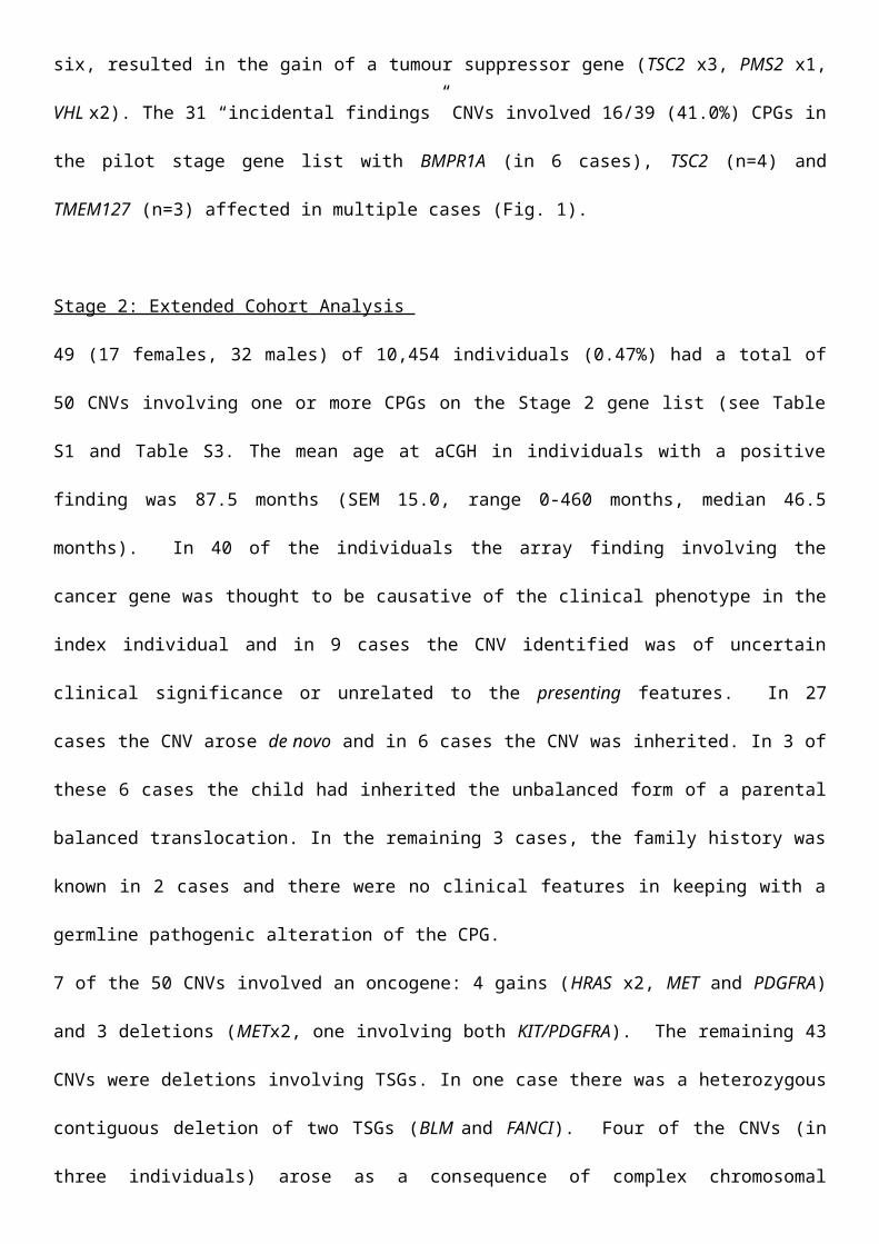
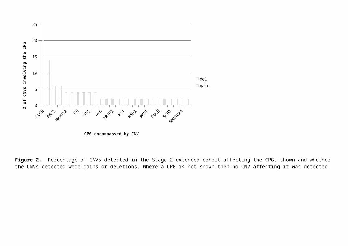
Stage 2: Extended Cohort Analysis
49 (17 females, 32 males) of 10,454 individuals (0.47%) had a total of 50 CNVs involving one or more
CPGs on the Stage 2 gene list (see Table S1 and Table S3. The mean age at aCGH in individuals with a
positive finding was 87.5 months (SEM 15.0, range 0-460 months, median 46.5 months). In 40 of the
individuals the array finding involving the cancer gene was thought to be causative of the clinical phenotype
in the index individual and in 9 cases the CNV identified was of uncertain clinical significance or unrelated
to the presenting features. In 27 cases the CNV arose de novo and in 6 cases the CNV was inherited. In 3 of
Page 7
these 6 cases the child had inherited the unbalanced form of a parental balanced translocation. In the
remaining 3 cases, the family history was known in 2 cases and there were no clinical features in keeping
with a germline pathogenic alteration of the CPG.
7 of the 50 CNVs involved an oncogene: 4 gains (HRAS x2, MET and PDGFRA) and 3 deletions (METx2,
one involving both KIT/PDGFRA). The remaining 43 CNVs were deletions involving TSGs. In one case
there was a heterozygous contiguous deletion of two TSGs (BLM and FANCI). Four of the CNVs (in three
individuals) arose as a consequence of complex chromosomal rearrangement resulting in gain of an
oncogene in three cases (HRAS x2, MET x1) and loss of a tumour suppressor gene (SDHA) in one. These 50
CNVs affected 24 of the 105 genes on the search list (24/105 = 22.9%) with CNVs affecting FLCN
accounting for 10/50 (20%) and of SDHA 7/50 (14%) (Fig. 2).
Joint Analysis of Stage 1 and Stage 2 data sets and CPG lists
Oncogene gains: 3 of 13,820 cases (0.02%) in the combined Stage 1 and Stage 2 cohorts had a CNV gain at
one or more of the 4 oncogenes (RET, PDGFRA, MET and KIT) in the Stage 1 gene list with CNV gains
occurring twice at the MET locus (including one individual with a complex rearrangement leading to gain)
and once at the PDGFRA locus. Three individuals had a deletion of one or more of these oncogenes
(including the one individual with the deletion of both MET and PDGFRA and two with a deletion of MET)
(Table S4).
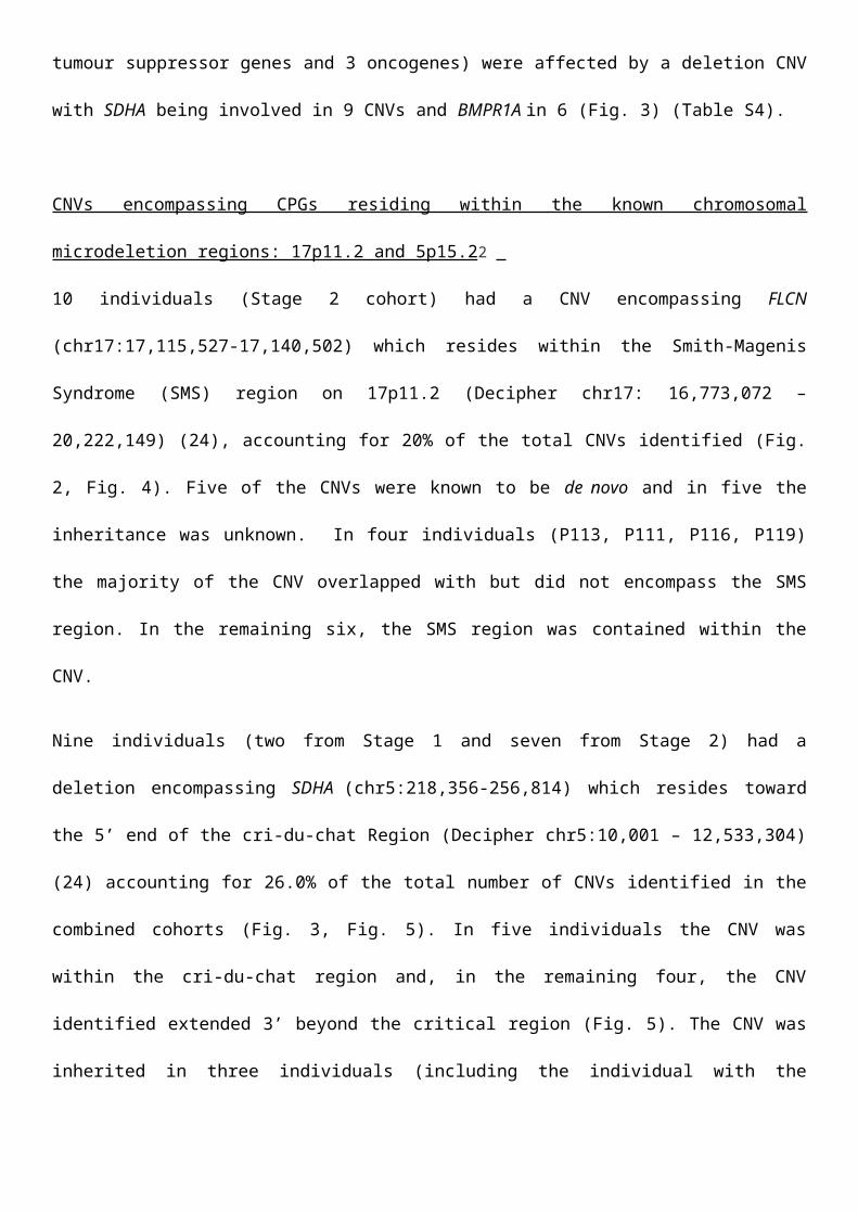
TSG losses: 30 of 13,820 (0.22%) individuals in the combined Stage 1 and 2 cohorts had a partial or whole
deletion involving one or more of 35 tumour suppressor genes (Table S4). In 22/30 cases the CNV
identified was thought to be causative of the child’s presenting features and was thought to be either
unrelated to, or of uncertain significance, in the remaining eight. In 17 cases the CNV identified was de
novo and was found to be inherited in 6 individuals (including one where the child had inherited the
unbalanced form of the parental balanced translocation). 15 of the 39 (38.5%) genes on the common search
list (12 tumour suppressor genes and 3 oncogenes) were affected by a deletion CNV with SDHA being
involved in 9 CNVs and BMPR1A in 6 (Fig. 3) (Table S4).
Page 8
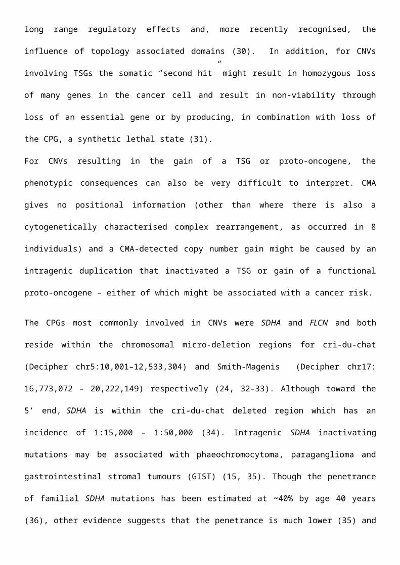
CNVs encompassing CPGs residing within the known chromosomal microdeletion regions: 17p11.2 and
5p15.22
10 individuals (Stage 2 cohort) had a CNV encompassing FLCN (chr17:17,115,527-17,140,502) which
resides within the Smith-Magenis Syndrome (SMS) region on 17p11.2 (Decipher chr17: 16,773,072 –
20,222,149) (24), accounting for 20% of the total CNVs identified (Fig. 2, Fig. 4). Five of the CNVs were
known to be de novo and in five the inheritance was unknown. In four individuals (P113, P111, P116,
P119) the majority of the CNV overlapped with but did not encompass the SMS region. In the remaining
six, the SMS region was contained within the CNV.
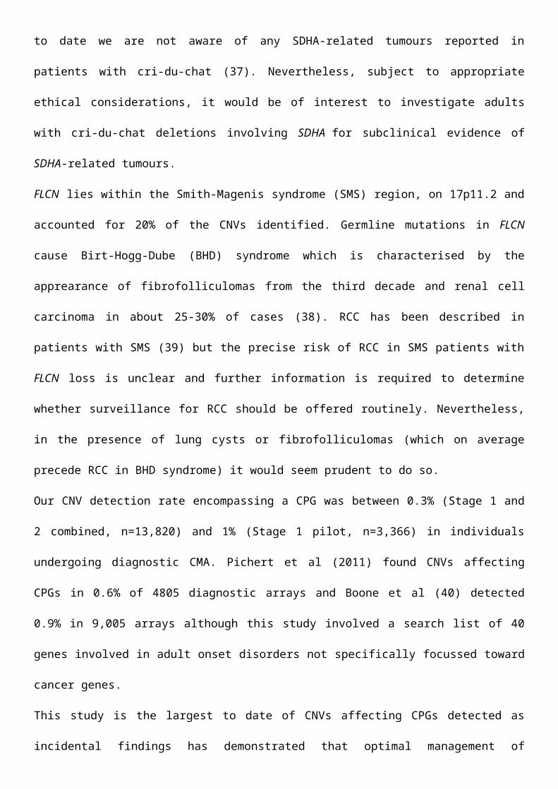
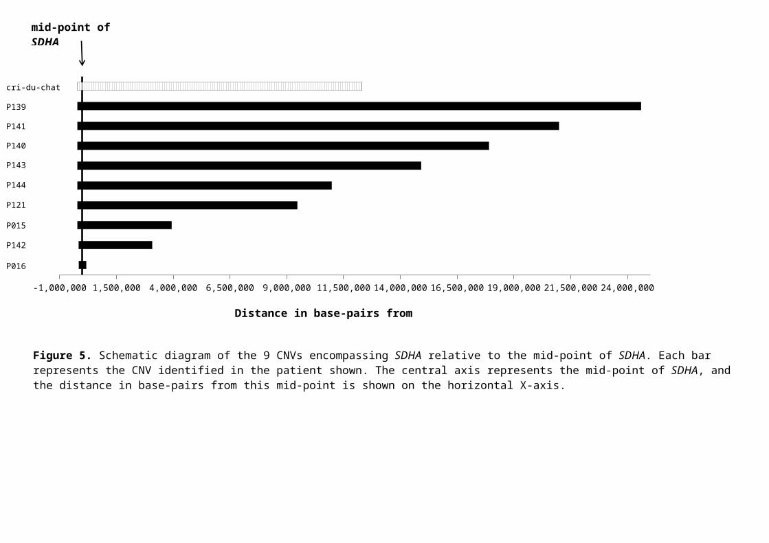
Nine individuals (two from Stage 1 and seven from Stage 2) had a deletion encompassing SDHA
(chr5:218,356-256,814) which resides toward the 5’ end of the cri-du-chat Region (Decipher chr5:10,001 –
12,533,304) (24) accounting for 26.0% of the total number of CNVs identified in the combined cohorts (Fig.
3, Fig. 5). In five individuals the CNV was within the cri-du-chat region and, in the remaining four, the CNV
identified extended 3’ beyond the critical region (Fig. 5). The CNV was inherited in three individuals
(including the individual with the unbalanced translocation leading to loss of SDHA and gain of HRAS), de
novo in two and inheritance was unknown in four individuals.
Page 9
Discussion
CMA testing is now routinely ordered for individuals presenting with undiagnosed learning difficulties
and/or developmental abnormalities and is often undertaken outwith the genetics clinic, for example in the
paediatric mainstream setting.
Whilst these investigations provide the opportunity for diagnosis, the CNVs identified may encompass or
involve genes where intragenic alteration or whole gene copy number losses are known to be associated with
predisposition to other condition(s) unrelated to the presenting features and can be classed as incidental
findings. Unlike other genome wide molecular genetic diagnostic strategies, such as whole exome and
genome sequencing for which results can be filtered in a gene specific manner, identified CNVs are usually
visible to the investigator. Incidental finding CNVs involving CPGs can present significant counselling
challenges as (i) whilst the phenotype and cancer risks of intragenic mutations in a CPG may be well defined
the risks associated with large CNVs are often unclear as deletion of additional in cis genes might modify
cancer risks (25); (ii) the known cancer risks associated with CPGs are for individuals ascertained because
of a family history and are likely to be lower for population-based ascertainment; and (iii) most CNVs
involved CPGs associated with later onset cancers whereas CMA is more commonly performed in a
paediatric setting (mean age at positive finding in our pilot and extended cohorts was 51.9m and 87.5m
respectively). Nevertheless CPG-CNVs cannot be ignored - as exemplified by two infants (P136 and P137,
ages at aCGH 0m and 3m respectively) with deletions of ~50Mb and 24.5Mb respectively encompassing
RB1 who subsequently developed clinical retinoblastoma after the CMA was requested. Whilst
retinoblastoma is highly penetrant at a young age (mean age diagnosis of bilateral retinoblastoma 15m) (26),
and the tumour penetrance for intragenic mutation of other CPGs is often more variable, this highlights that
CNVs encompassing a CPG may be of clinical consequence and should be considered as a paradigm for the
need to report such findings until more is known regarding their effects.
Indeed recent analysis of a range of CPGs showed that large deletions including whole gene deletions were
associated with fairly typical cancer predisposition compared to point mutations (27). Deletions of CPGs
with substantial childhood onset risks such as SMARCB1 (malignant rhabdoid tumour) and TP53 (brain and
sarcoma) also appear to be not infrequent and there is no evidence these deletions are less penetrant than
Page 10
point mutations (27). On the other hand, we also detected an inherited deletion encompassing BMPR1A
(P005) where there was no family history of polyposis. Whilst BMPR1A mutations are of lower penetrance
than RB1 (28), it likely that other factors influencing penetrance/expression are also involved. Varying
phenotypic consequences of large deletions encompassing disease-causing genes are a recognised challenge
(29) and the mechanisms underlying such variable phenotypic effects may include combinations of
underlying genomic architecture, long range regulatory effects and, more recently recognised, the influence
of topology associated domains (30). In addition, for CNVs involving TSGs the somatic “second hit” might
result in homozygous loss of many genes in the cancer cell and result in non-viability through loss of an
essential gene or by producing, in combination with loss of the CPG, a synthetic lethal state (31).
For CNVs resulting in the gain of a TSG or proto-oncogene, the phenotypic consequences can also be very
difficult to interpret. CMA gives no positional information (other than where there is also a cytogenetically
characterised complex rearrangement, as occurred in 8 individuals) and a CMA-detected copy number gain
might be caused by an intragenic duplication that inactivated a TSG or gain of a functional proto-oncogene –
either of which might be associated with a cancer risk.
The CPGs most commonly involved in CNVs were SDHA and FLCN and both reside within the
chromosomal micro-deletion regions for cri-du-chat (Decipher chr5:10,001–12,533,304) and Smith-Magenis
(Decipher chr17: 16,773,072 – 20,222,149) respectively (24, 32-33). Although toward the 5’ end, SDHA is
within the cri-du-chat deleted region which has an incidence of 1:15,000 – 1:50,000 (34). Intragenic SDHA
inactivating mutations may be associated with phaeochromocytoma, paraganglioma and gastrointestinal
stromal tumours (GIST) (15, 35). Though the penetrance of familial SDHA mutations has been estimated at
~40% by age 40 years (36), other evidence suggests that the penetrance is much lower (35) and to date we
are not aware of any SDHA-related tumours reported in patients with cri-du-chat (37). Nevertheless, subject
to appropriate ethical considerations, it would be of interest to investigate adults with cri-du-chat deletions
involving SDHA for subclinical evidence of SDHA-related tumours.
FLCN lies within the Smith-Magenis syndrome (SMS) region, on 17p11.2 and accounted for 20% of the
CNVs identified. Germline mutations in FLCN cause Birt-Hogg-Dube (BHD) syndrome which is
characterised by the apprearance of fibrofolliculomas from the third decade and renal cell carcinoma in
Page 11
about 25-30% of cases (38). RCC has been described in patients with SMS (39) but the precise risk of RCC
in SMS patients with FLCN loss is unclear and further information is required to determine whether
surveillance for RCC should be offered routinely. Nevertheless, in the presence of lung cysts or
fibrofolliculomas (which on average precede RCC in BHD syndrome) it would seem prudent to do so.
Our CNV detection rate encompassing a CPG was between 0.3% (Stage 1 and 2 combined, n=13,820) and
1% (Stage 1 pilot, n=3,366) in individuals undergoing diagnostic CMA. Pichert et al (2011) found CNVs
affecting CPGs in 0.6% of 4805 diagnostic arrays and Boone et al (40) detected 0.9% in 9,005 arrays
although this study involved a search list of 40 genes involved in adult onset disorders not specifically
focussed toward cancer genes.
This study is the largest to date of CNVs affecting CPGs detected as incidental findings has demonstrated
that optimal management of incidentally detected CPG-CNVs and will require systematic collection of long-
term follow up data and international data sharing. In particular detailed studies of the clinical significance
of SDHA and FLCN loss in patients with cri-du-chat and SMS would address the most frequently detected
CPG-CNVs. Though CMAs are routinely performed in children with learning disability, significant numbers
of adults with learning disability are likely not to have had high resolution CMA testing and routine
reinvestigation of such patients could provide important information on cancer risks. In addition, we are
compiling a database of adults with pathogenic CNVs and cancer and request that appropriate cases should
be notified to [email protected] .
Page 12
Figures and Legends
BMPR1A
TSC2
TMEM
127PTEN
RB1SD
HASD
HD
SMARCB1
TP53MET
MSH2
MSH6
NF1
PRKR1A0
5
10
15
20
25
del gain
CPG encompassed by CNV
% o
f CN
Vs
invo
lvin
g th
e CP
G
Figure 1. Percentage of CNVs detected in the Stage 1 pilot cohort affecting the CPG shown and whether the CNVs detected were gains or deletions. Where a CPG gene present in Table 1 is not shown then no CNV involving it was detected.
Page 13
FLCNPM
S2
BMPR1A FH
RB1APC
BRIP1
KITNSD
1PM
S1POLE
SDHB
SMARCA4
0
5
10
15
20
25
delgain
CPG encompassed by CNV
% o
f CN
Vs
invo
lvin
g th
e CP
G
Figure 2. Percentage of CNVs detected in the Stage 2 extended cohort affecting the CPGs shown and whether the CNVs detected were gains or deletions. Where a CPG is not shown then no CNV affecting it was detected.
Page 14
SDHA
BMPR1A
TMEM
127PM
S2MET
RB1APC
KITMAX
PDFRαPTEN
SDHB
SDHD
SMARCB1
TSC2
0
5
10
15
20
25
30
del
CPG encompassed by the CNV
% o
f CN
Vs
invo
lvin
g th
e CP
G
Figure 3. Percentage of deletion CNVs detected in the combined Stage 1 pilot and Stage 2 extended cohorts affecting the CPGs shown. Where a CPG is not shown then no deletion CNV affecting it was detected.
Page 15
Figure 4. Schematic diagram of the 10 CNVs encompassing FLCN relative to the mid-point of FLCN. Each bar represents the CNV identified in the patient shown. The central axis represents the mid-point of FLCN, and the distance in base-pairs from this mid-point is shown on the horizontal X-axis.
mid-point of FLCN
SMS
P113
P111
P116
P119
P110
P112
P114P115
P120
P118
-1.5MB
-1.0MB
-0.5MB
0.5MB 1.0MB 1.5MB 2.0MB 2.5MB 3.0MB 3.5MB
Distance in base-pairs from mid-point of FLCN
Page 16
-1,000,000 1,500,000 4,000,000 6,500,000 9,000,000 11,500,000 14,000,000 16,500,000 19,000,000 21,500,000 24,000,000
Figure 5. Schematic diagram of the 9 CNVs encompassing SDHA relative to the mid-point of SDHA. Each bar represents the CNV identified in the patient shown. The central axis represents the mid-point of SDHA, and the distance in base-pairs from this mid-point is shown on the horizontal X-axis.
mid-point of SDHA
cri-du-chat regionP139
P141
P144
P121
P015
P142
P016
P143
Distance in base-pairs from mid-point of SDHA
P140
Page 17
Table Legends
Table S1Cancer Predisposing Genes (CPGs). Corresponding omim gene and phenotype numbers are also given.
Table S2CNVs detected involving a CPG in the Stage 1 Pilot Cohort Analysis.* oncogene# presence of a complex chromosomal rearrangement Table S3CNVs detected involving a CPG in the Stage 2 Extended Cohort Analysis. * oncogene# presence of a complex chromosomal rearrangement
Table S4CNVs detected involving a CPG in the combined Stage 1 Pilot and Stage 2 Extended Cohorts using the CPG search list from the Stage 1 Pilot Cohort Analysis. * oncogene# presence of a complex chromosomal rearrangement
Page 18
Acknowledgements
This study did not receive any specific grant funding. Prof. Evans is an NIHR senior investigator and DGE
and ERW are supported by the Manchester NIHR Biomedical Research Centre. ERM acknowledges support
from NIHR, European Research Council Advanced Researcher Award and Cancer Research UK.
Page 19
References
1. Redon R, Ishikawa S, Fitch KR, Feuk L, Perry GH, Andrews TD, Fiegler H, Shapero MH, Carson AR,
Chen W, Cho EK. Global variation in copy number in the human genome. Nature 2006;444:444-54.
2. Miller DT, Adam MP, Aradhya S, Biesecker LG, Brothman AR, Carter NP, Church DM, Crolla JA,
Eichler EE, Epstein CJ, Faucett WA, Feuk L, Friedman JM, Hamosh A, Jackson L, Kaminsky EB, Kok k,
Krants ID, Kuhn RM, Lee C, Ostell JM, Rosenberg C, Scherer SW, Spinner NB, Stavropoulos DJ,
Tepperberg JH, Thorland EC, Vermeesch JR, Waggoner DK, Watson MS, Martin CL, Ledbetter. DH.
Consensus statement: chromosomal microarray is a first-tier clinical diagnostic test for individuals with
developmental disabilities or congenital anomalies. Am J Hum Genet 2010;86:749-64.
3. Pichert G, Mohammed SN, Ahn JW, Ogilvie CM, Izatt L. Unexpected findings in cancer predisposition
genes detected by array comparative genomic hybridisation: what are the issues? J Med Genet 2011;48::535-
9.
4. http://www.illumina.com/products/trusight_cancer.html. Accessed 21 June 2017.
5. Pellegata NS, Quintanilla-Martinez L, Siggelkow H, Samson E, Bink K, Höfler H, Fend F, Graw J,
Atkinson MJ. Germ-line mutations in p27Kip1 cause a multiple endocrine neoplasia syndrome in rats and
humans. Proc Natl Acad Sci USA 2006;103:15558-63.
6. Jafri M, Wake NC, Ascher DB, Pires DE, Gentle D, Morris MR, Rattenberry E, Simpson MA, Trembath
RC, Weber A, Woodward ER, Donaldson A, Blundell TL, Latif F, Maher ER. Germline mutations in the
CDKN2B tumor suppressor gene predispose to renal cell carcinoma. Cancer Discov 2015;5:723-9.
7. Smith J, Read ML, Hoffman J, Brown R, Bradshaw B, Campbell C, Cole T, Navas JD, Eatock F, Gundara
JS, Lian E, McMullan D, Morgan NV, Mulligan L, Morrison PJ, Robledo M, Simpson MA,Smith VE,
Page 20
Stewart S, Trembath RC, Sidhu S, Togneri FS, Wake NC, Wallis Y, Watkinson JC, Maher ER, McCabe CJ,
Woodward ER. Germline ESR2 mutation predisposes to medullary thyroid carcinoma and causes up-
regulation of RET expression. Hum Mol Genet 2016;25:1836-45.
8. Lorenzo FR, Yang C, Fui MN, Vankayalapati H, Zhuang Z, Huynh T, Grossmann M, Pacak K, Prchal JT.
A novel EPAS1/HIF2A germline mutation in a congenital polycythemia with paraganglioma. J Mol Med
(Berl) 2013; 91:507-12.
9. Ewing CM, Ray AM, Lange EM, Zuhlke KA, Robbins CM, Tembe WD, Wiley KE, Isaacs SD, Johng D,
Wang Y, Bizon C, Yan G, Gelzak M, Partin AW, Shanmugam V, Izatt T, Sinari S, Craig DW, Zheng SL,
Walsh PC, Montie JE, Xu J, Carpten JD, Isaacs WB, Cooney KA. Germline mutations in HOXB13 and
prostate-cancer risk. N Engl J Med 2012;366:141-9.
10. Chompret A, Kannengiesser C, Barrois M, Terrier P, Dahan P, Tursz T, Lenoir GM, Bressac-De
Paillerets B. PDGFRA germline mutation in a family with multiple cases of gastrointestinal stromal tumor.
Gastroenterology 2004;126:318-21.
11. Palles C, Cazier JB, Howarth KM, Domingo E, Jones AM, Broderick P, Kemp Z, Spain SL, Guarino E,
Salguero I, Sherborne A, Chubb D, Carvajal-Carmona LG, Ma Y, Kaur K, Dobbins S, Barclay E, Gorman
M, Martin L, Kovac MB, Humphray S; CORGI Consortium; WGS500 Consortium, Lucassen A, Holmes
CC, Bentley D, Donnelly P, Taylor J, Petridis C, Roylance R, Sawyer EJ, Kerr DJ, Clark S, Grimes J,
Kearsey SE, Thomas HJ, McVean G, Houlston RS, Tomlinson I. Germline mutations affecting the
proofreading domains of POLE and POLD1 predispose to colorectal adenomas and carcinomas. Nat Genet
2013;45:136-44.
12. Schneppenheim R, Frühwald MC, Gesk S, Hasselblatt M, Jeibmann A, Kordes U, Kreuz M, Leuschner
I, Subero JI, Obser T, Oyen F, Vater I, Siebert R. Germline nonsense mutation and somatic inactivation of
Page 21
SMARCA4/BRG1 in a family with rhabdoid tumor predisposition syndrome. Am J Hum Genet
2010;86:279-84.
13. Witkowski L, Carrot-Zhang J, Albrecht S, Fahiminiya S, Hamel N, Tomiak E, Grynspan D, Saloustros
E, Nadaf J, Rivera B, Gilpin C, Castellsagué E, Silva-Smith R, Plourde F, Wu M, Saskin A, Arseneault M,
Karabakhtsian RG, Reilly EA, Ueland FR, Margiolaki A, Pavlakis K, Castellino SM, Lamovec J, Mackay
HJ, Roth LM, Ulbright TM, Bender TA, Georgoulias V, Longy M, Berchuck A, Tischkowitz M, Nagel I,
Siebert R, Stewart CJ, Arseneau J, McCluggage WG, Clarke BA, Riazalhosseini Y, Hasselblatt M,
Majewski J, Foulkes WD. Germline and somatic SMARCA4 mutations characterize small cell carcinoma of
the ovary, hypercalcemic type. Nat Genet 2014;46:438-43.
14. Smith MJ, O'Sullivan J, Bhaskar SS, Hadfield KD, Poke G, Caird J, Sharif S, Eccles D, Fitzpatrick D,
Rawluk D, du Plessis D, Newman WG, Evans DG. Loss-of-function mutations in SMARCE1 cause an
inherited disorder of multiple spinal meningiomas. Nat Genet 2013;45:295-8.
15. Burnichon N, Brière JJ, Libé R, Vescovo L, Rivière J, Tissier F, Jouanno E, Jeunemaitre X, Bénit P,
Tzagoloff A, Rustin P, Bertherat J, Favier J, Gimenez-Roqueplo AP. SDHA is a tumor suppressor gene
causing paraganglioma. Hum Mol Genet 2010;19:3011-20.
16. Mossé YP, Laudenslager M, Longo L, Cole KA, Wood A, Attiyeh EF, Laquaglia MJ, Sennett R, Lynch
JE, Perri P, Laureys G, Speleman F, Kim C, Hou C, Hakonarson H, Torkamani A, Schork NJ, Brodeur GM,
Tonin GP, Rappaport E, Devoto M, Maris JM. Identification of ALK as a major familial neuroblastoma
predisposition gene. Nature 2008 16;455:930-5.
17. Bell DW, Gore I, Okimoto RA, Godin-Heymann N, Sordella R, Mulloy R, Sharma SV, Brannigan BW,
Mohapatra G, Settleman J, Haber DA. Inherited susceptibility to lung cancer may be associated with the
T790M drug resistance mutation in EGFR. Nat Genet 2005;37:1315-6.
Page 22
18. Aoki Y, Niihori T, Kawame H, Kurosawa K, Ohashi H, Tanaka Y, Filocamo M, Kato K, Suzuki Y, Kure
S, Matsubara Y. Germline mutations in HRAS proto-oncogene cause Costello syndrome. Nat Genet
2005;37:1038-40..
19. Blaydon DC, Etheridge SL, Risk JM, Hennies HC, Gay LJ, Carroll R, Plagnol V, McRonald FE, Stevens
HP, Spurr NK, Bishop DT, Ellis A, Jankowski J, Field J K, Leigh IM, South AP, Kelsell DP. RHBDF2
mutations are associated with tylosis, a familial esophageal cancer syndrome. Am J Hum Genet
2012;90:340-6.
20. Zuo L, Weger J, Yang Q, Goldstein AM, Tucker MA, Walker GJ, Hayward N, Dracopoli NC. Germline
mutations in the p16INK4a binding domain of CDK4 in familial melanoma. Nat Genet 1996;12:97-9.
21. Nishida T, Hirota S, Taniguchi M, Hashimoto K, Isozaki K, Nakamura H, Kanakura Y, Tanaka T,
Takabayashi A, Matsuda H, Kitamura Y. Familial gastrointestinal stromal tumours with germline mutation
of the KIT gene. Nat Genet 1998;19:323-4.
22. Schmidt L, Duh FM, Chen F, Kishida T, Glenn G, Choyke P, Scherer SW, Zhuang Z, Lubensky I, Dean
M, Allikmets R, Chidambaram A, Bergerheim UR, Feltis JT, Casadevall C, Zamarron A, Bernues
M, Richard S, Lips CJ, Walther MM, Tsui LC, Geil L, Orcutt ML, Stackhouse T, Lipan J, Slife L, Brauch
H, Decker J, Niehans G, Hughson MD, Moch H, Storkel S, Lerman MI, Linehan WM, Zbar B. Germline
and somatic mutations in the tyrosine kinase domain of the MET proto-oncogene in papillary renal
carcinomas. Nat Genet 1997;16:68-73.
23. Mulligan LM, Kwok JB, Healey CS, Elsdon MJ, Eng C, Gardner E, Love DR, Mole SE, Moore JK, Papi
L. Germ-line mutations of the RET proto-oncogene in multiple endocrine neoplasia type 2A. Nature
1993;363:458-60.
24. https://decipher.sanger.ac.uk/browser. Accessed 21 June 2017
Page 23
25. McNeill A, Rattenberry E, Barber R, Killick P, MacDonald F, Maher ER. Genotype–phenotype
correlations in VHL exon deletions. Am J Med Genet 2009;149A:2147-51.
26. Lohmann DR, Brandt B, Höpping W, Passarge E, Horsthemke B. The spectrum of RB1 germ-line
mutations in hereditary retinoblastoma. Am J Hum Genet 1996;58:940-9.
27. Smith MJ, Urquhart JE, Harkness EF, Miles EK, Bowers NL, Byers HJ, Bulman M, Gokhale C, Wallace
AJ, Newman WG, Evans DG. The contribution of whole Gene deletions and large rearrangements to the
mutation spectrum in inherited tumor predisposing syndromes. Hum Mutat 2016;37:250-6.
28. Aytac E, Sulu B, Heald B, O'malley M, LaGuardia L, Remzi FH, Kalady MF, Burke CA, Church JM.
Genotype‐defined cancer risk in juvenile polyposis syndrome. Br J Surg 2015;102:114-8.
29. Girirajan S, Eichler EE. Phenotypic variability and genetic susceptibility to genomic disorders. Hum Mol
Genet 2010;19:R176-87.
30. Ordulu Z, Kammin T, Brand H, Pillalamarri V, Redin CE, Collins RL, Blumenthal I, Hanscom C,
Pereira S, Crandall BF, Gerrol P, Hayden MA, Hussain N, Kanengisser-Pines B, Kantarci S, Levy B,
Macera MJ, Quintero-Rivera F, Spiegel E, Stevens B, Ulm JE, Warburton D, Wilkins-Haug LE,
Yachelevich N, Gusella JF, Talkowski ME, Morton CC. Structural Chromosomal Rearrangements Require
Nucleotide-Level Resolution: Lessons from Next-Generation Sequencing in Prenatal Diagnosis. Am J Hum
Genet 2016;99:1015-33.
31. Nijman S. Synthetic lethality: general principles, utility and detection using genetic screens in human
cells. FEBS lett 2011;585:1-6.
Page 24
32. Lejeune J, Lafourcade J, Berger R, Vialatte J, Boeswillwald M, Seringe P, Turpin R. 3 Cases of Partial
Deletion of the Short Arm of a 5 Chromosome. C R Hebd Seances Acad Sci 1963;257:3098-102.
33. Smith A, McGavran L, Robinson J, Waldstein G, Macfarlane J, Zonona J, Reiss J, Lahr M, Allen L,
Magenis E, Opitz JM, Reynolds JF. Interstitial deletion of (17)(p11. 2p11. 2) in nine patients. Am J Med
Genet 1986;24:393-414.
34. Mainardi PC, Perfumo C, Cali A, Coucourde G, Pastore G, Cavani S, Zara F, Overhauser J, Pierluigi M,
Bricarelli FD. Clinical and molecular characterisation of 80 patients with 5p deletion: genotype-phenotype
correlation. J Med Genet 2001;38:151-8.
35. Casey RT, Ascher DB, Rattenberry E, Izatt L, Andrews KA, Simpson HL, Challis B, Park SM, Bulusu
VR, Lalloo F, Pires DEV, West H, Clark GR, Smith PS, Whitworth J, Papathomas TG, Taniere P, Savisaar
R, Hurst LD, Woodward ER, Maher ER. SDHA related tumorigenesis: a new case series and literature
review for variant interpretation and pathogenicity. Mol Genet Genomic Med 2017;5:237-50.
36. Bausch B, Schiavi F, Ni Y, Welander J, Patocs A, Ngeow J, Wellner U, Malinoc A, Taschin E, Barbon
G, Lanza V, Söderkvist P, Stenman A, Larsson C, Svahn F, Chen JL, Marquard J, Fraenkel M, Walter MA,
Peczkowska M, Prejbisz A, Jarzab B, Hasse-Lazar K, Petersenn S, Moeller LC, Meyer A, Reisch N, Trupka
A, Brase C, Galiano M, Preuss SF, Kwok P, Lendvai N, Berisha G, Makay Ö, Boedeker CC, Weryha G,
Racz K, Januszewicz A, Walz MK, Gimm O, Opocher G, Eng C, Neumann HP; European-American-Asian
Pheochromocytoma-Paraganglioma Registry Study Group. Clinical characterization of the
pheochromocytoma and paraganglioma susceptibility genes sdha, tmem127, max, and sdhaf2 for gene-
informed prevention. JAMA oncology 2017 Apr 6. [Epub ahead of print]
37. Guala A, Spunton M, Kalantari S, Kennerknecht I, Danesino C. Neoplasia in Cri du Chat Syndrome
from Italian and German Databases. Case Rep Genet 2017; Apr 24;.
Page 25
38. Menko FH, Van Steensel MA, Giraud S, Friis-Hansen L, Richard S, Ungari S, Nordenskjöld M, vO
Hansen T, Solly J, Maher ER, European BHD Consortium. Birt-Hogg-Dubé syndrome: diagnosis and
management. Lancet Oncol 2009;10:1199-206.
39. Dardour L, Verleyen P, Lesage K, Holvoet M, Devriendt K. Bilateral renal tumors in an adult man with
Smith-Magenis syndrome: The role of the FLCN gene. Eur J Med Genet 2016;59:499-501.
40. Boone PM, Soens ZT, Campbell IM, Stankiewicz P, Cheung SW, Patel A, Beaudet AL, Plon SE, Shaw
CA, McGuire AL, Lupski JR. Incidental copy-number variants identified by routine genome testing in a
clinical population. Genet Med 2013;15:45-54.