Introduction Age-related macular degeneration, or AMD, is a disease that affects central vision. AMD is the leading cause of severe vision loss in people 60 and older.
AMD is “age-related” because it usually develops as a person gets older. More than 1.6 million American adults have advanced age-related macular degeneration.
Since AMD usually only affects central vision, people rarely go blind from AMD. However, AMD can make it difficult to perform daily activities that require fine central vision.
This reference summary explains what age-related macular degeneration is and how it is diagnosed and treated. The tutorial also discusses tips for preventing AMD.
Cornea
Iris
Lens
Vitreous
Retina
Macula
Anatomy This section reviews the anatomy of the eye. Our eyes are very sophisticated optical organs that collect light and focus it on the back of the eye, allowing us to see.
The cornea is the front, transparent part of the eye. It allows light to enter the eye.
Light hits the colored part of the eye, which is called the iris. The opening in the middle of the iris is called the pupil. The iris controls the amount of light entering the eye by changing the size of the pupil.
As light passes through the pupil, it goes through a clear lens. Like the lens of a camera, the lens of the eye focuses the light onto the back of the eye or the retina.
After reaching the lens and before reaching the retina, light rays travel through a transparent substance called the vitreous.
When light reaches the retina, the retina changes the light signals into electric signals. The electric signals are sent through the optic nerve to the brain, which turns the signals into images.
The center of the retina is the macula, which takes up less than 10% of the entire retina. In the center of the macula is the most sensitive part of the retina: the fovea. The fovea is smaller than the period at the end of this sentence. Light is focused onto the macula. There, millions of cells change the light into nerve signals that tell the brain what is being seen. This is called central vision.
You need a healthy macula for normal central vision. The macula is made of tightly packed, light-sensitive cells called rods and cones. These cells, especially the cones, are necessary for central vision. The rods and cones are nourished by a layer of blood vessels called the choroid.
The outermost surface of the retina is a layer of tissue called the retinal pigment epithelium (RPE). The RPE is an important passageway. Nutrients travel from the choroid to the retina through the RPE. As waste products are removed from the retina, they travel out to the choroid through the RPE.
With central vision, we are able to read, drive and perform other activities that require fine, sharp, straight-ahead vision. The eye only sees 20/20 at the fovea, so even a small scratch on the fovea could cause poor central vision. A scar on the fovea the size of a pinhead could cause 20/400 vision, which would prevent someone from driving or reading normal-sized print.
Causes Macular degeneration is a disease that causes the cells in the macula to die. The tendency to develop macular degeneration is inherited, but not everyone with a family history of AMD develops the disease.
There are two forms of macular degeneration: the less-severe dry form and the wet form. Macular degeneration almost always starts out as dry form. In dry AMD, cells in the macula start to die off much more quickly than would be expected in the “normal aging process.” As macular function deteriorates, central vision decreases over time. Dry AMD may initially affect only one eye, but in most cases, both eyes eventually become involved.
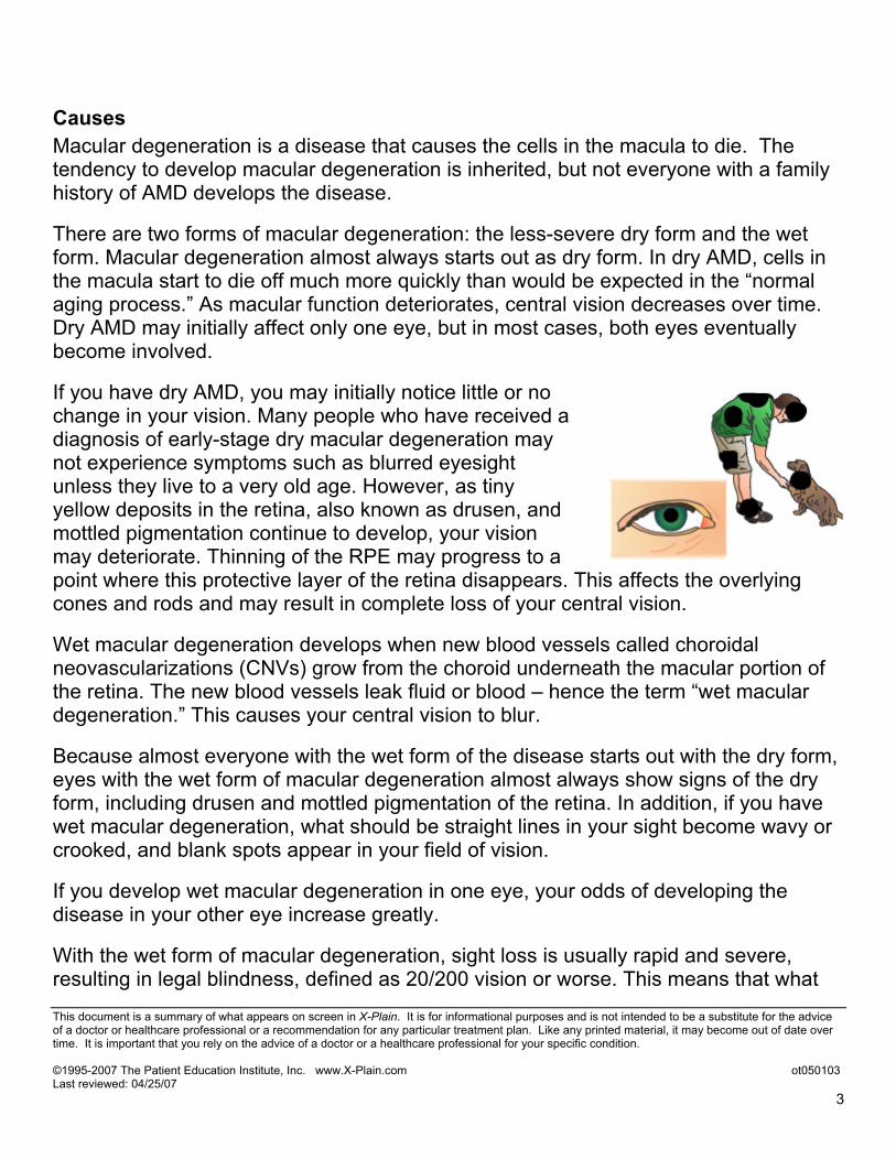
If you have dry AMD, you may initially notice little or no change in your vision. Many people who have received a diagnosis of early-stage dry macular degeneration may not experience symptoms such as blurred eyesight unless they live to a very old age. However, as tiny yellow deposits in the retina, also known as drusen, and mottled pigmentation continue to develop, your vision may deteriorate. Thinning of the RPE may progress to a point where this protective layer of the retina disappears. This affects the overlying cones and rods and may result in complete loss of your central vision.
Wet macular degeneration develops when new blood vessels called choroidal neovascularizations (CNVs) grow from the choroid underneath the macular portion of the retina. The new blood vessels leak fluid or blood – hence the term “wet macular degeneration.” This causes your central vision to blur.
Because almost everyone with the wet form of the disease starts out with the dry form, eyes with the wet form of macular degeneration almost always show signs of the dry form, including drusen and mottled pigmentation of the retina. In addition, if you have wet macular degeneration, what should be straight lines in your sight become wavy or crooked, and blank spots appear in your field of vision.
If you develop wet macular degeneration in one eye, your odds of developing the disease in your other eye increase greatly.
With the wet form of macular degeneration, sight loss is usually rapid and severe, resulting in legal blindness, defined as 20/200 vision or worse. This means that what
someone with normal vision can see from 200 feet, a person with 20/200 vision can see only from 20 feet.
Although some patients do not even realize that they have the condition until they develop the wet form, most patients with AMD learn that they have the condition when it is still in the dry form and they have few or no symptoms.
The risk of developing AMD increases with age. Results of a large study show that people in their 50s have a 2% chance of getting AMD. This risk rises to 30% after the age of 75.
Researchers don't know the exact causes of macular degeneration, but they have identified some contributing factors, including:
• smoking • family history of AMD • high cholesterol
Symptoms The first sign of macular degeneration may be a need for more light when you do close-up work. Fine newsprint may become harder to read and street signs more difficult to recognize.
You may eventually notice that, when you're looking at a grid, some of the straight lines appear distorted or crooked. Gray or blank spots may mask the center of your visual field. The condition usually develops gradually, but may sometimes progress rapidly, leading to severe vision loss in one or both eyes.
Macular degeneration affects your central vision, but not your peripheral vision. As a result, it does not cause total blindness. Still, the loss of clear central vision — critical for reading, driving, recognizing people's faces and doing detail work — greatly affects your quality of life. In most cases, the damage caused by macular degeneration can't be reversed, but early detection may help reduce the extent of vision loss.
Macular degeneration usually develops gradually and painlessly. The signs and symptoms of the disease may vary, depending on which of the two types of macular degeneration you have.
With dry macular degeneration, you may notice the following symptoms: • The need for increasingly bright illumination when reading or doing close work • Increasing difficulty adapting to low levels of illumination, such as when entering
a dimly lit restaurant • Printed words that appear
increasingly blurry • Colors that appear less bright • Difficulty recognizing faces • Gradual increase in the haziness of your
overall vision • Blurred or blind spot in the center of your
visual field, combined with a profound drop in your central vision acuity
• A need to scan your eyes all around an object to provide a more complete image • With wet macular degeneration, the following symptoms may appear, and they
may progress rapidly: • Visual distortions, such as straight lines appearing wavy or crooked, a doorway
or street sign that seems out of whack, or objects appearing smaller or farther away than they should
• Decrease in or loss of central vision • Central blurry spot
In either form of macular degeneration, your vision may falter in one eye while the other remains fine for years. You may not notice any or much change because your good eye compensates for the weak one. Your vision and lifestyle begin to be dramatically affected when this condition develops in both eyes.
Neither dry nor wet AMD causes any pain. The most common symptom of dry AMD is slightly blurred vision.
As dry AMD gets worse, it may become increasingly difficult to read, see television, or drive. Some patients may no longer meet driving requirements as the disease progresses. Low vision aids may be helpful to many patients with this stage of AMD. Sometimes the vision worsens in one eye before the other. People with dry AMD often are not significantly bothered by changes in their vision until the vision has decreased in both eyes.
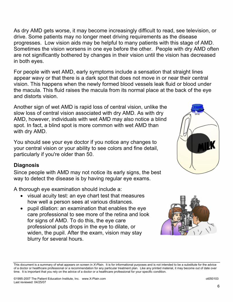
For people with wet AMD, early symptoms include a sensation that straight lines appear wavy or that there is a dark spot that does not move in or near their central vision. This happens when the newly formed blood vessels leak fluid or blood under the macula. This fluid raises the macula from its normal place at the back of the eye and distorts vision.
Another sign of wet AMD is rapid loss of central vision, unlike the slow loss of central vision associated with dry AMD. As with dry AMD, however, individuals with wet AMD may also notice a blind spot. In fact, a blind spot is more common with wet AMD than with dry AMD.
You should see your eye doctor if you notice any changes to your central vision or your ability to see colors and fine detail, particularly if you're older than 50.
Diagnosis Since people with AMD may not notice its early signs, the best way to detect the disease is by having regular eye exams.
A thorough eye examination should include a: • visual acuity test: an eye chart test that measures
how well a person sees at various distances. • pupil dilation: an examination that enables the eye
care professional to see more of the retina and look for signs of AMD. To do this, the eye care professional puts drops in the eye to dilate, or widen, the pupil. After the exam, vision may stay blurry for several hours.
A common sign of AMD is the presence of drusen, tiny yellow deposits in the retina that can be seen during an eye examination.
Drusen are not always an indicator of disease, but their presence may mean the eye is at risk of developing more severe AMD.
Several tests can be helpful in evaluating AMD.
An eye care professional may ask you to look at a small grid with a checkerboard pattern, called an Amsler grid. This test allows you to monitor your vision at home between visits to the eye doctor. You cover one eye and stare at a black dot in the center of the grid.
As you stare at the dot on the Amsler grid, if straight lines look wavy or lines seem to be missing, these could be signs of wet AMD.
If your eye care professional thinks you have wet AMD, you may need to have a test called fluorescein angiography. In this test, a special dye is injected into a vein in your arm.
Pictures are then taken as the dye passes through the blood vessels in the retina. The photos help the eye care professional examine leaking blood vessels and decide if they can be treated.
Since early signs of AMD may go unnoticed, the best way to detect AMD is by having frequent, thorough eye examinations BEFORE you experience any symptoms.
Treatment Options No treatment is available to reverse dry macular degeneration. But this doesn't mean you'll eventually lose all your sight. Dry macular degeneration usually progresses slowly, and many people with this condition are able to live relatively normal, productive lives, especially if only one eye is affected.
If you have dry AMD, you should: • have your eyes examined through dilated pupils
at least once a year • get an Amsler grid to quickly and inexpensively
evaluate your vision each day for signs of wet AMD. This test works best for people who still have good central vision
Check your vision by reading the newspaper, watching television, and just looking at people’s faces. Testing each eye separately is also very important. If you detect any changes, you should contact your eye doctor and have an eye exam right away.
Some treatment options are available for wet macular degeneration. But the success of the treatment in stopping further progression of the disease depends on the location and the extent of the abnormal blood vessels.
In most cases, the damage already caused by macular degeneration can't be reversed. The sooner the damage is detected, the better your chances that treatment will preserve what's left of your central vision.
Treatment possibilities for wet AMD also depend on the level of vision before treatment and certain characteristics of the disease as seen by fluorescein angiography testing.
Wet AMD that does not yet involve the fovea can sometimes be treated with laser surgery, where a high-energy beam of light is aimed directly onto leaking blood vessels.
If blood vessels keep leaking, more laser surgery may be needed. It is important to know that laser surgery is not a cure for AMD. Its objective is to prevent further vision loss in some patients.
The risk of new blood vessels growing back after laser treatment is fairly high.
For new blood vessels already under the center of vision, treatments other than laser surgery are possible. While these treatments do not dry up wet AMD in all patients, they do help some patients. It is important to realize that
none of these treatments reverse AMD. They only dry up the leakage in wet AMD and stabilize the vision. They do not work in all patients.
These treatments include: 1) Photodynamic therapy with Visudyne™. With this treatment, you receive
Visudyne dye through an IV. A retina specialist then administers a low-energy laser (sometimes referred to as a “cold laser”) to the area of the wet AMD to activate the Visudyne medication. Patients receiving this treatment must avoid sunlight and bright halogen lights for five days after the treatment to avoid the risk of serious sunburn.
2) Macugen® injection into the eye. Macugen is a medication that causes new blood vessel leakage to dry up in some patients with wet AMD. Macugen must be injected directly inside the eye with a small needle every six weeks for up to two years. There is a small risk of eye infection and retinal detachment with injection into the eye, so patients receiving this treatment require close monitoring. You should discuss this treatment with your doctor.
3) Macular translocation surgery is a treatment that can be used if the abnormal blood vessels are located directly under the fovea. To start this procedure, a surgeon detaches the retina, shifts the fovea away from the CNV and relocates it over healthy tissue. When the CNV is exposed, the surgeon can remove the CNV with tiny forceps or use a hot laser to destroy blood vessels without damaging the fovea. This surgery can be performed only if your vision loss is recent, the extent of CNV is limited and the tissue around the fovea is healthy.
If you have wet AMD, you should discuss treatment options with your doctor.
Coping with AMD There are ways to cope with impaired vision. Below are a few suggestions:
• Avoid driving in certain conditions. Don't drive at night, in heavy traffic, in bad weather or on a freeway.
• Seek help traveling. Use public transportation or ask family members to help, especially with night driving.
• Travel with others. Contact your local area agency on aging for a list of vans and shuttles, volunteer driving networks or ride shares.
• Have proper light in your home. This will help with reading and other activities.
• Remove home hazards. Eliminate throw rugs and other possible tripping hazards in your home.
• Ask friends and family members for help. Tell them about your vision problems so that they can help you perform certain tasks and help you recognize people.
• Don't become socially isolated. A common frustration of people with macular degeneration is the inability to recognize other people and greet them by name. If this happens to you, try asking people you know to say hi and to tell you their name when you meet them on the street or in other situations so you can greet them back.
• Take advantage of online networks. The Internet is a good source for support groups and resources for people with macular degeneration.
• Talk to your doctor. Ask your doctor about receiving professional help to make your home safer and more convenient for you to use.
Normal use of the eyes does not hurt vision. Even if you have lost sight because of AMD, you should not be afraid to use your eyes for reading, watching TV, and other daily activities.
Low vision aids, special lenses, or electronic systems that make images appear larger are available to help people make the most of their remaining vision.
Prevention You may be able to take steps to help slow down dry AMD and decrease the risk of conversion from dry to wet AMD.
Current recommendations for dry AMD are: • Take vitamin and mineral supplements. A recent study
demonstrated that a combination of vitamin C, vitamin E, beta carotene, zinc, and copper delay the progression of dry AMD and decrease the likelihood of dry AMD progressing to wet AMD 20-25% of the time in certain patients with AMD. Ask your eye doctor if you may benefit from the vitamins and minerals used in the study.
• Wear sunglasses that block out harmful ultraviolet light. Orange-, yellow- or amber-tinted lenses can help filter out both ultraviolet light and blue light. The main purpose of these glasses is to protect the surface of your eye and the skin of your eyelids. Look for glasses that filter 99 to 100 percent of ultraviolet A (UVA) and ultraviolet B (UVB) rays. UVA rays penetrate deeper, while UVB rays have a more superficial effect.
• Stop smoking. Smokers are more likely to develop macular degeneration than nonsmokers. Ask your doctor for help to stop smoking. People who smoke or who recently stopped smoking should not take beta carotene, as it increases their risk of lung cancer.
• Manage your other diseases. For example, if you have cardiovascular disease or high blood pressure, take your medication and follow your doctor's instructions for controlling the condition.
• Get regular eye exams. Early detection of macular degeneration increases your chances of preventing serious vision loss. If you're older than 40, get an exam every two to four years. If you’re older than 65, you should have an eye exam every year or two. If you have a family history of macular degeneration, have your eyes examined more frequently, perhaps annually.
• Screen your vision regularly. If you've received a diagnosis of early-stage macular degeneration, your doctor may suggest that you regularly monitor your vision at home with an Amsler grid. Doing so may help you to detect subtle changes in your vision at the earliest possible time and seek help promptly.
Conclusion Age-related macular degeneration is a serious eye disease.
A healthy diet, appropriate vitamin supplementation, and avoidance of smoking all help to slow down the progression of AMD.
Early detection and treatment, especially for wet AMD, can be crucial in preventing major vision loss. It is important for all older patients to have at least a yearly eye exam to screen for AMD. Once AMD is diagnosed, it is important to follow your doctor’s advice about the frequency of eye examinations so that treatable lesions can be detected early.


![Uveitic macular edema: a stepladder treatment paradigm€¦ · of macular edema [1,3–4], this review will focus on uveitic macular edema specifically. Uveitic macular edema Macular](https://static.documents.pub/doc/80x56/5ed770e44d676a3f4a7efe51/uveitic-macular-edema-a-stepladder-treatment-paradigm-of-macular-edema-13a4.jpg)


