40
Editorial Office Notes: RES-16-148.R1 ORIGINAL ARTICLE Received 22 February 2016 Invited to revise 13 April 2016 Revised 23 May 2016 Accepted 9 June 2016 Associate Editor: Conroy Wong

Editorial Office Notes:
RES-16-148.R1ORIGINAL ARTICLE
Received 22 February 2016Invited to revise 13 April 2016Revised 23 May 2016Accepted 9 June 2016Associate Editor: Conroy Wong
Bronchiectasis in yellow nail syndrome
Dr Woodfield G1, Dr Nisbet M1, Dr Jacob J2, Dr Mok W2, Dr Loebinger MR1, Prof
Hansell DM2, Prof Wells AU3, Prof Wilson R1
1. Host Defence Unit, Royal Brompton Hospital, London,
2. Radiology Department, Royal Brompton Hospital, London,
3. Interstitial Lung Disease, Royal Brompton Hospital, London, UK
Correspondence:
Professor R Wilson MD, FRCP
Consultant Physician
Host Defence Unit
Royal Brompton Hospital
Sydney Street
London SW3 6NP
UK
Email: [email protected]
Summary at a glance
Bronchiectasis in yellow nail syndrome (YNS) has distinct features which influence
clinical presentation and management. Bronchiectasis in YNS is milder than in
idiopathic bronchiectasis, with less upper and middle lobe disease, but increased
mucus plugging. A better understanding of aetiology may influence future research
and management decisions.
2
ABSTRACT
Background and objective: Yellow nail syndrome (YNS) is a rare and poorly
described disease process. In this case control study, clinical features and findings on
high resolution computed tomography [HRCT] were compared with idiopathic
bronchiectasis (IBx).
Methods: A review of all patients attending an adult bronchiectasis clinic between
2007 and 2013 identified 25 YNS patients. IBx patients were matched in a 2:1 ratio
for age, duration of symptoms and gender.
Results: Median age of onset was 53 years. There were 12 male and 23 Caucasian
YNS patients. Respiratory manifestations included chronic productive cough (100%),
chronic rhinosinusitis (88%), pleural effusions (20%), lymphoedema (12%). Chest
symptoms preceded yellow nails in the majority (68%). Abnormal nails persisted at
follow up in 23/25 patients but improved in 14.
In both disorders, there was symmetrical, predominantly lower lobe bronchiectasis on
HRCT. Extent (p=0.04), severity (p=0.03) and bronchial wall thickness (p=0.05)
scores were lower in YNS, with less upper and middle lobe disease. Multivariate
analysis showed an independent association with increased mucus plugging in YNS.
There was a similar prevalence of Pseudomonas aeruginosa infection and mild lung
function abnormalities.
Conclusion: Bronchiectasis in YNS is less severe than IBx but is associated with
increased mucus plugging, onset is in middle age and there is no female
predominance. Treatment targeted at improved secretion clearance may improve both
chest and nail symptoms, with consideration of longterm macrolide antibiotics.
3
Keywords: yellow nail syndrome, Pseudomonas aeruginosa, lymphoedema, pleural
effusion, idiopathic bronchiectasis
Short title: Bronchiectasis in yellow nail syndrome
4
INTRODUCTION
Yellow Nail Syndrome (YNS) was initially described by Samman and White in 1964
and the literature currently documents approximately 150 cases1. It is a rare clinical
syndrome characterised by a triad of yellow dystrophic nails, lymphoedema, and
pulmonary manifestations. These include chronic cough with sputum, recurrent
respiratory infections, pleural effusions, bronchiectasis and rhinosinusitis. It has been
previously accepted that two of the triad are adequate for diagnosis2. The complete
triad occurs in one third of patients3 where symptoms may occur years apart4. The
characteristic nail changes in YNS include yellow-green discolouration, slow growth,
cross-ridging, loss of cuticle, over-curvature, onycholysis and shedding1.
The aetiology of YNS remains undefined however the pathogenesis may involve
impaired lymphatic drainage5. Lymphangiography has shown hypoplastic lymphatic
systems in the legs and lungs of some patients1,5. This finding has not been confirmed
by lymphoscintigraphic studies and, given the reversibility in some, the underlying
cause may be functional rather than anatomical5. A small subset has
hypoalbuminaemia caused by enteric albumin loss suggesting increased
microvascular permeability6. The cause of bronchiectasis is unclear but may relate to
dysfunctional lymphatics and impaired secretion drainage, with subsequent increased
infection risk.
In 2008 Maldonado et al described 41 patients with YNS, mean age of onset 61 years.
18 patients (44%) had mainly symmetrical lower lobe bronchiectasis7. 17 (41%) had
chronic rhinosinusitis. By comparison a study of 165 consecutive bronchiectasis
5
patients referred to a single centre contained 4 patients with YNS and 43 with IBx.
The mean age of onset of IBx was 43 years with female predominance (65%), with a
predominantly lower lobe bronchiectasis and chronic rhinosinusitis8.
Patients with different bronchiectasis aetiologies have characteristic CT scan
appearances and clinical features. This case control study of patients referred to a
tertiary speciality hospital aims to further define the respiratory features of YNS by
comparing radiological findings, lung function and microbiology with a matched
cohort of IBx patients.
METHODS
YNS patients were identified by electronically searching all Royal Brompton Hospital
adult bronchiectasis clinic letters from 2007 to 2013. Patients were initially referred
for chronic productive cough with suspected or previously confirmed bronchiectasis,
with or without yellow nails. Diagnosis of YNS was made (often subsequently) after
development of yellow nails. Clinical features, microbiology, radiology, lung
function, progress and follow up duration were recorded. All patients had been
investigated using a protocol previously published [8]. This includes blood
investigations to exclude known aetiologies; nasal nitric oxide as a test for primary
ciliary dyskinesia (followed by light and electron microscopy of nasal epithelium if
indicated); sweat test (and/or genotyping) for cystic fibrosis8. The study was an
anonymised retrospective review of cases and neither patient consent nor ethical
6
approval was sought. All patients attending the Royal Brompton Hospital are
informed about the use of patient data for continuing research at the hospital.
IBx patients were defined as having had no history of serious infections predating
bronchiectasis, with normal or negative tests for other bronchiectasis aetiologies8. The
IBx comparison group was formulated by matching IBx patients to YNS patients in a
2:1 ratio, matched for age (within 10 years), duration of symptoms (within 5 years)
and, for lung function comparison, gender.
High resolution computed tomography (CT) scans were performed on initial
assessment. 1.5 mm sections were obtained at 10 mm intervals, supine and at full
inspiration. Bronchiectasis was defined according to accepted CT criteria9. All CT
scans were independently reviewed by the same two experienced observers, blinded
to clinical information. Observers awarded scores (grades) per lobe (lingula regarded
as a separate lobe) for eight variables: extent of bronchiectasis, severity of bronchial
dilation, bronchial wall thickness, mucus plugging, tree-in-bud pattern, mosaicism,
consolidation and septal thickening. Full scoring criteria are described in
Supplementary Appendix S1, and are similar to previous studies10-11. After initial
scoring, a consensus score was agreed between observers in cases with >1 point
discrepancy, where one result was zero, or where there was a >15% discrepancy for
Mosaicism or Consolidation. Consensus scores were used for data analysis.
Statistical analysis
HRCT variables were evaluated as follows:
1. Global scores (per scan) for all variables were computed as the sum of lobar
grades, with the exception of mosaic attenuation. See Appendix S1 for details
7
of scoring system. For example, the maximum grade per lobe for “extent”
was 3, therefore the maximum global extent score was 18 (assuming 6 lobes).
Global mosaic attenuation scores were calculated as the mean of lobar scores,
as mosaic attenuation was a continuous variable rather than a grade
(quantified as the percentage of total lung volume in each lobe). Mean scores
for the two observers were evaluated in all analyses of global variables. Lobar
grades were recorded in a table as shown in Appendix S2, for each observer.
2. Septal thickening and consolidation were seldom present and were not
evaluated further.
3. Regional scores were computed as upper lobe, middle lobe and lower lobe
scores using the same approach as for global scores (i.e. mean lobar scores for
mosaic attenuation, summed scores for all other variables).
Group comparisons for all variables were made using the Wilcoxon rank sum test. A
p value <0.05 indicated statistical significance. Stepwise logistic regression was used
to identify independent HRCT morphologic differences between YNS and IBx.
Lung function was compared between diseases, where YNS and IBx patients were
also sex matched (as well as for age and symptom duration). This meant that the
matched IBx lung function group and radiology group were not necessarily formed of
the same IBx patients. However, matched YNS/IBx patients were kept the same
where possible; 28 of the idiopathic patients used for lung function comparison were
also used for CT comparison (this meant that there were 16 IBx patients that featured
only in the PFT comparison group, and 10 that featured only in the CT comparison
group). The YNS group was consistent throughout. The lung function matched IBx
8
group was the comparator group used to compare microbiology findings between
diseases (as opposed the IBx radiology matched group used for CT comparison).
Lung function readings and microbiology data were recorded at initial assessment and
at every subsequent visit (in order to record any subsequent P. aeruginosa infection).
Lung function data were compared using an unpaired student t-test. Categorical
variables, namely presence or absence of P. aeruginosa, were compared using a chi-
squared test. P < 0.05 was regarded as being statistically significant.
RESULTS
Twenty five patients with YNS attended clinics at the Royal Brompton Hospital
between 2007 and 2013. The median referral age of YNS was 58.2 years (range 11.8-
77.7), and median age of first symptoms was 53 (range 4-75). 12 were male (48%).
23/25 (92%) were Caucasian, from the UK and Ireland. One patient was from Iraq
with Arabic origins, the other was Asian from Sri Lanka. All presented to the Royal
Brompton with chronic cough and recurrent infections. During follow up, 21 patients
suffered from chronic sinusitis, 10 had wheeze. Three patients had lymphoedema of
lower limbs, one with left arm involvement. Five patients had a history of pleural
effusion. Supplementary tables 1 and 2 describe YNS and IBx individual patient
characteristics in more detail.
22/25 patients (88%) had abnormal nails at referral, with the remaining 3 patients
developing yellow nails within two years. In the majority of patients (17, 68%), chest
9
symptoms preceded nail changes. Yellow nails had preceded chest symptoms by one
year in two patients, and occurred simultaneously in six. Two patients had a history of
malignancy (breast and melanoma), five patients had reflux, four had coronary artery
disease and two had cardiac failure. One patient had asbestos plaques. The proportion
of ex-smokers was similar between YNS and IBx (32% and 29% respectively). None
were current smokers.
Immunoglobulins were normal in all. Two patients had lymphopenia (lymphocyte
count of 0.7 x10⁹/L and 1.0x10⁹/L). Four patients had low albumin levels of 26-34g/L.
Seven patients had elevated IgE levels (>150 IU/mL). Two of these were diagnosed
with aspergillus sensitivity, with Aspergillus fumigatus RAST results of 14.7 and 9.74
IU/mL. Neither of these patients had peripheral eosinophilia nor clinical/radiological
evidence of ABPA.
The median follow-up was 6.2 years (range 0 to 19 years) in the YNS cohort, and 4.9
years (range 0 to 14.7 years) in the IBx cohort. Zero follow up was recorded where
patients were seen once only (1/25 YNS, 5/44 IBx). Initial referral dates ranged
between 1992-2013 for YNS, and 2001-2012 for IBx patients.
Microbiology
At initial referral, sputum cultured P. aeruginosa in 5/22 YNS patients (23%),
Haemophilus influenzae in five (23%), Streptococcus pneumoniae in two (9%), and
Moraxella catarrhalis in one patient (5%). Nine (41%) patients had no significant
bacterial growth. 3 patients’ initial results were unavailable. No mycobacteria were
cultured. By comparison, 12/44 (27%) IBx patients cultured P. aeruginosa. During
follow up, 9 further YNS and 12 further IBx patients became colonised (64% and
10

55% of cohort respectively). There was no significant difference in incidence of P.
aeruginosa at presentation (p=0.69), or during follow up (p=0.48) between diseases.
Table 1 compares microbiology between diseases.
Lung function studies
Lung function studies showed YNS to be a mildly obstructive lung disease, with no
significant difference in readings compared to IBx (Table 2).
Within the YNS group, patients colonised with P. aeruginosa at presentation had a
statistically lower mean FEV1 (p=0.013) and FVC (p=0.042) than those without
colonisation.
Radiology
Nineteen YNS patients had CT scans available, where seventeen showed
bronchiectasis. The remaining two patients scored zero for extent and severity of
bronchiectasis. However, both had bronchial wall thickening, chronic daily sputum
production and recurrent infections, with bronchiectasis diagnosed clinically. One
patient’s CT scan had been reported as mild bilateral lower lobe bronchiectasis by the
radiologist reporting it originally; the second patient had right lower lobe
bronchiectasis on a repeat scan 2 years later.
YNS patients had significantly lower extent (p=0.04), severity (p=0.03) and bronchial
wall thickness (p=0.05) scores than matched IBx patients, see Table 3. Differences in
scores for tree in bud, mosaicism and mucus plugging did not reach statistical
significance on initial analysis (Wilcoxons signed rank test). Consolidation was
present in only 4/19 (21%) of YNS and 13/38 (34%) of IBx patients. Septal
11
thickening was present in only 6/19 (32%) of YNS and 13/38 (34%) of IBx patients.
Median global scores for consolidation and septal thickening were zero, so they were
not included in Table 3.
Examination of a stepwise logistic regression model (with the HRCT variables listed
in Table 3 examined as initial covariates prior to stepwise evaluation) showed that
when compared to IBx, YNS was independently associated with less bronchial wall
thickness (OR 0.64; 95% CI 0.46-0.87; p<0.005) and higher mucus plugging scores
(OR 1.89; 95% CI 1.10 – 3.25; p=0.02) than IBx. Analyses of the distribution of
abnormalities on HRCT revealed that when compared to IBx, YNS was characterised
by lower scores for the extent and severity of bronchiectasis and overall bronchial
wall thickness (all p<0.05). However, lower lobe scores taken in isolation did not
differ between the two diseases for any HRCT variable. Thus, compared to IBx,
YNS is associated with sparing of the upper and middle lobes, with the abnormalities
being present in the lower lobes.
Management
All patients received physiotherapy tuition at presentation and throughout follow up.
Low dose antibiotic prophylaxis was introduced in 20 (80%) of YNS patients because
of poor symptom control and/or four exacerbations or more per year. Three of these
stopped antibiotics due to intolerance, side effects or non-response, leaving 17 on
long-term prophylaxis. Prophylaxis was oral azithromycin in 14/17 (82%) (usually
250mg three times weekly). This improved chest symptoms in the majority (9/14
=64%) of this group. One patient colonised with P. aeruginosa was taking colomycin
nebulisers additionally as dual prophylaxis. 3/17 took other forms of antibiotic
12
prophylaxis; 2 took colomycin alone (intolerant to azithromycin) with improvement in
symptoms, and one on regular erythromycin had no improvement. For the YNS group
as a whole, 16 (64%) had a subjective improvement in chest symptoms since first
review. Eleven of these were taking regular antibiotic prophylaxis and five patients
improved with physiotherapy alone. Figures 1a and 1b shows a CT scan of a YNS
patient before and after treatment with a long course of clarithromycin 250mg twice
daily and inhaled colomycin 1million units twice daily, started when pseudomonas
was isolated.
23/25 patients had persisting nail changes at the time of last review, where 17 had
improved over time. Seven of these also had improved chest symptoms over time.
Resolution of nail changes occurred in two patients with improvement in chest
symptoms (complete resolution in one). Figures 2a and 2b shows an example of one
patient’s nail changes before and after treatment with physiotherapy and episodic
antibiotics during infective exacerbations.
13
DISCUSSION
This study investigates YNS patients referred to a tertiary hospital respiratory clinic,
and compares them to matched IBx patients. IBx was chosen as the comparator group
because both groups have normal or negative tests for other aetiologies, present in a
similar way and are currently only differentiated by nail changes. A better
understanding of aetiology may influence future research and management decisions.
A recent multicentre study of 1258 bronchiectasis patients determined the aetiology in
60%, where this knowledge changed the patient’s management in 13% of cases12.
Additionally, the radiological and clinical findings in YNS bronchiectasis have been
poorly defined. In Varney et al’s series of 17 YNS patients, all had chronic cough and
the majority rhinosinusitis (83%)13. Lymphoedema was present in 76%, compared to
only 36% with lymphatic dysfunction in our series. Hoque et al. described eleven
YNS patients of whom only six had bronchiectasis14. Comparable with our cohort the
mean age of onset of symptoms was 53 years (range 27 to 69) but in contrast they
reported lymphoedema in 46% of cases and only 27% had sinusitis. A recent study of
165 consecutive bronchiectasis patients (43 with IBx) found the age average of onset
of IBx was 43, 65% women8. Other studies have shown similarly younger age of
onset for IBx and female predominance15-16. In our YNS cohort the average age of
onset was 53 years, with no gender predominance.
YNS has been associated with other conditions including autoimmune disorders [4],
immunodeficiency states17-18, nephrotic syndrome19, Guillain-Barre syndrome20, drugs
(D-penicillamine, gold) and cancers4,6,17,19,21-28. Three of our patients had a history of
malignancy. YNS has also been reportedly associated with lymphopenia, low IgA
levels and hypogammaglobulinaemia29. Two patients in our series had lymphopenia
14
but immunoglobulin levels were normal in all. These previously described
associations of YNS were therefore not seen in our cohort of patients presenting to a
chest clinic.
The notable CT finding of YNS in our study was symmetrical predominantly lower
lobe bronchiectasis, with significantly less upper and middle lobe disease, and
increased mucus plugging compared to IBx. Our cohort of YNS patients had
significantly milder disease than IBx in terms of total extent, severity and bronchial
wall thickening. However, this may be explained by disease being restricted to the
lower lobes in YNS. We therefore hypothesise that bronchiectasis in YNS is a
disorder of delayed mucociliary clearance due to abnormal lymphatic
structure/function, where gravity influences lower lobe disease development.
However, other factors such as frequency and severity of infections and the presence
of other pro-inflammatory influences (such as aspergillus sensitivity in two patients),
will also determine bronchiectasis development. Previous CT findings in YNS
include one case series of four patients all with bronchiectasis primarily in the middle
lobe, lingula and lower lobes30. Maldonado et al reported 41 YNS patients where 18
had bilateral lower lobe bronchiectasis on CT7. A possible explanation for milder
disease in YNS could be earlier referral (and therefore treatment) due to yellow nail
development. This explanation is however unlikely because YNS and IBx patients
were matched by length of symptoms at the time of CT/lung function tests.
The YNS group as a whole had mild abnormalities of lung function, although patients
with P. aeruginosa had significantly poorer FEV1 readings at presentation (p=0.013).
The association with P. aeruginosa with more severe airflow limitation has been
15
noted in bronchiectasis previously31-32. Lung function was comparable to the IBx
cohort, as was incidence of P. aeruginosa both at presentation and subsequently. This
was surprising considering less severe and less widespread bronchiectasis in YNS.
One explanation could be increased mucus plugging predisposing to bacterial
colonisation, or possibly higher use of antibiotic prophylaxis in this group.
Longterm prophylaxis with azithromycin improved chest symptoms (cough, sputum
and exacerbation frequency) in the majority (64%) of the YNS cohort taking it. This
suggests that it is a good choice where symptom control is poor with physiotherapy
and rescue antibiotics. Improvement with clarithromycin has been described in two
case studies of YNS33-34. However, one study focussed on nail improvement, whilst
the second described a case of eosinophilic bronchial disease in YNS, unlike in our
cohort.
Limitations
YNS and IBx cohorts were matched for age and length of symptoms. This system has
the inherent flaw of using patient recollection to determine the date of symptom onset.
The use of a small (five year) margin attempts to minimise poor recollection or
individual variability in perception of symptoms. There may be some patient selection
bias, as YNS patients may be referred earlier due to yellow nails rather than for
problematic respiratory symptoms. However, the majority of YNS patients in our
cohort were undiagnosed at referral. This study did not assess disease improvement
objectively, and relied on clinic letter reports of symptoms. This is a small cohort of
patients, as YNS is rare and we are a single centre. However, being a tertiary
respiratory centre has enabled us to match IBx patients
16
In conclusion, this study shows YNS as a distinct aetiology of bronchiectasis that may
relate to impaired mucus clearance. The bronchiectasis in YNS is usually mild and
involves predominantly lower lobes, often presenting at an older age than IBx.
Management should target mucus clearance. Longterm macrolide antibiotics may
provide symptomatic relief, and in some cases led to resolution of dystrophic nails.
Patients should be screened for impaired immunity, aspergillus sensitivity and
malignancy, although these findings were uncommon in our cohort.
17
Table 1- Comparison of microbiology between diseases at first clinic appointment
Initial Sputum culture results
Yellow Nail SyndromeFrequency of culture result
Idiopathic Bx frequencyFrequency of culture result
No growth/ Upper respiratory tract flora
9 8
Pseudomonas aeruginosa
5 12
Haemophilus Influenzae 5 12Streptococcus pneumonia
2 1
Moraxella cattarhalis 1 4Stenotrophomonas maltophilia
0 1
Staphylococcus aureus 0 3Coliforms 0 1Serratia marcesens 0 1Beta haemolytic streptococcus
0 1
Total samples =22 (3 patients not available)
Total samples = 44
The IBx group used for comparison was the IBx lung function test group (patients varied very slightly to the IBx CT comparison group)
18
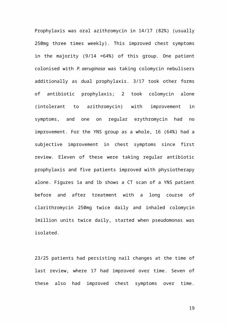
Table 2- Lung function tests in Yellow Nail Syndrome and Idiopathic Bronchiectasis
Yellow Nail
Syndrome
n= 22
Idiopathic
bronchiectasis
n= 44 P value
Mean FEV1 % of
predicted value
(Standard deviation)
83.86 (15.80) 76.50 (26.91) 0.251
Mean FEV1/FVC ratio
(Standard deviation) 0.70 (0.10) 0.67 (0.17) 0.413
Mean TLCOc % of
predicted value
(Standard deviation)
76.43 (15.52) 77.90 (16.70) 0.740
Student t test was used to compare data sets.
19
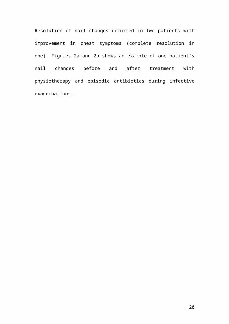
Table 3 High Resolution Computed Tomogram variables compared between Yellow
Nail Syndrome and Idiopathic Bronchiectasis.
HRCT variables
(max score per scan)
YNS -19 patientsMedian CT score
(range)
IBx -38 patientsMedian CT score
(range)P value
Extent of
bronchiectasis (18)
4
(0-14)
8
(0-16)
0.04
Severity of
bronchiectasis (24)
5.5
(0-13.5)
8
(0-18.5)
0.03
Wall thickness (24) 5.5
(1-13)
6.5
(1-17)
0.05
Tree in bud (12) 2.5
(0-7)
2.75
(0-8.5)
0.73
Mucus
Plugging (12)
2.0
(0-6)
2.0
(0-8)
0.80
Mosaic attenuation
(100%)
20
(0-65%)
25
(0-90%)
0.86
HRCT variables including maximum scores are expressed as the sum of lobar scores
for each patient, with the exception of mosaic attenuation (quantified as percentage of
lung volume). All lobar scores were averaged for two observers, before summing. See
Supplementary Appendix S1 for details of how each lobe was scored.
20
Figure legends
Figure 1- (A) 57 year old female with YNS at presentation. HRCT section through
the lower lobes showing cylindrical bronchiectasis, particularly in the left lower lobe
where there is an associated tree-in-bud pattern; there are also small foci of
consolidation in the left lower lobe and lingual. (B)- In the same patient 18 months
later, after treatment with a long course of clarithromycin 250mg twice daily and
inhaled colomycin 1million units twice daily, The appearances in the left lower lobe
have improved with resolution of the tree-in-bud pattern and consolidation.
Figure 2 – (A) 54 year old man with yellow nails at first presentation, demonstrating
discolouration, ridging and shedding. (B) - The same patient 1.5 years later
demonstrating complete resolution of nail changes after treatment with physiotherapy
alone.
21
REFERENCES
1. Samman P, White W. The “Yellow nail syndrome”. Br J Dermatol. 1964;76: 153-7.
2. Hershko A, Hirshberg B, Nahir M et al. Yellow Nail Syndrome. Postgrad Med J.
1997;73: 466-8.
3. Emerson P. Yellow nails, lymphoedema, and pleural effusions. Thorax. 1966; 21:
247-53.
4. Gupta A, Davies G, Haberman H. Yellow nail syndrome. Cutis. 1986; 37: 371-4.
5. Bull R, Fenton D, Mortimer P. Lymphatic function in the yellow nail syndrome. Br
J Dermatol. 1996; 134: 307-12.
6. D’Alessandro A, Muzi G, Monaco A, Filiberto S, Barboni A, Arbritti G. Yellow
nail syndrome: does protein leakage play a role? Eur Resp J. 2001; 17: 149-52.
7. Maldonado F, Tazelaar H, Wang C, Ryu J. Yellow nail syndrome: analysis of 41
consecutive patients. Chest. 2008: 134: 375-381
8. Shoemark A, Ozerovitch L, Wilson R. Aetiology in adult patients with
bronchiectasis. Respir Med. 2007; 101: 1163-70.
9. Naidich DP, McCauley DI, Khouri NF, Stitik FP, Siegelman SS. Computed
tomography of bronchiectasis. J Comput Assist Tomogr. 1982; 6: 437-44.
10. Horsley AR, Davies JC, Gray RD et al. Changes in physiological, functional and
structural markers of cystic fibrosis lung disease with treatment of a pulmonary
exacerbation. Thorax. 2013; 68: 532-539
11. Reiff DB, Wells AU, Carr DH, Cole PJ, Hansell DM. CT findings in
bronchiectasis: limited value in distinguishing between idiopathic and specific types.
Am J Roentgenol. 1995;165: 261-7.
12. Lonni S, Chalmers J, Goeminne P, McDonnell M, Dimakou K, De Soyza A,
Polverino E, Van De Kerkhove C, Rutherford R, Davison J, Rosales E, Pesci A,
22
Restrepo M, Torres A, Stefano A. Etiology of non-cystic fibrosis bronchiectasis in
adults and its correlation to disease severity. Ann Am Thorac Soc. 2015; 12: 1764-
1770.
13. Varney V, Cumberworth V, Sudderick R, Durham S, Mackay I. Rhinitis, sinusitis
and the yellow nail syndrome: a review of symptoms and response to treatment in 17
patients. Clin Otolaryngol. 1994; 19: 237-40.
14 . Hoque S, Mansour S, Mortimer P. Yellow nail syndrome: not a genetic disorder?
Eleven new cases and a review of the literature. Brit J Dermat. 2007; 1230-4.
15. Smith C. Prevots D, Adjemian J , Seitz A, Daniels M,
Czaja C , Milla C , Hall D , Holland S, Knowles M, Olivier K. Characterization Of
Idiopathic Bronchiectasis In Patients With And Without Pulmonary
Nontuberculous Mycobacterial Disease. Am J Respir Crit Care med. 2013;187;
A4535
16 . S. Fuschillo, A. De Felice and G. Balzano. Mucosal inflammation in idiopathic
bronchiectasis: cellular and molecular mechanisms. Eur Respir J. 2008; 31: 396–
406
17. Siegelman S, Heckman B, Hasson J. Lymphedema, pleural effusions and yellow
nails: associated immunologic deficiency. Dis Chest. 1969; 56: 114-7.
18. Scher R. Acquired immunodeficiency syndrome and yellow nails. J Am Acad
Dermatol. 1988; 18: 758-9.
19. Yanez S, Val-Bernal J, Fernandez-Llaca H. Yellow nails and minimal change
nephrotic syndrome. Nephron. 1999; 82: 180-2.
20. Woollons A, Darley C. Yellow nail syndrome following Guillian-Barre syndrome.
Clin Exp Dermatol. 1997; 22: 253-4.
23
21. Iqbal M, Rossoff L, Marzouf K, Steinberg H. Yellow nail syndrome: resolution of
yellow nails after successful treatment of breast cancer. Chest. 2000; 117: 1516-8.
22. Guin J, Elleman J. Yellow Nail syndrome: possible association with malignancy.
Arch Dermatol. 1979; 115: 734-5.
23. Thomas P, Sidhu B. Yellow nail syndrome and bronchial carcinoma. Chest. 1987;
92: 191.
24. Burrows N, Jones R. Yellow nail syndrome in association with carcinoma of the
gall bladder. Clin Exp Dermatol. 1991; 16: 471-3.
25. Hiller E, Rosenow E, Olsen A. Pulmonary manifestations of the yellow nail
syndrome. Chest. 1972; 61: 452-8.
26. Stosiek N, Paters K, Hiller D et al. Yellow nail syndrome in a patient with
mycosis fungoides. J Am Acad Dermatol. 1993; 28: 792-4.
27. Seve P, Thieblemont C, Dumontet C, Boufia F, Arnaud P, Hequet O, Espinouse
D, Salles G, Coiffier B. Skin lesions in malignancy. Case 3. Yellow nail syndrome in
non-Hodgkins lymphoma. J Clin Oncol. 2001; 19: 2100-1.
28. Ginarte M, Monteagudo B, Toribio J. Yellow nail syndrome and lung lymphoma.
Clin Exp Dermatol. 2004; 29: 423-36.
29. Bokszczanin A, Levinson A. Coexistent yellow nail syndrome and selective
antibody deficiency. Ann Allergy Asthma Immunol. 2003; 91: 496-500.
30. Wiggins J, Strickland B, Chung K. Detection of bronchiectasis by high-resolution
computed tomography in the yellow nail syndrome. Clin Radiol. 1991; 43: 377-9.
31. Davies G, Wells A, Doffman S, Watanabe S, Wilson R. The effect of
Pseudomonas aeruginosa on pulmonary function in patients with bronchiectasis. Eur
Respir J. 2006; 28: 974-9.
24
32. Evans SA, Turner SM, Bosch BL, Hardy CC, Woodhead MA. Lung Function in
bronchiectasis: the influence of Pseudomonas aeruginosa. European Resp J. 1996; 9:
1601-4
33. Suzuki M, Yoshizawa A, Sugiyama H, Ichimura Y, Morita A, Takasaki J, Naka
G, Hirano S, Izumi S, Takeda Y, Hoji M, Kobayashi N, Kudo K. A case of yellow
nail syndrome with dramatically improved nail discoloration by oral clarithromycin.
Case Resp Dermatol. 2011; 3:251-8.
34. Toyoshima M, Chinda K, Suda T. A case of yellow nail syndrome associated with
eosinophilic bronchial disease successfully treated with clarithromycin and
budesonide. Nihon Kokyuki Gakkai Zasshi. 2005; 43: 508-12.
25


