Zea mays Annexins Modulate Cytosolic Free Ca2+ andGenerate a Ca2+-Permeable Conductance W
Anuphon Laohavisit,a Jennifer C. Mortimer,a,1,2 Vadim Demidchik,a,1,3 Katy M. Coxon,a,4 Matthew A. Stancombe,a
Neil Macpherson,a Colin Brownlee,b Andreas Hofmann,c Alex A.R. Webb,a Henk Miedema,d Nicholas H. Battey,e
and Julia M. Daviesa,5
a Department of Plant Sciences, University of Cambridge, Cambridge CB2 3EA, United KingdombMarine Biological Association, Plymouth PL1 2PB, United Kingdomc Structural Chemistry Program, Eskitis Institute for Cell and Molecular Therapies, Griffith University, Nathan QLD 4111, Australiad BioMaDe, 9747 AG, Groningen, The Netherlandse School of Biological Sciences, University of Reading, Whiteknights, Reading RG6 6AS, United Kingdom
Regulation of reactive oxygen species and cytosolic free calcium ([Ca2+]cyt) is central to plant function. Annexins are small
proteins capable of Ca2+-dependent membrane binding or membrane insertion. They possess structural motifs that could
support both peroxidase activity and calcium transport. Here, a Zea mays annexin preparation caused increases in [Ca2+]cytwhen added to protoplasts of Arabidopsis thaliana roots expressing aequorin. The pharmacological profile was consistent
with annexin activation (at the extracellular plasma membrane face) of Arabidopsis Ca2+-permeable nonselective cation
channels. Secreted annexins could therefore modulate Ca2+ influx. As maize annexins occur in the cytosol and plasma
membrane, they were incorporated at the intracellular face of lipid bilayers designed to mimic the plasma membrane. Here,
they generated an instantaneously activating Ca2+-permeable conductance at mildly acidic pH that was sensitive to
verapamil and Gd3+ and had a Ca2+-to-K+ permeability ratio of 0.36. These results suggest that cytosolic annexins create a
Ca2+ influx pathway directly, particularly during stress responses involving acidosis. A maize annexin preparation also
demonstrated in vitro peroxidase activity that appeared independent of heme association. In conclusion, this study has
demonstrated that plant annexins create Ca2+-permeable transport pathways, regulate [Ca2+]cyt, and may function as
peroxidases in vitro.
INTRODUCTION
Annexins form amultigene, multifunctional family of amphipathic
proteins with a broad taxonomic distribution covering prokary-
otes, fungi, protists, plants, and higher vertebrates (Gerke and
Moss, 2002; Morgan et al., 2004, 2006). Found in all plants
studied to date and in all organs, these small (32 to 42 kD)
proteins can comprise up to 0.1% of total plant cell protein
(Delmer and Potikha, 1997; Clark et al., 2001; Moss andMorgan,
2004; Mortimer et al., 2008). The annexin C-terminal core is
constructed from four annexin repeats, each comprising five
short a-helices. The annexin repeat, of ;70 amino acids, con-
tains the conserved endonexin fold (K-G-X-G-T-{38}-D/E) and is
able to bind Ca2+ (Kourie and Wood, 2000; Figure 1). Calcium
enables the reversible binding of annexins to negatively charged
phospholipids, and the Ca2+ requirement for binding can be
reduced by acidic pH (Blackbourn et al., 1991). In animals,
annexins can be cytosolic, membrane associated, or membrane
inserted, depending on the prevailing conditions of cytosolic free
Ca2+ ([Ca2+]cyt), pH, and membrane oxidation (reviewed in Gerke
and Moss, 2002).
Isolated plant annexins can bind membranes (including se-
cretory vesicles, cell membranes, and endomembranes), GTP/
ATP, and F-actin (reviewed inMortimer et al., 2008). Their roles in
planta are poorly understood (Mortimer et al., 2008). Annexins
have been found colocalized with anatomical regions of high
secretion and growth rates (Blackbourn et al., 1991, 1992;
Blackbourn and Battey, 1993; Carroll et al., 1998; Clark et al.,
1995, 2001, 2005a, 2005b; Bassani et al., 2004). Maize (Zea
mays) annexins have been found to stimulate Ca2+-dependent
exocytosis in root cap cells (Carroll et al., 1998), the process
underlying cell expansion and plant growth (Carroll et al., 1998).
Annexin relocation from the cytosol to membranes can occur in
response to specific stimuli, such as touch (Thonat et al., 1997),
cold (Breton et al., 2000), and salinity (Lee et al., 2004), suggest-
ing a role in adaptive signaling. Indeed, Arabidopsis thaliana
annexin 1 (ANN1) expression is upregulated by peroxide, sali-
cylic acid (Gidrol et al., 1996), abscisic acid (Lee et al., 2004),
drought, cold, and salt stress (Cantero et al., 2006). The ann1
1 These authors contributed equally to this work.2 Current address: Department of Biochemistry, University of Cambridge,Tennis Court Road, Cambridge CB2 1QW, UK.3Current address: Department of Biological Sciences, University ofEssex, Colchester CO4 3SQ, UK.4Current address: Division of Cell Biology, Institute of Ophthalmology,University of London, 11-43 Bath Street, London EC1V 9EL, UK.5 Address correspondence to [email protected] author responsible for distribution of materials integral to thefindings presented in this article in accordance with the policy describedin the Instructions for Authors (www.plantcell.org) is: Julia M. Davies([email protected]).WOnline version contains Web-only data.www.plantcell.org/cgi/doi/10.1105/tpc.108.059550
The Plant Cell, Vol. 21: 479–493, February 2009, www.plantcell.org ã 2009 American Society of Plant Biologists
loss-of-function mutant is impaired in osmotolerance (Lee et al.,
2004) and primary root growth (Clark et al., 2005b), but the
precise roles of ANN1 in these processes remain to be deter-
mined.
Results from Arabidopsis, Brassica, and Capsicum point to
functions of annexin in ion transport and regulation of cellular
activity involving reactive oxygen species (ROS) (Gidrol et al.,
1996; Hofmann et al., 2000; Gorecka et al., 2005, 2007; Jami
et al., 2008). Initial characterization of recombinant ANN1 has
revealed the ability to form K+-permeable ion channels in planar
lipid bilayers (Gorecka et al., 2007) and in vitro peroxidase activity
(Gidrol et al., 1996; Gorecka et al., 2005). Recombinant Brassica
juncea ANN1 also appears to possess peroxidase activity (Jami
et al., 2008). Capsicum annuum (bell pepper) ANN24 has been
found previously to mediate passive Ca2+ transport across
liposome membranes (Hofmann et al., 2000), consistent with
Ca2+ channel formation, but effects on [Ca2+]cyt are unknown.
Peroxidase activity of plant annexins is thought to be bestowed
by a sequence strongly resembling the heme binding region of
horseradish peroxidase (Gidrol et al., 1996; Clark et al., 2001;
Gorecka et al., 2005). Site-directed mutagenesis of a key His
residue (His40) in the putative heme binding region abolished
peroxidase activity of recombinant At ANN1 (Gorecka et al.,
2005). However, to date there have been no reports of heme
binding to annexins fromplants nor has the in vivo electron donor
been identified. Neither the physiological significance of trans-
port and peroxidase activities nor the extent to which they are
shared by other annexins is known. Animal annexins are
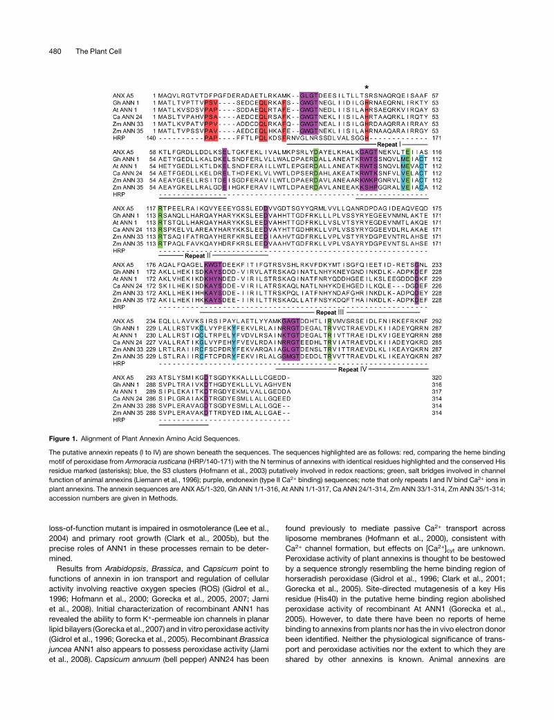
Figure 1. Alignment of Plant Annexin Amino Acid Sequences.
The putative annexin repeats (I to IV) are shown beneath the sequences. The sequences highlighted are as follows: red, comparing the heme binding
motif of peroxidase from Armoracia rusticana (HRP/140-171) with the N terminus of annexins with identical residues highlighted and the conserved His
residue marked (asterisks); blue, the S3 clusters (Hofmann et al., 2003) putatively involved in redox reactions; green, salt bridges involved in channel
function of animal annexins (Liemann et al., 1996); purple, endonexin (type II Ca2+ binding) sequences; note that only repeats I and IV bind Ca2+ ions in
plant annexins. The annexin sequences are ANX A5/1-320, Gh ANN 1/1-316, At ANN 1/1-317, Ca ANN 24/1-314, Zm ANN 33/1-314, Zm ANN 35/1-314;
accession numbers are given in Methods.
480 The Plant Cell
variously capable of forming Ca2+-permeable ion channels or
modulating the activity of existing channel proteins but lack the
heme binding motif (Liemann et al., 1996; Kourie and Wood,
2000). Plant annexins may therefore have more varied cellular
functions than their animal counterparts.
Plant cells contain suites of proteins regulating [Ca2+]cyt- and
ROS-mediated signaling. ROS act in defense and abiotic stress
responses (e.g., Zhang et al., 2003; Shin andSchachtman, 2004),
control of stomatal aperture (e.g., McAinsh et al., 1996), and
growth and development (e.g., Foreman et al., 2003; Liszkay
et al., 2004). ROSand [Ca2+]cyt appear to act sequentially in some
networks, most notably guard cell abscisic acid signaling (e.g.,
McAinsh et al., 1996; reviewed in Kwak et al., 2006) and root hair
polar growth (Foreman et al., 2003). Thus, a protein such as an
annexin with the ability to regulate these two key network
components could be a significant control point. Here, to avoid
adverse effects of His tags (Hofmann et al., 2000) and ensure
correct posttranslational modification, native annexins (ANN33
and ANN35) have been purified from maize to quantify peroxi-
dase activity using an Amplex Red assay.
Asmaize annexins can potentially formCa2+-permeable chan-
nels or regulate the activity of Ca2+ channel proteins, the ability of
maize annexins to modulate [Ca2+]cyt has also been addressed.
To do this, Arabidopsis root epidermal protoplasts have been
used as an assay system. These protoplasts have been used
extensively in the characterization of native plasma membrane
cation or anion channels and [Ca2+]cyt regulation (e.g., Demidchik
et al., 2002, 2003; Foreman et al., 2003; Diatloff et al., 2004). As
such, they are a useful screening tool for assessing which, if any,
channels are annexin regulated. This protoplast assay requires
annexin addition to the extracellular plasma membrane face.
While ANN33/35 are soluble proteins, they also exist in themaize
plasma membrane (Hochholdinger et al., 2005; Carletti et al.,
2008) and are predicted to be secreted (SecretomeP program;
Bendtsen et al., 2004). Thus, the protoplast assay also tests for
annexin interaction with a plasma membrane from the extracel-
lular face. To assess the likelihood of ANN33/35 interaction with
the plasma membrane from the intracellular membrane face and
test directly for Ca2+ transport function, we have incorporated the
maize annexins into planar lipid bilayers enriched with the maize
plasma membrane phospholipid, phosphatidylethanolamine
(see Bohn et al., 2001; Kukavica et al., 2007). The results ob-
tained place plant annexins as potential modulators of both plant
cell [Ca2+]cyt and ROSwith the ability to generate Ca2+-permeable
cation transport pathways.
RESULTS
Maize Annexins Contain a Putative Peroxidase Sequence
and Putative Salt Bridges
In silico analysis suggests that plant annexins could be capable
of both ion transport function and peroxidase activity. Sequence
alignments (Figure 1) reveal that the maize annexins (ANN33 and
ANN35) found previously to act as ATP/GTPases (McClung et al.,
1994) and stimulate exocytosis (Carroll et al., 1998) contain the
putative peroxidase sequence described in At ANN1 (Gidrol
et al., 1996; Clark et al., 2001; Gorecka et al., 2005). The ANN1
peroxidase sequence (P-A-P-{5}-Q-L-{3}-F-{17}-H) is identical
to that found in horseradish peroxidase (HRP). Overall, ANN33
and ANN35 from maize are 60.6 and 58.7% identical, respec-
tively, to At ANN1 in amino acid sequence, but the putative
peroxidase sequence differs from At ANN1 by only two amino
acids (P-P-V) in ANN33 and one (P-A-V) in ANN35. ANN33,
ANN35, and At ANN1 are 29.3, 28.8, and 31.1% identical,
respectively, to human annexin 5 (ANX A5), which exhibits
channel activity (Burger et al., 1994; Liemann et al., 1996). The
salt bridges (Asp92-Arg117 and Glu112-Arg271) of the hydro-
phobic pore of ANX A5 (which are involved in ion conduction and
selectivity) are perfectly conserved in these plant annexins
(Figure 1). Sequence comparison also shows that Ca ANN24
harbors both the peroxidase sequence (differing from At ANN1
by two amino acids; P-S-A) and the two salt bridges (100%
identical to ANX A5 despite an overall identity of 32.49%; Figure
1). These data suggest that transport and peroxidase activity
could both be exhibited by members of the plant annexin family,
spanning monocots and dicots.
An Annexin-Enriched Preparation Induces
[Ca2+]cyt Elevation
As sequence analysis revealed the presence of salt bridges
indicative of transport capacity in ANN33/35 (Figure 1), we tested
for the ability of an annexin-enriched preparation to induce
changes in [Ca2+]cyt. Calcium-dependent binding to liposomes
was used to produce an annexin preparation (see Supplemental
Figure 1 online) from maize etiolated coleoptiles as described by
Blackbourn et al. (1991) and Carroll et al. (1998). This preparation
was found previously to stimulate root cap exocytosis (Carroll
et al., 1998). The mean6 SE of total protein yield was 2476 139
mg from 40 g of coleoptiles, n = 3. In common with the maize
annexin preparation used previously by Carroll et al. (1998), two
nonannexin proteins (23 and 90 kD) were present in lower
abundance (see Supplemental Figure 1 online). These results
were reproduced in more than three independent trials, but while
the annexins were always the predominant proteins, the relative
amounts of the 90-kD contaminant varied with the maize variety
used (Mona resulted in greater abundance of the 90-kD band
than Earligold).
To test for modulation of [Ca2+]cyt by the annexin-enriched
preparation, protoplasts were isolated from fully expanded epi-
dermal cells of Arabidopsis roots constitutively expressing (apo)
aequorin as a [Ca2+]cyt reporter (Demidchik et al., 2002, 2003;
Dodd et al., 2006). With 10 mM CaCl2 in the assay solution,
application of the annexin preparation (5 mg/L) to protoplasts
caused an initial touch response followed by a transient increase
in [Ca2+]cyt lasting ;2 min (Figure 2A). The extracellular Ca2+
concentration is not representative of physiological conditions
but is used here to help resolve activity. The mean6 SE transient
increase in [Ca2+]cyt caused by 5 mg/L annexin preparation was
1.4260.15mM(n=5; Figure2B), and themeandurationwas332663 s. A lower concentration (2 mg/L) caused a smaller [Ca2+]cyttransient (Figure 2A, middle panel; 0.556 0.70 mMand 2396 59
s duration, n = 4; P < 0.01; Student’s t test). Preincubation with
0.1 mM Gd3+ as a cation channel blocker completely inhibited
Annexin and [Ca2+]cyt 481
the response to the 5 mg/L annexin preparation (Figure 2A,
bottom panel). Heat-inactivated annexin preparation was inef-
fective at elevating [Ca2+]cyt (Figure 2B, n = 5). The cation channel
blockers verapamil and nifedipine (100 mM) did not inhibit the
[Ca2+]cyt increase caused by the annexin preparation (5 mg/L), in
contrast with lanthanides (Gd3+ and La3+; Figure 2B, n = 5). This
pharmacological profile matches that of the Arabidopsis plasma
membrane Ca2+-permeable nonselective cation channels
(NSCCs), described previously in this cell type by Demidchik
et al. (2002) (i.e., Gd3+-sensitive, verapamil-insensitive). This
suggests that the annexin preparation was acting at the extra-
cellular membrane face to modulate the activity of the native
NSCC population or was generating a similar Ca2+-conducting
pathway.
Examination of dose–response effects revealed that 0.5 mg/L
was the lowest concentration of annexin preparation tested
capable of causing a statistically significant increase in [Ca2+]cyt:
Compared with the control (95 6 6 nM), 0.5 mg/L annexin
resulted in a peak [Ca2+]cyt of 120 6 9 nM, P = 0.065; n = 4. The
annexin preparation not only caused a dose-dependent transient
increase in [Ca2+]cyt but also increased the apparent steady state
basal [Ca2+]cyt after the transient (Figures 2A and 2C). The lowest
concentration of annexin preparation tested capable of causing
a statistically significant increase in basal [Ca2+]cyt was 1 mg/L
(control 92 6 6 nM; 1 mg/L annexin, [Ca2+]cyt 115 6 10 nM, P =
0.11; n = 4). This is consistent with the annexin preparation’s
stably altering [Ca2+]cyt by acting at the plasma membrane.
Further Purification of Annexins and Identification of
Contaminating Proteins
The effects of the annexin preparation on [Ca2+]cyt could be due
to, or influenced by, the contaminating proteins. To address this,
the ANN33/35 doublet was further purified using size exclusion
chromatography. As shown in Figure 3, this removed the 90-kD
contaminant. The annexin doublet was apparent after;5 min of
developing the silver stain and remained the only band apparent
after 30min, but overdeveloping (40min) revealed contamination
by the 23-kD protein. To date, we have not been able to separate
this protein from the annexin doublet. The 33- and 35-kD bands
were confirmed as themaize annexin doublet by matrix-assisted
laser-desorption ionization (MALDI) fingerprint. Peptide frag-
ments from the lighter band gave 43% identical coverage to
ANN33, and those from the heavier band gave 34% identical
coverage to ANN35. The final yield of highly purified ANN33/35
preparation was 33 6 6 mg (mean 6 SE; n = 5).
Figure 2. Annexin-Enriched Preparation Elevates [Ca2+]cyt in Arabidop-
sis Root Epidermal Protoplasts.
(A) Increase in [Ca2+]cyt. An annexin-enriched preparation added (arrow)
to the extracellular membrane face of Arabidopsis root epidermal pro-
toplasts (constitutively expressing aequorin) caused a touch-induced
transient increase in [Ca2+]cyt followed by a more sustained, dose-
dependent increase (top and middle panels). Preincubation with 0.1 mM
GdCl3 abolished the response (bottom panel).
(B) Effect of channel blockers on the increase in [Ca2+]cyt. Mean 6 SE
peak transient [Ca2+]cyt responses from 5 mg/L annexin preparation with
10 mM external Ca2+. Channel blockers were incorporated at 0.1 mM
(n = 5).
(C) Dose dependency of mean 6 SE peak transient [Ca2+]cyt response
and apparent steady state basal [Ca2+]cyt. Basal [Ca2+]cyt was deter-
mined 7 to 10 min after the end of the transient (n = 4).
Figure 3. Further Purification of ANN33/35.
Annexins were further purified using lipid binding (before) followed by
size exclusion chromatography (after). The gels were either Colloidal
Coomassie stained (C), silver stained (S), or used to perform protein gel
blot analysis (W) to probe for ANN33/35 using anti-maize annexin
antibody (Blackbourn et al., 1991). “Before” and “after” gels were run
separately but used protein from the same preparation. The annexin
doublet appeared on silver staining for 5 min, and the stains shown were
developed for 15 min. Two separate preparations from size exclusion
chromatography were used in an overdeveloped silver stain (40 min);
samples are shown alongside a control for buffer only. This shows the
presence of the p23 contaminant in the highly purified preparation. The
sizes of ANN33/35 are 33 and 35 kD, respectively. All lanes were loaded
with 3 mg protein.
482 The Plant Cell
MALDI fingerprint analysis was also used to explore the
identity of the 23- and 90-kD contaminants. The 23-kD protein
(p23) is currently absent from maize databases. However, the
peptide sequences matched a protein with a mass of ;19 kD
(p19) from another plant (resurrection grass [Sporobolus stapfia-
nus]; see Supplemental Figure 2 online). S. stapfianus p19 is 171
amino acids long and is associated with drought tolerance
(Blomstedt et al., 1998; http://www.ncbi.nlm.nih.gov). Gas-
phase Edman degradation chemistry was used previously to
identify a segment of p23 (Blackbourn et al., 1992). The se-
quence coverage of the 19-kD protein from S. stapfianus be-
came 47%when both results were combined (see Supplemental
Figure 2 online). p19 contains a C2 domain (see Supplemental
Figure 2 online). This is aCa2+-dependent domain found in awide
range of proteins implicated in signal transduction and mem-
brane trafficking (for review, see Nalefski and Falke, 1996).
Analysis of the maize p23 peptide fragments reveals the pres-
ence of part of the consensus C2 domain (DPYV-[X12]-K, where
X12 signifies 12 amino acids), suggesting that it may be a novel
C2 domain protein (see Supplemental Figure 2 online). Analysis
of the maize 90-kD band using MALDI fingerprint demonstrated
44% identical coverage of a Z. mays lipoxygenase. Plant lipoxy-
genases (LOX) are non-heme, iron-containing dioxygenases that
catalyze the hydroperoxidation of polyunsaturated fatty acids
into oxylipins (Porta and Rocha-Sosa, 2002). The sequence of
this lipoxygenase fromZ.mays is 76, 73, and 68% identical to Zm
LOX1, Zm LOX2, and Zm LOX4, respectively.
Highly Purified ZmANN33/35 Preparation Elevates [Ca2+]cyt
Root epidermal protoplasts of Arabidopsis constitutively ex-
pressing apoaequorin were used to investigate the ability of
highly purified ANN33/35 preparation to raise [Ca2+]cyt. Experi-
mental conditions were the same as those used to test the
annexin-enriched preparation (Figure 2). When highly purified
ANN33/35 preparation (5 mg/L) was added to protoplasts, a
transient increase in [Ca2+]cyt was observed (Figure 4A) that was
significantly greater than that caused by buffer alone (ANN33/35
mean6 SE, 0.536 0.05 mM [Ca2+]cyt, n = 3; buffer control 0.1860.07 mM [Ca2+]cyt, n = 3; P = 0.0116, Student’s t test; Figures 4B
and 4F). The elevation of [Ca2+]cyt by 5 mg/L highly purified
ANN33/35 preparation was lower than that effected by 5 mg/L of
the annexin-enriched preparation (0.536 0.05mMversus 1.4260.15 mM, respectively), whichmay reflect loss of activity incurred
during purification or the participation of the contaminant pro-
teins in the tests shown in Figure 2. Similarly, the mean 6 SE
duration of the transient increase in [Ca2+]cyt induced by highly
purified ANN33/35 preparation was shorter than that of the
annexin-enriched preparation (5 mg/L: 13 s versus 332 s, re-
spectively). In contrast with the annexin-enriched preparation,
highly purified ANN33/35 preparation at 5 mg/L did not signifi-
cantly increase basal [Ca2+]cyt (i.e., [Ca2+]cyt measured after the
transient increase). Mean 6 SE resting [Ca2+]cyt prior to ANN33/
35 addition was 98 6 25 nM (n = 12), and this returned to 120 633 nM (measured 105 to 150 s after annexin addition, n = 12;
buffer control, 117 6 48 nM, n = 16). However, in common with
the annexin-enriched preparation, elevation of [Ca2+]cyt by highly
purified ANN33/35 preparation was sensitive to Gd3+ but insen-
sitive to verapamil. Addition of 100 mM verapamil did not block
the transient annexin-induced [Ca2+]cyt increase (5 mg/L; 0.57 +
0.03 mM [Ca2+]cyt; n = 4; Figures 4C and 4F); this was not
significantly different to treatment with ANN33/35 alone (P =
0.522). Addition of 300 mMGd3+ prevented [Ca2+]cyt elevation by
5mg/L highly purified ANN33/35 preparation (0.29 + 0.01mM, n=
4; buffer, 0.26 + 0.02 mM, n = 4; P = 0.093, not significantly
different; Figures 4D and 4F). To confirm annexin involvement in
the [Ca2+]cyt elevation, the highly purified preparation was
immunoprecipitated (2:1 antibody: annexin [Carroll et al.,
1998]; 10 mg/mL maize antibody: 5 mg/mL maize annexin). As a
control, antibody alone or preimmune serum alone was added to
the protoplasts. These evoked a small transient, but their peak
values were not significantly greater than that caused by buffer
alone (antibody alone, 0.29 6 0.07 mM [Ca2+]cyt, n = 3; preim-
mune serum, 0.266 0.02mM [Ca2+]cyt, n = 3; buffer control 0.1860.07 mM [Ca2+]cyt, n = 3; P = 0.240, Student’s t test; Figures 4E
and 4F). The response evoked by immunoprecipitated annexin
preparation was not significantly different from antibody alone
(0.29 6 0.06 mM [Ca2+]cyt, n = 5; Figures 4E and 4F), thus
demonstrating the involvement of annexin in the [Ca2+]cyt re-
sponse. Overall, the data suggest that highly purified ANN33/35
preparation elevates [Ca2+]cyt in test protoplasts in a manner
similar to that of the annexin-enriched preparation. Zm ANN33/
35 at the extracellular plasma membrane face most likely acti-
vates the native Arabidopsis NSCC or generates a similar Ca2+-
permeable conductance.
HighlyPurifiedZmANN33/35PreparationFormsaCa2+- and
K+-Permeable Conductance in Planar Lipid Bilayers
Having used the protoplast [Ca2+]cyt assay as an indicator of
maize annexin transport function, the highly purified annexin
preparation was incorporated into planar lipid bilayers. This
reductionist assay tests for a protein’s ability to form an ionic
conductance and has been used successfully to address animal
annexin transport function (e.g., Burger et al., 1994; Liemann
et al., 1996). Phosphatidylethanolamine was used to form bila-
yers as it is a predominant phospholipid in maize plasma mem-
brane (Bohn et al., 2001; Kukavica et al., 2007) where ANN33 and
ANN35 are located (Carletti et al., 2008). Phosphatidylserine was
incorporated because annexin binding to phosphatidylserine is a
hallmark of this protein family, and cholesterol was used to
provide bilayer stability under the ionic conditions used. A mildly
acidic pHwas chosen to aid annexin incorporation (Langen et al.,
1998; Golczak et al., 2001; Gerke and Moss, 2002; Isas et al.,
2003; Gorecka et al., 2007). Ca2+ and K+ gradients qualitatively
reflected those across the plasmamembrane, although absolute
levels were higher, as required by the bilayer technique.
Highly purified ANN33/35 preparation (3 mg; the amount used
in the silver staining test of purity) was incorporated into planar
lipid bilayers of 5:3:2 of 1-palmitoyl 2-oleoyl phosphatidyl-
ethanolamine (POPE):cholesterol:1-palmitoyl 2-oleoyl phospha-
tidylserine (POPS), respectively. In all experiments, the results
were from at least three different protein purification prepara-
tions. ANN33/35 preparation was added to the cis-chamber
(cytosolic equivalent), which comprised 1 mM CaCl2, pH 6. The
(grounded) trans-chamber (extracellular equivalent) comprised
Annexin and [Ca2+]cyt 483
200 mM CaCl2, pH 6.0 (Figure 5A). Under these conditions, a
macroscopic conductance was observed in 6 out of 12 attempts
in response to a step voltage protocol (Figures 5B and 5C).With a
holding membrane voltage of 2150 mV, the time taken for the
activity to occur was between 40 and 60 min. Macroscopic
currents showed no clear time dependency (Figure 5C). As yet,
the conditions to support routine resolution of single channel
activity have not been elucidated. Themean current-voltage (I-V)
plot of the macroscopic conductance showed a largely linear
relationship between membrane voltage and current (Figure 5D),
with thecurrentmagnitude similar between inwardcurrent (–3064
pA at –200 mV; n = 6) and outward current (29 6 6 pA at + 200
mV; n = 6). Clearly, the annexin preparation would support influx
of Ca2+ at the hyperpolarized voltages observed for plant plasma
membranes. The mean reversal potential (Erev) from the I-V
relationship in Figure 5D was 96 3 mV (n = 6), which is closer to
the predicted equilibrium potential for Ca2+, ECa (+50 mV) than
ECl (–140mV). The permeability ratio of PCa/PCl can be calculated
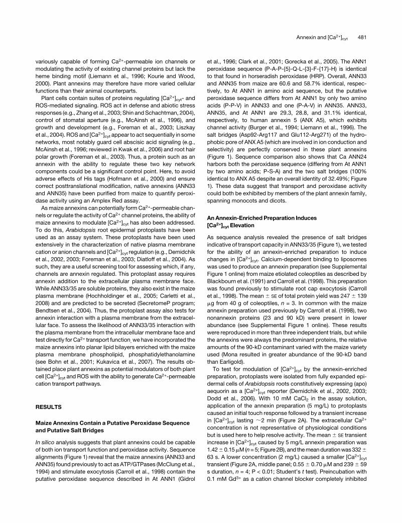
Figure 4. Effect of Highly Purified ANN33/35 Preparation on [Ca2+]cyt of Protoplasts from Mature Root Epidermal Cells.
(A) Example of an annexin-induced increase in [Ca2+]cyt. Highly purified maize annexin preparation (5 mg/L) added (arrow; closed circles) to the
extracellular membrane face of Arabidopsis root epidermal protoplasts (constitutively expressing aequorin) in 10 mM external Ca2+ caused a transient
increase in [Ca2+]cyt. The control experiment adding buffer alone is also shown (open circle).
(B) Mean 6 SE [Ca2+]cyt response to 5 mg/L Zm ANN33/35 (closed circle; n = 3) or buffer alone (open circle; n = 4).
(C)Mean6 SE [Ca2+]cyt response to 5mg/L ZmANN33/35 preparation (closed circle; n = 4) or buffer alone (open circle; n = 4) both in the presence of 100
mM verapamil (Vp).
(D)Mean6 SE [Ca2+]cyt response to 5 mg/L Zm ANN33/35 (closed circle; n = 4) or buffer alone (open circle; n = 4) both in the presence of 300 mMGd3+.
(E)Mean6 SE [Ca2+]cyt response to 5 mg/L immunoprecipitated Zm ANN33/35 (IP, closed circle; n = 3) or buffer containing equivalent anti-Zm ANN33/
35 antibody (open circle; n = 3).
(F)Mean6 SE peak [Ca2+]cyt responses from (B) to (E) and themean response to preimmune serum at an equivalent concentration to antibody (Ab) used
in (E) (n = 3).
484 The Plant Cell
from the Goldman equation with the assumption ICa + ICl = 0 at
Erev (Hille, 1992). PCa/PCl was calculated to be 5. This conduc-
tance was completely blocked by an application of 50 mM Gd3+
to the trans-compartment (n = 3; Figures 5C and 5D) within 5 min
after addition. That the blocker was acting at the opposite
membrane face to that exposed to the annexin shows that a
transbilayer pathway for ions had been formed. The conduc-
tance was also substantially blocked by an application of 50 mM
verapamil, causing a 94% reduction inmean current amplitude at
both –200 mV and +200 mV (n = 3; Figure 5D). Heat-inactivated
annexin preparation did not evoke a current (n = 3; Figure 5E). As
the highly purified preparation still contained p23, the prepara-
tion was subjected to immunoprecipitation (as described previ-
ously) with the anti-ANN33/35 antibody. No currents were
evoked over a 3.5 h time frame after adding either antibody
alone or immunoprecipitated preparation (3mg) to the cis-chamber
(n = 4 for each test; see Supplemental Figures 3A to 3C online).
This represents a 100% failure rate compared with the 50%
observed with normal protein and indicates annexin involvement
in current generation. Preimmune serum did not evoke current
(n=2). The sensitivity of the highly purified annexin preparation to
verapamil in the bilayer assay is in stark contrast with the
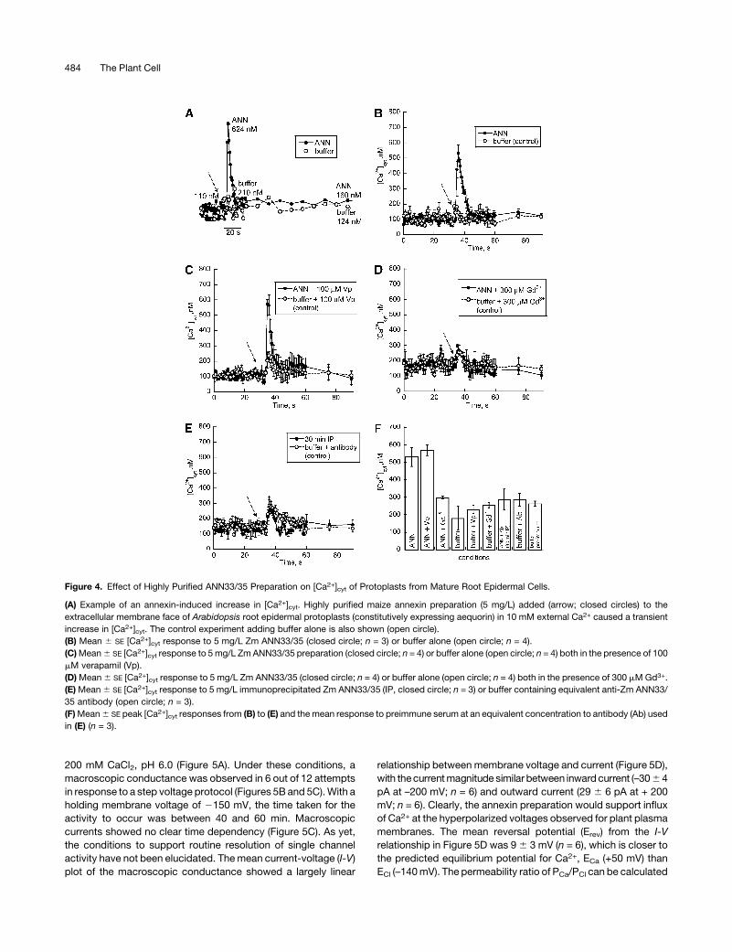
Figure 5. Highly Purified ANN33/35 Preparation Forms a Ca2+-Permeable Conductance in Planar Lipid Bilayers.
(A) Schematic of experimental conditions. Highly purified ANN33/35 preparation (3 mg) was added to the cis-chamber, while channel blockers were
added to the (grounded) trans-chamber. Chambers were separated by a POPE:cholesterol:POPS (5:3:2) bilayer formed across a 200-mm-diameter hole
in the septum (filled circle). With this configuration, positive charge flowing from trans to cis is plotted as negative current, while that flowing from cis to
trans is plotted as positive current.
(B) Schematic of the step voltage pulse protocol used to elicit current.
(C) Representative current traces recorded in response to the voltage protocol applied from a 0 mV baseline. Left panel, prior to annexin addition.
Middle panel, current generated by addition of highly purified ANN33/35 preparation. Right panel, effect of 50 mM Gd3+ in the trans-chamber.
(D) Mean 6 SE I-V relationships for conditions described in (A) and in response to the voltage protocol shown in (B). ANN33/35 (closed circles; n = 6),
with 50 mM Gd3+ (x; n = 3) or 50 mM verapamil (open circles; n = 3). ECa was + 50 mV, and ECl was –140 mV.
(E) Mean 6 SE I-V relationship for heat-inactivated ANN33/35 (n = 3). Conditions as in (A) and (B).
Annexin and [Ca2+]cyt 485
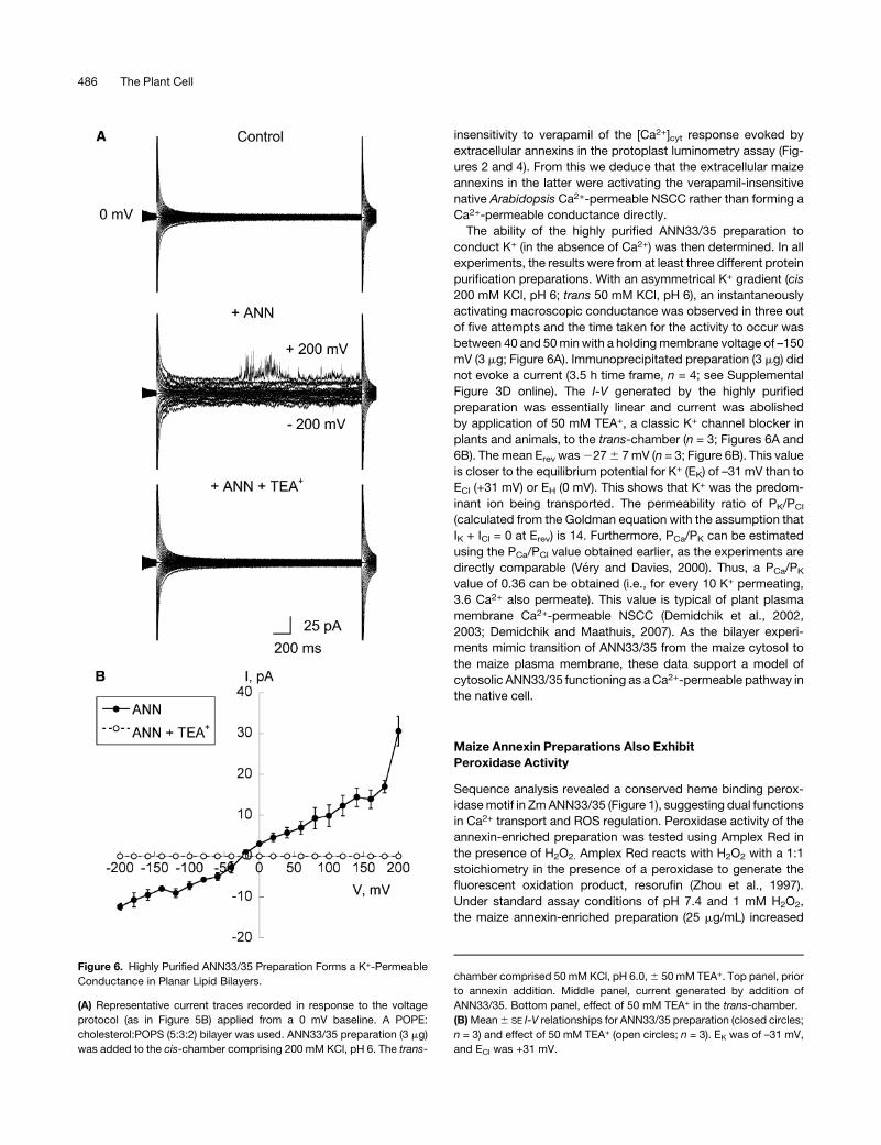
insensitivity to verapamil of the [Ca2+]cyt response evoked by
extracellular annexins in the protoplast luminometry assay (Fig-
ures 2 and 4). From this we deduce that the extracellular maize
annexins in the latter were activating the verapamil-insensitive
native Arabidopsis Ca2+-permeable NSCC rather than forming a
Ca2+-permeable conductance directly.
The ability of the highly purified ANN33/35 preparation to
conduct K+ (in the absence of Ca2+) was then determined. In all
experiments, the results were from at least three different protein
purification preparations. With an asymmetrical K+ gradient (cis
200 mM KCl, pH 6; trans 50 mM KCl, pH 6), an instantaneously
activating macroscopic conductance was observed in three out
of five attempts and the time taken for the activity to occur was
between 40 and 50min with a holdingmembrane voltage of –150
mV (3 mg; Figure 6A). Immunoprecipitated preparation (3 mg) did
not evoke a current (3.5 h time frame, n = 4; see Supplemental
Figure 3D online). The I-V generated by the highly purified
preparation was essentially linear and current was abolished
by application of 50 mM TEA+, a classic K+ channel blocker in
plants and animals, to the trans-chamber (n = 3; Figures 6A and
6B). The mean Erev was2276 7mV (n = 3; Figure 6B). This value
is closer to the equilibrium potential for K+ (EK) of –31 mV than to
ECl (+31 mV) or EH (0 mV). This shows that K+ was the predom-
inant ion being transported. The permeability ratio of PK/PCl
(calculated from the Goldman equation with the assumption that
IK + ICl = 0 at Erev) is 14. Furthermore, PCa/PK can be estimated
using the PCa/PCl value obtained earlier, as the experiments are
directly comparable (Very and Davies, 2000). Thus, a PCa/PK
value of 0.36 can be obtained (i.e., for every 10 K+ permeating,
3.6 Ca2+ also permeate). This value is typical of plant plasma
membrane Ca2+-permeable NSCC (Demidchik et al., 2002,
2003; Demidchik and Maathuis, 2007). As the bilayer experi-
ments mimic transition of ANN33/35 from the maize cytosol to
the maize plasma membrane, these data support a model of
cytosolic ANN33/35 functioning as aCa2+-permeable pathway in
the native cell.
Maize Annexin Preparations Also Exhibit
Peroxidase Activity
Sequence analysis revealed a conserved heme binding perox-
idasemotif in ZmANN33/35 (Figure 1), suggesting dual functions
in Ca2+ transport and ROS regulation. Peroxidase activity of the
annexin-enriched preparation was tested using Amplex Red in
the presence of H2O2. Amplex Red reacts with H2O2 with a 1:1
stoichiometry in the presence of a peroxidase to generate the
fluorescent oxidation product, resorufin (Zhou et al., 1997).
Under standard assay conditions of pH 7.4 and 1 mM H2O2,
the maize annexin-enriched preparation (25 mg/mL) increased
Figure 6. Highly Purified ANN33/35 Preparation Forms a K+-Permeable
Conductance in Planar Lipid Bilayers.
(A) Representative current traces recorded in response to the voltage
protocol (as in Figure 5B) applied from a 0 mV baseline. A POPE:
cholesterol:POPS (5:3:2) bilayer was used. ANN33/35 preparation (3 mg)
was added to the cis-chamber comprising 200 mM KCl, pH 6. The trans-
chamber comprised 50 mM KCl, pH 6.0,6 50 mM TEA+. Top panel, prior
to annexin addition. Middle panel, current generated by addition of
ANN33/35. Bottom panel, effect of 50 mM TEA+ in the trans-chamber.
(B)Mean6 SE I-V relationships for ANN33/35 preparation (closed circles;
n = 3) and effect of 50 mM TEA+ (open circles; n = 3). EK was of –31 mV,
and ECl was +31 mV.
486 The Plant Cell
fluorescence, thus demonstrating peroxidase activity, whereas
heat-inactivated annexin preparation (boiled for 10 min) did not
support an increase (Figure 7A). The mean 6 SE rate of fluores-
cence increase of the annexin-enriched preparation was 38 60.6 DF mg21 s21 (n = 3), an order of magnitude lower than that of
an HRP positive control (882 6 28 DF mg21 s21; n = 3); no
increase in fluorescence was observed with heat-inactivated
HRP (Figure 7B). The highly purified ANN33/35 preparation was
also tested under standard assay conditions (pH 7.4 and 1 mM
H2O2). The mean 6 SE rate of fluorescence increase generated
by the highly purified ANN33/35 preparation at 25 mg/mL (Figure
7C) was 456 2 DF mg21 s21 (n = 3), which supports the premise
that peroxidase activity of the annexin-enriched preparation was
generated by the annexins. Despite the presence of a heme
binding motif similar to that of HRP in maize annexins, peroxi-
dase activity appears independent of heme. We tested for the
presence of heme using absorbance spectroscopy but were
unable to detect the presence of heme in 5-mg protein samples
that had supported peroxidase activity (annexin-enriched prep-
aration). By contrast, cytochrome C oxidase as a positive heme
control could be diluted to 2.2 mM, an equivalent of 7.3 ng
annexin, and still exhibited a Soret absorbance peak (;410 nm)
indicative of a heme moiety (Margoliash and Frohwirt, 1959)
(Figure 7D).
DISCUSSION
We report that native maize annexins are likely to be multifunc-
tional proteins capable of peroxidase activity, elevation of [Ca2+]cyt,
and direct formation of a passive Ca2+- and K+-permeable con-
ductance. It has been estimated that at least 152 proteins in
Arabidopsis are involved in the regulation of ROS (Mittler et al.,
2004). Peroxidases comprise a superfamily of intra- or extracel-
lular heme-containing enzymes catalyzing a number of oxidative
reactions, using H2O2 as the electron acceptor. Peroxidases are
envisaged not only to protect the cell from ROS toxicity but also
play a role in signaling, biotic and abiotic stress responses, auxin
metabolism, and cell wall modification (reviewed in Passardi
et al., 2005). Recombinant Arabidopsis ANN1 purified from
Escherichia coli or Nicotiana benthamiana expression systems
has peroxidase activity in vitro (Gidrol et al., 1996; Gorecka
et al., 2005). A preliminary study found that recombinant Ca
ANN24 purified from E. coli also has peroxidase activity in vitro
(Mortimer, 2007), and recombinant B. juncea ANN1 also exhibits
peroxidase activity (Jami et al., 2008). Such findings clearly argue
for the inclusion of the annexin protein family in considerations
of ROS.
Annexin peroxidase activity is reliant on a region of the first
annexin repeat (centering on a conservedHis residue; His40) that
has similarity to the ;30–amino acid heme binding domain of
plant peroxidases (Gidrol et al., 1996; Clark et al., 2001; Gorecka
et al., 2005). Mutagenesis of His40 in At ANN1 abolished perox-
idase activity (Gorecka et al., 2005). Zm ANN33/35 contain the
conserved His (Figure 1). Spectral analysis of the annexin-
enriched preparation failed to detect heme (Figure 7D). The
observation that maize annexin preparations supported perox-
idase activity in the apparent absence of an associated heme
points to this His residue as being the structural basis for this
enzymatic activity in annexins or as being essential for maintain-
ing the overall structure of the annexin molecule. It is possible
that annexin peroxidases play a similar role to heme-free gluta-
thione peroxidases (a subgroup of the peroxiredoxins) that
catalyze the reduction of lipid peroxides and H2O2 to prevent
membrane peroxidation and act in (a)biotic stress signal trans-
duction (reviewed in Rouhier and Jacquot, 2005). Peroxidases
are distributed throughout roots, and the known ANN33/35
expression in the root elongation zone (Carroll et al., 1998;
Bassani et al., 2004) could reflect a role in elongation growth,
possibly by limiting H2O2 accumulation (Pnueli et al., 2003). This
part of the maize root is also the site of ROS accumulation during
stress responses, for example aluminum stress (Jones et al.,
2006). By extension, loss of peroxidase function could contribute
to the short root phenotype of the ANN1 loss-of-function mutant
(Clark et al., 2005b; Mortimer, 2007).
Purification of Zm ANN33/35 by lipid binding also resolves 23-
kD (p23) and 90-kD (p90) proteins. The association of p23 with
lipids is readily explained by the presence of a (partial) C2
Figure 7. Peroxidase Activity of Maize Annexin Preparations Measured
Using Amplex Red.
(A) Time course. Increase in fluorescence (arbitrary units [AU]) supported
by 25 mg/mL annexin-enriched preparation at pH 7.4 in the presence of
1 mM H2O2 (closed circles) was abolished by heat inactivation (open
circles).
(B) Time course of HRP activity (0.095 mg/mL) using conditions de-
scribed in (A). Heat-inactivated HRP (open circles).
(C) Time course of fluorescence increase supported by 25 mg/mL highly
purified ANN33/35 preparation at pH 7.4 in the presence of 1 mM H2O2
(closed circles); heat inactivated ANN33/35 preparation (open circles).
(D) Heme detection. Absorption spectrum shows peroxidase-competent
annexin-enriched preparation (5 mg) has no Soret peak (;410 nm)
indicative of a heme group, in contrast with either 2.2 mM (7.3 ng) or 22
mM (73 ng) cytochrome C oxidase (CytC).
Annexin and [Ca2+]cyt 487
domain. The C2 domainmediates Ca2+-dependent phospholipid
binding (Nalefski and Falke, 1996). Although few C2-containing
proteins have been characterized in plants, the domain has been
shown to be essential for plasma membrane association of a
mung bean (Vigna radiata) phospholipase C (Kim et al., 2004) and
a novel bell pepper protein (Kim et al., 2008). To date, plant C2
domain proteins appear to be involved in (a)biotic stress re-
sponses and development (Kim et al., 2004, 2008; Yang et al.,
2008), suggesting that the novel maize p23 protein could be
involved in such Ca2+-dependent processes. The assignment of
the p90 as a putative lipoxygenase helps explain its presence in
the preparation. Maize lipoxygenases are known to be wound
inducible and wound responsive (e.g., Cho et al., 2007). It is
possible that the presence of p90 was due to wounding of maize
coleoptiles on harvesting. Given that lipoxygenases are com-
partmentalized on membranes (Porta and Rocha-Sosa, 2002), it
is likely that they are obtained during the annexin lipid binding
purification process.
Plant annexins have been postulated to act as ion channels
(Hofmann et al., 2000). To date, Ca ANN24 has been found to
mediate passive Ca2+ flux into liposomes by an unknown mech-
anism (Hofmann et al., 2000), andwhile At ANN1 has been shown
to support K+ channel activity in planar lipid bilayers, its ability to
transport Ca2+ is unknown (Gorecka et al., 2007). In common
with At ANN1 and Ca ANN24, maize annexins conserve the salt
bridges that could confer transport function (Figure 1). Modula-
tion of [Ca2+]cyt bymaize annexin preparations demonstrated the
capacity to form and/or regulate Ca2+ transport pathways in
native membranes. Immunoprecipitation experiments demon-
strated annexin involvement but cannot preclude the possibility
of p23 activity. As highly purified Zm ANN33/35 preparation
formed a verapamil-sensitive Ca2+-permeable conductance in
bilayers but caused a verapamil-insensitive elevation of [Ca2+]cytin Arabidopsis, the findings point to Zm ANN33/35 regulation of
the native Arabidopsis plasma membrane verapamil-insensitive
NSCC (Demidchik et al., 2002) at the extracellular membrane
face to increase [Ca2+]cyt.
There are no reports to date of Zm ANN33/35 in the cell wall
and so function at the extracellular plasma membrane face in
vivo must remain speculative. However, high percentages of
plant cell wall proteins appear to be nonclassical secretory
proteins and can be predicted using the SecretomeP program
(Bendtsen et al., 2004). Maize proteins predicted to be secreted
by SecretomeP have now been identified in cell wall fractions
(Zhu et al., 2006). ANN33 and ANN35 are predicted to be
secreted as are the two predominantly expressed annexins of
Arabidopsis, ANN1 and ANN2. Although they lack the classic
N-terminal signal peptide, both ANN1 and ANN2 have indeed
been detected in cell walls (Kwon et al., 2005; Bayer et al., 2006).
It is therefore possible that, in some cells, annexins could affect
Ca2+ transport and [Ca2+]cyt by acting at the extracellular plasma
membrane face. There are precedents from animal cells. Animal
annexins can be secreted to operate in the extracellular space. In
its extracellular form, the channel-forming annexin ANXA5 reg-
ulates Polycystin-1 (a transient receptor potential channel) at its
extracellular N terminus (Markoff et al., 2007), while extracellular
ANXA1 causes [Ca2+]cyt increase by activating plasma mem-
brane G-protein-coupled receptors (Babbin et al., 2006). Gen-
erally, the known abundance of annexins in the cytosol and their
ability to relocate to plasma and endomembranes (Thonat et al.,
1997; Breton et al., 2000; Lee et al., 2004; reviewed in Mortimer
et al., 2008) place them predominantly as intracellular regulators
of [Ca2+]cyt. In this respect, annexins, as regulators of plant
channels, can be viewed as being similar to calmodulin. Cal-
modulin acts at the cytosolic plasma membrane face to regulate
cyclic nucleotide-gated channels (Hua et al., 2003) but can also
act extracellularly to regulate plasmamembrane hyperpolarization-
activated Ca2+-permeable channels (Shang et al., 2005).
ANN33/35 are known soluble proteins in maize that also exist
in the plasma membrane and possibly tonoplast (Hochholdinger
et al., 2005; Carletti et al., 2008). A function at the intracellular
plasma membrane face has already been demonstrated by
Carroll et al. (1998) who showed an increase in maize root cap
plasma membrane capacitance on adding the annexin-enriched
preparation to protoplast cytosol. Here, the bilayer experiments
mimicked movement of ANN33/35 from the cytosol to the
plasma membrane and showed the ability of ANN33/35 to
generate a Ca2+ influx pathway at slightly acidic pH (pH 6). This
agrees with animal studies in which acidic pH promotes channel
activity (Langen et al., 1998; Golczak et al., 2001; Isas et al., 2003)
and the ability of At ANN1 to from a K+-permeable conductance
at pH 5.8 (Gorecka et al., 2007). Immunoprecipitation experi-
ments confirmed annexin involvement in conductance formation
but, strictly, the possibility remains that the conductance was
formed by the low level p23 contaminant with the aid of the
annexins or that both p23 and annexins were required. A mac-
roscopic conductancewas observedmore frequently than single
channel events, and this has also been reported for animal
annexins (Arispe et al., 1996). Blockage by Gd3+ is also consis-
tent with the pharmacology of animal annexin channels (Kourie
and Wood, 2000), but Ca2+ permeability is low compared with
animal counterparts (PCa/PK 0.36 versus 4.34 for single channel
ANXA5; Burger et al., 1994; Liemann et al., 1996). Site-directed
mutagenesis of ANXA5 salt bridges dramatically decreases Ca2+
permeability to 0.87 (Burger et al., 1994), suggesting that subtle
sequence differences in plant annexins could cause low Ca2+
permeability. This possibility and the mode of conductance
formation by plant annexins now need to be determined.
The channel proteins of the maize plasma membrane are in-
creasingly well understood at the molecular level. Inward and
outward K+-selective rectifiers are known to be encoded by mem-
bers of the Shaker family (Philippar et al., 2003; Buschenschutz
et al., 2005; Su et al., 2005). The genetic identities of the less
selective and instantaneously activating channels remain un-
known. The K+-permeable, instantaneously activating conduc-
tance formed by the ANN33/35 preparation resembles themaize
MgC conductance that has been characterized by patch clamp
electrophysiology in guard and subsidiary cell plasma mem-
branes (Wolf et al., 2005). However, unlike MgC, the ANN33/35
conductance is sensitive to block by TEA+. The sensitivity of the
ANN33/35 conductance to verapamil also distinguishes this
conductance from those characterized in maize plasma mem-
brane vesicles by 45Ca2+ flux analysis (Marshall et al., 1994). It
may be that ANN33/35 (6 p23) underpins the instantaneous
and TEA+-sensitive K+-permeable conductance observed in
root and suspension cell plasma membranes (Ketchum et al.,
488 The Plant Cell
1989; Roberts, 1998). Analysis of loss-of-functionmutants is now
needed to resolve in planta transport function, which at the
plasmamembrane could support Ca2+ influx for signaling and K+
flux for voltage regulation.
Annexin expression is dynamic and responsive to stress
conditions known to increase [Ca2+]cyt and ROS, such as salinity,
cold, and nutrient deprivation (reviewed in Mortimer et al., 2008).
Such conditions cause relocation of annexins from the cytosol to
membranes, sometimes also causing annexin insertion into
membranes (Breton et al., 2000; Lee et al., 2004). The findings
here (that annexins can act as peroxidases, modulate [Ca2+]cyt,
and form a Ca2+-permeable K+ conductance) firmly place the
dynamic behavior of annexins in a signaling context. ANN33/35
are expressed in roots, coleoptiles, and egg cells (Blackbourn
et al., 1991; Carroll et al., 1998; Bassani et al., 2004: Okamoto
et al., 2004; Hochholdinger et al., 2005; Carletti et al., 2008)
where [Ca2+]cyt and ROS regulation will be pivotal to develop-
ment and adaptation. The amount of ANN33/35 in root plasma
membrane is now known to increase in response to environ-
mental conditions (Carletti et al., 2008), consistent with a signal-
ing role. Additionally, that acidic pH supports a Ca2+-permeable
conductance by ANN33/35 (6p23) suggests that it could func-
tion in Ca2+ signaling in response to stress-induced cytosolic
acidosis. For example, anoxia causes root tip cytosolic acidosis
in maize, and while the Ca2+ signal for metabolic adaptation
appears to emanate from mitochondria, the Ca2+ signal for an
adaptive localized cell death comes from the plasma membrane
(Chang et al., 2000; Subbaiah et al., 2000).
The molecular identities of higher plant Ca2+-permeable chan-
nels are now being established. The impact of loss of function on
[Ca2+]cyt is used to associate Ca2+ transport with a gene, while
heterologous expression can permit Ca2+ permeation of the
gene product to be ascertained, provided that expression does
not affect native conductances. Combining these approaches
has confidently identified TPC1 (Two Pore Channel 1) and
CNGC2 (Cyclic Nucleotide-Gated Channel 2) as Ca2+-permeable
channels in Arabidopsis (Leng et al., 2002; Peiter et al., 2005; Ali
et al., 2007). Other CNGCs and members of the glutamate re-
ceptor family are strong candidates as potential Ca2+-permeable
channels (Meyerhoff et al., 2005; Urquhart et al., 2007; Tapken
and Hollmann, 2008), while the Arabidopsis Stelar K+ Outward
Rectifier has also been found to be Ca2+ permeable (Gaymard
et al., 1998). Bilayers provide unequivoval evidence for Ca2+
transport by a single protein. The determination here of Ca2+
transport by maize annexins in bilayers clearly adds this family of
proteins to the suite of higher plant Ca2+ transporters. With eight
annexins in Arabidopsis (Cantero et al., 2006) and nine in rice
(Oryza sativa; Moss and Morgan, 2004) to explore, these enig-
matic, multifunctional proteins now need to be considered in the
networks translating cellular signals.
METHODS
Plant Growth
Seeds of maize (Zea mays cv Mona [Pioneer Hi-Bred International] or
Earligold [Moles Seeds]) were soaked overnight in aerated distilled water.
Seeds were grown in the dark on damp vermiculite in propagators for 6 to
9 d at room temperature. Arabidopsis thaliana [Columbia-0 wild type and
constitutively expressing cytosolic (apo)aequorin, driven by the cauli-
flowermosaic virus 35S promoter] was grown aseptically at 228C for 10 to
15d (16-hdaylength; 100mmol/m2/s irradiance) on full-strengthMurashige-
Skoog medium (Duchefa) with 1% (w/v) sucrose and 0.3% (w/v) Phytagel
(Sigma-Aldrich).
Annexin Production
Maize etiolated coleoptiles (40 g) were ground with liquid nitrogen in a
mortar and pestle to a fine powder. The purification protocol was based
on Blackbourn et al. (1991). Homogenization buffer (0.15 M NaCl, 10 mM
HEPES, 10 mM EDTA, and 1 mM PMSF, pH 7.4) was added and the
mixture filtered through two layers of muslin. After centrifugation (30 min)
to remove remaining debris (30,000g), the supernatant was added to
liposomes. Thesewere prepared frombovine brain lipid (Folch fraction IV;
Sigma-Aldrich) dissolved in chloroform:methanol (2:1; 15 mg/mL) and
then formed by the addition of liposome buffer (50 mM HEPES, 150 mM
NaCl, 5 mM EGTA, and 1 mM PMSF, pH 7.5) and brief vortexing.
Alternatively, liposomes were prepared from soybean asolectin (200 mg;
Fluka) dissolved in (2:1) chloroform:methanol to give a final concentration
of 13.3 mg/mL. CaCl2 was added to liposomes to a final concentration of
10 mM, and the suspension was incubated on ice for 1 h, followed by
centrifugation for 30 min (30,000g). The supernatant was discarded and
the pellet was resuspended in wash buffer (0.15 M NaCl, 10 mM HEPES,
10 mM CaCl2, and 1 mM PMSF, pH 7.4). Following centrifugation for 30
min (30,000g), the pellet was resuspended in wash buffer lacking NaCl.
The centrifugation step was repeated, and the pellet was resuspended in
elution buffer (0.15 M NaCl, 10 mM HEPES, 15 mM EDTA, and 1 mM
PMSF, pH 7.4). After a final centrifugation step (1 h, 100,000g), the
supernatant was collected and added to bovine brain or asolectin
liposomes; the entire process was then repeated. After the final centrif-
ugation, the buffer was exchanged to 10 mM K-PO4 buffer, pH 7.4, and
the sample concentrated using a centricon (molecular weight cutoff 10
kD; Amicon). All steps were performed at 48C. The concentrated sample
was then loaded onto a gel filtration column (Superose 12; Amersham)
attached to an FPLC system. The column was equilibrated with 10 mM
K-PO4 buffer, pH 7.4. Maize annexins (ANN33/35) were eluted with this
buffer at a flow rate of 0.4 mL/min. Fractions of 1 mL were collected and
tested for ANN33/35 by SDS-PAGE and protein gel blot analysis. All
proteins were exchanged into buffer (10mMK-PO4 buffer, pH 7.4) prior to
use. In immunoprecipitation studies, the maize annexin preparation was
incubated for 30 min at room temperature with anti-ANN33/35 antibody
(Blackbourn et al., 1991) in a 2:1 ratio (Carroll et al., 1998). Antibody or
preimmune serum alone was added as a control. Protein concentration
was estimated using Protein Assay Reagent (Bio-Rad) calibrated against
BSA (Sigma-Aldrich).
Gel Electrophoresis and Immunoblotting
Samples were incubated for 10 min at 908C in buffer (10% [w/v] sucrose,
5% [v/v] b-mercaptoethanol, 2% [w/v] SDS, 0.001% [w/v] bromophenol
blue, and 50 mM Tris-HCl, pH 6.8). Samples were first run through a 4%
(w/v) acrylamide stacking gel at 70 V and then through a 12% (w/v)
acrylamide resolving gel at 150 V for 70 min. Gels were either immuno-
blotted or stained using either Colloidal Coomassie Brilliant Blue (Sigma-
Aldrich) or Silver Stain Plus (Bio-Rad). For silver staining, gels were fixed
overnight to enhance detection and then washed twice for 20 min with
distilled water to ensure that all acetic acid in the gel during the fixative
step was washed away to prevent its slowing down band resolution on
staining. Staining was conducted for up to 40 min. For immunoblotting,
proteins were electroblotted onto polyvinylidene fluoride (GE Healthcare)
using transfer buffer (25 mM Tris, 200 mM glycine, and 20% [v/v]
methanol) at 48C, 60 V, for 3 h. Membranes were blocked by incubating
Annexin and [Ca2+]cyt 489
overnight at 48C in TBS-T (20 mM Tris, 150 mM NaCl, and 0.1% [w/v]
Tween 20, pH 7.6) containing 5% (w/v) skimmed milk powder (Sains-
burys). Following washing in TBS-T, membranes were incubated with
primary antibody (1:5000 dilution of the anti-ANN33/35 antibody de-
scribed in Blackbourn et al., 1991) for 1 h, at room temperature, with
shaking then after washing for a further 1 h with secondary antibody
(1:5000 peroxidase-linked anti-rabbit IgG; Invitrogen). Proteins were
visualized using enhanced chemiluminesence (ECL-plus; GE Healthcare)
according to the manufacturer’s instructions.
Protein Verification
Protein identity was determined by mass spectrometry. Samples were
subjected toSDS-PAGEwith slightmodification.All solutions forSDS-PAGE
weremembrane filtered. Gels were stained with Coomassie Brilliant Blue or
Colloidal Coomassie blue (Sigma-Aldrich) and destained in filter-sterilized
10% (v/v)methanol once. The gelwas stored in 10% (v/v)methanol prior to
in-gel digestion andMALDI peptidemass fingerprint analysis (Protein and
Nucleic Acid Chemistry Facility; University of Cambridge). Protein iden-
tifications were based on multiple peptide matches.
[Ca2+]cyt Determination
Protoplasts were isolated from fully expanded root epidermal cells as
described by Demidchik et al. (2002) and suspended in holding solution
(HS) comprising 5mMKCl, 2mMCaCl2, 1mMMgCl2, 10mMsucrose, 10
mM glucose, and 2 mM MES, pH 5.7, with Tris (290-300 mOsM).
Protoplasts were incubated in HS with 4 mg/mL coelentrazine (free
base; NanoLight Technology) for 3 h in the dark (288C) and thenwashed in
recording solution (10 mM CaCl2 unless stated otherwise; osmotic
potential and pH were as in HS, colentrazine was maintained at 4 mg/
mL) before being placed in luminometer cuvettes. Annexin preparation
was added at 0.1 to 10 mg/L; heat-inactivated annexin was used as a
control. Channel blockers (Sigma-Aldrich) were incorporated into the
assay prior to annexin addition. Luminometry and calibration to convert
luminescent values to [Ca2+]cyt were performed as described by Dodd
et al. (2006).
Planar Lipid Bilayers
Lipids were obtained from Avanti Polar Lipids or Sigma-Aldrich. These
were POPE, POPS, and cholesterol. Lipids (25 mg/mL) were mixed in the
proportions required. Bilayers were composed of POPE:cholesterol:
POPS (5:3:2). Lipid dispersions (10mg/mL)were prepared by evaporating
the chloroform under a stream of nitrogen and resuspending the lipid in
n-decane after 15 min (Sigma-Aldrich). Bilayers were formed by the
planar lipid bilayer technique. Bilayers were formed across a 200-mm-
diameter aperture in the septum by painting lipid dispersion across the
(lipid-pretreated) septum. In Ca2+-permeability experiments, the cis- and
trans-chambers were filled with 0.5 mL 10 mM MES/bis Tris propane
(BTP), 1 mM CaCl2, pH 6.0, 2 mL 10 mM MES/BTP, and 200 mM CaCl2,
pH 6.0. In K+-permeability experiments, the cis-chamber comprised 200
mM KCl and 10 mM MES/BTP, pH 6.0, and the trans-comprised 50 mM
KCl and 10 mM MES-/TP, pH 6.0. The cis-compartment was connected
to the headstage of the Axopatch 200B amplifier (Axon Instruments), and
the trans-compartment was connected to the signal ground using agar-
salt bridges (3 M KCl and 1% [w/v] agar) and Ag/AgCl electrodes. Bilayer
formation was monitored electronically by capacitance measurements
with their final capacitance ranging from 150 to 200 pF and their
resistance from 2 to 5 GV. Following stable bilayer formation, 3 mg of
annexin protein was added to the cis-chamber. The bilayer was held at
2150 mV (cis-negative) to aid protein insertion, and the cis-chamber was
magnetically stirred intermittently. Experiments were performed at room
temperature (20 to 248C). Antagonists were added to the trans-chamber
as indicated in Results. Filter and sample frequency were 1 and 5 kHz,
respectively, and data analysis was performed with Clampfit software
(version 8). Positive current deflections indicated movement of positive
charge from the cis- to the trans-chamber, while negative current defec-
tions indicated themovement of positive charge from the trans- to the cis-
chamber. Holding potentials were all corrected for (measured) liquid
junctions. Liquid junctions were measured and corrected. Permeability
ratios were calculated using the Goldman equation with the assumption
at the reversal potential (Erev), IX + IY + IZ = 0where X, Y, and Z are different
species of ions (Very and Davies, 2000). Curve fitting of data was
performed using SigmaPlot (SPSS Science).
Peroxidase Assay and Heme Analysis
Assay buffer (50 mM K-phosphate, pH 7.4) was used to dilute all
solutions. All assays (final volume 100 mL) were performed at room
temperature in the dark in a black 96-well plate (Greiner Bio-One) and
comprised 50 mM Amplex Red (10-acetyl-3,7-dihydroxyphenoxazine;
Invitrogen), 1 mM H2O2 (Sigma-Aldrich), and protein (ANN33/35 prepa-
ration or HRP Type VI; Sigma-Aldrich). Resorufin fluorescence was
measured using a 560-nm excitation filter and a 590-nm emission filter
in a FLUO-star plate-reader (BMG). Proteins heated for 10 min at 958C
were used as negative controls. Fluorescence values from identical
assays without protein were subtracted from all data. To assay for heme
content, absorption spectra were obtained using a Thermospectronic UV
visible spectrophotometer. Samples were analyzed in 100-mL capacity
UVettes (Eppendorf).
Phylogenetic Analysis
Amino acid sequence alignment was performed using ClustalW (default
parameters; Larkin et al., 2007) and edited in JalView (http://www.jalview.
org; Clamp et al., 2004). Prediction of secretion was performed using
SecretomeP (www.cbs.dtu.dk/services/secretomeP;Bendtsenet al., 2004).
Accession Numbers
Sequence data from this article can be found in the Arabidopsis Genome
InitiativeorGenBank/EMBL/NCBIRefSeqdata libraries under the following
accession numbers: gb:CAA10210 (Ca ANN24), gb:AAC33305 (Gh ANN),
gb:CAA66900 (ZmANN33), gb:CAA66901 (ZmANN35), gb:ABC59685 (Zm
LOX1), gb:ABC59686 (ZmLOX2), gb:ABC59687 (ZmLOX4), gb:CAA00083
(Armoracia rusticana horseradish peroxidase; HRP), gb:CAA71759 (Spor-
obolus stapfianus hypothetical protein), RefSeq:NP_174810 (At1g35720,
At ANN1), RefSeq:NP_201307 (At5g65020, At ANN2), RefSeq:NP_001145
(humanannexin 5 Anx5), RefSeq:NP_005630 (humansynaptotagmin I; Syn
I), RefSeq:NP_796376 (human synaptotagmin II; Syn II), and RefSeq:
NP_002730 (human protein kinase C isoform gamma; PKC g).
Supplemental Data
The following materials are available in the online version of this article.
Supplemental Figure 1. Purification of ANN33/35 from Etiolated
Maize Coleoptiles by Lipid Affinity.
Supplemental Figure 2. Analysis of the Maize 23-kD Protein and 19-
kD Protein from Sporobolus stapfianus.
Supplemental Figure 3. Immunoprecipitation of Zm ANN33/35 Pre-
vents Conductance Formation in Planar Lipid Bilayers.
ACKNOWLEDGMENTS
This work was supported by the Biotechnology and Biological Science
Research Council, the Isaac Newton Trust, the Cambridge Overseas
490 The Plant Cell
Trusts, and the University of Cambridge Department of Plant Sciences
Research Fund. We thank A.G. Smith’s group for equipment access, L.
Packman for assistance in protein identification, P.J. White for guidance
on bilayers, and John Banfield for bilayer technical assistance. We thank
Stephen Chivasa, Peter Hepler, and Joan Rigau for helpful discussions
on protein localization and the referees, whose insight improved this
work.
Received March 20, 2008; revised December 11, 2008; accepted Feb-
ruary 2, 2009; published February 20, 2009.
REFERENCES
Ali, R., Lemtiri-Chlieh, F., Tsaltas, D., Leng, Q., von Bodman, S., and
Berkowitz, G.A. (2007). Death don’t have no mercy and neither does
calcium: Arabidopsis Cyclic Nucleotide Gated Channel2 and innate
immunity. Plant Cell 19: 1081–1095.
Arispe, N., Rojas, E., Genge, B.R., Licia, N.Y.W., and Wuthier, R.E.
(1996). Similarity in calcium channel activity of Annexin V and matrix
vesicles in planar lipid bilayers. Biophys. J. 71: 1764–1775.
Babbin, B.A., Lee, W.Y., Parkos, C.A., Winfree, L.M., Akylldiz, A.,
Perretti, M., and Nusrat, A. (2006). Annexin 1 regulates SKCO-15 cell
invasion by signalling through formyl peptide receptors. J. Biol. Chem.
281: 19588–19599.
Bassani, M., Neumann, P.M., and Gepstein, S. (2004). Differential
expression profiles of growth-related genes in the elongation zone of
maize primary roots. Plant Mol. Biol. 56: 367–380.
Bayer, E.M., Bottrill, A.R., Walshaw, J., Vigouroux, M., Naldrett,
M.J., Thomas, C.L., and Maule, A.J. (2006). Arabidopsis cell wall
proteome defined using multidimensional protein identification tech-
nology. Proteomics 6: 301–311.
Bendtsen, J.D., Jensen, L.J., Blom, N., von Heijne, G., and Brunak,
S. (2004). Feature-based prediction of non-classical and leaderless
protein secretion. Protein Eng. Des. Sel. 17: 349–356.
Blackbourn, H.D., Barker, P.J., Huskisson, N.S., and Battey, N.H.
(1992). Properties and partial protein-sequence of plant annexins.
Plant Physiol. 99: 864–871.
Blackbourn, H.D., and Battey, N.H. (1993). Annexin-mediated secre-
tory vesicle aggregation in plants. Physiol. Plant. 89: 27–32.
Blackbourn, H.D., Walker, J.H., and Battey, N.H. (1991). Calcium-
dependent phospholipid-binding proteins in plants. Planta 184: 67–73.
Blomstedt, C.K., Gianello, R.D., Gaff, R.D., Hamill, J.D., and Neale,
A.D. (1998). Differential gene expression in desiccation-tolerant and
desiccation-sensitive tissue of the resurrection grass, Sporobolus
stapfianus. Aust. J. Plant Physiol. 25: 937–946.
Bohn, M., Heinz, E., and Luthje, S. (2001). Lipid composition and
fluidity of plasma membranes isolated from corn (Zea mays L.) roots.
Arch. Biochem. Biophys. 387: 35–40.
Breton, G., Vazquez-Tello, A., Danyluk, J., and Sarhan, F. (2000). Two
novel intrinsic annexins accumulate in wheat membranes in response
to low temperature. Plant Cell Physiol. 41: 177–184.
Burger, A., Voges, D., Demange, P., Ruiz Perez, C., Huber, R., and
Berendes, R. (1994). Structural and electrophysiological analysis of
Annexin V mutants. J. Mol. Biol. 237: 479–499.
Buschenschutz, K., Marten, I., Becker, D., Philippar, K., Ache, P.,
and Hedrich, R. (2005). Differential expression of K+ channels be-
tween guard cells and subsidiary cells within the maize stomatal
complex. Planta 222: 968–976.
Cantero, A., Barthakur, S., Bushart, T.J., Chou, S., Morgan, R.O.,
Fernandez, M.P., Clark, G.B., and Roux, S.J. (2006). Expression
profiling of the Arabidopsis annexin gene family during germination,
de-etiolation and abiotic stress. Plant Physiol. Biochem. 44: 13–24.
Carletti, P., Masi, A., Spolaore, B., De Laureto, P.P., De Zorzi, M.,
Turetta, L., Ferretti, M., and Nardi, S. (2008). Protein expression
changes in maize roots in response to humic substances. J. Chem.
Ecol. 34: 804–818.
Carroll, A.D., Moyen, C., Van Kesteren, P., Tooke, F., Battey, N.H.,
and Brownlee, C. (1998). Ca2+, annexins, and GTP modulate exocy-
tosis from maize root cap protoplasts. Plant Cell 10: 1267–1276.
Chang, W.W.P., Huang, L., Shen, M., Webster, C., Burlingame, A.L.,
and Roberts, J.K.M. (2000). Patterns of protein synthesis and toler-
ance of anoxia in root tips of maize seedlings acclimated to a low-
oxygen environment, and identification of proteins by mass spectrometry.
Plant Physiol. 122: 295–317.
Cho, K., Jang, S., Huon, T., Park, S., and Han, O. (2007). Biochemical
characterization of the dual positional specific maize lipoxygenase
and the dependence of lagging and initial burst phenomenon on pH,
substrate and detergent during pre-steady state kinetics. J. Biochem.
Mol. Biol. 40: 100–106.
Clamp, M., Cuff, J., Searle, S.M., and Barton, G.J. (2004). The Jalview
Java Alignment Editor. Bioinformatics 20: 426–427.
Clark, G., Cantero-Garcia, A., Butterfield, T., Dauwalder, M., and
Roux, S.J. (2005b). Secretion as a key component of gravitropic
growth: Implications for annexin involvement in differential growth.
Gravit. Space Biol. Bull. 18: 113–114.
Clark, G.B., Lee, D.W., Dauwalder, M., and Roux, S.J. (2005a).
Immunolocalization and histochemical evidence for the association
of two different Arabidopsis annexins with secretion during early
seedling growth and development. Planta 220: 621–631.
Clark, G.B., Sessions, A., Eastburn, D.J., and Roux, S.J. (2001).
Differential expression of members of the annexin multigene family in
Arabidopsis. Plant Physiol. 126: 1072–1084.
Clark, G.B., Turnwald, S., Tirlapur, U.K., Haas, C.J., von der Mark,
K., Roux, S.J., and Scheuerlein, R. (1995). Polar distribution of
annexin-like proteins during phytochrome-mediated initiation and
growth of rhizoids in the ferns Dryopteris and Anemia. Planta 197:
376–384.
Delmer, D.P., and Potikha, T.S. (1997). Structures and functions of
annexins in plants. Cell. Mol. Life Sci. 53: 546–553.
Demidchik, V., Bowen, H.C., Maathuis, F.J.M., Shabala, S.N., Tester,
M.A., and Davies, J.M. (2002). Arabididopsis thaliana root non-
selective cation channels mediate calcium uptake and are involved
in growth. Plant J. 32: 799–808.
Demidchik, V., and Maathuis, F.J.M. (2007). Physiological roles of
nonselective cation channels in plants: from salt stress to signalling
and development. New Phytol. 175: 387–404.
Demidchik, V., Shabala, S.N., Coutts, K.B., Tester, M.A., and Davies,
J.M. (2003). Free oxygen radicals regulate plasma membrane Ca2+ &
K+-permeable channels in plant root cells. J. Cell Sci. 116: 81–88.
Diatloff, E., Roberts, M., Sanders, D., and Roberts, S.K. (2004).
Characteristics of anion channels in the plasma membrane of Arabi-
dopsis epidermal root cells and the identification of a citrate-permeable
channel induced by phosphate starvation. Plant Physiol. 136: 4136–
4149.
Dodd, A.N., Kyed Jakobsen, M., Baker, A.J., Telzerow, A., Hou,
S.-W., Laplaze, L., Barrot, L., Poethig, R.S., Haseloff, J.P., and
Webb, A.A.R. (2006). Time of day modulation of Ca2+ signals in
Arabidopsis. Plant J. 48: 962–973.
Foreman, J., Demidchik, V., Bothwell, J.H.F., Mylona, P., Miedema,
H., Torres, M.A., Linstead, P., Costa, S., Brownlee, C., Jones, J.D.
G., Davies, J.M., and Dolan, L. (2003). Reactive oxygen species
produced by NADPH oxidase regulate plant cell growth. Nature 422:
442–446.
Annexin and [Ca2+]cyt 491
Gaymard, F., Pilot, G., Lacombe, B., Bouchez, D., Bruneau, D.,
Boucherez, J., Michaux-Ferriere, N., Thibaud, J.-B., and Sentenac,
H. (1998). Identification and disruption of a plant Shaker-like outward
channel involved in K+ release into the xylem sap. Cell 94: 647–655.
Gerke, V., and Moss, S.E. (2002). Annexins: From structure to function.
Physiol. Rev. 82: 331–371.
Gidrol, X., Sabelli, P.A., Fern, Y.S., and Kush, A.K. (1996). Annexin-like
protein from Arabidopsis thaliana rescues DoxyR mutant of Esche-
richia coli from H2O2 stress. Proc. Natl. Acad. Sci. USA 93: 11268–11273.
Golczak, M., Kicinska, A., Bandorowicz-Pikula, J., Buchet, R.,
Szewczyk, A., and Pikula, S. (2001). Acidic pH-induced folding of
annexin VI is a prerequisite for its insertion into lipid bilayers and
formation of ion channels by the protein molecules. FASEB J. 16:
1083–1085.
Gorecka, K., Thouverey, C., Buchet, R., and Pikula, S. (2007).
Potential role of annexin AnnAt1 from Arabidopsis thaliana in pH-
mediated cellular response to environmental stimuli. Plant Cell Physiol.
48: 792–803.
Gorecka, K.M., Konopka-Postupolska, D., Hennig, J., Buchet, R.,
and Pikula, S. (2005). Peroxidase activity of annexin 1 from Arabi-
dopsis thaliana. Biochem. Biophys. Res. Commun. 336: 868–875.
Hille, B. (1992). Ionic channels of excitable membranes. (Sinauer:
Sunderland, MA) 2nd ed.
Hochholdinger, F., Woll, K., Guo, L., and Schnable, P.S. (2005). The
accumulation of abundant soluble proteins changes early in the
development of the primary roots of maize (Zea mays L.). Proteomics
5: 4885–4893.
Hofmann, A., Delmer, D.B., and Wlodawer, A. (2003). The crystal
structure of annexin Gh1 from Gossypium hirsutum reveals an unusual
S3 cluster. Eur. J. Biochem. 270: 2557–2564.
Hofmann, A., Proust, J., Dorowski, A., Schantz, R., and Huber, R.
(2000). Annexin 24 from Capsicum annuum X-ray structure and
biochemical characterization. J. Biol. Chem. 275: 8072–8082.
Hua, B.G., Mercier, R.W., Zielinski, R.F., and Berkowitz, G.A. (2003).
Functional interaction of calmodulin with a plant cyclic nucleotide
gated cation channel. Plant Physiol. Biochem. 41: 945–954.
Isas, J.M., Patel, D.R., Jao, C., Jayasinghe, S., Cartailler, J.P.,
Haigler, H.T., and Langen, R. (2003). Global structural changes in
annexin 12. The role of phospholipid, Ca2+, and pH. J. Biol. Chem.
278: 30227–30234.
Jami, S.K., Clark, G.B., Turlapati, S.W., Handley, C., Roux, S.J., and
Kirti, P.B. (2008). Ectopic expression of an annexin from Brassica
juncea confers tolerance to abiotic stress treatments in transgenic
tobacco. Plant Physiol. Biochem. 46: 1019–1030.
Jones, D.L., Blancaflor, E.B., Kochian, L.V., and Gilroy, S. (2006).
Spatial coordination of aluminium uptake, production of reactive
oxygen species, callose production and wall rigidification in maize
roots. Plant Cell Environ. 29: 1309–1319.
Ketchum, K.A., Shrier, A., and Poole, R.J. (1989). Characterization of
potassium-dependent currents in protoplasts of corn suspension
cells. Plant Physiol. 89: 1184–1192.
Kim, Y.-C., Kim, S.-Y., Choi, D., Ryu, C.-M., and Park, J.M. (2008).
Molecular characterization of a pepper C2 domain-containing SRC2
protein implicated in resistance against host and non-host pathogens
and abiotic stresses. Planta 227: 1169–1179.
Kim, Y.J., Kim, J.E., Lee, J.H., Lee, M.H., Jung, H.W., Bahk, Y.Y.,
Hwang, B.K., Hwang, I., and Kim, W.T. (2004). The Vr-PLC3 gene
encodes a putative plasma membrane-localized phosphoinositide-
specific phospholipiase C whose expression is induced by abiotic
stress in mung bean (Vigna radiate L.). FEBS Lett. 556: 127–136.
Kourie, J.I., and Wood, H.B. (2000). Biophysical and molecular prop-
erties of annexin-formed channels. Prog. Biophys. Mol. Biol. 73:
91–134.
Kukavica, B., Quartacci, M.F., Veljovic-Jovanovic, S., and Navari-Izzo,
F. (2007). Lipid composition of pea (Pisum sativum L.) and maize (Zea
mays L.) root plasma membrane and membrane-bound peroxidase and
superoxide dismutase. Arch. Biol. Sci. 59: 295–302.
Kwak, J.M., Nguyen, V., and Schroeder, J.I. (2006). The role of
reactive oxygen species in hormonal responses. Plant Physiol. 141:
323–329.
Kwon, H., Yokoyama, R., and Nishitani, K. (2005). A proteomic
approach to apoplastic proteins involved in cell wall regeneration in
protoplasts of Arabidopsis suspension-cultured cells. Plant Cell
Physiol. 46: 843–857.
Langen, R., Isas, J.M., Hubbell, W.L., and Haigler, H.T. (1998). A
transmembrane form of annexin XII detected by site-directed spin
labelling. Proc. Natl. Acad. Sci. USA 95: 14060–14065.
Larkin, M.A., et al. (2007). Clustal W and Clustal X version 2.0.
Bioinformatics 23: 2947–2948.
Lee, S., Lee, E.J., Yang, E.J., Lee, J.E., Park, A.R., Song, W.H., and
Park, O.K. (2004). Proteomic identification of annexins, calcium-
dependent membrane binding protein that mediate osmotic stress
and abscisic acid signal transduction in Arabidopsis. Plant Cell 16:
1378–1391.
Leng, Q., Mercier, R.W., Hua, B.G., Fromm, H., and Berkowitz, G.A.
(2002). Electrophysiological analysis of cloned cyclic nucleotide-gated
ion channels. Plant Physiol. 128: 400–408.
Liemann, S., Benz, J., Burger, A., Voges, D., Hofmann, A., Huber, R.,
and Gottig, P. (1996). Structural and functional characterisation of the
voltage sensor in the ion channel human annexin V. J. Mol. Biol. 258:
555–561.
Liszkay, A., van der Zalm, E., and Schopfer, P. (2004). Production of
reactive oxygen intermediates (O2˙-, H2O2, and ˙OH·) by maize roots
and their role in wall loosening and elongation growth. Plant Physiol.
136: 3114–3123.
Margoliash, E., and Frohwirt, N. (1959). Spectrum of horse-heart
cytochrome C. Methods Enzymol. 71: 570–572.
Markoff, A., Bogdanova, N., Knop, M., Ruffer, C., Kenis, H., Lux, P.,
Reutelingsperger, C., Todorov, V., Dworniczak, B., Horst, J., and
Gerke, V. (2007). Annexin A5 interacts with Polycystin-1 and inter-
feres with the Polycystin-1 stimulated recruitment of E-cadherin into
adherens junctions. J. Mol. Biol. 369: 954–966.
Marshall, J., Corzo, A., Leigh, R.A., and Sanders, D. (1994). Mem-
brane potential-dependent calcium transport in right-side-out plasma
membrane vesicles from Zea mays L. roots. Plant J. 5: 683–694.
McAinsh, M.R., Clayton, H., Mansfield, T.A., and Hetherington, A.M.
(1996). Changes in stomatal behaviour and guard cell cytosolic free
calcium in response to oxidative stress. Plant Physiol. 111: 1031–
1042.
McClung, A.D., Carroll, A.D., and Battey, N.H. (1994). Identification
and characterization of ATPase activity associated with maize (Zea
mays) annexins. Biochem. J. 30: 709–712.
Meyerhoff, O., Muller, K., Roelfsma, M.R.G., Latz, A., Lacombe, B.,
Hedrich, R., Dietrich, P., and Becker, D. (2005). AtGLR3.4, a
glutamate receptor channel-like gene is sensitive to touch and cold.
Planta 222: 418–427.
Mittler, R., Vanderauwera, S., Gollery, M., and Van Breusegem, F.
(2004). Reactive oxygen gene network of plants. Trends Plant Sci. 9:
490–498.
Morgan, R.O., Martin-Almedina, S., Garcia, M., Jhoncon-Kooyip, J.,
and Fernandez, M. (2006). Deciphering function and mechanism of
calcium-binding proteins from their evolutionary imprints. Biochim.
Biophys. Acta 1763: 1238–1249.
Morgan, R.O., Martin-Almedina, S., Iglesias, J.M., Gonzalez-Florez,
M.I., and Fernandez, M.P. (2004). Evolutionary perspective on annexin
calcium-binding domains. Biochim. Biophys. Acta 1742: 133–140.
492 The Plant Cell
Mortimer, J.C. (2007). Plant Annexins: Calcium-Binding Peroxidases.
PhD dissertation (Cambridge, UK: University of Cambridge).
Mortimer, J.C., Laohavisit, A., Macpherson, N., Webb, A., Brownlee,
C., Battey, N.H., and Davies, J.M. (2008). Annexins: Multi-functional
components of growth and adaptation. J. Exp. Bot. 59: 533–544.
Moss, S.E., and Morgan, R.O. (2004). The annexins. Genome Biol.
5: 219.
Nalefski, E.A., and Falke, J.J. (1996). The C2 domain calcium-binding
motif: Structural and functional diversity. Protein Sci. 5: 2375–2390.
Okamoto, T., Higuchi, K., Shinkawa, T., Isobe, T., Lorz, H., Koshiba,
T., and Kranz, E. (2004). Identification of major proteins in maize egg
cells. Plant Cell Physiol. 45: 1406–1412.
Passardi, F., Cosio, C., Penel, C., and Dunand, C. (2005). Peroxidases
have more functions than a Swiss army knife. Plant Cell Rep. 24: 255–265.
Peiter, E., Maathuis, F.J.M., Mills, L.N., Knight, H., Pelloux, J.,
Hetherington, A.M., and Sanders, D. (2005). The vacuolar
Ca2+-activated channel TPC1 regulates germination and stomatal
movement. Nature 434: 404–408.
Philippar, K., Buschenschutz, K., Abshagen, M., Fuchs, I., Geiger,
D., Lacombe, B., and Hedrich, R. (2003). The K+ channel KZM1
mediates potassium uptake into the phloem and guard cells of the C4
grass Zea mays. J. Biol. Chem. 278: 16973–16981.
Pnueli, L., Liang, H., Rozenberg, M., and Mittler, R. (2003). Growth
suppression, altered stomatal responses, and augmented induction of
heat shock proteins in cytosolic ascorbate peroxidase (Apx1)-defi-
cient Arabidopsis plants. Plant J. 34: 187–203.
Porta, H., and Rocha-Sosa, M. (2002). Plant lipoxygenases. Physio-
logical and molecular features. Plant Physiol. 130: 12–21.
Roberts, S.K. (1998). Regulation of K+ channels in maize roots by water
stress and abscisic acid. Plant Physiol. 116: 145–153.
Rouhier, N., and Jacquot, J.P. (2005). The plant multigenic family of
thiol peroxidases. Free Radic. Biol. Med. 38: 1413–1421.
Shang, Z.L., Ma, L.G., Zhang, H.L., He, R.R., Wang, X.C., Cui, S.J.,
and Sun, D.Y. (2005). Calcium influx into lily pollen grains through a
hyperpolarization activated Ca2+-permeable channel which can be
regulated by extracellular CaM. Plant Cell Physiol. 46: 598–608.
Shin, R., and Schachtman, D.P. (2004). Hydrogen peroxide mediates
plant root cell response to nutrient deprivation. Proc. Natl. Acad. Sci.
USA 101: 8827–8832.
Su, Y.H., North, H., Grignon, C., Thibaud, J.-B., Sentenac, H., and
Very, A.-A. (2005). Regulation by external K+ in a maize inward shaker
channel targets transport activity in the high concentration range.
Plant Cell 17: 1532–1548.
Subbaiah, C.C., Kollipara, K.P., and Sachs, M.M. (2000). A Ca2+-
dependent cysteine protease is associated with anoxia-induced root
tip death in maize. J. Exp. Bot. 51: 721–730.
Tapken, D., and Hollmann, M. (2008). Arabidopsis thaliana glutamate
receptor ion channel function demonstrated by ion pore transplanta-
tion. J. Mol. Biol. 383: 36–48.
Thonat, C., Mathieu, C., Crevecoeur, M., Penel, C., Gaspar, T., and
Boyer, N. (1997). Effects of a mechanical stimulation on localization of
annexin-like proteins in Bryonia dioica internodes. Plant Physiol. 114:
981–988.
Urquhart, W., Gunawardena, A.H.L.A.N., Moeder, W., Ali, R., Berkowitz,
G.A., and Yoshioka, K. (2007). The chimeric cyclic nucleotide-gated ion
channel ATCNGC11/12 constitutively induces programmed cell death in
a Ca2+-dependent manner. Plant Mol. Biol. 65: 747–761.
Very, A.-A., and Davies, J.M. (2000). Hyperpolarization-activated
calcium channels at the tip of Arabidopsis root hairs. Proc. Natl.
Acad. Sci. USA 97: 9801–9806.
Wolf, T., Guinot, D.R., Hedrich, R., Dietrich, P., and Marten, I. (2005).
Nucleotides and Mg2+ ions differentially regulate K+ channels and
non-selective cation channels present in cells forming the stomatal
complex. Plant Cell Physiol. 46: 1682–1689.
Yang, W.Q., Lai, Y., Li, M.-N., Xu, W.-Y., and Xue, Y.-B. (2008). A
novel C2-domain phospholipids-binding protein, OsPB1, is required
for pollen fertility in rice. Mol. Plant 1: 770–785.
Zhang, W., Wang, C., Qin, C., Wood, T., Olafsdottir, G., Welti, R., and
Wang, X. (2003). The oleate-stimulated phospholipiase D, PLDd, and
phosphatidic acid decrease H2O2-induced cell death in Arabidopsis.
Plant Cell 15: 2285–2295.
Zhou, M., Diwu, Z., Panchuk-Voloshina, N., and Haugland, R.P.
(1997). A stable nonfluorescent derivative of resorufin for the fluoro-
metric determination of trace hydrogen peroxide: Applications in
detecting the activity of phagocyte NADPH oxidase and other oxi-
dases. Anal. Biochem. 253: 162–168.
Zhu, J., Chen, S., Alvarez, S., Asirvatham, V.S., Schachtman, D.P.,
Wu, Y., and Sharp, R.E. (2006). Cell wall proteome in the maize
primary root elongation zone. I. Extraction and identification of
water soluble and lightly ionically bound proteins. Plant Physiol. 140:
311–325.
Annexin and [Ca2+]cyt 493
DOI 10.1105/tpc.108.059550; originally published online February 20, 2009; 2009;21;479-493Plant Cell
Battey and Julia M. DaviesNeil Macpherson, Colin Brownlee, Andreas Hofmann, Alex A.R. Webb, Henk Miedema, Nicholas H.
Anuphon Laohavisit, Jennifer C. Mortimer, Vadim Demidchik, Katy M. Coxon, Matthew A. Stancombe,-Permeable Conductance2+ and Generate a Ca2+ Annexins Modulate Cytosolic Free CaZea mays
This information is current as of June 20, 2019
Supplemental Data /content/suppl/2009/02/12/tpc.108.059550.DC1.html
References /content/21/2/479.full.html#ref-list-1
This article cites 89 articles, 26 of which can be accessed free at:
Permissions https://www.copyright.com/ccc/openurl.do?sid=pd_hw1532298X&issn=1532298X&WT.mc_id=pd_hw1532298X
eTOCs http://www.plantcell.org/cgi/alerts/ctmain
Sign up for eTOCs at:
CiteTrack Alerts http://www.plantcell.org/cgi/alerts/ctmain
Sign up for CiteTrack Alerts at:
Subscription Information http://www.aspb.org/publications/subscriptions.cfm
is available at:Plant Physiology and The Plant CellSubscription Information for
ADVANCING THE SCIENCE OF PLANT BIOLOGY © American Society of Plant Biologists